Surgical Strategy for the Treatment of Facial Clefts §

1. Introduction
- at the junction between the lateral nasal processes and the mid-nasal processes;
- between the maxillary processes where the palate is formed;
- between the maxillary and mandibular processes.
- formation of the body of the sphenoid;
- formation of the anterior and medial cranial fossae;
- reduction of the interorbital distance;
- union of the two nasal halves;
- development of the naso-maxillary complex;
- elongation of the mandibular branch.
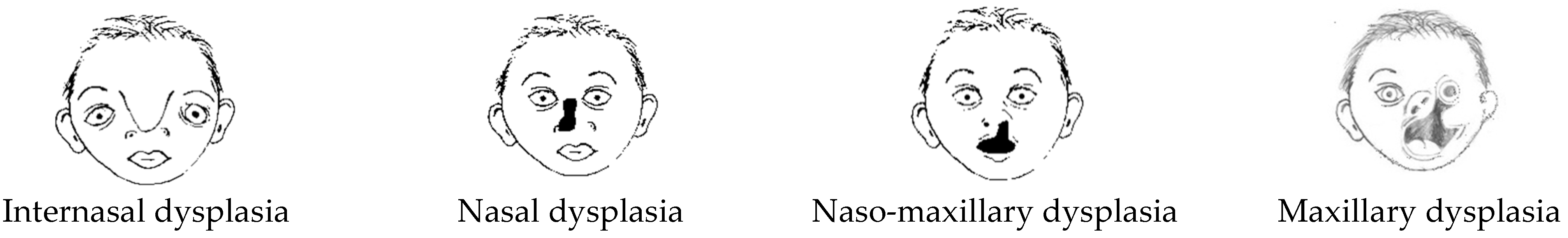
- internasal;
- nasal;
- naso-maxillary;
- maxillary—the maxillary location can be subdivided into median and lateral clefts;
- malar dysplasia/or zygomatic (Treacher Collins Syndrome).
- -
- resection of abnormal or excessive tissues;
- -
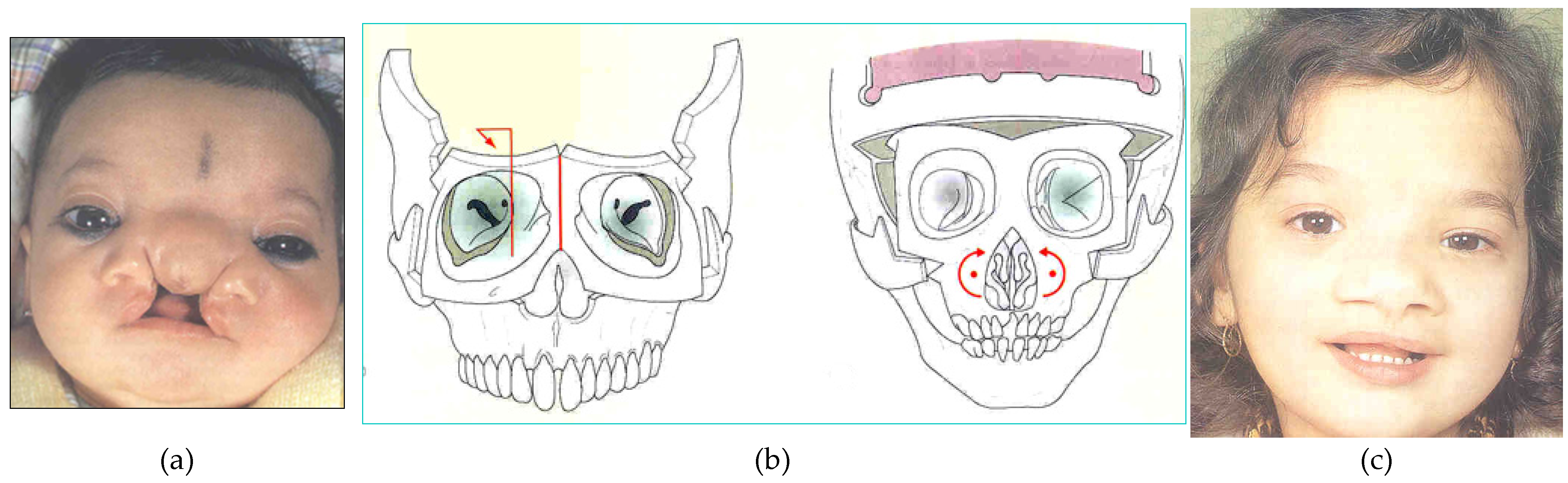
- correction of hypertelorism by medial rotation of the orbits;
- -
- restoration of the nasal pyramid.
- -
- elongation and expansion of the orifices of the nasal cavities;
- -
- reconstructions of the nasal spine.

- nasal aplasia;
- nasal aplasia with the presence of a proboscis;
- nasal cleft;
- nasal duplication.
- -
- resection of abnormal or excessive tissues;
- -
- correction of hypertelorism by medial rotation of the orbits;
- -
- restoration of the nasal pyramid.
- -
- closure of the surrounding fragments of nasal mucosa by mobilizing and bringing the flaps closer;
- -
- reconstruction of the nasal integuments [17].
- -
- resection of abnormal or excessive tissues;
- -
- correction of hypertelorism by medial rotation of the orbits;
- -
- increase in maxillary volume.
- -
- dissection of the medial and lateral flaps of the cleft;
- -
- approach and fixation of the muscular and mucous structures to the periosteum;
- -
- elongation and interdigitation of the skin flaps of the cleft.
- -
- resection of abnormal or excessive tissues;
- -
- correction of hypertelorism, when present, by medial rotation of the orbits;
- -
- increase in maxillary volume if there is a deficit.
- -
- dissection of the medial and lateral flaps of the cleft; correction of the macrostomia with interdigitation of the orbicularis oris and its fixation;
- -
- fixation of the muscular and mucous structures and to the periosteum;
- -
- elongation and interdigitation of the skin flaps of the cleft.


2. Surgical Strategy
- -
- skeletal;
- -
- muscular;
- -
- cutaneous.
- -
- the determination of surgical times (timing) relating to corrections;
- -
- the programming of skin incisions.
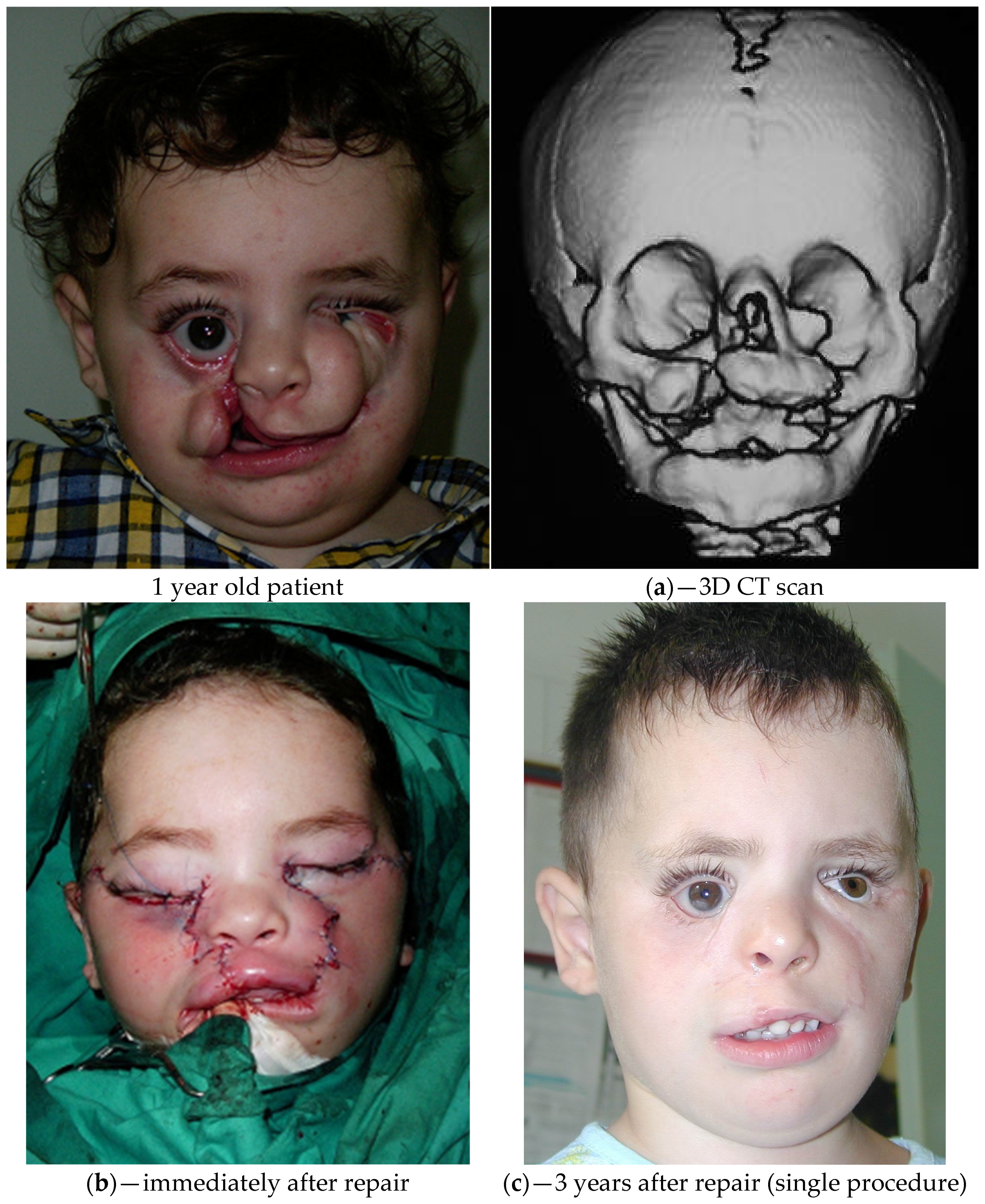
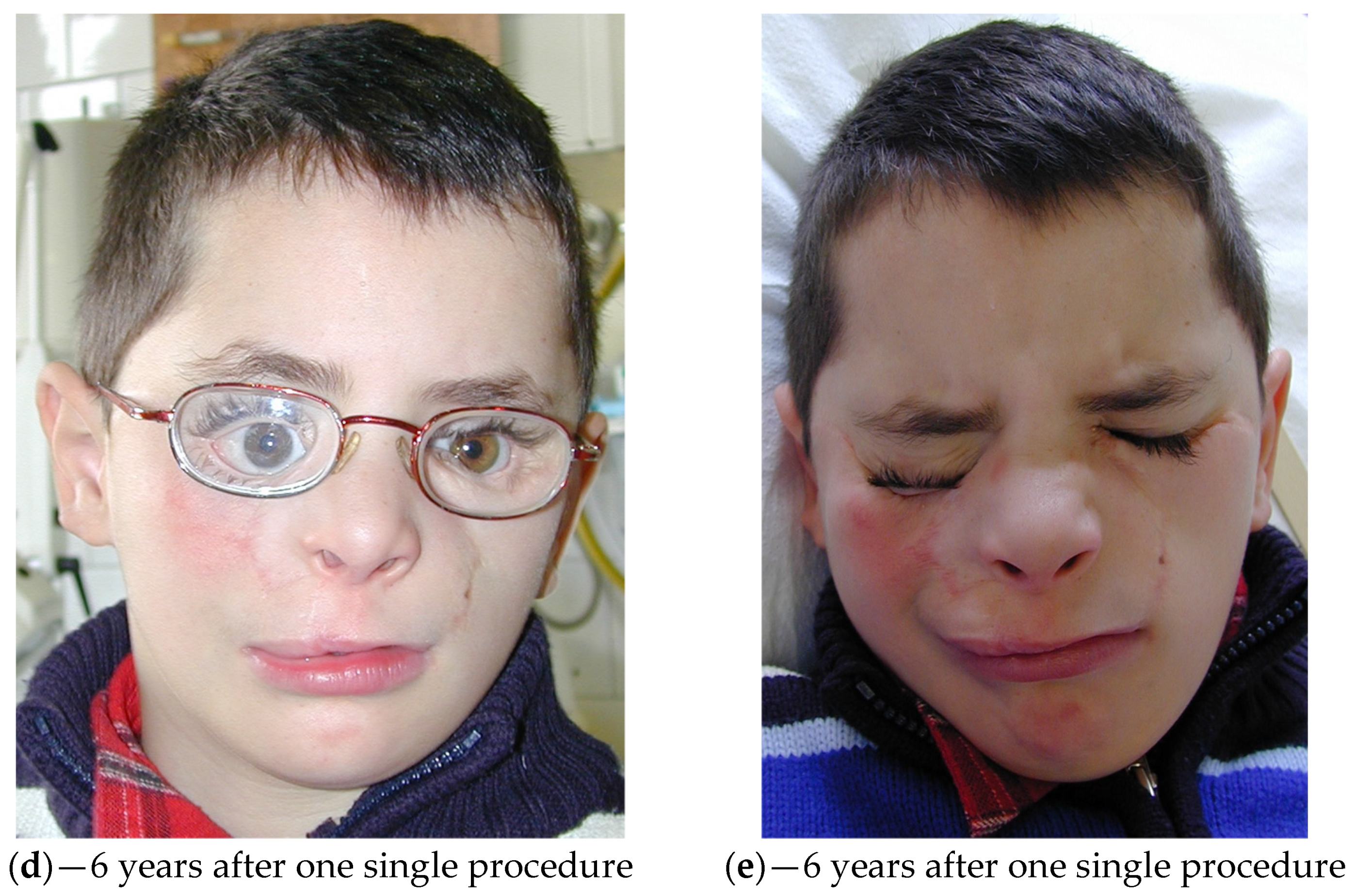
2.1. Skeletal Level
2.2. Muscular Level
- -
- contraction of subcutaneous scar tissue in the first two post-operative months;
- -
- traction of the orbicular muscle through the canthal ligaments;
- -
- traction generated by inadequate dissection of the periosteal tissues of the orbital structures;
- -
- inadequate fixation of bone structures in the midline;
- -
- inadequate exemption of the tissues contained within the orbital cavity.
2.3. Skin Level
2.4. Programming of Skin Incisions
3. Discussion
4. Conclusions
Funding
Acknowledgments
Conflicts of Interest
References
- Leslie, E.J.; Marazita, M.L. Genetics of Cleft Lip and Cleft Palate. Am. J. Med. Genet. C Semin. Med. Genet. 2013, 163, 246–258. [Google Scholar] [CrossRef] [Green Version]
- Kruszka, P.; Li, D.; Harr, M.H.; Wilson, N.R.; Swarr, D.; McCormick, E.M.; Chiavacci, R.M.; Li, M.; Martinez, A.F.; Hart, R.A.; et al. Mutations in SPECC1L, encoding sperm antigen with calponin homology and coiled-coil domains 1-like, are found in some cases of autosomal dominant Opitz G/BBB syndrome. J. Med. Genet. 2015, 52, 104–110. [Google Scholar] [CrossRef] [Green Version]
- Yoon, A.J.; Pham, B.N.; Dipple, K.M. Genetic Screening in Patients with Craniofacial Malformations. J. Pediatr. Genet. 2016, 5, 220–224. [Google Scholar] [CrossRef] [Green Version]
- Mayou, B.J.; Fenton, O.M. Oblique facial clefts caused by amniotic bands. Plast. Reconstr. Surg. 1981, 68, 675–681. [Google Scholar] [CrossRef] [PubMed]
- Moore, M.H. Rare craniofacial clefts. J. Craniofac. Surg. 1996, 7, 408–411. [Google Scholar] [CrossRef]
- Roosenboom, J.; Hens, G.; Mattern, B.C.; Shriver, M.D.; Claes, P. Exploring the Underlying Genetics of Craniofacial Morphology through Various Sources of Knowledge. BioMed Res. Int. 2016, 2016, 3054578. [Google Scholar] [CrossRef] [PubMed] [Green Version]
- Weinberg, S.M.; Cornell, R.; Leslie, E.J. Craniofacial Genetics: Where Have We Been and Where Are We Going? PLoS Genet. 2018, 14, e1007438. [Google Scholar] [CrossRef] [Green Version]
- Wilkie, A.O.; Morriss-Kay, G.M. Genetics of craniofacial development and malformation. Nat. Rev. Genet. 2001, 2, 458–468. [Google Scholar] [CrossRef] [PubMed]
- Cohen, M.M. Malformations of the craniofacial region: Evolutionary, embryonic, genetic, and clinical perspectives. Am. J. Med. Genet. 2002, 115, 245–268. [Google Scholar] [CrossRef]
- Dixon, M.J.; Marazita, M.L.; Beaty, T.H.; Murray, J.C. Cleft lip and palate: Synthesizing genetic and environmental influences. Nat. Rev. Genet. 2011, 12, 167–178. [Google Scholar] [CrossRef] [PubMed]
- Fogh-Andersen, P. Genetic and Non-Genetic Factors in the Etiology of Facial Clefts. Scand. J. Plast. Reconstruct. Surg. Hand Surg. 1967, 1, 22–29. [Google Scholar] [CrossRef]
- Hunt, J.A.; Hobar, P.C. Common Craniofacial Anomalies: Facial Clefts and Encephaloceles. Plast. Reconstr. Surg. 2003, 112, 606–616. [Google Scholar] [CrossRef] [PubMed]
- Fearon, J.A. Rare Craniofacial Clefts: A surgical Classification. J. Craniofac. Surg. 2008, 19, 110–112. [Google Scholar] [CrossRef] [PubMed] [Green Version]
- Tessier, P. Anatomical classification of facial, cranio-facial and latero-facial clefts. J. Maxillofac. Surg. 1976, 4, 69–92. [Google Scholar] [CrossRef]
- Van der Meulen, J.C.H.; Vaandrager, J.M. Facial Clefts. World J. Surg. 1989, 13, 373–383. [Google Scholar] [CrossRef] [PubMed]
- Van der Meulen Jacques, C.H.; Gilbert, P.; Roddi, R. Orbital surgery. In Ocular Plastic Surgery; Mosby-Wolfe: Barcelona, Spain, 1996. [Google Scholar]
- Roddi, R.; van der Meulen Jacques, C.H. Encephalocele. In Ocular Plastic Surgery; Mosby-Wolfe: Barcelona, Spain, 1996. [Google Scholar]
- Roddi, R.; van der Meulen Jacques, C.H. Treacher Collins. In Ocular Plastic Surgery; Mosby-Wolfe: Barcelona, Spain, 1996. [Google Scholar]
- Friede, H.; Johanson, B. Adolescent Facial Morphology of Early Bone-Grafted Cleft Lip and Palate Patients. Scand. J. Plast. Reconstruct. Surg. Hand Surg. 1982, 16, 41–53. [Google Scholar] [CrossRef] [PubMed]
- Kamerer, D.B.; Caparosa, R.J. Temporal Bone Encephalocele: Diagnosis and treatment. Laryngoscope 1982, 92, 878–882. [Google Scholar] [CrossRef]
- Marchac, D.; Arnaud, E. Midface surgery from Tessier to distraction. Childs Nerv. Syst. 1999, 15, 681–694. [Google Scholar] [CrossRef]
- Patipa, M.; Wilkins, R.B.; Guelzow, K.W. Surgical management of congenital eyelid coloboma. Ophthalmic. Surg. 1982, 13, 212–216. [Google Scholar] [CrossRef]
- Tabrizi, R.; Ozkan, T.B.; Mohammadinejad, C.; Minaee, N. Orbital Floor Reconstruction. J. Craniofac. Surg. 2010, 21, 1142–1146. [Google Scholar] [CrossRef]
- Tardy, M.E., Jr.; Denneny, J., III; Fritsch, M.H. The Versatile Cartilage Autograft in Reconstruction of the Nose and Face. Laryngoscope 1985, 95, 523–533. [Google Scholar] [CrossRef] [PubMed]


Disclaimer/Publisher’s Note: The statements, opinions and data contained in all publications are solely those of the individual author(s) and contributor(s) and not of MDPI and/or the editor(s). MDPI and/or the editor(s) disclaim responsibility for any injury to people or property resulting from any ideas, methods, instructions or products referred to in the content. |
© 2023 by the authors. Licensee MDPI, Basel, Switzerland. This article is an open access article distributed under the terms and conditions of the Creative Commons Attribution (CC BY) license (https://creativecommons.org/licenses/by/4.0/).
Share and Cite
Roddi, R.; Oo, A.L.; Pepe, E.; Naing, E.E.; Sung, S.B.H. Surgical Strategy for the Treatment of Facial Clefts. Surg. Tech. Dev. 2023, 12, 34-42. https://doi.org/10.3390/std12010002
Roddi R, Oo AL, Pepe E, Naing EE, Sung SBH. Surgical Strategy for the Treatment of Facial Clefts. Surgical Techniques Development. 2023; 12(1):34-42. https://doi.org/10.3390/std12010002
Chicago/Turabian StyleRoddi, Roberto, Aung Lwin Oo, Ernesto Pepe, Ei Ei Naing, and Shalom Biak Hlei Sung. 2023. "Surgical Strategy for the Treatment of Facial Clefts" Surgical Techniques Development 12, no. 1: 34-42. https://doi.org/10.3390/std12010002