
Surg. Tech. Dev., Volume 11, Issue 3 (December 2022) – 3 articles
Cover Story (view full-size image):
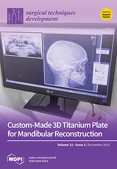
The development of new prototyping systems provides accurate 3D biomodels on which surgery can be simulated, especially in cases of ameloblastoma, in which the safety margin is vital for the clinical outcome. The objective of this paper was to report a clinical case of employing these methodologies for reconstruction after an extensive mandibular resection. A case of follicular ameloblastoma of the mandible is depicted in the following paper, where a 3D biomodel was used throughout the surgery. A 3D-printed patient-specific titanium implant was manufactured and placed intraoperatively for reconstruction. The treatment had satisfactory postoperative results without complications. Titanium implants, being bioinert, customizable, and easily workable, especially with the help of 3D virtual planning techniques, can be considered ideal alloplastic materials for mandibular reconstruction. View this paper
- Issues are regarded as officially published after their release is announced to the table of contents alert mailing list.
- You may sign up for e-mail alerts to receive table of contents of newly released issues.
- PDF is the official format for papers published in both, html and pdf forms. To view the papers in pdf format, click on the "PDF Full-text" link, and use the free Adobe Reader to open them.
Previous Issue
Next Issue