
Extramedullary T-lymphoblastic Crisis in a Myelodysplastic/Myeloproliferative Neoplasm with a t(12;22)/MN1::ETV6 Translocation

 and
and 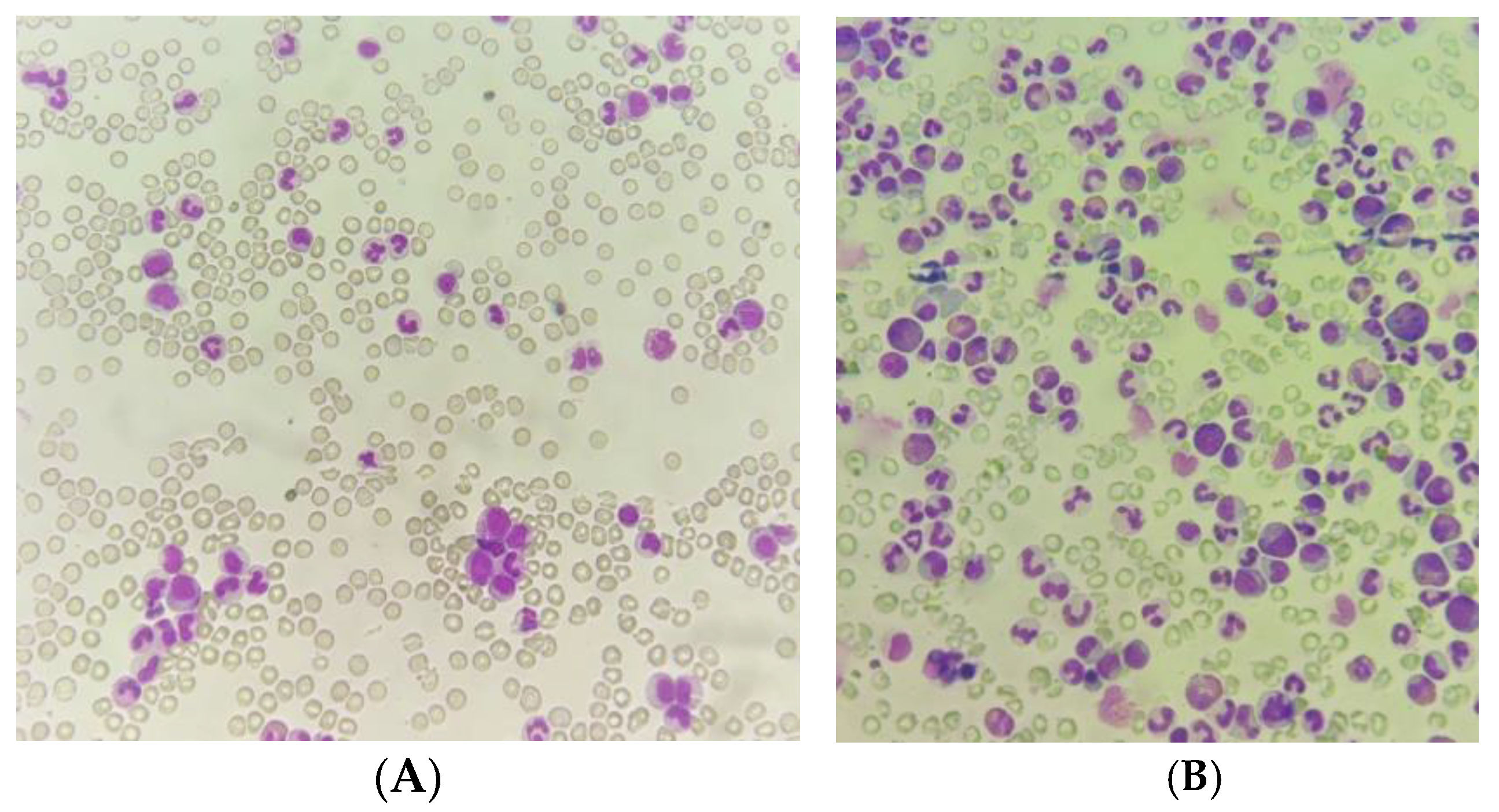
Abstract
:1. Introduction
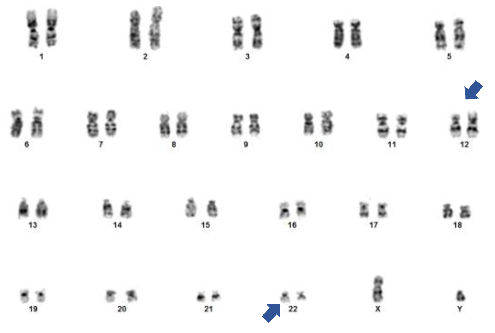
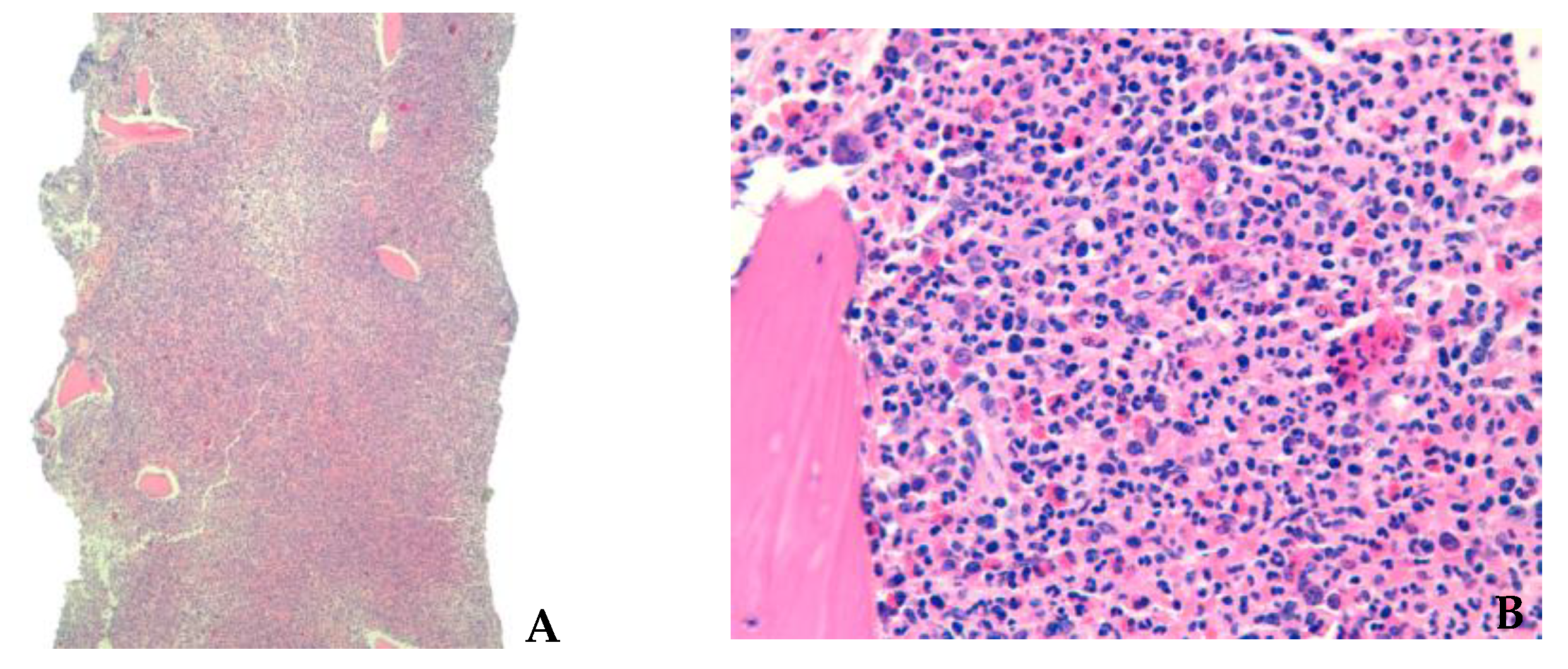
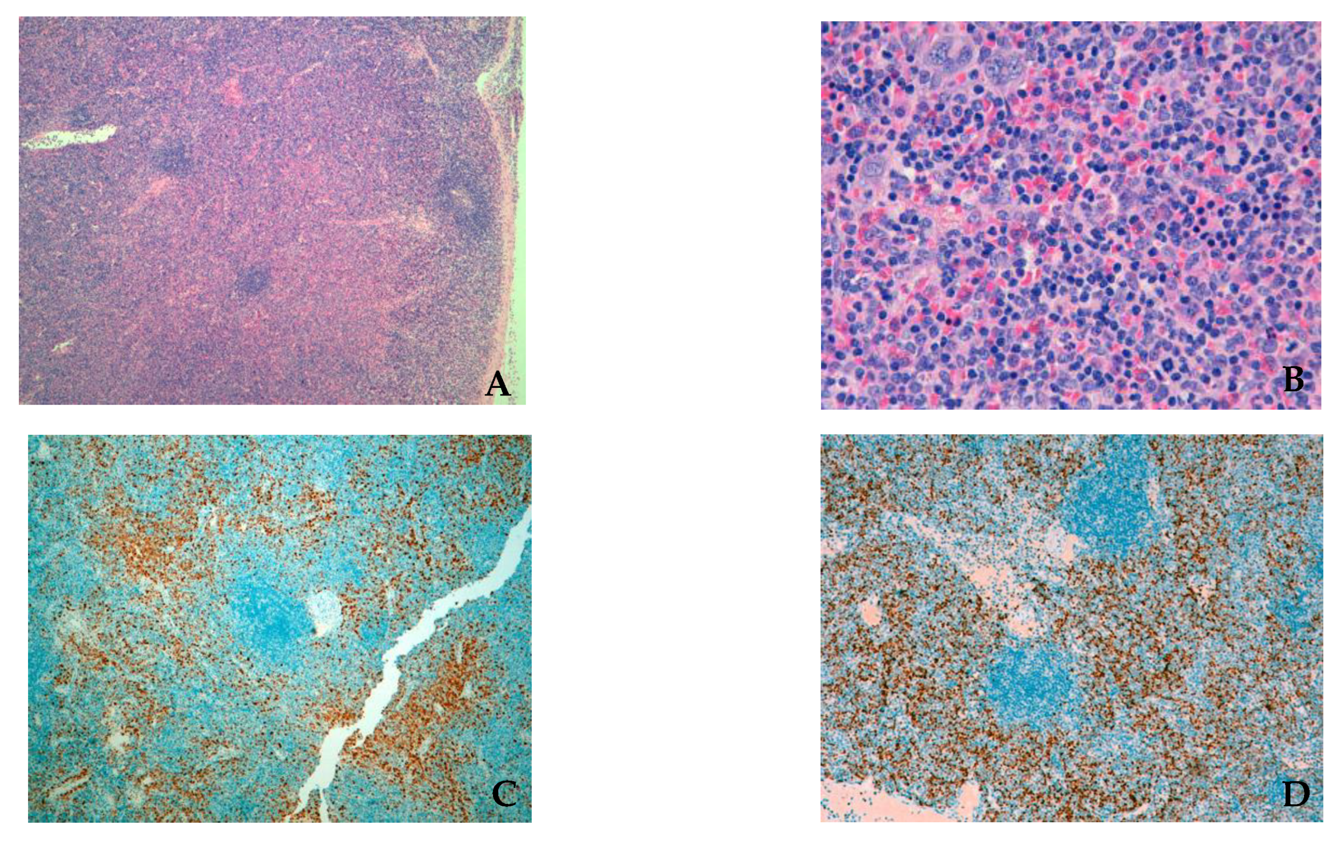
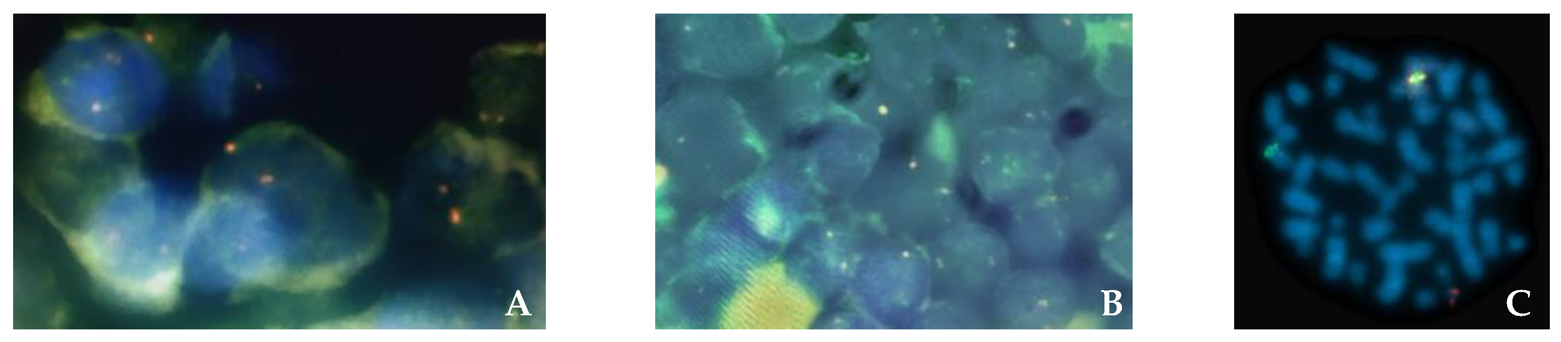
2. Case Report
3. Discussion
Supplementary Materials
Author Contributions
Funding
Institutional Review Board Statement
Informed Consent Statement
Data Availability Statement
Conflicts of Interest
References
- Palomo, L.; Meggendorfer, M.; Hutter, S.; Twardziok, S.; Ademà, V.; Fuhrmann, I.; Fuster-Tormo, F.; Xicoy, B.; Zamora, L.; Acha, P.; et al. Molecular landscape and clonal architecture of adult myelodysplastic/myeloproliferative neoplasms. Blood 2020, 136, 1851–1862. [Google Scholar] [CrossRef] [PubMed]
- Khoury, J.D.; Solary, E.; Abla, O.; Akkari, Y.; Alaggio, R.; Apperley, J.F.; Bejar, R.; Berti, E.; Busque, L.; Chan, J.K.C.; et al. The 5th edition of the World Health Organization Classification of Haematolymphoid Tumours: Myeloid and Histiocytic/Dendritic Neoplasms. Leukemia 2022, 36, 1703–1719. [Google Scholar] [CrossRef] [PubMed]
- Arber, D.A.; Orazi, A.; Hasserjian, R.P.; Borowitz, M.J.; Calvo, K.R.; Kvasnicka, H.-M.; Wang, S.A.; Bagg, A.; Barbui, T.; Branford, S.; et al. International Consensus Classification of Myeloid Neoplasms and Acute Leukemias: Integrating morphologic, clinical, and genomic data. Blood 2022, 140, 1200–1228. [Google Scholar] [CrossRef]
- Döhner, H.; Wei, A.H.; Appelbaum, F.R.; Craddock, C.; DiNardo, C.D.; Dombret, H.; Ebert, B.L.; Fenaux, P.; Godley, L.A.; Hasserjian, R.P.; et al. Diagnosis and management of AML in adults: 2022 recommendations from an international expert panel on behalf of the ELN. Blood 2022, 140, 1345–1377. [Google Scholar] [CrossRef] [PubMed]
- Grosveld, G.C. MN1, a novel player in human AML. Blood Cells Mol. Dis. 2007, 39, 336–339. [Google Scholar] [CrossRef] [Green Version]
- Heuser, M.; Beutel, G.; Krauter, J.; Döhner, K.; von Neuhoff, N.; Schlegelberger, B.; Ganser, A. High meningioma 1 (MN1) expression as a predictor for poor outcome in acute myeloid leukemia with normal cytogenetics. Blood 2006, 108, 3898–3905. [Google Scholar] [CrossRef] [Green Version]
- Langer, C.; Marcucci, G.; Holland, K.B.; Radmacher, M.D.; Maharry, K.; Paschka, P.; Whitman, S.P.; Mrózek, K.; Baldus, C.D.; Vij, R.; et al. Prognostic importance of mn1 transcript levels, and biologic insights from MN1-associated gene and microRNA expression signatures in cytogenetically normal acute myeloid leukemia: A cancer and leukemia group b study. J. Clin. Oncol. 2009, 27, 3198–3204. [Google Scholar] [CrossRef] [Green Version]
- Haferlach, C.; Kern, W.; Schindela, S.; Kohlmann, A.; Alpermann, T.; Schnittger, S.; Haferlach, T. Gene expression of BAALC, CDKN1B, ERG, and MN1 adds independent prognostic information to cytogenetics and molecular mutations in adult acute myeloid leukemia. Genes Chromosom. Cancer 2011, 51, 257–265. [Google Scholar] [CrossRef]
- Schwind, S.; Marcucci, G.; Kohlschmidt, J.; Radmacher, M.D.; Mrózek, K.; Maharry, K.; Becker, H.; Metzeler, K.; Whitman, S.P.; Wu, Y.-Z.; et al. Low expression of MN1 associates with better treatment response in older patients with de novo cytogenetically normal acute myeloid leukemia. Blood 2011, 118, 4188–4198. [Google Scholar] [CrossRef]
- De Braekeleer, E.; Douet-Guilbert, N.; Morel, F.; Le Bris, M.-J.; Basinko, A.; De Braekeleer, M. ETV6 fusion genes in hematological malignancies: A review. Leuk. Res. 2012, 36, 945–961. [Google Scholar] [CrossRef]
- Shao, H.; Cen, J.; Chen, S.; Qiu, H.; Pan, J. Myeloid neoplasms with t(12;22)(p13;q12)/MN1-EVT6: A systematic review of 12 cases. Ann. Hematol. 2017, 97, 417–424. [Google Scholar] [CrossRef] [PubMed]
- Wang, T.; Chen, X.; Hui, S.; Ni, J.; Yin, Y.; Cao, W.; Zhang, Y.; Wang, X.; Ma, X.; Cao, P.; et al. Ectopia associated MN1 fusions and aberrant activation in myeloid neoplasms with t(12;22)(p13;q12). Cancer Gene Ther. 2020, 27, 810–818. [Google Scholar] [CrossRef] [Green Version]
- Callen, D.F.; Hull, Y.J.; Toogood, I.R.; Fioretos, T.; Heim, S.; Mandahl, N.; Mitelman, F. New chromosomal rearrangement, t(12;22)(p13;q12), in acute nonlymphocytic leukemia. Cancer Genet. Cytogenet. 1991, 51, 255–258. [Google Scholar] [CrossRef] [PubMed]
- Van Kessel, A.G.; Stellink, F.; Van Gaal, J.; Van De Klundert, W.; Siepman, A.; Oosten, H. Translocation (12;22)(p13;q12) as sole karyotypic abnormality in a patient with nonlymphocytic leukemia. Cancer Genet. Cytogenet. 1994, 72, 105–108. [Google Scholar] [CrossRef]
- Rosenzweig, J.; Pillai, P.M.; Prockop, S.; Benayed, R.; Brodersen, L.; Najfeld, V.; Loken, M.; Zhang, Y.; Shukla, N. Acute myeloid leukemia with an MN1-ETV6 fusion in a young child with Down syndrome. Mol. Case Stud. 2022, 8, a006167. [Google Scholar] [CrossRef]
- Kantarjian, H.; Thomas, D.; O’Brien, S.; Cortes, J.; Giles, F.; Jeha, S.; Bueso-Ramos, C.E.; Pierce, S.; Shan, J.; Koller, C.; et al. Long-term follow-up results of hyperfractionated cyclophosphamide, vincristine, doxorubicin, and dexamethasone (Hyper-CVAD), a dose-intensive regimen, in adult acute lymphocytic leukemia. Cancer 2004, 101, 2788–2801. [Google Scholar] [CrossRef]
- Parker, J.E.; Pagliuca, A.; Mijovic, A.; Cullis, J.O.; Czepulkowski, B.; Rassam, S.M.B.; Samaratunga, I.R.; Grace, R.; Gover, P.A.; Mufti, G.J. Fludarabine, cytarabine, G-CSF and idarubicin (FLAG-IDA) for the treatment of poor-risk myelodysplastic syndromes and acute myeloid leukaemia. Br. J. Haematol. 1997, 99, 939–944. [Google Scholar] [CrossRef]
- Estey, E.; Thall, P.; Andreeff, M.; Beran, M.; Kantarjian, H.; O’Brien, S.; Escudier, S.; Robertson, L.E.; Koller, C.; Kornblau, S. Use of granulocyte colony-stimulating factor before, during, and after fludarabine plus cytarabine induction therapy of newly diagnosed acute myelogenous leukemia or myelodysplastic syndromes: Comparison with fludarabine plus cytarabine without granulocyte colony-stimulating factor. J. Clin. Oncol. 1994, 12, 671–678. [Google Scholar] [CrossRef]
- Heuser, M.; Yun, H.; Berg, T.; Yung, E.; Argiropoulos, B.; Kuchenbauer, F.; Park, G.; Hamwi, I.; Palmqvist, L.; Lai, C.K.; et al. Cell of origin in AML: Susceptibility to MN1-induced transformation is regulated by the meis1/abdb-like hox protein complex. Cancer Cell 2011, 20, 39–52. [Google Scholar] [CrossRef] [Green Version]
- Kawagoe, H.; Grosveld, G.C. MN1-TEL myeloid oncoprotein expressed in multipotent progenitors perturbs both myeloid and lymphoid growth and causes T-lymphoid tumors in mice. Blood 2005, 106, 4278–4286. [Google Scholar] [CrossRef] [Green Version]
- Chen, S.; Xue, Y.; Zhu, X.; Wu, Y.; Pan, J. Minimally differentiated acute myeloid leukemia with t(12;22)(p13;q11) translocation showing primary multidrug resistance and expressing multiple multidrug-resistant proteins. Acta Haematol. 2007, 118, 38–41. [Google Scholar] [CrossRef] [PubMed]
- Libbrecht, C.; Xie, H.M.; Kingsley, M.C.; Haladyna, J.N.; Riedel, S.S.; Alikarami, F.; Lenard, A.; McGeehan, G.M.; Ernst, P.; Bernt, K.M. Menin is necessary for long term maintenance of meningioma-1 driven leukemia. Leukemia 2021, 35, 1405–1417. [Google Scholar] [CrossRef] [PubMed]
- Issa, G.C.; Ravandi, F.; DiNardo, C.D.; Jabbour, E.; Kantarjian, H.M.; Andreeff, M. Therapeutic implications of menin inhibition in acute leukemias. Leukemia 2021, 35, 2482–2495. [Google Scholar] [CrossRef] [PubMed]


Disclaimer/Publisher’s Note: The statements, opinions and data contained in all publications are solely those of the individual author(s) and contributor(s) and not of MDPI and/or the editor(s). MDPI and/or the editor(s) disclaim responsibility for any injury to people or property resulting from any ideas, methods, instructions or products referred to in the content. |
© 2023 by the authors. Licensee MDPI, Basel, Switzerland. This article is an open access article distributed under the terms and conditions of the Creative Commons Attribution (CC BY) license (https://creativecommons.org/licenses/by/4.0/).
Share and Cite
Freitas, A.C.; Maia, T.; Desterro, J.; Pierdomenico, F.; Nunes, A.; Ferreira, I.; Cabeçadas, J.; Gomes da Silva, M. Extramedullary T-lymphoblastic Crisis in a Myelodysplastic/Myeloproliferative Neoplasm with a t(12;22)/MN1::ETV6 Translocation. Hematol. Rep. 2023, 15, 212-219. https://doi.org/10.3390/hematolrep15010022
Freitas AC, Maia T, Desterro J, Pierdomenico F, Nunes A, Ferreira I, Cabeçadas J, Gomes da Silva M. Extramedullary T-lymphoblastic Crisis in a Myelodysplastic/Myeloproliferative Neoplasm with a t(12;22)/MN1::ETV6 Translocation. Hematology Reports. 2023; 15(1):212-219. https://doi.org/10.3390/hematolrep15010022
Chicago/Turabian StyleFreitas, Ana Carolina, Tiago Maia, Joana Desterro, Francesca Pierdomenico, Albertina Nunes, Isabelina Ferreira, José Cabeçadas, and Maria Gomes da Silva. 2023. "Extramedullary T-lymphoblastic Crisis in a Myelodysplastic/Myeloproliferative Neoplasm with a t(12;22)/MN1::ETV6 Translocation" Hematology Reports 15, no. 1: 212-219. https://doi.org/10.3390/hematolrep15010022