Review

Production of Recombinant Biopharmaceuticals in Chlamydomonas reinhardtii

by
Cesar Andres Diaz Arias
1,*,
Caroline Frere Martiniuc de Oliveira
2,
João Vitor Dutra Molino
1,3,
Livia Seno Ferreira-Camargo
2,
Marcelo Chuei Matsudo
2 and
João Carlos Monteiro de Carvalho
1,*
1
Department of Biochemical and Pharmaceutical Technology, Faculty of Pharmaceutical Sciences, University of São Paulo, Av. Prof. Lineu Prestes 580, Bl. 16, São Paulo 05508-900, SP, Brazil
2
Center of Natural and Human Sciences, Federal University of ABC, Av. dos Estados, 5001, Santo André 09210-580, SP, Brazil
3
California Center for Algae Biotechnology, Division of Biological Sciences, University of California, San Diego, CA 92093, USA
*
Authors to whom correspondence should be addressed.
Int. J. Plant Biol. 2023, 14(1), 39-52; https://doi.org/10.3390/ijpb14010004
Submission received: 28 October 2022 / Revised: 16 December 2022 / Accepted: 20 December 2022 / Published: 29 December 2022
(This article belongs to the Special Issue Microalgae as a Powerful Tool for Biopharming Development)

Abstract

:
This review aimed to present Chlamydomonas reinhardtii as an alternative for heterologous protein production, especially for biopharmaceuticals, and its general characteristics when compared with other expression systems. The need to produce heterologous proteins for industrial interest, therapeutic ends, and diagnostic kits has led to the development of recombinant microalgal technology. This technology presents some interesting features, such as rapid growth and low transgene dispersion compared to plants, the ability to fold complex proteins compared to bacteria, and low production costs compared to other expression systems, such as yeast and mammalian cells. Overall, C. reinhardtii heterologous protein expression is coming of age with several research groups focused on developing an optimal producer strain.

1. Introduction

Microalgae have been used as an energy source for human and animal consumption, producing fatty acids and secondary metabolites, such as pigments and biomass [1,2,3,4]. There are many microalgae species around the world. However, only a few are successfully used for commercialization, including Chlorella and Spirulina [5,6]. Besides, Dunaliella salina and Haematococcus pluvialis have been demonstrated as great species for β-carotene and astaxanthin production [7].
As recombinant DNA technology advanced, the genomes of several microalgae species were sequenced, unveiling a myriad of DNA sequences exploitable for heterologous protein expression [8,9,10]. Among all microalgae species, Chlamydomonas reinhardtii is widely used in various studies, such as photosynthesis, cellular motility, and phototaxis. As plants, microalgae present three genomes and protocols for genetic modification of the nucleus, mitochondria, and chloroplast [11,12], which allows for successful results when genetically modified. Many algae industries are developing algae-based products [1], and one of them (Lumen Bioscience—Seattle, WA, USA) announced this year that they are hacking Arthrospira platensis to generate biologics. However, as far as we are aware, no industry is commercializing biopharmaceuticals produced by Chlamydomonas reinhardtii.
C. reinhardtii presents several advantages over other systems with its lower production costs and rapid growth, as well as being a microorganism generally regarded as safe (GRAS) by the US FDA (United States Food and Drug Administration).
The genus Chlamydomonas is distributed worldwide and may be found in a diversity of habitats (e.g., freshwater, marine, and brackish waters, sewage ponds, snow, agricultural and forest soils, deserts, and peat bogs), but the first strain of Chlamydomonas reinhardtii was probably isolated from a soil sample collected in a potato field, in Massachusetts State, USA, in 1945 [13,14]. More recently, strains of C. reinhardtii have been newly isolated from palm oil mill effluent in Malaysia [15] and freshwater samples in Korea [16,17]. Interestingly, Hong et al. [16] found that the C. reinhardtii isolated from freshwater samples was able to grow in a broad range of pH values (pH 4.0–12.0) and temperatures (10–30 °C). Although it is isolated from soil samples, it can successfully grow in liquid or solid media, and several recipes are proposed for that purpose [13]. Different culture medium compositions and culture collections may be found at http://www.chlamy.org (accessed on 1 December 2022).
This photosynthetic microorganism can also be cultivated in closed photobioreactors, reducing transgene dispersion risk [18,19]. It can produce complex proteins with post-translational modifications, such as disulfide bonds and N-glycosylations [20]. Although Chlamydomonas represents a tremendous potential technology, there is no optimal strain that unifies efficient heterologous protein production on a commercial scale, which depends on the cell components, cultivation processes, and protein extraction and purification [1].
In addition to developing robust gene expression systems, cultivation processes that allow high biomass concentration and productivity are also needed [21], including developing suitable photobioreactors. In this sense, several types of photobioreactors have been evaluated for the photoautotrophic cultivation of Chlamydomonas reinhardtii, such as bubble column photobioreactors [22], reflector-coated LED-photobioreactors [23], horizontal-dual bladed bioreactors [24], tubular photobioreactors with polypropylene-based tubes [25], airlift photobioreactors [26], biocoil photobioreactors made of 16 m-long polyvinyl tubes [27], and high-density polyethylene bags ranged on racks within a greenhouse [28]. There is also the possibility of performing heterotrophic growth in a mechanically stirred bioreactor [29].
This review aimed to present Chlamydomonas reinhardtii as a platform for heterologous protein production compared to other expression systems and its efficient heterologous protein production in the biopharmaceutical biotechnology application.

2. Chlamydomonas reinhardtii: The Microalga

The Chlamydomonas genus is commonly identified through morphological criteria. Among species, measures such as size and body shape, as well as shape and position of the chloroplast and flagellar length, are applied [30]. It is a biflagellate and unicellular green alga, widely used as a photosynthetic model organism for studies in different areas, including genetics, biochemistry, and cell biology. Some of these areas focus their studies on the similarity of algae flagella and basal bodies to mammal cells, despite their evolutionary divergence 109 years ago [31]. Approximately 500 species of Chlamydomonas are described and can be found in different ecological niches. For example, Chlamydomonas nivalis was found in land samples, first isolated by GM Smith in 1945 at the glaciers [30].
Chlamydomonas reinhardtii is easy to grow and presents low production costs, requiring only sunlight, minimal media, and CO2 to grow at scale [32]. It can grow heterotrophically using acetate as a carbon source without needing light or in mixotrophic culture, in which both energy sources are used. Moreover, this unicellular microalga is considered a fast-growing microorganism, with a generation time of less than 10 h; it can be cultivated in liquid and solid media, with neutral pH, without adding vitamins and cofactors [33]. Depending on the culture conditions, C. reinhardtii can divide up to five times to produce 32 daughter cells [34]. In a usual cycle of 12 h light and 12 h dark, the growth occurs in the light phase, and the cellular division occurs in the dark phase. If nutrients do not limit the growth, C. reinhardtii divides synchronously during the period of darkness into successive mitotic divisions, generating four new cells [34].
This microalga is considered a powerful platform for the study of gene expression [12] since it is a model organism with completely sequenced genomes [32] that present beneficial specific characteristics, such as a GC-rich nuclear genome (62–68%) and an AT-rich chloroplast genome [35].

3. Chlamydomonas reinhardtii Genomic Knowledge

The nuclear genome has an estimated size of approximately 121 Mb, where the DNA is randomly inserted and frequently generates DNA deletions in the inserted region, which causes some difficulties for mutants’ analyses and, mainly, low heterologous protein expression [30].
Chlamydomonas’s chloroplast genome is approximately 195 kb, can be reliably transformed through homologous recombination, and has an inverted repeat structure similar to some terrestrial plants. Yet, the order of the genes differs significantly [30]. Compared to the nuclear genome, the genetic content in the chloroplast is more straightforward, with approximately 160 times fewer genes, with a reduced size [36,37,38]. Despite its simpler genome, the chloroplast hosts a more considerable diversity of proteins (~3000), with most imported from the nuclear genome. The diverse set of proteins harbors a range of protein chaperones and protein disulfide isomerases that assist the correct folding of complex proteins, such as a full-length human monoclonal antibody [39,40,41]. Contrary to the nucleus, it is amenable to homologous recombination, simplifying genetic modification strategies. By homologous recombination, two of the most frequent chloroplast genome site transformations are the psbA and psbH gene sites [41,42,43]. The chloroplast occupies half the cell volume, accumulating more proteins [44].
The nucleus and chloroplast genomes have different codon usages since, in the chloroplast, there is a higher occurrence of nucleotides adenine and thymine in the third position, while in the nucleus, guanine and cytosine are predominant. Green fluorescent protein (GFP) was increased 80-fold in protein accumulation, while in the nucleus there was only a 5-fold increase when the recombinant genes were previously codon-optimized [32]. This microalga expression levels range from 0.16 to 5% of total soluble protein.
Heterologous protein expression levels depend on various factors, for example, recombinant protein codon optimization, promoter sequence, untranslated regions (UTRs), gene silencing, proteases, and other molecules in the cultivation media. Another critical factor affecting expression is the generated clones that are not maintained under selective conditions [45,46].

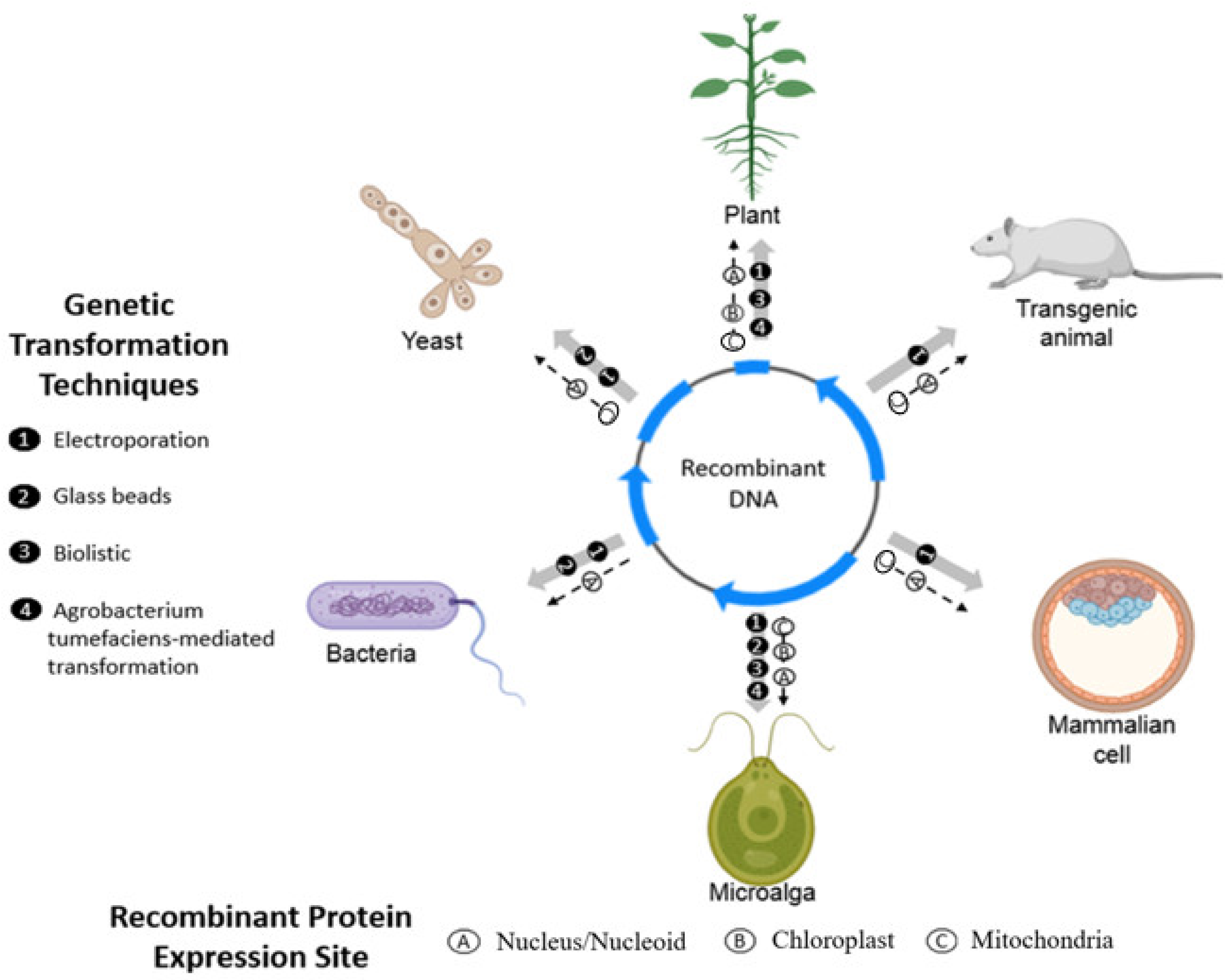
3.1. Transformation Methods in Chlamydomonas reinhardtii

Microalgae genetic transformation is based on the permeabilization of the microorganism membrane, so the DNA is introduced into the cell. For this purpose, some techniques are proposed as electroporation, glass beads agitation, biolistic systems, and Agrobacterium tumefaciens-mediated transformation. It is essential to mention that the preferred transformation methods in the nucleus are agitation with glass beads and electroporation, while the chloroplast is biolistic [47].
In 1988, the first stable chloroplast transformation in C. reinhardtii was successfully performed [48]. C. reinhardtii has been used as the platform for heterologous protein production in microalgae [47]. However, compared to the nucleus, the chloroplast cannot perform, for example, protein glycosylation, a significant post-translational modification in many therapeutic proteins. It also does not present a protein secretory pathway to the culture media, increasing the purification step cost, since it requires an additional step for cell lysing [41,49,50].
Despite its exciting features, such as complex post-translational modification and secretion capacity, nuclear expression has low protein expression levels. Nevertheless, in the last decade, expression vector development has presented some progress, using techniques such as codon optimization and the development of appropriate expression vectors [51]. Despite the big leap required for protein expression in microalgae to attain economic interest, recombinant strategies have presented a yield increase. For instance, the employment of RuBisCo endogenous introns increased the expression levels of heterologous proteins up to 450% [47], evidencing the gain yet to be achieved by further development. Some endeavors with other microalgae species have also been made for protein expression. Other species are yet to be evaluated, which places C. reinhardtii as the best-evaluated and model species for this purpose [52].

3.2. Chloroplast Gene Regulation and Nuclear Gene Regulation

The promoter is an essential element to start the transcription process, and its choice is critical for the expression of heterologous proteins. The most applied ones for gene expression in microalgae are the following: RbcS2, Hsp70, psaD, and psbA from the genus Chlamydomonas, frequently used in Chlamydomonas sp., NR from Thalassiosira pseudonana, Nos from Agrobacterium tumefaciens, 35S Cauliflower mosaic virus that is commonly used in dinoflagellates, and Fcp from Phaeodactylum tricornutum and Thalassiosira pseudonana [32,53,54].
Some fusion promoters, such as HSP70A (heat shock protein 70A)/RBCS2 and HSP70A/β2TUB (β2 tubulin), have been shown to enhance the expression of heterologous proteins in the nucleus [32]. Some inducible promoters, such as NR, encoding a nitrate reductase, have also been studied for protein expression. In this case, the promoter activity is suppressed with the presence of ammonium in the medium and activated when ammonium is replaced by nitrate [55].
As well as the promoters, UTRs (untranslated regions) have been recognized as crucial factors for the heterologous gene expression in the C. reinhardtii chloroplast. There are some commonly used UTRs, such as atpA, rbcL, and psbD [56,57,58,59,60,61]. Still, the most successful one is the psbA UTR in combination with the psbA promoter [62], which resulted in 5% of heterologous M-SAA (Mammary-Associated Serum Amyloid) from total protein accumulation, as shown in Table 1.
In addition to the genetic machinery strategy, a chemical approach was also attempted as an alternative for heterologous protein folding. Increasing concentrations of the diselenide selenocystamine were added to C. reinhardtii cultivation medium and, when comparing the accumulation of three different proteins expressed in the chloroplast, the ones containing disulfide bonds resulted in significant accumulation increases when compared to the protein without disulfide bonds. These experiments suggest that the disulfide bond formation could be a rate-limiting step for protein accumulation [63].
Table 1. Summary of biomolecules expressed in Chlamydomonas reinhardtii, its genetically transformed organelle, expression percentage (%) from total soluble protein, concentration in milligrams per liter (mg/L), and the cloning vector.
Heterologous Protein*** (%) from Total Soluble ProteinExpression
(mg/L)
Vector
14FN3 (fourteenth human fibronectin type III domain) (ch) **3UN *psbA/D1-deficient strain (Rasala et al., 2010) [43]
VEGF (human vascular endothelial growth factor) (ch) **2UN *psbA/D1-deficient strain (Rasala et al., 2010) [43]
HMGB1 (high mobility group protein B1) (ch) **2.5UN *psbA/D1-deficient strain (Rasala et al., 2010) [43]
Endolysins—Pal and Cpl111.3 mg/g of cell dry weightpsbH insertion site/pSRSap vector/psaA promoter (Stoffels et al., 2017) [64]
Growth HormoneUN *0.5psbH insertion site/pASapI vector/psaA and atpA promoter (Wannathong et al., 2016) [65]
D2 fibronectin-binding domain of Staphylococcus aureus fused with the cholera toxin B subunit (CTB)0.71.6 mg/g of cell dry weighttscA insertion site/p463 and p464 vectors/rbcL and atpA promoters(Dreesen, Hamri and Fussenegger 2010) [66]
14FN3 (fourteenth human fibronectin type III domain) (ch) **0.210.0416S atpA/photosynthetic strain (Rasala et al., 2010) [43]
Xylanase (nu) **0.25UN *Ble 2A expression system
(Rasala et al., 2012) [67]
Immunotoxin (αCD22HCH23PE40) (ch) **0.2–0.3UN *psbA/D1-deficient strain
(Tran et al., 2013) [42]
CD22 (Single chain antibody (scFv) directed against the CD22 cell surface antigen) (ch) **0.6–0.7UN *psbA/D1-deficient strain
(Tran et al., 2013) [42]
CD22Gel (CD22-scFv genetically linked to gelonin, a ribosome-inactivating protein from G. multiflorum) (ch) **0.2–0.3UN *psbA/D1-deficient strain
(Tran et al., 2013) [42]
CD22CH23Gel (CD22-scFv genetically fused to the hinge and constant domains of an IgG1 and to gelonin to create a chimeric protein homodimer) (ch) **0.1–0.2UN *psbA/D1-deficient strain
(Tran et al., 2013) [42]
M-SAA (bovine mammary-associated serum amyloid) (ch) **5UN *psbA/D1-deficient strain (Manuell et al., 2007) [62]
Immunotoxin (αCD22HCH23PE40) (ch) **3.0–4.00.3–0.4psbA/D1-deficient strain
(Manuell et al., 2007) [62]
CtxB-Pfs25 [a chimeric protein consisting of the 25-kDa Plasmodium falciparum surface protein (Pfs25) fused to the subunit of the
cholera toxin (CtxB)] (ch) **
0.09UN *psbA/D1-deficient strain
(Gregory et al., 2013) [54]
PFs25 (25-kDa Plasmodium falciparum surface protein) (ch) **0.5125psbA/D1-deficient strain
(Gregory et al., 2013) [54]
PFs28 (28-kDa Plasmodium falciparum surface protein) (ch) **0.250psbA/D1-deficient strain
(Gregory et al., 2012) [68]
NCQ synthetic protein containing bioactive peptides that present different activities (antihypertensive, antioxidant, antimicrobial, opioid, and hypocholesterolemic) (ch) **0.16–2.4UN *tscA insertion site/rbcL or atpA promoters
(Campos-Quevedo et al., 2013) [60]
Human glutamic acid decarboxylase 65 (Hgad65) (ch) **0.25–0.3UN * psbA exon V—5s rRNA intergenic region/pXW-GAD-His vector/rbcL promoter
(Wang et al., 2008) [59]
A fusion protein comprising foot and mouth disease virus VP1 protein and the cholera toxin B subunit (CTBVP1) (ch) **3–4≈40 **** chlL insertion site/pACTBVP1 vector/atpA promoter
(Sun et al., 2003) [56]
A fusion protein comprising the D2 fibronectin binding domain of staphylococcus aureus and cholera toxin B subunit (CTBD2) (ch) **0.7260tscA insertion site/rbcL promoter
(Dreesen, Hamri and Fussenegger 2010) [66]
Structural protein E2 from classical swine fever virus (ch) **1.5–2.0UN *chlL insertion site/p64E2 vector/atpA promoter
(He et al., 2007) [58]
The human gene for erythropoietin (nu) **UN *0.1random insertion site/Expression cassette in the plasmid pEPO6/HSP70A/RBCS2 tandem promoter
(Eichler-Stahlberg et al., 2009) [47]
HSV8-1sc and HSV8-scFv, a human IgA anti-herpesmonoclonal, large single-chain (Lsc) antibody and a single-chain fragment variable (scFv)antibody (ch) **0.5UN *p322-atpA-HSV8/or p322-rbcL-HSV8 vectors/atpA or rbcL promoters
(Mayfield, Franklin, and Lerner 2003) [69]
Human tumor necrosis factor-related apoptosis-inducing ligand (TRAIL) (ch) **0.43–0.67UN *chlL insertion site/p64TRAIL vector/atpA promoter
(Yang et al., 2006) [57]
Full-length IgG1 human monoclonal antibody (mAb) against anthrax protective antigen 83 (PA83); potentially blocks the effects of anthrax toxin (ch) **UN *100 µg of purified protein per 1 g of dry algal biomasspsbA/D1-deficient strain
(Tran et al., 2009) [41]
White spot syndrome virus VP28 protein (ch) **0.2 to 20.9% total cellular protein (0.1 to 10.5%TSP)UN *psbA and upstream of 16S insertion sites/pBA155 or pSR229 vectors/psbA and atpA promoters
(Surzycki et al., 2009) [70]
P. falciparum surface protein Pfs48/45 C-terminal domain (ch) **UN *UN *psbA/D1 deficient strain
(Jones et al., 2013) [53]
Human Papillomavirus type 16 E7 protein, attenuated mutant (E7GGG) (ch) **0.12UN *Insertion site/pCG2 vector/psbD promoter
(Demurtas et al., 2013) [61]
Angiotensin II fused to Hepatitis B virus capsid antigen (HbcAg) (nu) **0.05UN *Random insertion site/HbcAgI vector cassette/CaMV 35S promoter
(Soria-Guerra et al., 2014) [71]
**** Result assuming that one gram of cell per liter is produced; *** Expression (%) from total soluble protein; ** Organelle Expression (ch) ** = Chloroplast; (nu) ** = Nucleus; * Uninformed = UN *.

4. Heterologous Protein Expression Systems

Using living organisms in bioprocesses for obtaining products is an important section of the current economy, moving €2 trillion and employing more than 22 million people in 2015 in Europe [72]. In the United States alone, the bioeconomy handled USD 369 billion and 4 million direct jobs in 2013, with around 2% annual job growth [73]. In this context, the microalgal bioeconomy represents a steadily growing market, seizing in 1999 an estimated 1000 tons of dry weight production, and a 500% increase in production over the next five years, reaching a market value of €1 billion. In 2011, the global microalgae market increased to €2.4 billion, producing 9000 tons of dry weight. However, compared to some commodities, such as wheat, the production of microalgae represents only 0.001% by weight. In addition, despite its possible application in other niches, more than 75% of the production volume of microalgae-based products is directed toward the market of healthy foods, such as food supplements [74].
The literature has well-established platforms to produce therapeutic proteins such as those in bacteria, yeast, plants, transgenic animals, mammary cells, and microalgae [9,75,76]. All of these platforms have established and efficient transformation techniques.
As described in this review, microalgae have various technical choices: electroporation, biolistic, agrobacterium, and glass beads. However, not all of these simple techniques are used on other platforms. In addition, while the microalgae can be transformed in the nucleus, chloroplast, or mitochondria, some other platforms can be transformed only in the nucleus. The transformation techniques and possible insertion regions for each platform are described in Figure 1.
Bacteria and yeasts are traditional and successful systems that are frequently used [77,78]. They present advantages such as well-characterized and easy-to-manipulate genomes, as well as simple and inexpensive cultivation, compared to mammalian cells. Bacteria, however, present some disadvantages, such as the absence of protein post-translational modifications such as glycosylation, among other modifications necessary for the assembly of more complex proteins [79].
Yeasts can perform these modifications because they contain the eukaryotic organism machinery [80]. However, these systems generally produce hyperglycosylated proteins that can generate an immune response in the treated organisms and, consequently, have low or no therapeutic activity [81]. To overcome these problems, mammalian, plant, and insect cells have been studied and applied for the approved therapeutic protein production [82].
Although mammalian cells are a good expression platform to produce complex heterologous proteins, their necessary culture media are expensive, the cultures are prone to contamination, and the cells are susceptible to shear stress [83]. Insect cells are somewhat more tolerant to osmotic changes than mammalian cells, and they express high yields of recombinant proteins ranging from 0.1 to 50% of total proteins. However, they demand complex nutrients for cultivation, and the cultures are easily contaminated, usually due to infections caused by Baculovirus [83].
N-glycosylation is one of the most important post-translational modifications in the maturation process of proteins in eukaryotes systems. These modifications occur in the endoplasmic reticulum (ER) and the Golgi complex. This process is similar in animals, insects, or plants. N-glycosylation is required for some proteins because it influences their stability, solubility, folding and biological activity. A modification in the structure of a glycan could generate changes in the activity or the lifetime of the heterologous glycoprotein compared to the native one [84,85].
Proteins’ N-glycosylation differs in animals and plants. This post-translational modification in plant proteins begins in the ER. The first modification consists of the transference of an oligosaccharide precursor at the amino-terminal end of an asparagine residue constituted of glucose, mannose, and N-acetylglucosamine (Glc3Man9GlcNAc2). In humans, the presence of β-terminal (1, 4) and sialic acid terminal galactose glycoproteins residues is typical. Diversely, plant recombinant proteins have β (1, 2)-xylose and α (1, 3)-fucose residues. Thus, the process of humanizing glycoproteins expressed in plants requires the elimination or prevention of the fucose, xylose residues, and galactose and sialic acid addition [84,86].
In C. reinhardtii, glycoproteins found in the chloroplast contain α (1, 3)-fucose and β (1, 2)-xylose residues, which indicates that these proteins perform complex N-glycan reactions. The reactions occur by the action of fucosyltransferases and xylosyltransferases localized in the Golgi. It appears that glycoproteins in Chlamydomonas are transported to the chloroplast through the endomembrane system as it occurs in Arabidopsis [86]. Subsequent in silico analysis of C. reinhardtii revealed the presence of α-Mannosidase I, α—Mannosidase II, β (1, 2)—xylosiltransferase, α (1, 3)—fucosyltransferase and an α(1,6)-fucosyltransferase. An absence of orthologous sequences of N-acetylglucosaminyltransferase (GnT) suggests that the N-glycan process is more straightforward in C. reinhardtii when compared with other organisms [86]. The fucose residues attached to the N-glycan via an α (1, 3)- linkage, together with the absence of an orthologous sequence of GnT I in strains of the C. reinhardtii genome, suggested the existence of a fucosylation pathway independent of GnT I. This pathway has been previously described in the nematode Caenorhabditis elegans [86].
As photosynthetic organisms, plants were used as a vaccine production platform. In 2005, the World Health Organization (WHO) concluded that the current vaccine development and evaluation guidelines could be applied to plants. However, badly reported incidents such as those with Prodigene Corp. in 2009 began to cause public concern. The US Department of Agriculture fined the company and forced several products designed for oral vaccines to be discarded [87]. In addition, plant-made vaccines can be risky to human health because they can cause oral tolerance, allergenicity, inconsistent dosage, worker exposure, and accidental exposure to antigens. Even if these risks are controllable [88], those facts generate a need for vaccine standardization and processing to ensure non-toxicity, stability, and antigen content, resulting in additional cost and time for these products’ commercialization [87]. Although the panorama is not very promising, new technologies, such as using C. reinhardtii for vaccine production, are emerging. When compared to plants, transformed microalgae lineages are very stable, easier, and faster to obtain; there is much less risk of gene flow, as it is already reported for a variety of plant crops [89]. Microalgae can be grown in closed photobioreactors, reducing the risk of predator or pathogen attack. The purification process is potentially more straightforward since the biomass volume is lower and it is compressed of only one cell type [90].
Besides, C. reinhardtii, similar to other microalgae, does not need arable soil because it grows in liquid media, it grows at a higher rate with a generation time of approximately 8 h, and the heterologous protein expression can be achieved in a few weeks. When comparing the purification process, plants offer more difficult purification processes than C. reinhardtii [55]. Because it is a eukaryotic system, C. reinhardtii can produce heterologous proteins that require post-translational modifications, such as disulfide bond formation and some glycosylation. Also, being part of the green algae group, it is generally regarded as safe (GRAS).
Many obstacles still need to be overcome to increase the expression levels of heterologous proteins in microalgae as the expression platform. Chlamydomonas reinhardtii is the chosen expression system because of all the advantages previously described. However, since expression levels are low, genetic engineering development has been done to improve and generate, for example, fully functional recombinant proteins, including antibodies, at economically viable levels [41,91,92]. The expression system choice depends on the advantages related to the system production cost (culture media, bioreactor, process time, man labor), possible problems of culture contamination, and the protein of interest yield and complexity, when compared to the other existing platforms. In this sense, Arias et al. [22] evaluated the semicontinuous cultivation process for increasing the heterologous mCherry protein productivity by Chlamydomonas reinhardtii, employing a bubble column photobioreactor.
It is essential to notice that the expression of the recombinant protein may also be affected by regulatory mechanisms such as protein synthesis rates or the mechanisms of degradation. In bacteria, proteases are required to cleave and degrade abnormal proteins [93], which is an obstacle when using this system for recombinant protein expression. The concern is the same as in photosynthetic microorganisms, since the protease activity was shown to degrade truncated D1 protein in the chloroplast and inhibit vaccine expression in microalgae [94].

5. Chlamydomonas reinhardtii Potential for Pharmaceutical Proteins Biotechnology

More than 20 therapeutic proteins have been successfully expressed in microalgae, mainly in C. reinhardtii, which has been shown as a versatile expression platform. In a study conducted in 2010, this microalga was investigated for the chloroplast expression of seven recombinant human proteins, including human erythropoietin, human fibronectin, human interferon B1, human proinsulin, endothelial growth factor (VEGF), and the B1 mobility protein (HMGB1). The mRNA expression levels were compared for each protein. It was observed that at least five of the seven proteins were detected through Western blotting, and VEGF and HMGB1, respectively, accumulated up to 3% and 2.5% of total soluble protein [43]. Various biomolecules have been expressed in C. reinhardtii: antimicrobial, nutraceutical, growth hormone, and industrial enzyme [43,64,65]. Even though these are all commercially attractive biomolecules, C. reinhardtii presents emerging characteristics that make this organism unique as a heterologous expression platform. Its chloroplast has been shown as an excellent environment for producing immunotoxins, vaccines, and antibodies that are difficult to make in other systems [68,69].
Immunotoxins are antibodies that can be genetically or chemically fused to eukaryotic active toxins. These chimeric molecules deliver the toxin to targeted cancer cells, initiating apoptosis. The genetically fused immunotoxins cannot be produced and accumulate in traditional eukaryotic systems such as CHO cells, yeasts, or insect cells, and the toxin would then inhibit these cells’ proliferation. Interestingly, algae the chloroplast, due to its prokaryotic origin and the presence of nuclear-imported proteins, is capable of handling complex protein expression, such as the expression of immunotoxins, without deleterious effects, owed to the toxin portion of the protein [42].
Table 1 presents many biomolecules that have been successfully expressed in Chlamydomonas reinhardtii. The current data in this table show that the psbA promoter is widely used for this microalgae chloroplast transformation. This promoter is a powerful photoinhibitor since it inhibits the production of the D1 protein essential to the photosystem II activity. The highest heterologous protein expression was 20.9% of the total protein percentage [70], but it is relevant to note that the expression average is 2.1% (Table 1). On the other hand, the lowest expression was 0.05% when the CaMV35S promoter was used for the expression of Angiotensin II fused to Hepatitis B virus capsid antigen (HBcAg) [71], followed by Gregory et al., 2013 [54] and Tran et al., 2013 [42] (Table 1), who expressed, respectively, CtxB-Pfs25 (0.09%) and CD22CH23Gel (0.1–0.2%), which are both heterologous genes driven by psbA promoter. It is important to note that most of the proteins were cloned in the chloroplast, and only three were in the nucleus.
A large single-chain antibody (lsc) directed against a glycoprotein of the herpes simplex virus D (HSV8) was expressed with the promoters atpA and rbcL in the chloroplast [69]. In this plastid, the lsc antibody forms disulfide bonds, and even though it contains no apparent posttranslational modifications, it was demonstrated by ELISA assays that it binds to herpes simplex virus D (HSV8) [69].
The C. reinhardtii chloroplast is an excellent alternative for producing recombinant proteins for vaccines (Table 1). It was observed that rats fed for five weeks with the transformed microalgae expressing D2 protein, a binding domain of Staphylococcus aureus fibronectin, fused with cholera toxin B promoted 80% protection in mice against infection. Some studies performed with other molecules have presented satisfactory results. However, they still need more investigations to be used with humans [66].
In addition to the lower protein expression in the C. reinhardtii nucleus, some biomolecules were demonstrated to accumulate and be active. One is erythropoietin, a small hormone exhibiting two disulfide bonds and specific N- and O-glycosylation at four positions in its mature form. To be successfully expressed, the human gene encoding for the erythropoietin protein was artificially divided into two exons and adapted to the nuclear codon usage of C. reinhardtii and to being secreted into the culture medium (Table 1) [47].
Another protein expressed in the nucleus was produced by Rasala et al. [67]. They demonstrated that strains of C. reinhardtii transformed with ble-2A-GFP were resistant to zeocin and accumulated high levels of cytosolic monomeric GFP, which was detected by fluorescence analysis. This ble-2A vector was used to express a gene encoding for the industrial enzyme xylanase that resulted in a significant increase (about 100-fold) in this enzyme activity.
Table 1 gathers great information on C. reinhardtii as a potential platform for heterologous protein expression, including expression and soluble protein percentage, as well as vectors and insertion sites. As mentioned before, Chlamydomonas reinhardtii is a model organism for many studies on cell physiology, biochemistry, genetics, and cell biology. Still, it has yet to be a ready platform for heterologous proteins. There is still research to be developed tackling different vectors and involved proteins to reach an optimal desired strain able to express higher levels on this cheap and sustainable platform.

6. Conclusions

From food to medicine, living organisms are valuable sources of current challenges. Microalgae have shown to be promising in this field due to the various transformation techniques that can be applied and the different genomes that can be transformed. Most of the studies targeted Chlamydomonas reinhardtii as the model organism for its ease of cultivation and genetic manipulation. Regarding the production of the therapeutic protein, both the nucleus and the chloroplast showed their abilities in the studies discussed. Despite the advantages, when compared to other platforms, the microalgae’s main challenge to be overcome is their low protein production yield, which in a not distant future can be approached through scale-up using bioreactors and process optimization, improving genetic engineering to increase protein production.

Author Contributions

Conceptualization, J.C.M.d.C. and J.V.D.M.; Investigation, C.A.D.A. and C.F.M.d.O.; Data curation, C.A.D.A., C.F.M.d.O. and L.S.F.-C.; Writing—original draft preparation, C.A.D.A., C.F.M.d.O. and L.S.F.-C.; writing-review and editing, C.A.D.A., C.F.M.d.O., J.V.D.M., L.S.F.-C., M.C.M. and J.C.M.d.C.; visualization, C.A.D.A., C.F.M.d.O., J.V.D.M., L.S.F.-C. and M.C.M.; supervision, J.C.M.d.C.; project administration, J.C.M.d.C.; funding acquisition, J.C.M.d.C. and L.S.F.-C. All authors have read and agreed to the published version of the manuscript.

Funding

This project was supported by: FAPESP—Fundação de Amparo à Pesquisa do Estado de São Paulo (projects 2016/12992-6 and 13/18224-2) and CAPES—Coordenação de Aperfeiçoamento de Pessoal de Nível (Finance Code 001).

Institutional Review Board Statement

Not applicable.

Informed Consent Statement

Not applicable.

Data Availability Statement

Not applicable.

Conflicts of Interest

The authors declare no conflict of interest.

References

  1. Walker, T.L.; Purton, S.; Becker, D.K.; Collet, C. Microalgae as bioreactors. Plant Cell Rep. 2005, 24, 629–641. [Google Scholar] [CrossRef] [PubMed]
  2. Abdul Razack, S.; Duraiarasan, S.; Mani, V. Biosynthesis of silver nanoparticle and its application in cell wall disruption to release carbohydrate and lipid from C. vulgaris for biofuel production. Biotechnol. Rep. 2016, 11, 70–76. [Google Scholar] [CrossRef] [PubMed] [Green Version]
  3. Vargas, S.R.; dos Santos, P.V.; Giraldi, L.A.; Zaiat, M.; Calijuri, M.C. Anaerobic phototrophic processes of hydrogen production by different strains of microalgae Chlamydomonas sp. FEMS Microbiol. Lett. 2018, 365, fny073. [Google Scholar] [CrossRef] [PubMed] [Green Version]
  4. López, G.; Yate, C.; Ramos, F.A.; Cala, M.P.; Restrepo, S.; Baena, S. Production of Polyunsaturated Fatty Acids and Lipids from Autotrophic, Mixotrophic and Heterotrophic cultivation of Galdieria sp. strain USBA-GBX-832. Sci. Rep. 2019, 9, 10791. [Google Scholar] [CrossRef] [PubMed] [Green Version]
  5. Ötleş, S.; Pire, R. Fatty acid composition of Chlorella and Spirulina microalgae species. J. AOAC Int. 2001, 84, 1708–1714. [Google Scholar] [CrossRef] [Green Version]
  6. Abou-Shanab, R.A.I.; Matter, I.A.; Kim, S.N.; Oh, Y.K.; Choi, J.; Jeon, B.H. Characterization and identification of lipid-producing microalgae species isolated from a freshwater lake. Biomass Bioenergy 2011, 35, 3079–3085. [Google Scholar] [CrossRef]
  7. Cezare-Gomes, E.A.; Mejia-da-Silva, L.d.C.; Pérez-Mora, L.S.; Matsudo, M.C.; Ferreira-Camargo, L.S.; Singh, A.K.; Carvalho, J.C.M. Potential of Microalgae Carotenoids for Industrial Application. Appl. Biochem. Biotechnol. 2019, 188, 602–634. [Google Scholar] [CrossRef]
  8. Wang, D.; Ning, K.; Li, J.; Hu, J.; Han, D.; Wang, H.; Zeng, X.; Jing, X.; Zhou, Q.; Su, X.; et al. Nannochloropsis Genomes Reveal Evolution of Microalgal Oleaginous Traits. PLoS Genet. 2014, 10, e1004094. [Google Scholar] [CrossRef]
  9. Wang, L.; Yang, L.; Wen, X.; Chen, Z.; Liang, Q.; Li, J.; Wang, W. Rapid and high-efficiency transformation of Chlamydomonas reinhardtii by square-wave electroporation. Biosci Rep. 2019, 39, BSR20181210. [Google Scholar] [CrossRef] [Green Version]
  10. Satjarak, A.; Paasch, A.E.; Graham, L.E.; Kim, E. Complete chloroplast genome sequence of phagomixotrophic green alga Cymbomonas tetramitiformis. Genome Announc. 2016, 4, e00551-16. [Google Scholar] [CrossRef]
  11. Scaife, M.A.; Nguyen, G.T.D.T.; Rico, J.; Lambert, D.; Helliwell, K.E.; Smith, A.G. Establishing Chlamydomonas reinhardtii as an industrial biotechnology host. Plant J. 2015, 82, 532–546. [Google Scholar] [CrossRef]
  12. Rasala, B.A.; Mayfield, S.P. Photosynthetic biomanufacturing in green algae; production of recombinant proteins for industrial, nutritional, and medical uses. Photosynth Res. 2015, 123, 227–239. [Google Scholar] [CrossRef]
  13. Harris, E.H.; Stern, D.B.; Witman, G.B. (Eds.) Chapter 8—Chlamydomonas in the Laboratory. In The Chlamydomonas Sourcebook; Academic Press: London, UK, 2009; Volume 2, pp. 241–302. [Google Scholar]
  14. Sasso, S.; Stibor, H.; Mittag, M.; Grossman, A. The natural history of model organisms from molecular manipulation of domesticated Chlamydomonas reinhardtii to survival in nature. Elife 2018, 7, e39233. [Google Scholar] [CrossRef]
  15. Ding, G.; Yaakob, Z.; Takriff, M.S.; Salihon, J. Biomass production and nutrients removal by a newly-isolated microalgal strain Chlamydomonas sp in palm oil mill effluent (POME). Int. J. Hydrogen Energy 2016, 41, 4888–4895. [Google Scholar] [CrossRef]
  16. Hong, J.W.; Jeong, J.; Kim, S.H.; Kim, S.; Yoon, H.S. Isolation of a Korean domestic microalga, Chlamydomonas reinhardtii KNUA021, and analysis of its biotechnological potential. J. Microbiol. Biotechnol. 2013, 23, 375–381. [Google Scholar] [CrossRef] [Green Version]
  17. Kim, E.K.; Cho, D.H.; Suh, S.I.; Lee, C.J.; Kim, H.S.; Suh, H.H. Isolation and Characterization of the Indigenous Microalgae Chlamydomonas reinhardtii K01 as a Potential Resource for Lipid Production and Genetic Modification. J. Life Sci. 2022, 32, 202–209. [Google Scholar]
  18. Contreras-Flores, C.; Peña-Castro, J.M.; Flores-Cotera, L.B.; Cañizares-Villanueva, R.O. Avances en el diseño conceptual de fotobiorreactores para el cultivo de microalgas. Interciencia 2003, 28, 450–456. [Google Scholar]
  19. Yan, N.; Fan, C.; Chen, Y.; Hu, Z. The Potential for Microalgae as Bioreactors to Produce Pharmaceuticals. Int. J. Mol. Sci. 2016, 17, 962. [Google Scholar] [CrossRef] [Green Version]
  20. Rosales-Mendoza, S.; Paz-Maldonado, L.M.T.; Soria-Guerra, R.E. Chlamydomonas reinhardtii as a viable platform for the production of recombinant proteins: Current status and perspectives. Plant Cell Rep. 2012, 31, 479–494. [Google Scholar] [CrossRef]
  21. Fields, F.J.; Ostrand, J.T.; Mayfield, S.P. Fed-batch mixotrophic cultivation of Chlamydomonas reinhardtii for high-density cultures. Algal Res. 2018, 33, 109–117. [Google Scholar] [CrossRef]
  22. Arias, C.A.D.; Matsudo, M.C.; Ferreira-Camargo, L.S.; Molino, J.V.D.; Mayfield, S.P.; Carvalho, J.C.M. Semicontinuous system for the production of recombinant mCherry protein in Chlamydomonas reinhardtii. Biotechnol. Prog. 2021, 37, e3101. [Google Scholar] [CrossRef] [PubMed]
  23. Ajayan, K.; Anjula, K.; Syamasurya, A.; Harilal, C. Energy efficient technology for enhanced growth and lipid production in Chlamydomonas reinhardtii through additional reflector coated LED photo-bioreactor. Biochem. Eng. J. 2019, 144, 81–88. [Google Scholar]
  24. Duman, E.; Kose, A.; Celik, Y.; Oncel, S. Design of a horizontal-dual bladed bioreactor for low shear stress to improve hydrodynamic responses in cell cultures: A pilot study in Chlamydomonas reinhardtii. Biochem. Eng. J. 2021, 169, 107970. [Google Scholar] [CrossRef]
  25. Baki, A.E.; Baroty, E. Optimization cultivtion of Chlamydomonas reinhardtii in a tubular photobioreactor (2000 Liter) for biomass and green bioenergy (biodiesel) production. Not. Bot. Horti Agrobot. Cluj-Napoca 2020, 48, 1439–1457. [Google Scholar]
  26. Banerjee, S.; Ray, A.; Das, D. Optimization of Chlamydomonas reinhardtii cultivation with simultaneous CO2 sequestration and biofuels production in a biorefinery framework. Sci. Total Environ. 2021, 762, 143080. [Google Scholar] [CrossRef]
  27. Kong, Q.X.; Li, L.; Martinez, B.; Chen, P.; Ruan, R. Culture of Microalgae Chlamydomonas reinhardtii in Wastewater for Biomass Feedstock Production. Appl. Biochem. Biotechnol. 2010, 160, 9–18. [Google Scholar] [CrossRef]
  28. Gimpel, J.A.; Hyun, J.S.; Schoepp, N.G.; Mayfield, S.P. Production of recombinant proteins in microalgae at pilot greenhouse scale. Biotechnol. Bioeng. 2015, 112, 339–345. [Google Scholar] [CrossRef]
  29. Torres-Tiji, Y.; Fields, F.; Yang, Y.; Heredia, V.; Horn, S.J.; Keremane, S.R.; Jin, M.M.; Mayfield, S.P. Optimized production of a bioactive human recombinant protein from the microalgae Chlamydomonas reinhardtii grown at high density in a fed-batch bioreactor. Algal Res. 2022, 66, 102786. [Google Scholar] [CrossRef]
  30. Harris, E.H. Chlamydomonas as a model organism. Annu. Rev. Plant Physiol. Plant Mol. Biol. 2001, 52, 363–406. [Google Scholar] [CrossRef] [Green Version]
  31. Silflow, C.D.; Lefebvre, P.A. Assembly and motility of eukaryotic cilia and flagella. Lessons from Chlamydomonas reinhardtii. Plant Physiol. 2001, 127, 1500–1507. [Google Scholar] [CrossRef]
  32. Specht, E.; Miyake-Stoner, S.; Mayfield, S. Micro-algae come of age as a platform for recombinant protein production. Biotechnol. Lett. 2010, 32, 1373–1383. [Google Scholar] [CrossRef] [Green Version]
  33. Tam, L.W.; Lefebvre, P.A. Cloning of flagellar genes in Chlamydomonas reinhardtii by DNA insertional mutagenesis. Genetics 1993, 135, 375–384. [Google Scholar] [CrossRef] [PubMed]
  34. Cross, F.R.; Umen, J.G. The Chlamydomonas cell cycle. Plant J. 2015, 82, 370–392. [Google Scholar] [CrossRef]
  35. Franklin, S.; Ngo, B.; Efuet, E.; Mayfield, S.P. Development of a GFP reporter gene for Chlamydomonas reinhardtii chloroplast. Plant J. 2002, 30, 733–744. [Google Scholar] [CrossRef]
  36. Maul, J.E.; Lilly, J.W.; Cui, L.; dePamphilis, C.W.; Miller, W.; Harris, E.H.; Stern, D.B. The Chlamydomonas reinhardtii Plastid Chromosome: Islands of Genes in a Sea of Repeats. Plant Cell 2002, 14, 2659–2679. [Google Scholar] [CrossRef]
  37. Gallaher, S.D.; Fitz-Gibbon, S.D.; Strenkert, D.; Purvine, S.O.; Pellegrini, M.; Merchant, S.S. High-throughput sequencing of the chloroplast and mitochondrion of Chlamydomonas reinhardtii to generate improved de novo assemblies, analyze expression patterns and transcript speciation, and evaluate diversity among laboratory strains and wild isolate. Plant J. 2018, 93, 545–565. [Google Scholar] [CrossRef] [Green Version]
  38. Merchant, S.S.; Prochnik, S.E.; Vallon, O.; Harris, E.H.; Karpowicz, S.J.; Witman, G.B.; Terri, A.; Salamov, A.; Fritz-Laylin, L.K.; Maréchal-Drouard, L.; et al. The Chlamydomonas genome reveals the evolution of key animal and plant functions. Science 2007, 318, 245–250. [Google Scholar] [CrossRef] [Green Version]
  39. Kim, J.; Mayfield, S.P. Protein disulfide isomerase as a regulator of chloroplast translational activation. Science 1997, 278, 1954–1957. [Google Scholar] [CrossRef]
  40. Schroda, M. The Chlamydomonas genome reveals its secrets: Chaperone genes and the potential roles of their gene products in the chloroplast. Photosynth. Res. 2004, 82, 221–240. [Google Scholar] [CrossRef]
  41. Tran, M.; Zhou, B.; Pettersson, P.L.; Pettersson, P.L.; Gonzalez, M.J.; Mayfield, S.P. Synthesis and assembly of a full-length human monoclonal antibody in algal chloroplasts. Biotechnol. Bioeng. 2009, 104, 663–673. [Google Scholar] [CrossRef]
  42. Tran, M.; Van, C.; Barrera, D.J.; Pettersson, P.L.; Peinado, C.D.; Bui, J.; Mayfield, S.P. Production of unique immunotoxin cancer therapeutics in algal chloroplasts. Proc. Natl. Acad. Sci. USA 2013, 110, E15–E22. [Google Scholar] [CrossRef] [PubMed] [Green Version]
  43. Rasala, B.A.; Muto, M.; Lee, P.A.; Jager, M.; Cardoso, R.M.F.; Behnke, C.A.; Kirk, P.; Hokanson, C.A.; Crea, R.; Mendez, M.; et al. Production of therapeutic proteins in algae, analysis of expression of seven human proteins in the chloroplast of Chlamydomonas reinhardtii. Plant Biotechnol. J. 2010, 8, 719–733. [Google Scholar] [CrossRef] [Green Version]
  44. Engel, B.; Schaffer, M.; Cuellar, L.; Villa, E.; Plitzko, J.M.; Baumeister, W. Native architecture of the Chlamydomonas chloroplast revealed by in situ cryo-electron tomography. Elife 2015, 4, e04889. [Google Scholar] [CrossRef]
  45. Cutolo, E.A.; Mandalà, G.; Dall’osto, L.; Bassi, R. Harnessing the Algal Chloroplast for Heterologous Protein Production. Microorganisms 2022, 10, 743. [Google Scholar] [CrossRef] [PubMed]
  46. Doron, L.; Segal, N.; Shapira, M. Transgene Expression in Microalgae—From Tools to Applications. Front. Plant. Sci. 2016, 7, 505. [Google Scholar] [CrossRef]
  47. Eichler-Stahlberg, A.; Weisheit, W.; Ruecker, O.; Heitzer, M. Strategies to facilitate transgene expression in Chlamydomonas reinhardtii. Planta 2009, 229, 873–883. [Google Scholar] [CrossRef]
  48. Boynton, J.E.; Gillham, N.W.; Harris, E.H.; Hosler, J.P.; Johnson, A.M.; Jones, A.R.; Randolph-Anderson, D.L.; Robertson, D.; Klein, T.M.; Shark, K.B.; et al. Chloroplast transformation in Chlamydomonas with high velocity microprojectiles. Science 1988, 240, 1534–1538. [Google Scholar] [CrossRef]
  49. Dauvillée, D.; Delhaye, S.; Gruyer, S.; Slomianny, C.; Moretz, S.E.; d’Hulst, C.; Long, C.A.; Ball, S.G.; Tomavo, S. Engineering the chloroplast targeted malarial vaccine antigens in Chlamydomonas starch granules. PLoS ONE 2010, 5, e15424. [Google Scholar] [CrossRef]
  50. Lehtimäki, N.; Koskela, M.M.; Mulo, P. Posttranslational modifications of chloroplast proteins: An emerging field. Plant Physiol. 2015, 168, 768–775. [Google Scholar] [CrossRef] [Green Version]
  51. Heitzer, M.; Zschoernig, B. Construction of modular tandem expression vectors for the green alga Chlamydomonas reinhardtii using the Cre/lox-system. Biotechniques 2007, 43, 324–332. [Google Scholar] [CrossRef]
  52. Carrier, G.; Garnier, M.; Le Cunff, L.; Bougaran, G.; Probert, I.; De Vargas, C.; Corre, E.; Cadoret, J.-P.; Saint-Jean, B. Comparative transcriptome of wild type and selected strains of the microalgae Tisochrysis lutea provides insights into the genetic basis, lipid metabolism and the life cycle. PLoS ONE 2014, 9, e86889. [Google Scholar] [CrossRef] [PubMed]
  53. Jones, C.S.; Luong, T.; Hannon, M.; Tran, M.; Gregory, J.A.; Shen, Z.; Briggs, S.P.; Mayfield, S.P. Heterologous expression of the C-terminal antigenic domain of the malaria vaccine candidate Pfs48/45 in the green algae Chlamydomonas reinhardtii. Appl. Microbiol. Biotechnol. 2013, 97, 1987–1995. [Google Scholar] [CrossRef] [PubMed]
  54. Gregory, J.A.; Topol, A.B.; Doerner, D.Z.; Mayfield, S. Alga-produced cholera toxin-Pfs25 fusion proteins as oral vaccines. Appl. Environ. Microbiol. 2013, 79, 3917–3925. [Google Scholar] [CrossRef] [Green Version]
  55. Gong, Y.; Hu, H.; Gao, Y.; Xu, X.; Gao, H. Microalgae as platforms for production of recombinant proteins and valuable compounds: Progress and prospects. J. Ind. Microbiol. Biotechnol. 2011, 38, 1879–1890. [Google Scholar] [CrossRef] [PubMed]
  56. Sun, M.; Qian, K.; Su, N.; Chang, H.; Liu, J.; Shen, G. Foot-and-mouth disease virus VP1 protein fused with cholera toxin B subunit expressed in Chlamydomonas reinhardtii chloroplast. Biotechnol. Lett. 2003, 25, 1087–1092. [Google Scholar] [CrossRef] [PubMed]
  57. Zonggi, Y.; Yinü, L.; Feng, C.; Dong, L.; Zhifang, Z.; Yanxin, L.; Dexian, Z.; Yong, W.; Guifang, S. Expression of human soluble TRAIL in Chlamydomonas reinhardtii chloroplast. Chin. Sci. Bull. 2006, 51, 1703–1709. [Google Scholar] [CrossRef]
  58. He, D.M.; Qian, K.X.; Shen, G.F.; Zhang, Z.-F.; Li, Y.-N.; Su, Z.-L.; Shao, H.-B. Recombination and expression of classical swine fever virus (CSFV) structural protein E2 gene in Chlamydomonas reinhardtii chroloplasts. Colloids Surf. B Biointerfaces 2007, 55, 26–30. [Google Scholar] [CrossRef]
  59. Wang, X.; Brandsma, M.; Tremblay, R.; Maxwell, D.; Jevnikar, A.M.; Huner, N.; Ma, S. A novel expression platform for the production of diabetes-associated autoantigen human glutamic acid decarboxylase (hGAD65). BMC Biotechnol. 2008, 8, 87. [Google Scholar] [CrossRef] [Green Version]
  60. Campos-Quevedo, N.; Rosales-Mendoza, S.; Paz-Maldonado, L.M.T.; Martínez-Salgado, L.; Guevara-Arauza, J.C.; Soria-Guerra, R.E. Production of milk-derived bioactive peptides as precursor chimeric proteins in chloroplasts of Chlamydomonas reinhardtii. Plant Cell Tissue Organ Cult. 2013, 113, 217–225. [Google Scholar] [CrossRef]
  61. Demurtas, O.C.; Massa, S.; Ferrante, P.; Venuti, A.; Franconi, R.; Giuliano, G. A Chlamydomonas-Derived Human Papillomavirus 16 E7 Vaccine Induces Specific Tumor Protection. PLoS ONE 2013, 8, e61473. [Google Scholar] [CrossRef] [Green Version]
  62. Manuell, A.L.; Beligni, M.V.; Elder, J.H.; Siefker, D.T.; Tran, M.; Weber, A.; McDonald, T.L.; Mayfield, S.P. Robust expression of a bioactive mammalian protein in Chlamydomonas chloroplast. Plant Biotechnol. J. 2007, 5, 402–412. [Google Scholar] [CrossRef]
  63. Ferreira-Camargo, L.S.; Tran, M.; Beld, J.; Burkart, M.; Mayfield, S.P. Selenocystamine improves protein accumulation in chloroplasts of eukaryotic green algae. AMB Express 2015, 5, 39. [Google Scholar] [CrossRef] [Green Version]
  64. Stoffels, L.; Taunt, H.N.; Charalambous, B.; Purton, S. Synthesis of bacteriophage lytic proteins against Streptococcus pneumoniae in the chloroplast of Chlamydomonas reinhardtii. Plant Biotechnol. J. 2017, 15, 1130–1140. [Google Scholar] [CrossRef] [Green Version]
  65. Wannathong, T.; Waterhouse, J.C.; Young, R.E.; Economou, C.K.; Purton, S. New tools for chloroplast genetic engineering allow the synthesis of human growth hormone in the green alga Chlamydomonas reinhardtii. Appl. Microbiol. Biotechnol. 2016, 100, 5467–5477. [Google Scholar] [CrossRef]
  66. Dreesen, I.A.J.; Hamri, G.C.E.; Fussenegger, M. Heat-stable oral alga-based vaccine protects mice from Staphylococcus aureus infection. J. Biotechnol. 2010, 145, 273–280. [Google Scholar] [CrossRef]
  67. Rasala, B.A.; Lee, P.A.; Shen, Z.; Briggs, S.P.; Mendez, M.; Mayfield, S.P. Robust expression and secretion of Xylanase1 in Chlamydomonas reinhardtii by fusion to a selection gene and processing with the FMDV 2A peptide. PLoS ONE 2012, 7, e43349. [Google Scholar] [CrossRef] [Green Version]
  68. Gregory, J.A.; Li, F.; Tomosada, L.M.; Cox, C.J.; Topol, A.B.; Vinetz, J.M.; Mayfield, S. Algae-produced Pfs25 elicits antibodies that inhibit malaria transmission. PLoS ONE 2012, 7, e37179. [Google Scholar] [CrossRef] [Green Version]
  69. Mayfield, S.P.; Franklin, S.E.; Lerner, R.A. Expression and assembly of a fully active antibody in algae. Proc. Natl. Acad. Sci. USA 2003, 100, 438–442. [Google Scholar] [CrossRef] [Green Version]
  70. Surzycki, R.; Greenham, K.; Kitayama, K.; Dibal, F.; Wagner, R.; Rochaix, J.-D.; Ajam, T.; Surzycki, S. Factors effecting expression of vaccines in microalgae. Biologicals 2009, 37, 133–138. [Google Scholar] [CrossRef]
  71. Soria-Guerra, R.E.; Ramírez-Alonso, J.I.; Ibáñez-Salazar, A.; Govea-Alonso, D.O.; Paz-Maldonado, L.M.T.; Bañuelos-Hernández, B.; Korban, S.S.; Rosalez-Mendoza, S.S. Expression of an HBcAg-based antigen carrying angiotensin II in Chlamydomonas reinhardtii as a candidate hypertension vaccine. Plant Cell Tissue Organ Cult. 2014, 116, 133–139. [Google Scholar] [CrossRef]
  72. Cristóbal, J.; Matos, C.T.; Aurambout, J.P.; Manfredi, S.; Kavalov, B. Environmental sustainability assessment of bioeconomy value chains. Biomass Bioenergy 2016, 89, 159–171. [Google Scholar] [CrossRef]
  73. Golden, J.S.; Handfield, R.B.; Daystar, J.; McConnell, T.E. An Economic Impact Analysis of the US Biobased Products Industry: A Report to the Congress of the United States of America. Ind. Biotechnol. 2015, 11, 201–209. [Google Scholar] [CrossRef]
  74. Chacón-Lee, T.L.; González-Marino, G.E. Microalgae for “Healthy” Foods—Possibilities and Challenges. Compr. Rev. Food Sci. Food Saf. 2010, 9, 655–675. [Google Scholar] [CrossRef] [PubMed]
  75. Mi, Y.; Zhu, Z.; Qian, G.; Li, Y.; Meng, X.; Xue, J.; Chen, Q.; Sun, W.; Shi, Y. Inducing hairy roots by Agrobacterium rhizogenes-mediated transformation in tartary buckwheat (Fagopyrum tataricum). J. Vis Exp. 2020, 157, e60828. [Google Scholar] [CrossRef] [PubMed]
  76. Morin, N.; Czerwiec, Q.; Nicaud, J.M.; Neuvéglise, C.; Rossignol, T. Transforming Candida hispaniensis, a promising oleaginous and flavogenic yeast. Yeast 2020, 37, 348–355. [Google Scholar] [CrossRef]
  77. Gasser, B.; Mattanovich, D. Antibody production with yeasts and filamentous fungi: On the road to large scale. Biotechnol. Lett. 2007, 201, 201–212. [Google Scholar] [CrossRef]
  78. Aizemberg, R.; Terrazas, W.; Ferreira-Dias, S.; Valentini, S.R.; Gattás, E.A.L. Optimal conditions for Biomass and Recombinant Glycerol Kinase Production Using the Yeast Pichia pastoris. Food Technol. Biotechnol. 2011, 49, 329–335. [Google Scholar]
  79. Potvin, G.; Zhang, Z. Strategies for high-level recombinant protein expression in transgenic microalgae: A review. Biotechnol. Adv. 2010, 28, 910–918. [Google Scholar] [CrossRef]
  80. Fingerut, E.; Gutter, B.; Meir, R.; Heliahoo, D.; Pitcovski, J. Vaccine and adjuvant activity of recombinant subunit B of E. coli enterotoxin produced in yeast. Vaccine 2005, 23, 4685–4696. [Google Scholar] [CrossRef]
  81. Serrano, A.G. Estudio de la Producción Heteróloga de una Lipasa del Hongo Rhizopus Oryzae en la Levadura Metilotrófica Pichia Pastoris. Ph.D. Thesis, Universitat Autònoma de Barcelona, Barcelona, Spain, 2001. [Google Scholar]
  82. Walsh, G. Biopharmaceutical benchmarks 2014. Nat. Biotechnol. 2014, 32, 992–1000. [Google Scholar] [CrossRef]
  83. Schirrmann, T.; Al-Halabi, L.; Dübel, S.; Hust, M. Production systems for recombinant antibodies. Front. Biosci. 2008, 13, 4576–4594. [Google Scholar] [CrossRef] [Green Version]
  84. Lerouge, P.; Cabanes-Macheteau, M.; Rayon, C.; Fischette-Lainé, A.C.; Gomord, V.; Faye, L. N-glycoprotein biosynthesis in plants: Recent developments and future trends. Plant Mol. Biol. 1998, 38, 31–48. [Google Scholar] [CrossRef]
  85. Park, J.J.; Wang, H.; Gargouri, M.; Deshpande, R.R.; Skepper, J.N.; Holguin, F.O.; Juergens, M.T.; Shachar-Hill, Y.; Hicks, L.M.; Gang, D.R. The response of Chlamydomonas reinhardtii to nitrogen deprivation: A systems biology analysis. Plant J. 2015, 81, 611–624. [Google Scholar] [CrossRef]
  86. Ariás Gomez, C.L. Caracterización de la Ruta de N-Glicosilación de Proteínas en el Sistema de Endomembranas del Alga Verde Chlamydomonas reinhardtii: Distribución al Cloroplasto de N-Glicoproteínas. Ph.D. Thesis, Universidad Autónoma de Madrid, Madrid, Spain, 2012. [Google Scholar]
  87. Rybicki, E.P. Plant-produced vaccines: Promise and reality. Drug Discov. Today 2009, 14, 16–24. [Google Scholar] [CrossRef]
  88. Kirk, D.D.; McIntosh, K.; Walmsley, A.M.; Peterson, R.K.D. Risk analysis for plant-made vaccines. Transgenic Res. 2005, 14, 449–462. [Google Scholar] [CrossRef]
  89. Busconi, M.; Baldi, G.; Lorenzoni, C.; Fogher, C. Gene flow from transgenic rice to red rice (Oryza sativa L.) in the field. Plant Biol. 2014, 16, 22–27. [Google Scholar] [CrossRef]
  90. Yao, J.; Weng, Y.; Dickey, A.; Wang, K.Y. Plants as factories for human pharmaceuticals: Applications and challenges. Int. J. Mol. Sci. 2015, 16, 28549–28565. [Google Scholar] [CrossRef]
  91. Mayfield, S.P.; Franklin, S.E. Expression of human antibodies in eukaryotic micro-algae. Vaccine 2005, 23, 1828–1832. [Google Scholar] [CrossRef]
  92. Ren, H.; Dong, B.; Fan, Z.; Meng, D. Prokaryotic expression and purification of Chlamydomonas reinhardtii intraflagellar transport protein 46(IFT46) and preparation of polyclonal antibody. Shengwu Gongcheng Xuebao/Chin. J. Biotechnol. 2016, 12, 1124–1132. [Google Scholar] [CrossRef]
  93. Lara, A. Produccio n de proteinas recombinantes en Escherichia coli. Rev. Mex Ing. Quim. 2011, 10, 209–223. [Google Scholar]
  94. Preiss, S.; Schrader, S.; Johanningmeier, U. Rapid, ATP-dependent degradation of a truncated D1 protein in the chloroplast. Eur. J. Biochem. 2001, 268, 4562–4569. [Google Scholar] [CrossRef] [PubMed]
Figure 1. The main platforms used for the therapeutic proteins’ expression. Numbers 1 to 4 indicate the possible genetic transformation method used on each platform, all of which are also used on Chlamydomonas reinhardtii. Letters A to C indicate the organelles that can be genetically modified on each platform, which is also possible on microalgae.
Ijpb 14 00004 g001
Disclaimer/Publisher’s Note: The statements, opinions and data contained in all publications are solely those of the individual author(s) and contributor(s) and not of MDPI and/or the editor(s). MDPI and/or the editor(s) disclaim responsibility for any injury to people or property resulting from any ideas, methods, instructions or products referred to in the content.

