Article

Assessment of Genetic Stability on In Vitro Propagation of Ardisia crenata var. bicolor Using ISSR Markers

1
Southwest Research Center for Engineering Technology of Landscape Architecture (State Forestry and Grassland Administration), College of Landscape Architecture and Horticulture Sciences, Southwest Forestry University, Kunming 650224, China
2
Biotechnology and Germplasm Resources Institute, Yunnan Academy of Agricultural Sciences, Kunming 650205, China
*
Authors to whom correspondence should be addressed.
Int. J. Plant Biol. 2023, 14(1), 218-227; https://doi.org/10.3390/ijpb14010018
Submission received: 26 December 2022 / Revised: 11 January 2023 / Accepted: 27 January 2023 / Published: 30 January 2023

Abstract

:
Ardisia crenata var. bicolor is a multi-purpose plant and has important ornamental and medicinal properties. Conventional methods of propagating the species from seeds and cuttings have low efficiency because of the recalcitrant properties of seeds and low survival rate of high-quality cuttings. This work aims to study the in vitro regeneration protocol for direct organogenesis from nodal segments of A. crenata var. bicolor on Murashige and Skoog (MS) medium, with different combinations and concentrations of plant growth regulators (PGRs). The treatments used for the establishment and proliferation of shoots included MS medium supplemented with different concentrations of Benzyl-aminopurine (BAP) and indole-3-butyric acid (IBA). For rooting, IBA was used in combination with naphthaleneacetic acid (NAA) in full- and half-strength MS media. Maximum shoot establishment (76.67%) and the highest shoot length (6.6 cm) were observed on MS medium with 1.0 mg·L−1 BAP with 0.5 mg·L−1 IBA, while BAP at 1.0 mg·L−1 with 0.25 mg·L−1 IBA obtained the highest shoot proliferation (4.5 ± 1.53). The best rooting response (83.33%) was achieved on half-strength MS including 1.0 mg·L−1 IBA with 0.25 mg·L−1 NAA, and the maximum survival rate of 84.4% was observed after acclimatization under 75% shading. To define their genetic stability, using eleven primers of ISSR markers to assess the genetic stability of the unstable leaf color samples compared with their mother plant, the ISSR markers demonstrated a level of genetic polymorphism in plantlets, but without other morphological variations. This indicates the genetic resemblance to the mother plant and the reliability of this protocol for the efficient micropropagation of A. crenata var. bicolor.

1. Introduction

Ardisia crenata var. bicolor is a member of the Primulaceae family with highly ornamental properties like bicolor leaves and clustered red berries [1,2]. The fruits can adhere to the maternal plant for twelve months or longer in a weak-light environment because of their low requirements for water and nutrients, which makes it a good candidate for ornamental houseplants and urban landscapes. Moreover, the root, stem, and leaf of A. crenata var. bicolor contain a wide array of bioactive compounds, which have anti-tumor, anti-cancer, and anti-inflammatory properties [2,3,4,5,6] and are commonly used in traditional medicine to treat tuberculosis and bronchitis. This plant plays a key role as a traditional folk herbal medicine in the ethnic minority region of southwest China and is used as an antidote for snake poison. Because of its ornamental and medicinal values, demand for A. crenata var. bicolor is increasing, and the species faces a high risk of extinction due to over-exploitation, so mass propagation is needed.
Despite the phytochemical constituents and pharmacological properties of A. crenata var. bicolor being applied widely, few studies have been reported on its in vitro culture. In present horticultural practice, A. crenata var. bicolor is propagated by conventional methods like seeding and cutting. Nevertheless, A. crenata var. bicolor has a large number of seeds with recalcitrant properties which restrict the rapid reproduction of the plant. Roh et al. [7] showed that propagation through seeds needed thirteen weeks or longer to achieve a germination rate of 80% at 25.8 °C, and commercial high-quality plants cannot be obtained until four years later. In addition, the seeds had a low survival rate due to susceptibility to dehydration in dry storage, even failing to germinate under dry conditions for 90 days [8,9], though they can be stored at low temperatures (approximately 5 °C) for a period of time with a higher and faster germination rate [10]. In addition, although cutting seedlings saves time, with 76% of seedlings growing roots after 45 days and the ability to obtain commercial seedlings in less than two years, only 31% to 40% of cuttings result in high-quality plants [7]. Therefore, an alternative technique like in vitro propagation is expected to provide a large number of seedlings in a short time.
In recent decades, in vitro propagation for large-scale woody plants has made major advances due to researcher efforts [11,12]. Many important species need to be urgently protected and judiciously commercialized [13,14,15], and the number is still increasing. During the period of culture, because of the variable reaction of plants to an in vitro environment, it is frequently accompanied by genetic variations, which may be affected by the cultural conditions such as medium composition, type, and concentration of hormones [16,17]. Therefore, the purpose of in vitro propagation is to produce morphologically and genetically identical copies of the mother plant and assess its genetic stability. Research shows that direct shoot induction without callus formation is a proper way to propagate plantlets, while nodal segments as the first choice of explants can activate the axillary bud meristems on the medium with plant growth regulators (PGRs) [18]. It also shows that directly regenerated plants are more stable and have a lower risk of genetic variability than plants raised from calluses [19,20]. The inter-simple sequence repeat (ISSR) molecular marker has the advantages of simplicity and cost effectiveness and the fact that it does not require knowledge of the DNA sequence information of the sample, which has been widely used in the genetic homogeneity assessment of in vitro regenerated plantlets and mother plants [11,21,22,23,24] and the genetic analysis of ornamental traits [25]. Because of the instability of leaf color in A. crenata var. bicolor, which has different degrees of transition from green to purple to purplish red during development, the leaf color may be related to external light conditions or genetic changes. Therefore, the aims of this work were to establish an efficient, rapid plant propagation protocol for A. crenata var. bicolor through nodal segments of senescent maternal plants and to define the genetic stability between the regenerated plantlets and the mother plant using ISSR marker analysis.

2. Materials and Methods

2.1. Plant Materials and Establishment of Culture Conditions

Explants of nodal segments (1.5–2.0 cm in length) were excised from the transverse stems of senescent maternal plants (about six to seven years old) in the greenhouse of Southwest Forestry University, Yunnan, China (Figure 1A). The explants were first washed thoroughly under running tap water for 30 min and then treated with several drops of tween-20 and 1.0 g·L−1 of mancozeb (Sichuan Guoguang Agrochemical Co., Ltd., Chengdu, China) for 5 min, followed by three successive washings with distilled water. Thereafter, explants were disinfected with surface-sterilizing agents under sterile conditions in a laminar airflow bench. Nodal segments were disinfected with 75% ethanol for 45 s, then 0.1% mercuric chloride (w/v) for 8–10 min, and they were finally rinsed thrice with sterilized double-distilled water to eliminate contamination. After washing, nodal segments were placed on the sterile filter paper, and surface moisture was removed. Each explant was placed in a single glass tube (150 × 25 mm) containing 10 mL of MS medium [26] with 20 mg·L−1 casein hydrolysate, 100 mg·L−1 phloroglucinol with 3% (w/v) sucrose, and 0.7% (w/v) agar. The pH was adjusted to 5.8 with 1.0 N NaOH or HCl before autoclaving. The MS medium, casein hydrolysate, phloroglucinol, and different concentrations of PGRs including Benzyl-aminopurine (BAP), indole-3-butyric acid (IBA), and naphthaleneacetic acid (NAA) used in this study were purchased from Sigma-Aldrich (St. Louis, MO, USA). The glass tubes were wrapped with aluminum foil and autoclaved at 121 °C for 20 min, and then the sterilized explants (size 1.5–2.0 cm) were inoculated on basal MS medium. The cultures were incubated in the culture room at 25 ± 2 °C under a 16/8 h (light/dark) cycle, with light intensity provided by cool white fluorescent lights (1500–3000 lx). Two weeks later, nodal segment explants without any contamination were subcultured on solid MS medium of the same composition and sterilization.

2.2. PGR Treatments for Shoot Establishment and Proliferation and Root Induction

For shoot establishment and induction, the MS medium was supplemented with different concentrations of BAP (0, 0.5, 1.0, and 1.5 mg·L−1) and IBA (0, 0.25, 0.5, and 1.0 mg·L−1), all with 0.2% activated charcoal (AC). The percentage of explant establishment (established explants/explants × 100%) was determined 30 days post-culturing, while shoot length (cm) and shoot proliferation (shoots/explants) were recorded after seven weeks. The results of visual observations were recorded every five days during the period of culture, and the effects of different PGRs on shoot establishment and number of shoots were evaluated. For rooting, in vitro developed single shoots were transferred to the full- and half-strength MS media with different IBA (0, 0.25, 0.5, and 1.0 mg·L−1) and NAA (0, 0.25, and 0.5 mg·L−1) concentrations. The plants for rooting in hormone-free MS medium were used as the control. Cultures were incubated under the same conditions as above, and the rooting percentage (number of rootings/explants × 100%) and average root numbers (root numbers/number of rooted explants) were recorded after six weeks of incubation. All the experiments were conducted in triplicate, with 20 explants in each group.

2.3. Plantlet Acclimatization

The rooted plantlets were removed from the culture tubes after six weeks and washed carefully with tap water to eliminate the medium residue and prevent fungal contamination. Regenerated plantlets were transferred to black nursery poly bags containing the autoclaved peat, perlite, and vermiculite mixture (2:1:1, v:v:v) and were kept under greenhouse conditions of 25 ± 2 °C and 80–90% relative humidity. To maintain the humidity, plants were covered with a transparent polythene sheet and sunshade net and removed after one week for further growth. The survival rate of transplanting was determined after five weeks.

2.4. DNA Extraction and Genetic Homogeneity Assessment

Genomic DNA was extracted from the leaves of seven randomly selected plantlets with different transition colors and from the mother plant by the modified cetyl trimethyl ammonium bromide (CTAB) method [27]. The concentration of DNA was evaluated using agarose gel (1.0%) electrophoresis. The samples of DNA were then stored at −20 °C. A total of forty-five ISSR primers (Shuoyangbiotech Co. Ltd., Kunming, China) were used for initial screening, from which eleven were finally selected (Table 1). PCR amplification was performed in a total of 20 μL reaction mixture containing 7.5 μL of master mix (Takara Bio Inc., Shiga, Japan), 10.5 μL of dd H2O, 1 μL of template DNA (50–60 ng·μL−1), and 1 μL of ISSR primer (30 ng·μL−1). The reaction condition was optimized, and then the amplification was performed in an Eppendorf thermal cycler (Stevenage, UK) using the following program: 94 °C for 5 min in the initial denaturation, followed by thirty-three cycles of denaturation at 94 °C for 45 s, annealing at 49–53 °C for 45 s, and extension at 72 °C for 1 min, with a final extension at 72 °C for 10 min. The PCR product was resolved by electrophoresis on 1% agarose gel in 1 × TBE buffer, stained with 7% ethidium bromide, and photographed in Bio-Doc Analyze Gel Documentation System (Biometra, Germany). A 100–2000 bp DNA ladder (Takara Bio Inc., Shiga, Japan) was used to estimate the length of the amplified products.

2.5. Statistical Analysis

Experiments were performed in a completely randomized design in triplicate. Data in percentages were subjected to an arcsin transformation prior to statistical analysis by SPSS software (version 21.0), and significant differences were determined by Duncan’s test, with p < 0.05. The results were shown as mean ± standard errors of the three replicates.

3. Results and Discussion

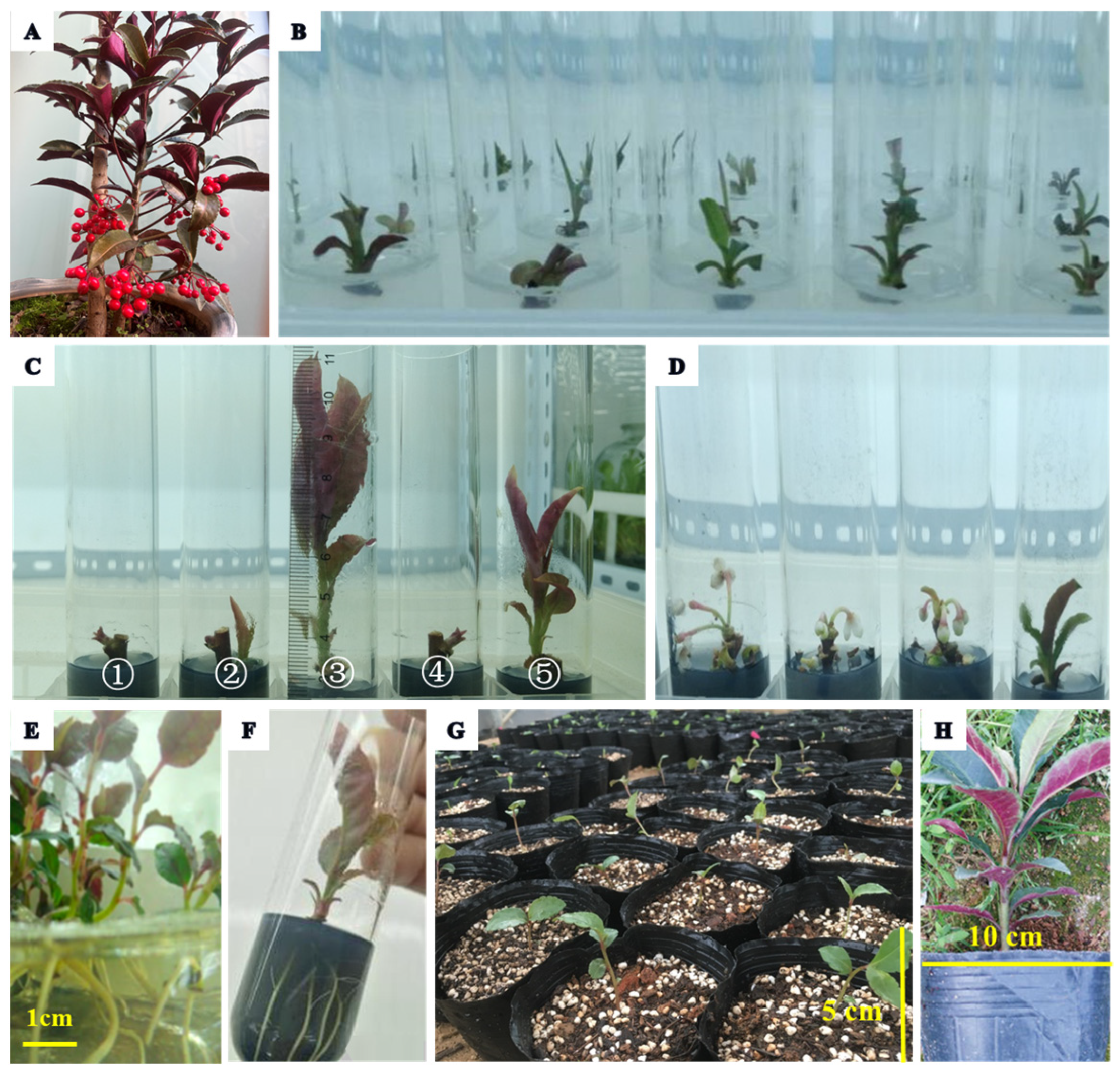
3.1. Shoot Establishment and Proliferation

One of the most important groups of PGRs is the cytokinins, which play a decisive role in shoot regeneration and multiplication [28]. Among them, BAP has been considered to be more favorable and provide a higher multiplication rate than other cytokinins [29], as well as being more easily metabolized in plant tissue. Aakriti et al. [30] also proved that BAP alone has the best bud induction, and for this reason it is widely used for in vitro culturing [14,31]. Jogam et al. [22] showed that BAP alone on MS medium could induce the direct regeneration of shoots from nodal meristems of Artemisia vulgaris. Based on all the experiments, the lowest establishment rate (11.67%) and the longest induction time (four weeks post-culturing) were detected in the hormone-free conditions, while the MS medium with IBA alone showed a low induction, and there were no significant differences among different treatments (p < 0.05) (Table 2). In addition, although an increase in BAP concentration on the MS medium without IBA caused a positive effect on the establishment of explants of A. crenata var. bicolor, the induction rate was still low. Therefore, the sterilized nodal segments of A. crenata var. bicolor started to respond fourteen days post-culturing on the basal MS medium with different concentrations of BAP and IBA, and the direct organogenesis occurred without callus formation on the epidermal layer of the explants and showed no morphogenic variation (Table 2, Figure 1B). The best performance in terms of the explants, with 76.67% establishment rate and 6.6 cm shoot length, was achieved on the MS medium supplemented with 1.0 mg·L−1 BAP and 0.5 mg·L−1 IBA (Figure 1C➂); the highest mean shoot number was 4.5, where BAP was at 1.0 mg·L−1 with 0.25 mg‧L−1 IBA (Figure 1C➄).
In short, a decrease in the establishment rate and few shoots were observed with lower concentrations of BAP and IBA. Higher concentrations of BAP and IBA, on the contrary, induced an earlier budding response and even the blooming of buds or albino leaves, but not necrosis (Figure 1D). In contrast, due to the combined effect, intermediate concentrations of BAP and IBA were able to obtain the highest numbers of shoots. The results were consistent with Francesca et al. [32] that the explants achieved the best growth on MS medium supplemented with BAP and IBA. Similarly, the maximum proliferation percent and the highest shoot number with the length of Petunia hybrida Vilm. Cv. “Bravo” were obtained in the PGR combination with both 0.5 mg·L−1 IBA and BAP [33]. Therefore, the combined application of BAP and IBA or IAA showed a better effect on the shoot regeneration of most plants than their individual use, and in combination they could significantly improve the maximum number of shoots per explant and shoot length [22,34,35]. The results were consistent with previous works that have argued for the crucial role of an optimal BAP and IBA ratio for shoot induction and proliferation.

3.2. Root Induction

The quality of root induction is significantly influenced by the medium type and strength and the concentration of auxins (IBA, IAA, and NAA) [36]. Aakriti et al. [30] revealed that full- or half-strength MS media without any PGRs failed to induce a rooting reaction. However, lower rooting percentages (8.33% and 11.67%, respectively) occurred in the hormone-free conditions in this study, which may be related to the endogenous level of auxins in the tissue surface. Yang et al. [37] suggested that supplementing with NAA alone on MS medium was found to be more optimal for inducing rhizogenesis than IBA, particularly in terms of root number and root length. However, Nongdam and Tikendra [38] found that IBA is more effective for rooting formation as compared to NAA; the result was consistent with Jung et al. [39] that a higher rooting rate was observed on half-strength MS. In this study, although the individual addition of IBA or NAA could also induce the root formation in A. crenata var. bicolor explants, the results were not satisfactory for rooting. Therefore, the robust in vitro shoots of A. crenata var. bicolor were shifted for root induction onto full- or half-strength MS basal media containing different concentrations of IBA and NAA. The results showed a significant difference in the rooting percentage and root numbers among the treatment groups (Table 3). With the increase in IBA levels (up to 1.0 mg‧L−1), the frequency of root induction was enhanced; in particular, the highest rooting percentages (71.67% and 83.33%, respectively) and root numbers (4.2 and 4.5, respectively) were reached in full- and half-strength MS medium supplemented with 0.25 mg‧L−1 NAA, but this gradually decreased with the increased concentration of NAA (Figure 1E,F). Sunil et al. [40] observed the maximum response on the rooting medium of Cicer arietinum supplemented with 0.10 and 0.50 mg‧L−1 of IBA and IAA. Comparatively, the combined application of IBA and NAA showed a better effect on root formation than their individual use and was more effective in a half-strength MS medium.

3.3. Acclimatization

A common problem of propagation in vitro is how to adapt to external conditions, which reduce the mortality of vulnerable plantlets caused by fungal and bacterial contamination, which is a major challenge for in vitro propagation [41]. In this study, all the regenerated plantlets were kept inside a greenhouse to adapt to an open environment for one month, and then the well-developed complete plantlets were removed from the culture medium and treated with 1.0 g·L−1 antifungal agent, then transferred to the nursery black poly bags containing the autoclaved peat, perlite, and vermiculite mixture (2:1:1, v:v:v). In addition, a different shading net and gradually increasing light irradiance could increase the survival rate of plantlets [42]. In this study, the plantlets were then placed under 75% shading, 50% shading, and natural light irradiance (0%) conditions for acclimatization (Table 4, Figure 1G,H) and continually kept at 25 ± 2 °C and 80–90% relative humidity under greenhouse conditions. Plantlets kept for a month under 75% shading achieved the highest survival rate of 84.4%, followed by 50% shading with 63.3% survival. The survival rate dropped to 20.0% when plantlets were kept under natural light irradiance for a month, indicating that the degree of shading drastically affected the survival rate of the acclimatized A. crenata var. bicolor plantlets in greenhouse conditions.

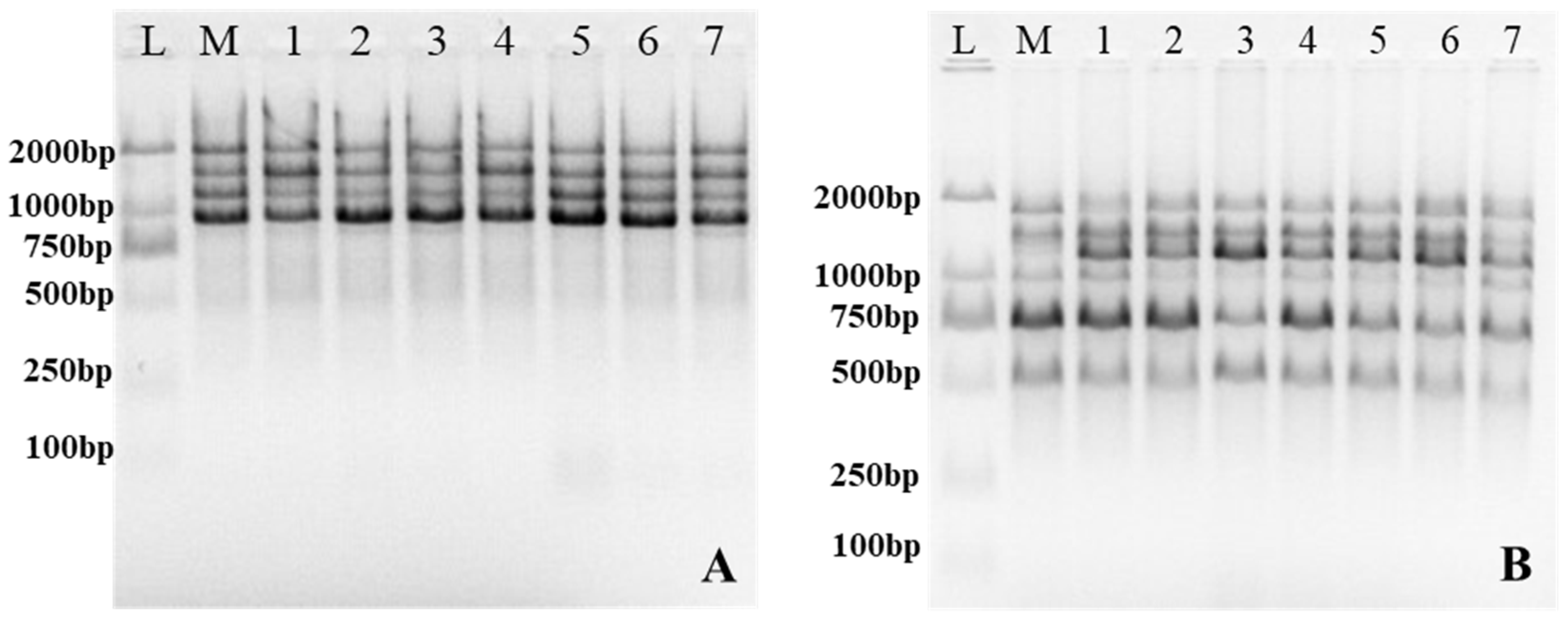
3.4. Assessment of Genetic Stability by ISSR

During the cultivation of A. crenata var. bicolor plantlets, no other morphological variations were observed except for some green and purple changes on the back of the leaves and stems. Based on the morphological changes, we further confirmed the genetic stability between typical plantlets and their mother plant; a comparison of seven A. crenata var. bicolor plantlets and their mother plant was carried out using a set of eleven screened ISSR primers. The number of amplified fragments is shown in Table 1. A total of eighty-three bands of clear and reproducible amplification products was produced, with an average of 7.5 bands per ISSR primer, seventy-five of which were monomorphic (90.36%), while the remaining eight bands were polymorphic (9.64%). The results indicated a level of genetic polymorphism of in-vitro-cultured plants (Figure 2, Supplementary Figure S1). Previous reports have also verified some degree of genetic instability of plantlets under long-term in vitro culture [43], which also increased with successive subcultures [44]. Therefore, we need to combine a level of genetic polymorphism with field planting and further assess the morphological variations [37]. In addition, the morphological performance of plantlets is affected by in vitro culture conditions such as light intensity and light quality [45,46]. In this study, all the leaf colors of the A. crenata var. bicolor plantlets gradually deepened to purplish red with the enhancement of natural light irradiance after acclimatization. They maintained the same traits as their mother plants, which may be related to the light intensity and light quality. The results of this study demonstrate the possibilities of nodal segments for producing genetically stable plants, and the high genetic stability which was demonstrated has great significance for the expansion and breeding of A. crenata var. bicolor.

4. Conclusions

Our findings show that the MS medium supplemented with PGRs provided better conditions for establishment rate and shoot proliferation. The combined application of IBA and BAP had better influence on shoot induction and increasing shoot growth of A. crenata var. bicolor explants than their individual use, and it was found that the higher the concentrations of BAP and IBA used, the earlier the budding response. Moreover, rooting could be induced, regardless of the medium strength, the auxin type (IBA, NAA) and concentration, or whether the explants were transferred onto a hormone-free medium. The incorporation of IBA and NAA into a half-strength MS medium more effectively promoted root formation, which gave significantly better results than incorporation into a full-strength medium. In this study, although there were some changes in the leaf color of the A. crenata var. bicolor plantlets, ISSR-marker-based genetic homogeneity results revealed a level of genetic polymorphism in plantlets, but the plantlets maintained the same morphological traits as their mother plants during development. Such relative higher genetic stability plays a positive role in expanding the reproduction coefficient and improving the germplasm quality of A. crenata var. bicolor.

Supplementary Materials

The following supporting information can be downloaded at: https://www.mdpi.com/article/10.3390/ijpb14010018/s1, Figure S1: ISSR profiles generated by PCR amplification.

Author Contributions

X.A., Conceptualization, Data curation, Formal analysis, Methodology, Writing—original draft; Y.W., Methodology, Formal analysis, Visualization; B.W., Formal analysis, Project administration, Resources, Supervision. All authors have read and agreed to the published version of the manuscript.

Funding

Partial financial support was received by the Doctoral Research Fund Project of Southwest Forestry University (CN) (Grant number 111806) and the National Natural Science Foundation of China (NO. 31960069).

Institutional Review Board Statement

Not applicable.

Informed Consent Statement

Not applicable.

Data Availability Statement

Not applicable.

Conflicts of Interest

The authors declare no conflict of interest.

References

  1. Chen, C.; Pipoly, J. Flora of China (Myrsinaceae through Loganiaceae); Science Press: Beijing, China; Missouri Botanical Garden Press: St. Louis, MO, USA, 1996; Volume 15, pp. 1–38. [Google Scholar]
  2. Podolak, I.; Mynarski, A.; Wróbel, D.; Grabowska, K.; Galanty, A. Bioactive benzoquinones content variability in red-berry and white-berry varieties of Ardisia crenata Sims. and assessment of cytotoxic activity. Nat. Prod. Res. 2019, 35, 157–161. [Google Scholar] [CrossRef] [PubMed]
  3. Hideka, K.; Elvira, D.M. The genus Ardisia: A novel source of health-promoting compounds and phytopharmaceuticals. J. Ethnopharmacol. 2005, 96, 347–354. [Google Scholar] [CrossRef]
  4. Ma, L.; Li, W.; Wang, H.; Kuang, X.; Li, Q.; Wang, Y.; Xie, P.; Koike, K. A simple and rapid method to identify and quantitatively analyze triterpenoid saponins in Ardisia crenata using ultrafast liquid chromatography coupled with electrospray ionization quadrupole mass spectrometry. J. Pharmaceut. Biomed. 2015, 102, 400–408. [Google Scholar] [CrossRef]
  5. Liu, D.L.; Zhang, X.; Zhao, Y.; Wang, N.; Yao, X. Three new triterpenoid saponins from the roots of Ardisia crenata and their cytotoxic activities. Nat. Pro. Res. 2016, 30, 2694–2703. [Google Scholar] [CrossRef] [PubMed]
  6. Raina, H.; Soni, G.; Jauhari, N.; Sharma, N.; Bharadvaja, N. Phytochemical importance of medicinal plants as potential sources of anticancer agents. Turk. J. Bot. 2014, 38, 1027–1035. [Google Scholar] [CrossRef]
  7. Roh, M.S.; Lee, A.K.; Suh, J.K. Production of high quality Ardisia plants by stem tip cuttings. Sci. Hortic. 2005, 104, 293–303. [Google Scholar] [CrossRef]
  8. Yang, Q.H.; Ye, W.H.; Wang, Z.M.; Yin, X.J. Seed germination physiology of Ardisia crenata var. bicolor. Seed Sci. Technol. 2009, 37, 291–302. [Google Scholar] [CrossRef]
  9. Lee, A.K.; Slovin, J.P.; Suh, J.K. Dehydration intolerant seeds of Ardisia species accumulate storage and stress proteins during development. Hortic. Environ. Biote. 2012, 53, 530–538. [Google Scholar] [CrossRef]
  10. Tezuka, T.; Yokoyama, H.; Tanaka, H.; Shiozaki, S.; Oda, M. Seed and Embryo Germination in Ardisia crenata. J. Bot. 2012, 2012, 679765. [Google Scholar] [CrossRef] [Green Version]
  11. Rohela, G.K.; Jogam, P.; Shabnam, A.A.; Shukla, P.; Abbagani, S.; Ghosh, M.K. In Vitro regeneration and assessment of genetic fidelity of acclimated plantlets by using ISSR markers in PPR-1 (Morus sp.): An economically important plant. Sci. Hortic. 2018, 241, 313–321. [Google Scholar] [CrossRef]
  12. Lin, J.; Zou, J.; Zhang, B.; Que, Q.; Zhang, J.; Chen, X. An efficient in vitro propagation protocol for direct or ganogenesis from root explants of a multi-purpose plant, Broussonetia papyrifera (L.) L’Hér. ex Vent. Ind. Crop. Prod. 2021, 170, 113686. [Google Scholar] [CrossRef]
  13. Cui, Y.; Deng, Y.; Zheng, K.; Hu, X.; Zhu, M.; Deng, X.; Xi, R. An efficient micropropagation protocol for an endangered ornamental tree species (Magnolia sirindhorniae Noot. & Chalermglin) and assessment of genetic uniformity through DNA markers. Sci. Rep. 2019, 9, 9634. [Google Scholar] [CrossRef] [PubMed] [Green Version]
  14. Demétrio, C.A.; Jacob, J.F.d.O.; Ambrosano, G.B.; Oliveira, Ê.T.D.; Rodrigues, P.H.V. In Vitro propagation of Cambuci (Campomanesia phaea): An endangered exotic fruit and ornamental plant from Brazilian Atlantic Forest. Plant Cell Tiss. Org. 2021, 145, 203–208. [Google Scholar] [CrossRef]
  15. Thakur, M.; Sharma, V.; Luharch, R. Propagation of plum (Prunus salicina L.) cultivar Frontier in vitro through control of shoot tip necrosis (STN) and validation of genetic integrity using ISSR markers. Plant Physiol. Rep. 2021, 26, 238–246. [Google Scholar] [CrossRef]
  16. Sathish, D.; Vasudevan, V.; Theboral, J.; Elayaraja, D.; Appunu, C.; Siva, R.; Manickavasagam, M. Efficient direct plant regeneration from immature leaf roll explants of sugarcane (Saccharum officinarum L.) using polyamines and assessment of genetic fidelity by SCoT markers. In Vitro Cell Dev. Biol.-Plant 2018, 54, 399–412. [Google Scholar] [CrossRef]
  17. Bednarek, P.T.; Orłowska, R. Plant tissue culture environment as a switch-key of (epi) genetic changes. Plant Cell Tiss. Org. 2020, 140, 245–257. [Google Scholar] [CrossRef] [Green Version]
  18. Choudhary, D.; Rai, M.K.; Shekhawat, N.S.; Kataria, V. In vitro propagation of Farsetia macrantha Blatt. & Hallb.: An endemic and threatened plant of Indian Thar Desert. Plant Cell Tiss. Org. 2020, 142, 519–526. [Google Scholar] [CrossRef]
  19. Bhatia, R.; Singh, K.P.; Sharma, T.R.; Jhang, T. Evaluation of the genetic fidelity of in vitro-propagated gerbera (Gerbera jamesonii Bolus) using DNA-based markers. Plant Cell Tiss. Org. 2011, 104, 131–135. [Google Scholar] [CrossRef]
  20. Saleh, A.; Reza, M. Establishment of an efficient in vitro propagation protocol for Sumac (Rhus coriaria L.) and confirmation of the genetic homogeneity. Sci. Rep. 2021, 11, 173. [Google Scholar] [CrossRef]
  21. Faisal, M.; Alatar, A.A.; El-Sheikh, M.A.; Abdel-Salam, E.M.; Qahtan, A.A. Thidiazuron induced in vitro morphogenesis for sustainable supply of genetically true quality plantlets of Brahmi. Ind. Crop. Prod 2018, 118, 173–179. [Google Scholar] [CrossRef]
  22. Jogam, P.; Sandhya, D.; Shekhawat, M.S.; Alok, A.; Abbagani, S.; Allini, V.R. Genetic stability analysis using DNA barcoding and molecular markers and foliar micro-morphological analysis of in vitro regenerated and in vivo grown plants of Artemisia vulgaris L. Ind. Crop. Prod. 2020, 151, 112476. [Google Scholar] [CrossRef]
  23. Thakur, M.; Sharma, V.; Chauhan, A. Genetic fidelity assessment of long term in vitro shoot cultures and regenerated plants in Japanese plum cvs Santa Rosa and Frontier through RAPD, ISSR and SCoT markers. S. Afr. J. Bot. 2021, 140, 428–433. [Google Scholar] [CrossRef]
  24. Rajendra, P.A.; Bee, L.C.; Sreeramanan, S. Assessment of genetic stability on in vitro and ex vitro plants of Ficus carica var. black jack using ISSR and DAMD markers. Mol. Biol. Rep. 2021, 48, 7223–7231. [Google Scholar] [CrossRef]
  25. Kostas, S.; Hatzilazarou, S.; Pipinis, E.; Vasileiadis, A.; Magklaras, P.; Smyrnioudis, I.; Vasilakis, T.; Chazakis, M.; Anastasiadi, V.; Ziogou, F.; et al. Propagation of Pistacia lentiscus var. Chia genotypes and determination of their ornamental traits combined with a genetic analysis using ISSR Markers. Agronomy 2021, 11, 205. [Google Scholar] [CrossRef]
  26. Murashige, T.; Skoog, F. A Revised Medium for Rapid Growth and Bio Assays with Tobacco Tissue Cultures. Physiol. Plantarum 1962, 15, 473–479. [Google Scholar] [CrossRef]
  27. Doyle, J.J. A rapid DNA isolation procedure for small quantities of fresh leaf tissue. Phytochem. Bull. 1987, 19, 11–15. [Google Scholar]
  28. Aremu, A.O.; Bairu, M.W.; Dolezal, K.; Finnie, J.F.; Staden, J.V. Topolins: A panacea to plant tissue culture challenges? Plant Cell Tiss. Org. 2012, 108, 1–16. [Google Scholar] [CrossRef]
  29. Alexander, T.J.; Esmeralda, R.; GómezAldapa, C.A.; VillagómezIbarra, J.R.; FalfánCortes, R.N.; AcevedoSandoval, O.A.; CastroRosas, J. Isolation and molecular identification of Serratia strains producing Chitinases, Glucanases, Cellulases, and Prodigiosin and determination of their antifungal effect against Colletotrichum Siamense and Alternaria Alternata in vitro and on Mango fruit. Int. J. Plant Biol. 2022, 13, 281–297. [Google Scholar] [CrossRef]
  30. Aakriti, B.; Harminder, S.; Amber, S.; Puneet, K. In vitro propagation and cytological analysis of Sophora mollis Royle: An endangered medicinal shrub. J. Genet. Eng. Biotechn. 2021, 19, 40. [Google Scholar] [CrossRef]
  31. Agarwal, T.; Gupta, A.K.; Patel, A.K.; Shekhawat, N.S. Micropropagation and validation of genetic homogeneity of Alhagi maurorum using SCoT, ISSR and RAPD markers. Plant Cell Tiss. Org. 2015, 120, 313–323. [Google Scholar] [CrossRef]
  32. Francesca, G.; Loreto, B.; Ricardo, C.; Mónica, C. Micropropagation of Citronella mucronata D. Don, a Vulnerable Chilean Endemic Tree Species. Plants 2022, 11, 2425. [Google Scholar] [CrossRef]
  33. Farooq, I.; Qadri, Z.A.; Rather, Z.A.; Nazki, I.T.; Banday, N.; Rafiq, S.; Masoodi, K.Z.; Noureldeen, A.; Mansoor, S. Optimization of an improved, efficient and rapid in vitro micropropagation protocol for Petunia hybrida Vilm. Cv. “Bravo”. Saudi. J. Biol. Sci. 2021, 28, 3701–3709. [Google Scholar] [CrossRef] [PubMed]
  34. Khatun, F.; Hoque, M.E.; Huq, H.; Adil, M.; Ashraf-Uz-Zaman, K.; Rabin, M.H. Effect of BAP and IBA on in vitro regeneration of local banana Variety of Sabri. Biotechnol. J. Inter. 2017, 18, 1–10. [Google Scholar] [CrossRef]
  35. Dimitrova, A.M.; Yordanova, P.Z.; Kapchina-Toteva, M.V. Influence of 6-benzylaminopurine and indole-3-butyric acid on in vitro propagation and secondary metabolites accumulation in Lamium album from lozen mountain. Acta Hortic. 2012, 955, 315–320. [Google Scholar] [CrossRef]
  36. Khajuria, A.K.; Bisht, N.; Bhagat, N. In vitro organogenesis and plant regeneration of Thymus serpyllum L.: An important aromatic medicinal plant. In Vitro Cell Dev. Biol. -Plant 2020, 56, 652–661. [Google Scholar] [CrossRef]
  37. Yang, L.; Chao, L.; Su, X.; Wang, C.; Dong, C.; Chen, S. High-frequency in vitro plantlet regeneration in Lilium davidii var. unicolour Salisb, an important edible and medicinal plant, and confirmation of genetic fidelity of regeneration plantlets using ISSR markers. Plant Biotechnol. Rep. 2021, 15, 435–446. [Google Scholar] [CrossRef]
  38. Nongdam, P.; Tikendra, L. Establishment of an efficient in vitro regeneration protocol for rapid and mass propagation of Dendrobium chrysotoxum lindl. Using seed culture. Sci. World J. 2014, 2014, 740150. [Google Scholar] [CrossRef] [Green Version]
  39. Jung, W.; IllMin, C.; SeungHyun, K.; HeeYeon, C.; Yeon, Y.C.; Kumar, G.B. Direct Shoot Organogenesis from Lycium chinense Miller Leaf Explants and Assessment of Genetic Stability Using ISSR Markers. Agronomy 2021, 11, 503. [Google Scholar] [CrossRef]
  40. Sunil, S.; Robinson, J.; KarthickBalan, S.; Anandhaprabhakaran, M.; Balakrishnan, V. In vitro regeneration and induction of multiple shooting in Cicer arietinum L. using cotyledonary nodal explants. Afr. J. Biotechnol. 2015, 14, 1129–1138. [Google Scholar] [CrossRef]
  41. Mbah, E.I.; Wakil, S.M. Elimination of bacteria from in vitro yam tissue cultures using antibiotics. J. Plant Pathol. 2012, 94, 53–58. [Google Scholar]
  42. Hatzilazarou, S.; Kostas, S.; Joachim, M.; Economou, A. Regeneration of Viburnum dentatum L. from alginate-encapsulated shoot explants after short-term cold storage and assessment of genetic stability using ISSR analysis. Agronomy 2020, 10, 1660. [Google Scholar] [CrossRef]
  43. Samarina, L.; Gvasaliya, M.; Koninskaya, N.; Rakhmangulov, R.; Efremov, A.; Kiselyova, N.; Ryndin, A.; Hanke, M.-V. A comparison of genetic stability in tea [Camellia sinensis (L.) Kuntze] plantlets derived from callus with plantlets from long-term in vitro propagation. Plant Cell. Tiss. Org. 2019, 138, 467–474. [Google Scholar] [CrossRef]
  44. Soares, D.M.M.; Sattler, M.C.; Ferreira, M.F.D.S.; Praça-Fontes, M.M. Assessment of genetic stability in three generations of in vitro propagated Jatropha Curcas, L. plantlets using ISSR markers. Trop. Plant Biol. 2016, 9, 229–238. [Google Scholar] [CrossRef]
  45. Manivannan, A.; Soundararajan, P.; Park, Y.G.; Wei, H.; Kim, S.H.; Jeong, B.R. Blue and red light-emitting diodes improve the growth and physiology of in vitro-grown carnations ‘Green Beauty’ and ‘Purple Beauty’. Hortic. Environ. Biote. 2017, 58, 12–20. [Google Scholar] [CrossRef]
  46. Chung, G.J.; Lee, J.H.; Oh, M.M. Growth and acclimation of in vitro-propagated ‘M9’ apple rootstock plantlets according to light intensity. Hortic. Environ. Biote. 2020, 61, 501–510. [Google Scholar] [CrossRef]
Figure 1. In Vitro propagation via organogenesis of A. crenata var. bicolor. (A) Plant of A. crenata var. bicolor; (B) Shoot induction; (C) Different PGR combinations of BAP and IBA: ➀ 0.0 mg·L−1 BAP + 1.00 mg·L−1 IBA; ➁ 0.5 mg·L−1 BAP + 1.00 mg·L−1 IBA; ➂ 1.0 mg·L−1 BAP + 0.50 mg·L−1 IBA; ➃ 1.5 mg·L−1 BAP + 1.00 mg·L−1 IBA; ➄ 1.0 mg·L−1 BAP + 0.25 mg·L−1 IBA; (D) Abnormal flower buds’ differentiation; (E,F) Root induction with different concentrations of IBA and NAA; (G) Regenerated plantlets, transferred to nursery black poly bags and covered with sunshade net; and (H) Transplantation for six months from the end of the acclimatization period.
Ijpb 14 00018 g001
Figure 2. ISSR profiles generated by PCR amplification. Lane L: Molecular marker (2000 bp); lane M: Mother plant; lane 1–7: In-vitro-raised plants. (A) PCR-amplified banding pattern obtained with ISSR-1 primer. (B) ISSR-4.
Ijpb 14 00018 g002
Table 1. Details of ISSR primers used in this work.
NO.Primer CodePrimer Sequence (5′–3′)Annealing
Temperature (°C)
Total BandsPolymorphic BandsPercentage of Polymorphic Bands (%)
1ISSR-1(AG)8T504000.00
2ISSR-2(AG)8C517000.00
3ISSR-3(GA)8C528112.50
4ISSR-4(GA)8A49.5800.00
5ISSR-5(CA)8G497114.28
6ISSR-6(AC)8G51.5900.00
7ISSR-7(TG)8C49.57228.57
8ISSR-8(AG)8YT5012216.67
9ISSR-9(GA)8YC52700.00
10ISSR-10(AC)8YG507114.28
11ISSR-11BDB(CA)7537114.28
Table 2. Effect of BAP and IBA combinations on in vitro propagation of A. crenata var. bicolor explants.
PGRs (mg·L−1)Nodal Segment
BAPIBAEstablishment Rates (%) Mean ± SENo. of Shoots/Explant Mean ± SEShoot Length (cm) Mean ± SE
0.00.0011.67 ± 0.01 d1.0 ± 0.58 e1.4 ± 0.10 h
0.2513.33 ± 0.03 d1.1 ± 0.00 e2.0 ± 0.13 g
0.5013.33 ± 0.05 d1.3 ± 1.53 e2.1 ± 0.13 g
1.0015.00 ± 0.08 d1.4 ± 1.53 e2.2 ± 0.25 g
0.50.0018.33 ± 0.08 d1.5 ± 2.08 e2.3 ± 0.09 g
0.2530.00 ± 0.06 cd1.6 ± 2.08 e2.5 ± 0.05 g
0.5041.67 ± 0.08 bc2.2 ± 6.11 d3.0 ± 0.23 f
1.0048.33 ± 0.06 b2.7 ± 1.53 bc3.4 ± 0.21 ef
1.00.0026.67 ± 0.03 cd1.6 ± 2.52 e2.1 ± 0.16 g
0.2560.00 ± 0.03 a4.5 ± 1.53 a4.4 ± 0.24 cd
0.5076.67 ± 0.02 a2.9 ± 2.52 b6.6 ± 0.20 a
1.0053.33 ± 0.04 ab2.8 ± 2.00 bc5.8 ± 0.09 b
1.50.0035.00 ± 0.05 c1.3 ± 0.58 e2.5 ± 0.13 g
0.2556.67 ± 0.06 ab2.5 ± 1.53 c3.7 ± 0.15 de
0.5065.00 ± 0.03 a3.0 ± 3.51 b4.8 ± 0.13 c
1.0053.33 ± 0.02 ab2.2 ± 1.53 d4.0 ± 0.16 d
Different lowercase letters in each column indicate significant difference by Duncan’s multiple range tests at p < 0.05.
Table 3. Effects of different IBA and NAA combinations on root induction of A. crenata var. bicolor explants.
PGRs (mg·L−1)Full-Strength MSHalf-Strength MS
IBANAAPercentage of Rooting (%) Mean ± SERoot Number Mean ± SEPercentage of Rooting (%) Mean ± SERoot Number Mean ± SE
00.008.33 ± 0.03 e1.0 ± 0.00 c11.67 ± 0.08 d1.3 ± 0.58 d
0.2518.33 ± 0.18 d1.3 ± 0.16 c30.00 ± 0.30 c1.4 ± 0.22 d
0.5020.00 ± 0.20 d1.5 ± 0.41 c33.33 ± 0.33 c2.0 ± 0.42 cd
0.250.0021.67 ± 0.22 d1.1 ± 0.27 c30.00 ± 0.30 c1.3 ± 0.39 d
0.2543.33 ± 0.43 b1.7 ± 0.44 c55.00 ± 0.55 c2.0 ± 0.05 cd
0.5055.00 ± 0.55 ab2.2 ± 0.47 b65.00 ± 0.65 b2.0 ± 0.04 cd
0.50.0026.67 ± 0.27 cd1.2 ± 0.05 c31.67 ± 0.32 c1.5 ± 0.42 d
0.2556.67 ± 0.57 ab3.7 ± 0.58 a63.33 ± 0.63 b4.0 ± 0.27 ab
0.5063.33 ± 0.63 ab3.3 ± 0.43 ab65.00 ± 0.65 b3.1 ± 0.68 bc
1.00.0033.33 ± 0.33 c1.3 ± 0.30 c36.67 ± 0.37 c1.6 ± 0.19 d
0.2571.67 ± 0.72 a4.2 ± 0.55 a83.33 ± 0.83 a4.5 ± 0.50 a
0.5068.33 ± 0.68 ab3.4 ± 0.64 ab70.00 ± 0.70 b3.2 ± 0.88 b
Different lower-case letters in each column indicate significant difference by Duncan’s multiple range test at p < 0.05.
Table 4. Influence of shading treatments during acclimatization of in-vitro-produced plantlets of A. crenata var. bicolor.
TreatmentSubstrate Mixture
Peat:Vermiculite:Perlite
ShadingSurvival Rate (%)
2:1:175%84.4 ± 0.13 a
2:1:150%63.3 ± 0.15 b
2:1:10%20.0 ± 0.03 c
Different letters in each column indicate significant difference by Duncan’s multiple range test at p < 0.05.
Disclaimer/Publisher’s Note: The statements, opinions and data contained in all publications are solely those of the individual author(s) and contributor(s) and not of MDPI and/or the editor(s). MDPI and/or the editor(s) disclaim responsibility for any injury to people or property resulting from any ideas, methods, instructions or products referred to in the content.

