Article

Pharmacological Effects of Lactobacillus casei ATCC 7469 Fermented Soybean and Green Microalgae, Chlorella vulgaris, on Diabetic Rats

by
Asmaa Abdella
1,*,
Fatma I. Abou-Elazm
2 and
Shaymaa Wagdy El-Far
3
1
Department of Industrial Biotechnology, Genetic Engineering and Biotechnology Research Institute, University of Sadat City, Sadat City 32897, Egypt
2
Department of Microbiology and Immunology, Faculty of Pharmacy, Misr University for Science and Technology, Giza 32361, Egypt
3
Division of Pharmaceutical Microbiology, Department of Pharmaceutics and Industrial Pharmacy, College of Pharmacy, Taif University, P.O. Box 11099, Taif 21944, Saudi Arabia
*
Author to whom correspondence should be addressed.
Microbiol. Res. 2023, 14(2), 614-626; https://doi.org/10.3390/microbiolres14020043
Submission received: 18 April 2023 / Revised: 23 April 2023 / Accepted: 26 April 2023 / Published: 30 April 2023

Abstract

:
Type 2 diabetes mellitus (T2DM) is a complex, multifactorial metabolic disorder mainly characterized by chronic hyperglycemia. It has become a significant, serious disease worldwide, presenting a huge risk to human life and health. This study aimed to investigate the hypoglycemic effect of Lactobacillus casei ATCC 7469 fermented soy flour extract and Chlorella vulgaris extract on Sprague-Dawley rats with T2DM induced by low-dose streptozotocin administration (STZ) compared to pioglitazone as a reference drug. Treatment with Lactobacillus casei ATCC 7469 fermented soy flour and Chlorella vulgaris resulted in a significant improvement in body weight, glucose tolerance, blood glucose level, and insulin resistance (p < 0.05). It also resulted in a significant decrease in total cholesterol (T.C), triglycerides (T.G), low-density lipoprotein (LDL), and very low-density lipoprotein (VLDL) (p < 0.05) and a significant increase in high-density lipoprotein (HDL) (p < 0.05). It also resulted in the improvement of liver oxidative biomarkers. Moreover, it prevented pancreatic histopathological changes. Lactobacillus casei ATCC 7469 fermented soy flour extract and Chlorella vulgaris extract had hypoglycemic, hypolipidemic, and antioxidant activity similar to pioglitazone.

1. Introduction

T2DM is a complicated multifactorial metabolic disease characterized primarily by hyperglycemia caused by insulin resistance and insufficient insulin production caused by pancreatic beta-cell malfunction [1]. Complications of T2DM include cardiovascular disease, diabetic neuropathy, nephropathy, and retinopathy [2]. There are many drugs for the treatment of T2DM, such as sulfonylureas, thiazolidinediones, biguanides, glinides, meglitinide, sodium-glucose cotransporter (SGLT2) inhibitors, and α-glucosidase inhibitors, and dipeptidyl peptidase IV (DPP-4) inhibitors, which inhibits glucagon secretion such as vildagliptin [3]. There are many side effects related to antidiabetic drugs, such as hypoglycemia, weight gain, nausea, skin reactions caused by sulfonylurea treatment, and heart failure caused by thiazolidinediones treatment [4,5].
Soybean contains many active constituents, such as complex carbohydrates, proteins, and dietary fiber [6]. Complex carbohydrates and dietary fiber contribute to low glycemic indices, which are beneficial to patients with T2DM [7]. The main constituents of soybeans are isoflavones, which are extremely beneficial to human health. Isoflavones are classified into four classes (malonyl glucoside, acetyl glucoside, glucoside, and aglycone) and three kinds (daidzein, genistein, and glycitein). Beneficial effects include anti-cancer, anti-arteriosclerosis, anti-inflammatory, antioxidant, estrogenic characteristics, anti-allergenic properties, and lung disease relief [8]. Soy-based products have been shown to be useful in the treatment of T2DM and hyperlipidemia [9]. The biofunctional characteristics of soybean are enhanced by microbial fermentation as a result of the rise in free isoflavones and peptides [10]. According to Kim et al. [11], fermented soybean has hypolipidemic, hypoglycemic, and anti-inflammatory properties. The surface polysaccharides and short-chain fatty acid metabolites of probiotics may have the ability to lower blood sugar levels, stimulate the immune system, lower cholesterol levels, avoid respiratory and intestinal diseases, and reduce inflammatory responses [12]. Long et al. [13] stated that soybeans fermented with probiotics have hypoglycemic benefits in T2DM patients. Yu et al. [14] found that fermented soybean can help with glucose metabolic disorders by inhibiting digesting enzymes, facilitating glucose transporter 4 translocation, increasing muscular glucose utilization, reducing hepatic gluconeogenesis, and improving pancreatic dysfunction. Hu et al. [15] found that Lactobacillus fermentum HFY02 fermented soybean milk can increase antioxidant factor content and antioxidant enzyme activity.
Chlorella vulgaris is a spherical, unicellular eukaryotic microorganism [16]. Chlorella vulgaris is high in protein, lipid-soluble vitamins, choline, and minerals [17]. Chlorella is a promising functional food because of its high level of nutrients, such as vitamins, polyunsaturated fatty acids, minerals, dietary fiber, and protein [18]. According to Lee et al. [19], Chlorella vulgaris exerts a hypolipidemic impact in Wistar rats on a high-fat diet. According to Yuh et al. [20], chlorella increased and sustained the hypoglycemic effects of administered insulin in streptozocin (STZ)-induced diabetic mice. Chlorella could be acting either by increasing glucose uptake without stimulating insulin secretion or suppressing hepatic gluconeogenesis and glycogenolysis [20].
To the best of our knowledge, it is the first study to evaluate the additive hypolipidemic, hypoglycemic, and antioxidant effects of a combination of Lactobacillus casei ATCC 7469 fermented soy flour extract and Chlorella vulgaris extract on rats with streptozotocin-induced diabetic rats.

2. Materials and Methods

2.1. Bacterial Strains

Lactobacillus casei ATCC 7469 was obtained from the American Type Culture Collection (Manassas, VA, USA). It was cultured in MRS broth (Sigma-Aldrich, Cairo, Egypt) at 37 °C for 24 h. After cultivation, the cells were harvested by centrifugation at 3000× g for 20 min (RWD High-Speed Benchtop Refrigerated Centrifuge RWD Life Science Inc., St. Petersburg, FL, USA) and washed with sterile distilled water. The cells were lyophilized (Benchtop Freeze Dryer, LYO60B-1S (Bioevopeak Inc., Chestnut Ridge, New York, NY, USA) and stored at −80 °C.

2.2. Preparation of the Soy Flour Fermentation Extracts

Glycerol-preserved Lactobacillus casei ATCC 7469 was cultured in MRS medium at 37 °C for 8 h. Then, 2% (v/v) Lactobacillus casei was inoculated into the fermentation medium (5 g of defatted soy flour, yeast extract 10, KH2PO4.2H2O 2.0, MgSO4. 7H2O 0.1, MnSO4. 4HfO 0.05, and 50 mL of distilled water) at pH = 7 and incubated at 37 °C for 24 h. Finally, the supernatant was collected by centrifugation (16,099× g, 30 min, 4 °C). The extract was freeze-dried (Benchtop Freeze Dry-er, LYO60B-1S (Bioevopeak Inc.). All chemicals were purchased from Fisher Scientific Egypt.

2.3. Algae Cultivation

Chlorella vulgaris was isolated from water samples collected from the Red Sea, Hurghada, Egypt. The isolation was carried out through a serial dilution technique followed by plating on a modified BG-11 medium. The microalga identification was based on Algae Base Bellinger and Sigee) [21]. They were cultivated on (BG11) media pH 7 at 25 ± 2 °C, with continuous illumination (3600 Lux), and shaken by hand twice daily for 15 days [22]. Cells were collected by centrifugation (RWD High-Speed Benchtop Refrigerated Centrifuge RWD Life Science Co., Ltd. (11,180× g, 20 min, 4 °C) and freeze-dried with a (Benchtop Freeze Dryer, LYO60B-1S (Bioevopeak Inc.),

2.4. Preparation of Chlorella vulgaris Extract

Chlorella vulgaris extract was prepared by grinding algal materials, soaking in 20 folds (w/v) of sterilized deionized water (DW), and vortexing with a magnetic stirrer for 24 h at 45 °C and 1006× g. The algal residues were discarded by filtration, and the clear extract obtained was freeze-dried into powder before use.

2.5. Experimental Design for Rats Feeding with Lactobacillus Casei ATCC 7469 Fermented Soy Flour Extract and Chlorella vulgaris Extract

2.5.1. Animals

Seven-week-old male Sprague–Dawley rats (110 ± 10 g) were obtained from the Faculty of Veterinary Medicine, University of Sadat City, Sadat City, Egypt. All animal handling procedures, sample collection, and disposal were according to the regulations of the Institutional Animal Care and Use Committee (IACUC), Faculty of Veterinary Medicine, University of Sadat City, Egypt, under ethical approval number VUSC-014-1-23. All experiments were performed in accordance with relevant guidelines and regulations. Rats were randomly housed in polypropylene cages at ambient humidity (60 ± 5%), temperature (22 ± 2 °C), and light period of 12:00 to 12:00 and permitted to acclimatize for 7 days before the experiment. During the 7-day acclimation period, the animals were fed diet ad libitum daily.
After 7 days of acclimatization, we randomly selected (n = 10) to form a control group (NC group) and induced diabetes in the other mice (n = 50) by injecting 40 mg/kg STZ intraperitoneally in citrate buffer (pH 4.5), while mice in the control group received buffer alone [23]. The fasting blood glucose (FBG) concentrations of these mice were subsequently measured, and those with concentrations ≥ 7 mmol/L were defined as having diabetes [24]. Diabetic rats were then randomly divided into five groups (n = 10/group). All the groups underwent the following treatments for 7 weeks:
D-G1: Diabetic rats fed a regular diet.
D-G2: Diabetic rats fed a regular diet and administered (10 mg/kg B.W.) of Chlorella vul-garis extract once daily by gavage.
D-G3: Diabetic rats fed a regular diet and administered (1.0 g/kg B.W.) Lactobacillus casei ATCC 7469 fermented soy flour extract once daily by gavage.
D-G4: Diabetic rats fed a regular diet and administered (10 mg/kg B.W.) of Chlorella vul-garis extract and (1.0 g/kg B.W. of Lactobacillus casei ATCC 7469 fermented soy flour extract once daily by gavage.
D-G5: Diabetic rats fed a regular diet and administered (10 mg/kg B.W.) of pioglitazone once daily by gavage.
The regular diet consists of wheat flour 22.5%, soybean powder 25%, essential fatty acids 0.6%, vitamins (A 0.6 mg/kg of diet, D 1000 IU/kg of diet, E 35 mg/kg of diet, niacin 20 mg/kg of diet, riboflavin 0.8 mg/1000 kcal of diet, thiamin 4 mg/kg of diet, B6 50 µg/kg of diet, and B12 7 mg/kg of diet) and minerals (calcium 5 g/kg of diet, phosphorus 4 g/kg of diet, fluoride 1 mg/kg of diet, iodine 0.15 mg/kg of diet, chloride 5 mg/kg of diet, iron 35 mg/kg of diet, copper 5 mg/kg of diet, magnesium 800 mg/kg of diet, potassium 35 mg/kg of diet, manganese 50 mg/kg of diet, and sulfur 3 mg/kg of diet) [25].

2.5.2. Termination of the Experiment

At the end of the 7-week experimental period, rats were fasted overnight, weighed, and sacrificed by decapitation under anesthesia using xylazine HCl (10 mg/kg/BW) and ketamine HCl (50 mg/kg/BW).

2.5.3. Blood Sampling and Biochemical Parameters

Blood was collected using sodium fluoride as an anticoagulant and centrifuged at 1006× g for 30 min. The sera were quickly removed and kept at −20 °C until used for biochemical investigation. Serum triglyceride (T.G.) concentration was determined according to the method of Fossati and Prencipe [26]. Serum total cholesterol (T.C.) concentration was determined according to the method of Deeg and Ziegenohrm [27]. Serum HDL concentration was measured according to the method of Burstein et al. [28]. Serum LDL concentration was determined according to Friedewald et al. [29]. Serum very low-density lipoprotein (VLDL) was determined according to Norbert [30]. T.G., cholesterol, LDL, VLDL, and HDL assay kits were purchased from (Asan and Youngdong Pharmaceutical Co., Seoul, Korea). Fasting blood glucose levels were monitored by collecting blood from the tail vein and analyzing it with an Accu-Check Glucose Analyser ((Roche Group, Indianapolis, IN, USA). Insulin was determined by ELISA kits (Abcam, Waltham, MA, USA). The liver was blotted, weighed, and homogenized with phosphate buffer saline to estimate superoxide dismutase (SOD) and catalase (CAT) enzymes. The activity of hepatic superoxide dismutase (SOD) was measured according to the method of Marklund and Marklund [31]. Liver catalase (CAT) was determined according to the technique of (Cohen et al. [32]. SOD and CAT assay kits were purchased from (Thermo Fisher Scientific, Waltham, MA, USA).

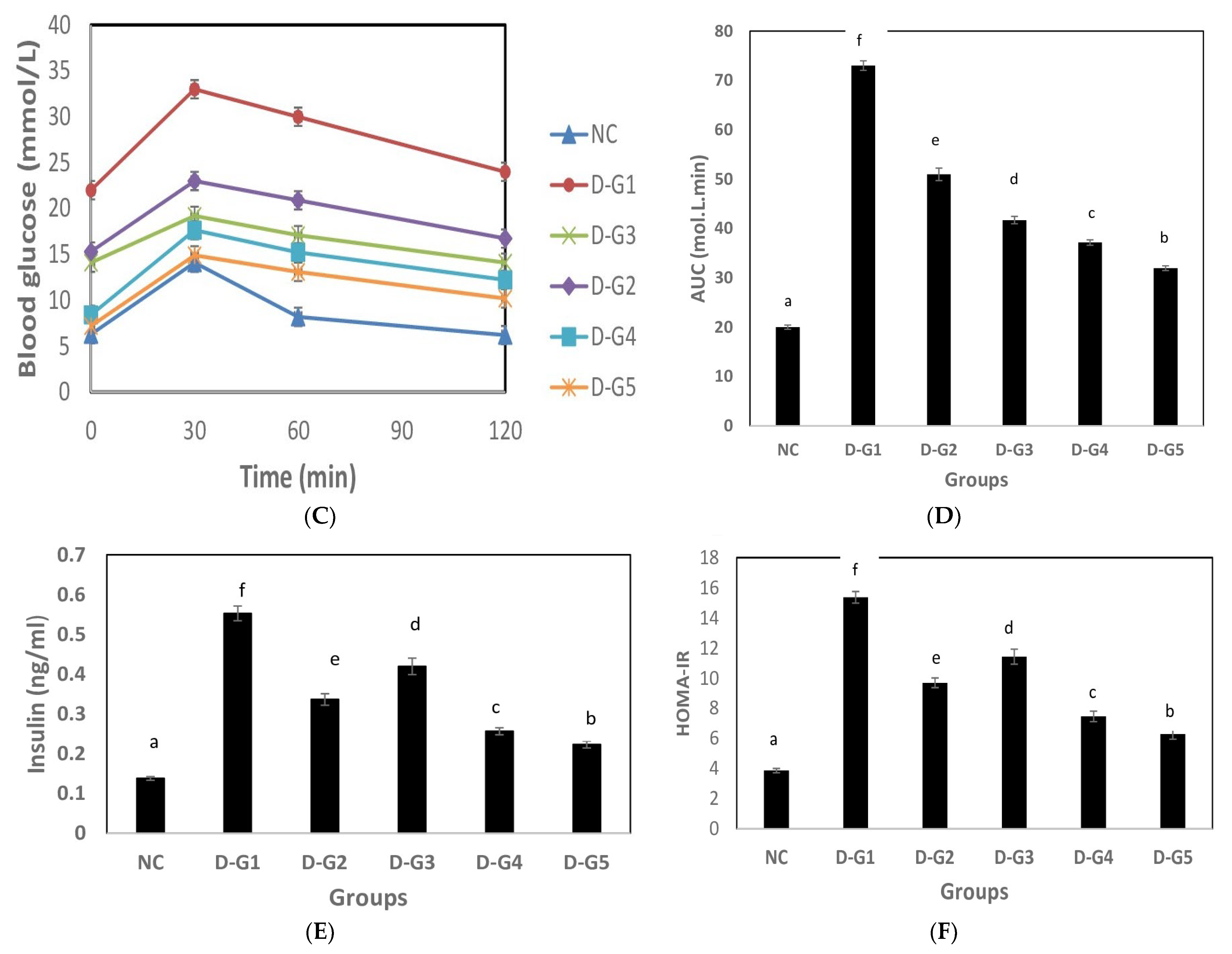
2.5.4. Oral Glucose Tolerance Test (OGTT)

At the end of the 7th week of the study, and after an overnight fast, 2 g/kg glucose was administered orally to all the mice, and blood samples were collected from each at 0, 30, 60, and 120 min afterward for the measurement of blood glucose using a glucose meter.

2.5.5. Histopathological Examination

For adequate fixation, pancreatic tissues were cut and kept in 10% formalin. These tissues were prepared, and embedding in paraffin wax was performed. Sections having a thickness of 5–6 microns were cut and stained with hematoxylin and eosin. All tissue slices were evaluated under the microscope using the Bancroft technique [33].

2.6. Statistical Analyses

The data were analyzed using one-way analysis of variance (ANOVA) Version IA (C). PC-STAT, programme coded by the University of Georgia, Athens, GA, USA. p < 0.05 was considered significant [34].

3. Results and Discussion

3.1. Effect on Lipid Profile

Diabetic rats (D-G1) had significantly greater T.C., T.G., LDL, and VLDL and lower HDL than other groups (p < 0.05). Treatments of diabetic rats with Chlorella vulgaris extract (D-G2) and Lactobacillus casei ATCC 7469 fermented soy flour extract (D-G3) significantly decreased levels of T.C., T.G., LDL, and VLDL in diabetic rats (D-G1), while they were still significantly higher than those of control (NC) or diabetic rats treated with pioglitazone (D-G5) (p < 0.05), while diabetic rats treated with a combination of Chlorella vulgaris extract and Lactobacillus casei ATCC 7469 fermented soy flour extract (D-G4) had significantly lower levels of T.C., T.G., LDL, and VLDL, even more so than those of diabetic rats treated with pioglitazone (D-G5) (p < 0.05) (Table 1).
Diabetes increases adipose tissue lipolysis in the absence of insulin, as well as free fatty acid mobilization from peripheral depots because insulin inhibits the hormone-sensitive lipase [35]. It has been proposed that the severe hyperlipidemia observed in STZ-diabetic rats fed a high-fat diet is owing to an increase in fat absorption through the gut, which in turn is due to an aberrant increase in small intestine acyl-coenzyme A: cholesterol acyl-transferase (ACAT) activity [36].
According to Cherng and Shih [37], the effects of Chlorella pyrenoidosa on blood lipids may be attributed to a reduction in fat absorption in the digestive tract. According to Lee et al. [19], the hypolipidemic impact of chlorella may be associated with the dietary fiber content of chlorella powder. Dietary fiber may have a hypolipidemic effect due to direct interference and modification of fat and glucose absorption in the intestine. Zhao et al. [38] discovered that Chlorella powder can drastically lower cholesterol in rats fed a high-fat diet by boosting fecal bile acid levels. Chlorella may improve lipid metabolism by modulating the expression of the SREBP-1c, ACC, and HMGCR genes, all of which are involved in the regulation of liver fat formation [39].
Lipid metabolism has been found to be significantly impacted by soy protein and isoflavones [40]. Soy protein or isoflavones have been proven to improve liver and blood lipid profiles by lowering triglycerides, total, and low-density lipoprotein (LDL) cholesterol levels and elevating the ratio of high-density lipoprotein (HDL)/LDL cholesterol [41]. The liver’s lipogenesis can be influenced by soy protein due to the activation of PPARs transcription factors involved in lipid metabolism, fatty acid oxidation, and glucose homeostasis by soy protein [42].
According to Zhang et al. [12], soymilk fermentation with B. bifidum, L. casei, and L. plantarum had a substantial impact on liver damage, weight loss, fat index, and hyperlipidemia. Through raising fecal bile acid excretion, Lactobacillus had a cholesterol-lowering impact [43]. The small intestine’s deconjugation of bile and binding to bile acid has been proposed as a possible explanation for lactic acid bacteria’s fecal bile acid excretion [44]. Higher excretion of bile acids may be the result of their deconjugation in the small intestine [45]. By expressing 7-alpha-hydroxylase (CYP7A1), a crucial enzyme in bile acid metabolism, Lactobacillus boosted hepatic bile acid production and fecal bile acid excretion [46]. Moreover, Lactobacillus decreased triglycerides by upregulating the expression of PPAR, F.X., and ApoAV and by raising apoA-V levels [47].

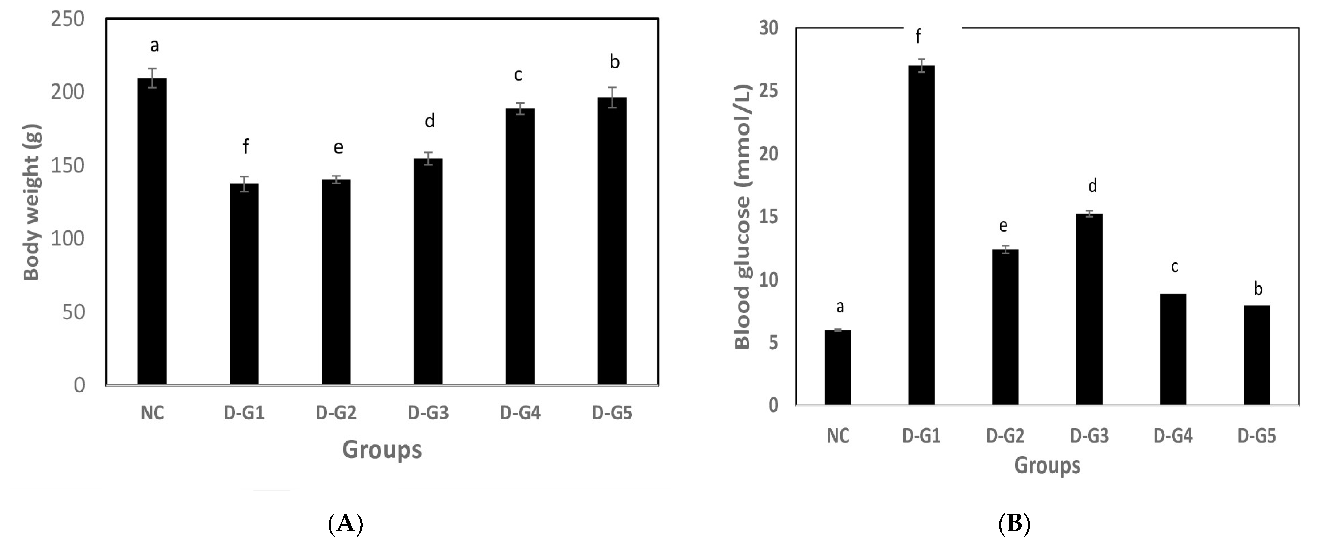
3.2. Hypoglycemic Effect

Diabetic rats (D-G1) had significantly lower body weight than other groups (p < 0.05). Treatments of diabetic rats with Chlorella vulgaris extract (D-G2) and Lactobacillus casei ATCC 7469 fermented soy flour extract (D-G3) significantly increased the body weight of diabetic rats (D-G1), while it was still significantly lower than that of control (CG) or diabetic rats treated with pioglitazone (D-G5) (p < 0.05). Diabetic rats (D-G1) had significantly higher blood glucose levels than other groups (p < 0.05). Treatments of diabetic rats with Chlorella vulgaris extract (D-G2) and Lactobacillus casei ATCC 7469 fermented soy flour extract (D-G3) significantly decreased the blood glucose of diabetic rats (D-G1), while it was still significantly higher than that of control (CG) or diabetic rats treated with pioglitazone (D-G5) (p < 0.05). Glucose tolerance was seriously impaired in group D-G1. The glucose area under the curve (AUC) in group D-G1 rats was significantly larger than that of group NC (p < 0.05). The glucose AUC was significantly lower (33, 44, and 50%) following administration of Chlorella vulgaris extract (D-G2), Lactobacillus casei ATCC 7469 fermented soy flour extract (D-G3), or their combination (D-G4), respectively, compared to group D-G1, but still significantly higher than the control group (NC), and diabetic rats administered pioglitazone (D-G5) (p ˂ 0.05). Diabetic rats (D-G1 group) had higher blood insulin and HOMA-IR than the control group (CG) (p < 0.05). However, feeding with Chlorella vulgaris extract D-G2, Lactobacillus casei ATCC 7469 fermented soy flour extract D-G3, or their combination D-G4 significantly decreased blood insulin, and HOMA-IR compared to the HF-G1 group (p < 0.05) (Figure 1).
Injection of small doses of STZ can damage islet β cells, which induces T2DM [48]. Hyperglycemia, characterized by fasting blood glucose (FBG) values and the oral glucose tolerance test (OGTT), is an important indicator of T2DM. HOMA-IR is usually used as a method for estimating insulin resistance 49This index is linked to insulin and fasting blood glucose levels. Type 2 diabetes is characterized by insulin resistance and relatively low insulin production [49].
Chlorella’s hypoglycemic effects may be caused by either decreasing hepatic glycogenolysis and gluconeogenesis or by boosting glucose absorption while inhibiting insulin secretion [50]. Hepatic triglyceride (TG) and non-esterified free fatty acid (NEFA) contents were significantly lower in the chlorella-fed groups, and these findings appear to be related to the low HOMA-IR of those groups. Reduced plasma non-esterified free fatty acid (NEFA) concentrations may enhance insulin’s ability to decrease hepatic glucose synthesis, increase glucose uptake, and alleviate type 2 diabetes’ high insulin concentration [37]. Additionally, some research has demonstrated that regular administration of chlorella reduced insulin resistance in animal models of diabetes or obesity [38]. An increase in the expression of glucose transporter 4 (GLUT4) via the activation of protein kinase B (Akt) phosphorylation in skeletal muscle is involved as a mechanism behind the chlorella intake-induced reduction in insulin resistance (IR) [50].
Consumption of soy protein decreased hyperinsulinemia and the irritability of the pancreatic Langerhans islets, according to Noriega-López et al. [51]. At micromolar concentrations, soy isoflavone genistein enhances the production of insulin induced by glucose in cell lines and pancreatic islets through a cAMP-dependent protein kinase pathway [52]. Soy isoflavones have inhibitory effects on α-glucosidase and amylase enzymes, which result in hydrolyzing dietary starch and raising blood glucose levels [53].
Probiotics have been demonstrated to decrease insulin resistance using different mechanisms. They increase the expression of adhesion proteins in the intestinal epithelium and decrease intestinal permeability, which results in reduced systemic inflammation and decreased insulin resistance [54]. They have been shown to increase liver natural killer T (NKT) cells, which have anti-diabetic actions against insulin resistance. They can also regulate gene expression and lower NF-kB binding activity, which reduces insulin resistance and inflammation [55]. Probiotics also inhibit pro-inflammatory cytokines, which can reduce insulin resistance [56]. Vallianou et al. [57] stated that probiotics induce the production of incretin hormones such as GLP-1, which, due to the insulinotropic properties of these hormones, can normalize insulin resistance in diabetic rats.

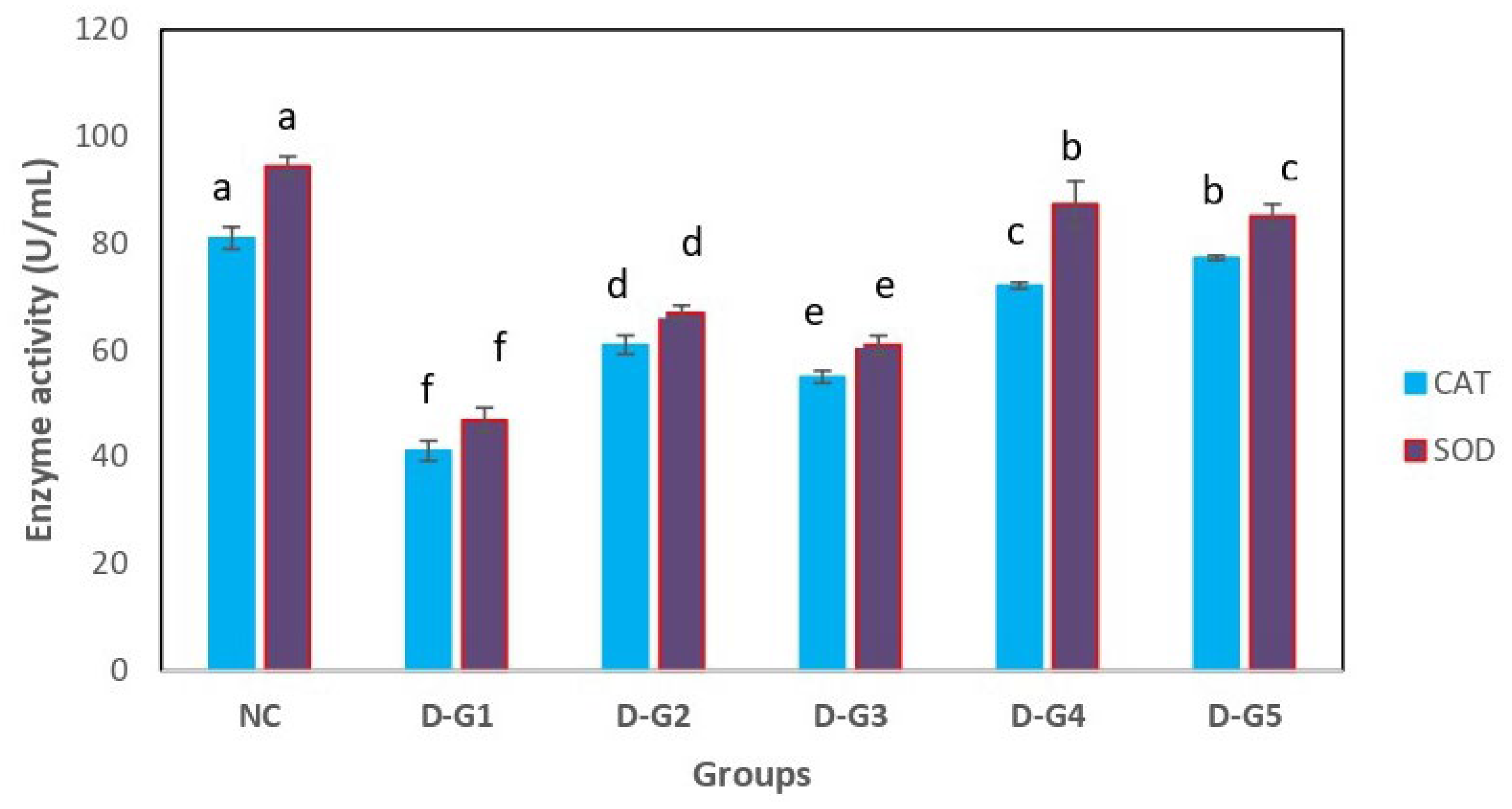
3.3. Effect on Antioxidant Parameters

Diabetic rats (D-G1group) had significantly lower CAT and SOD enzymes than other groups (p < 0.05). Feeding with Chlorella vulgaris extract D-G2, Lactobacillus casei ATCC 7469 fermented soy flour extract D-G3 or their combination D-G4 significantly increased levels of CAT, SOD enzymes than the diabetic group (p < 0.05), while they had significantly lower levels than the control group (CG) (p < 0.05) (Figure 2).
Oxidative stress and the oxidation of LDL-cholesterol and other lipoproteins increase in STZ-induced diabetes mellitus [58]. Insulin resistance in diabetic rats increases the release of free fatty acids from stored triglycerides, and increased oxidation of FFAs due to a lack of insulin stimulation of malonyl CoA production causes increased production of superoxide by the mitochondrial electron transport chain [59]. It has been shown that STZ promotes the production of H2O2 in pancreatic cells both in vitro and in vivo, which results in superoxide (O2) and hydroxyl radical (OH) damage to DNA, proteins, and lipids. STZ causes the organism to produce nitrogen monoxide (NO), which causes the destruction of the β-cells in the pancreatic islets. Diabetes is caused by STZ therapy because it lowers intracellular NADP levels and prevents the synthesis of proinsulin. The key factor contributing to the increased oxidative stress in experimental diabetes is higher blood glucose levels [60].
Probiotics were found to cause significant improvements in the blood total antioxidant activity (TAA) and total antioxidant status (TAS) of diabetic rats [61]. Probiotics can capture metal ions that catalyze oxidation processes, such as ferrous and cupric ions [62]. Probiotics can make use of their antioxidant enzymatic systems, such as superoxide dismutase and catalase [63]. Many metabolites with antioxidant characteristics, including GSH, butyrate, and folate, can be produced by probiotics [64]. Bahreini-Esfahani et al. [65] stated that by controlling the Nrf2-Keap1-ARE, mitogen-activated protein kinase, nuclear factor-jB, and protein kinase C pathways, probiotics can protect against oxidative stress. Probiotics can also control the ROS-producing enzymes, which reduce the activity of the cytochrome P450, cyclooxygenase, and NADPH oxidase enzymes.
It has been discovered that soy isoflavones act as protective antioxidants, preventing the formation of free radicals and reactive oxygen species (ROS) by quenching active singlet oxygen, trapping and neutralizing radicals before they can damage cells, and decomposing hydrogen peroxide without producing radicals [66]. Yoon et al. [67] stated that isoflavone supplementation may result in the upregulation of SOD and catalase activity, counteracting oxidative stress.
Chlorella vulgaris is a rich source of polyphenolic compounds, which result in enhanced antioxidant properties [68]. According to Shimada et al. [69], probiotics enhanced reduced glutathione concentrations, which resulted in the removal of hydrogen peroxide. Glutathione, a non-enzyme antioxidant, has a complex metabolic role and is capable of hydrolyzing hydrogen peroxides.
According to Ismail et al. [70], microalgae are protected against oxidative stress through a network of gene interactions associated with the upregulation of Sod1. The Sod1 gene collaborates with several other genes for the detoxification of reactive oxygen species and cellular prooxidants. Another antioxidant gene that is increased because of Chlorella vulgaris treatment is glutathione peroxidase (gpx1). The gene, which is also a protein-coding gene, is a member of the peroxidase family. When the gene is expressed, organic hydroperoxides are reduced, and hydrogen peroxide is detoxified [71].

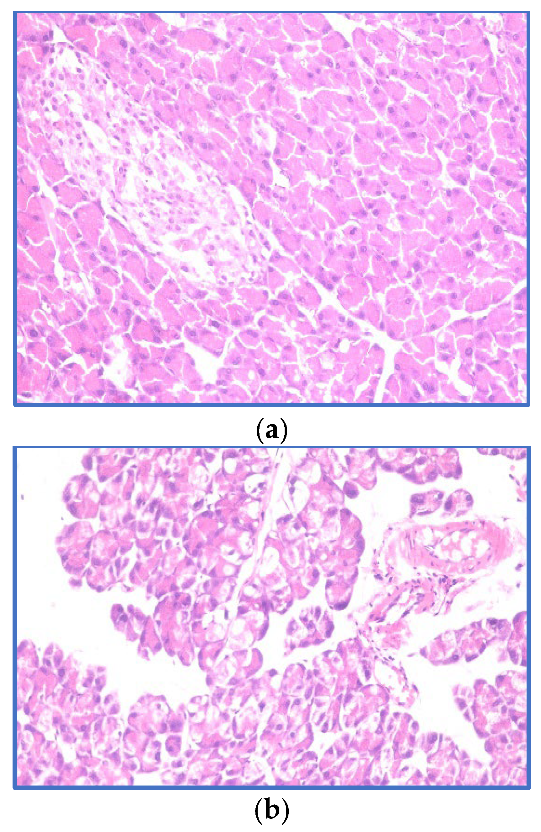
3.4. Histopathological Examination of the Pancreas

The exocrine and endocrine tissues in the pancreas of the normal control group (NC) had normal histological structures, according to microscopic analysis. The cellular com-ponents of the islets of Langerhans were arranged normally, and the acinar structure indicated typical protein-rich eosinophilic contents. The islets had a lighter stain than the acinar cells in the area. Pyramidal cells with basal nuclei and apical acidophilic cyto-plasm make up the acinar cells. The acinar cells that make up the exocrine portion of the pancreas are tightly packed together and organized into tiny lobules. Activated intralobular and interlobular connective tissue septa divide the pancreatic lobules (Figure 3a). Diabetic rats on a regular diet (D-G1) developed pancreatic lobule atrophy and acinar epithelial lining vacuolation. Diabetes causes pathological alterations in both exocrine and endocrine components in diabetic rats. The acinar cells were enlarged, and almost all of them had tiny vacuoles. Flattened epithelium bordered the interlobular ducts. Islet cells are nearly extinct. (Figure 3b). The pancreas of diabetic animals treated by Chlorella vulgaris and Lactobacillus fermented soy flour extract (D-G4) had normal pancreatic lobule morphology. The islets of Langerhans cells showed a little enlargement. There is no major cellular damage in the exocrine acini (Figure 3c). The treatment with Chlorella vulgaris and Lactobacillus fermented soy flour extract may have a liver protective effect, according to the H&E staining data.
According to Zeng et al. [72], there was a significant decrease in the number of islets of Langerhans in the diabetes group compared to the normal group. The diabetic rats had irregular islet shapes and atrophy, whereas the normal group rats had round or oval cell clusters interspersed in healthy acinar cells with obvious borders. Degenerative alterations such as nuclear shrinkage or karyolysis, cell necrosis, and vacuolization were seen. The rats treated with L. paracasei strain NL41 improved their histological abnormalities, with an increase in the number of islets, better form, and less cell damage. According to Long et al. [13], the normal group’s islet cells were present in high numbers, were oriented properly, and had normal morphology, with regular, distinct cell borders and normal cytoplasm. Islet cells in the DM group were uneven in structure, shape, and distribution, with irregularly shaped nuclei. Yet, when compared to the DM group, the diabetic group treated with Lactobacillus had more islet cells with a somewhat normal shape. Lactobacillus inhibits the formation of disease in pancreatic cells, which is related to improved kidney function and metabolic conditions overall.

4. Conclusions

The present study has clearly demonstrated that the deleterious clinical manifestations accompanying STZ-induced hyperglycemia in rats can be ameliorated by Chlorella vulgaris extracts and dried soy flour fermentation extracts. It may offer novel insights into the potent hypolipidemic, hypoglycemic, and antioxidant activities of Chlorella vulgaris extracts and dried soy flour fermentation extracts. The potential mechanisms of this effect might be related to decreasing insulin resistance, reducing oxidative stress, and protecting beta-cell function. These effects might be beneficial to further extend the application of soy flour as a potential prebiotic or dietary supplement for diabetes treatment. The hypolipidemic, hypoglycemic, and antioxidant mechanisms of Chlorella vulgaris extracts and lactobacillus fermented soy flour extracts need further investigation and validation of their efficacy through human clinical trials. The cytotoxicity of Chlorella vulgaris will be assessed in future investigations.

Author Contributions

Conceptualization, methodology, writing, A.A.; Methodology, F.I.A.-E.; Writing, Funding, S.W.E.-F. All authors have read and agreed to the published version of the manuscript.

Funding

The research received no external funding.

Institutional Review Board Statement

Not applicable.

Informed Consent Statement

Not applicable.

Data Availability Statement

Available when required.

Conflicts of Interest

The authors declare no conflict of interest.

References

  1. Takeda, Y.; Fujita, Y.; Honjo, J.; Sakagami, H.; Takiyama, Y.; Makino, Y.; Abiko, A.; Kieffer, T.J.; Haneda, M. Reduction of both beta cell death and alpha cell proliferation by dipeptidyl peptidase-4 inhibition in a streptozotocin-induced model of diabetes in mice. Diabetologia 2012, 55, 404. [Google Scholar] [CrossRef] [PubMed]
  2. Zhao, Y.; Jiang, Z.; Guo, C. New hope for type 2 diabetics: Targeting insulin resistance through the immune modulation of stem cells. Autoimmun Rev. 2011, 11, 137–142. [Google Scholar] [CrossRef]
  3. Mcclean, P.L.; Vadivel, P.; Emilie, F.; Christian, H.L. The diabetes drug liraglutide prevents degenerative processes in a mouse model of Alzheimer’s disease. J. Neurosci. 2011, 31, 6587–6594. [Google Scholar] [CrossRef] [PubMed]
  4. Sola, D.; Rossi, L.; Schianca, G.P.C.; Maffioli, P.; Bigliocca, M. Sulfonylureas and their use in clinical practice. Arch. Med. Sc. 2015, 4, 840–848. [Google Scholar] [CrossRef]
  5. Rizos, C.V.; Elisaf, M.; Mikhailidis, D.P.; Liberopoulos, E.N. How safe is the use of thiazolidinediones in clinical practice? Expert Opin. Drug Saf. 2009, 8, 15–32. [Google Scholar] [CrossRef] [PubMed]
  6. Abd El Latif, M.A.; Mohamed, N.H.; Zaki, N.L.; Abbas, M.S.; Sobhy, H.M. Effects of Soybean Isoflavone on Lipid Profiles and Antioxidant Enzyme Activity in Streptozotocin Induced Diabetic Rats. Glob. J. Pharmacol. 2014, 8, 378–384. [Google Scholar]
  7. Chang, J.H.; Kim, M.S.; Kim, T.W.; Lee, S.S. Effects of soybean supplementation on blood glucose, plasma lipid levels, and erythrocyte antioxidant enzyme activity in type 2 diabetes mellitus patients. Nutr. Res. Pract. 2008, 2, 152–157. [Google Scholar] [CrossRef]
  8. Shu, G.; Shi, X.; Chen, H.; Ji, Z.; Meng, J. Optimization of nutrient composition for producing ACE inhibitory peptides from goat milk fermented by Lactobacillus bulgaricus LB6, Probiotics Antimicrob. Proteins 2019, 11, 723–729. [Google Scholar]
  9. Wagner, J.D.; Zhang, L.; Shadoan, M.K.; Kavanagh, K.; Chen, H.; Tresnasari, K.; Kaplan, J.R.; Adams, M.R. Effects of soy protein and isoflavones on insulin resistance and adiponectin in male monkeys. Metabolism 2008, 57, 24–31. [Google Scholar] [CrossRef]
  10. Sanjukta, S.; Rai, A.K.; Muhammed, A.; Jeyaram, K.; Talukdar, N.C. Enhancement of antioxidant properties of two soybean varieties of Sikkim Himalayan region by proteolytic Bacillus subtilis fermentation. J. Funct. Foods 2015, 14, 650–658. [Google Scholar] [CrossRef]
  11. Kim, J.H.; Jia, Y.; Lee, J.G.; Nam, B.; Lee, J.H.; Shin, K.S.; Hurh, B.S.; Choi, Y.H.; Lee, S.J. Hypolipidemic and antiinflammation activities of fermented soybean fibers from meju in C57BL/6 J mice. Phytother. Res. 2014, 28, 1335–1341. [Google Scholar] [CrossRef] [PubMed]
  12. Zhang, X.L.; Wu, Y.F.; Wang, Y.S.; Wang, X.Z.; Piao, C.H.; Liu, J.M.; Liu, Y.L.; Wang, Y.H. The protective effects of probiotic-fermented soymilk on high fat diet-induced hyperlipidemia and liver injury. J. Funct. Foods 2017, 30, 220–227. [Google Scholar] [CrossRef]
  13. Long, X.S.; Liao, S.T.; Li, E.N.; Pang, D.R.; Li, Q.; Liu, S.C.; Hu, T.G.; Zou, Y.X. The hypoglycemic effect of freeze-dried fermented mulberry mixed with soybean on type 2 diabetes mellitus. Food Sci. Nutr. 2021, 9, 3641–3654. [Google Scholar] [CrossRef]
  14. Yu, S.; Wang, W.; Li, S.; Li, J.; Zhao, R.; Liu, D.; Wu, J. Glucoregulatory Properties of Fermented Soybean Products. Fermentation 2023, 9, 254. [Google Scholar] [CrossRef]
  15. Hu, T.; Chen, R.; Qian, Y.; Ye, K.; Long, X.; Park, K.Y.; Zhao, X. Antioxidant effect of Lactobacillus fermentum HFY02-fermented soy milk on D-galactose-induced aging mouse mode. Food Sci. Hum. Wellness 2022, 11, 1362–1372. [Google Scholar] [CrossRef]
  16. Stramarkou, M.; Papadaki, S.; Kyriakopoulou, K.; Krokida, M. Effect of drying and extraction conditions on the recovery of bioactive compounds from Chlorella vulgaris. J. Appl. Phycol. 2017, 29, 2947–2960. [Google Scholar] [CrossRef]
  17. Bito, T.; Okumura, E.; Fujishima, M.; Watanabe, F. Potential of Chlorella as a Dietary Supplement to Promote Human Health. Nutrients 2020, 12, 2524. [Google Scholar] [CrossRef]
  18. Daugherty, B.L. Histamine H4 antagonism: A therapy for chronic allergy? Br. J. Pharmacol. 2014, 142, 5. [Google Scholar] [CrossRef]
  19. Lee, H.S.; Park, H.J.; Kim, M.K. Effect of Chlorella vulgaris on lipid metabolism in Wistar rats fed high fat diet. Nutr. Res. Pract. 2008, 2, 204–210. [Google Scholar] [CrossRef]
  20. Jeong, H.; Kwon, H.J.; Kim, M.K. Hypoglycemic effect of Chlorella vulgaris intake in type 2 diabetic Goto-Kakizaki and normal Wistar rats. Nutr. Res. Pract. 2009, 3, 23–30. [Google Scholar] [CrossRef]
  21. Bellinger, E.G.; Sigee, D.C. Freshwater Algae: Identification, Enumeration and Use as Bioindicators, 2nd ed.; John Wiley & Sons: Hoboken, NJ, USA, 2015. [Google Scholar]
  22. Stainer, R.Y.; Kunisawa, R.; Mandel, M.; Cohen-Bazire, G. Purification and properties of unicellular blue-green algae (Order Chroococcales). Bacteriol. Rev. 1971, 35, 171–205. [Google Scholar] [CrossRef]
  23. Dong, J.; Liang, Q.; Niu, Y.; Jiang, S.; Zhou, L.I.; Wang, J.; Ma, C.; Kang, W. Effects of nigella sativa seed polysaccharides on type 2 diabetic mice and gut microbiota. Int. J. Biol. Macromol. 2020, 159, 725–738. [Google Scholar] [CrossRef] [PubMed]
  24. Islam, S.; Choi, H. Dietary red chilli (Capsicum frutescens L.) is insulinotropic rather than hypoglycemic in type 2 diabetes model of rats. Phytother. Res. 2008, 22, 1025–1029. [Google Scholar] [CrossRef] [PubMed]
  25. Assinewe, V.A.; Baum, B.R.; Gagnon, D.; Arnason, J.T. Phytochemistry of Wild Populations of Panax quinquefolius L. (North American Ginseng). J. Agric. Food Chem. 2003, 51, 4549–4553. [Google Scholar] [CrossRef]
  26. Fossati, P.; Prencipe, L. Serum triglycerides are determined colorimetrically with a Nenzyme that produces hydrogen peroxide. Clin. Chem. 1982, 28, 2077–2080. [Google Scholar] [CrossRef] [PubMed]
  27. Deeg, R.; Ziegenohrm, J. Kinetic enzymatic method for automated determination of total cholesterol in serum. J. Clin. Chem. 1983, 29, 1798–1802. [Google Scholar] [CrossRef]
  28. Burstein, M.; Selvenick, H.R.; Morfin, R. Rapid method for the isolation of lipoproteins from human serum by precipitation with polyanions. J. Lipid Res. 1970, 11, 583–595. [Google Scholar] [CrossRef] [PubMed]
  29. Friedewald, W.T.; Levy, R.I.; Fredrickson, D.S. Estimation of the concentration of low-density lipoprotein cholesterol in plasma, without use of the preparative ultracentrifuge. Clin. Chem. 1972, 18, 499–502. [Google Scholar] [CrossRef]
  30. Norbert, W.T. Clinical Guide to Laboratory Tests, 3rd ed.; W.B. Saunders Company: Philadelphia, PA, USA, 1995. [Google Scholar]
  31. Marklund, S.; Marklund, G. Involvement of the superoxide anion radical in the autoxidation of pyrogallol and a convenient assay for superoxide dismutase. Eur. J. Biochem. 1974, 47, 469–474. [Google Scholar] [CrossRef]
  32. Cohen, G.; Dembiec, D.; Marcus, J. Measurement of catalase activity in tissue. Anal. Biochem. 1970, 34, 30–38. [Google Scholar] [CrossRef]
  33. Bancroft, G.D.; Stevens, A.; Turner, D.R. Theory and Practice of Pathological Technique, 4th ed.; Churchill Livingston: New York, NY, USA, 1996. [Google Scholar]
  34. Snedcor, G.W.; Cochran, W.G. Statistical Methods, 7th ed.; The Iowa State University Press: Ames, IA, USA, 1982; p. 507. [Google Scholar]
  35. Zhao, J.; Wu, Y.Y.; Rong, X.L.; Zheng, C.W.; Guo, J. Anti-Lipolysis Induced by Insulin in Diverse Pathophysiologic Conditions of Adipose Tissue. Diabetes Metab. Syndr. Obes. Targets Ther. 2020, 13, 1575–1585. [Google Scholar] [CrossRef] [PubMed]
  36. Prangthipa, P.; Surasianga, R.; Charoensiria, R.; Leardkamolkarn, V.; Komindr, S.; Yamborisuta, U.; Vanavichitd, A.; Kongkachuichai, R. Amelioration of hyperglycemia, hyperlipidemia, oxidative stress and inflammation in steptozotocin-induced diabetic rats fed a high fat diet by riceberry supplement. J. Funct. Foods 2013, 5, 195–203. [Google Scholar] [CrossRef]
  37. Cherng, J.Y.; Shih, M.F. Potential hypoglycemic effects of Chlorella in streptozotocin-induced diabetic mice. Life Sci. 2005, 78, 980–990. [Google Scholar]
  38. Zhao, C.; Wu, Y.J.; Yang, C.F.; Liu, B.; Huang, Y.F. Hypotensive, hypoglycemic and hypolipidemic effects of bioactive compounds from microalgae and marine microorganisms. Int. J. Food Sci. Technol. 2015, 50, 1705–1717. [Google Scholar] [CrossRef]
  39. Chen, J.; Gong, S.; Wan, X.; Gao, X.; Wang, C.; Zeng, F.; Zhao, C.; Liu, B.; Huang, Y. Hypolipidemic properties of Chlorella pyrenoidosa organic acids via AMPK/HMGCR/SREBP-1c pathway in vivo. Food Sci. Nutr. 2021, 9, 459–468. [Google Scholar] [CrossRef] [PubMed]
  40. Squadrito, F.; Marini, H.; Bitto, A.; Altavilla, D.; Polito, F.; Adamo, E.B.; D’Anna, R.; Arcoraci, V.; Burnett, B.P.; Minutoli, L.; et al. Genistein in the metabolic syndrome: Results of a randomized clinical trial. J. Clin. Endocrinol. Metab. 2013, 98, 3366–3374. [Google Scholar] [CrossRef]
  41. Moradi, M.; Daneshzad, E.; Azadbakht, L. The effects of isolated soy protein, isolated soy isoflavones and soy protein containing isoflavones on serum lipids in postmenopausal women: A systematic review and meta-analysis. Crit. Rev. Food Sci. Nutr. 2020, 60, 3414–3428. [Google Scholar] [CrossRef]
  42. Singh, B.P.; Vij, S.; Hati, S. Functional significance of bioactive peptides derived from soybean. Peptides 2014, 54, 171–179. [Google Scholar] [CrossRef]
  43. Wang, J.Y.; Dang, N.; Sun, P.; Xia, J.; Zhang, C.; Pang, S. +e effects of metformin on fibroblast growth factor 19, 21 and fibroblast growth factor receptor 1 in high-fat diet and streptozotocin induced diabetic rats. Endocr. J. 2017, 64, 543–552. [Google Scholar] [CrossRef]
  44. Tsai, C.C.; Lin, P.-P.; Hsieh, Y.-M.; Zhang, Z.-Y.; Wu, H.-C.; Huang, C.-C. Cholesterol-Lowering Potentials of Lactic Acid Bacteria Based on Bile-Salt Hydrolase Activity and Effect of Potent Strains on Cholesterol Metabolism In Vitro and In Vivo. Sci. World J. 2014, 2014, 690752. [Google Scholar] [CrossRef]
  45. Hamouda, R.A.; Hamza, H.A.; Salem, M.L.; Kamal, S.; Alhasani, R.H.; Alsharif, I.; Mahrous, H.; Abdella, A. Synergistic Hypolipidemic and Immunomodulatory Activity of Lactobacillus and Spirulina platensis. Fermentation 2022, 8, 220. [Google Scholar] [CrossRef]
  46. Ridlon, J.M.; Kang, D.J.; Hylemon, P.B.; Bajaj, J.S. Bile acids and the gut microbiome. Curr. Opin. Gastroenterol. 2014, 30, 332–338. [Google Scholar] [CrossRef] [PubMed]
  47. Chiang, J.Y.L. Bile acid metabolism and signaling in liver disease and therapy. Liver Res. 2017, 1, 3–9. [Google Scholar] [CrossRef] [PubMed]
  48. Pegah, A.; Abbasi-Oshaghi, E.; Khodadadi, I.; Mirzaei, F.; Tayebina, H. Probiotic, and resveratrol normalize GLP-1 levels and oxidative stress in the intestine of diabetic rats. Metab. Open 2021, 10, 100093. [Google Scholar] [CrossRef] [PubMed]
  49. Gayoso-Diz, P.; Otero-González, A.; Rodriguez-Alvarez, M.X.; Gude, F.; García, F.; De Francisco, A. Insulin resistance (HOMA-IR) cut-off values and the metabolic syndrome in a general adult population: Effect of gender and age: EPIRCE cross-sectional study. BMC Endocr. Disord. 2013, 13, 47. [Google Scholar] [CrossRef]
  50. Vecina, J.F.; Oliveira, A.; Araújo, T.; Baggio, S.R.; Torello, C.O.; Saad, M.J.A.; Queiroz, M.L.D.S. Chlorella modulates insulin signaling pathway and prevents high-fat diet-induced insulin resistance in mice. Life Sci. 2014, 95, 45–52. [Google Scholar] [CrossRef]
  51. Noriega-Lopez, L.; Tovar, A.R.; Gonzalez-Granillo, M.; Hernandez-Pando, R.; Escalante, B.; Santillan-Doherty, P.; Torres, N. Pancreatic insulin secretion in rats fed a soy protein high fat diet depends on the interaction between the amino acid pattern and isoflavones. J. Biol. Chem. 2007, 282, 20657–20666. [Google Scholar] [CrossRef]
  52. Liu, D.; Zhen, W.; Yang, Z.; Carter, J.D.; Si, H.; Reynolds, K.A. Genistein acutely stimulates insulin secretion in pancreatic β-cells through cAMP-dependent protein kinase pathway. Daibetes 2006, 55, 1043–1050. [Google Scholar] [CrossRef]
  53. Rasouli, H.; Hosseini-Ghazvini, S.M.; Adibi, H.; Khodarahmi, R. Differential α-amylase/α-glucosidase inhibitory activities of plant-derived phenolic compounds: A virtual screening perspective for the treatment of obesity and diabetes. Food Funct. 2017, 8, 1942–1954. [Google Scholar] [CrossRef]
  54. Gomes, A.C.; Bueno, A.A.; de Souza RG, M.; Mota, J.F. Gut microbiota, probiotics, and diabetes. Nutr. J. 2014, 13, 60. [Google Scholar] [CrossRef]
  55. Ma, X.; Hua, J.; Li, Z. Probiotics improve high fat diet-induced hepatic steatosis and insulin resistance by increasing hepatic NKT cells. J. Hepatol. 2008, 49, 821–830. [Google Scholar] [CrossRef] [PubMed]
  56. Rajkumar, H.; Mahmood, N.; Kumar, M.; Varikuti, S.R.; Challa, H.R.; Myakala, S.P. Effect of probiotic (VSL# 3) and omega-3 on lipid profile, insulin sensitivity, inflammatory markers, and gut colonization in overweight adults: A randomized, controlled trial. Mediat. Inflamm. 2014, 2014, 348959. [Google Scholar]
  57. Vallianou, N.; Liu, J.; Dalamaga, M. What are the key points in the association between the gut microbiome and nonalcoholic fatty liver disease? Metabol Open 2019, 1, 9–10. [Google Scholar] [CrossRef] [PubMed]
  58. Jafarnejad, A.; Bathale, S.Z.; Nakhjavani, M.; Hyassan, M.Z. Effect of spermine on lipid profile and HDL functionality in the streptozotocin-induced diabetic rat model. Life Sci. 2008, 82, 301–307. [Google Scholar] [CrossRef]
  59. Du, X.; Edelstein, D.; Obici SHigham, N.; Zou, M.-H.; Brownlee, M. Insulin resistance reduces arterial prostacyclin synthase and eNOS activities by increasing endothelial fatty acid oxidation. J. Clin. Investig. 2006, 116, 1071–1082. [Google Scholar] [CrossRef]
  60. Damasceno, D.C.; Volpato, G.T.; Calderon, I.M.P.; Rudg, M.V.C.R. Oxidative stress and diabetes in pregnant rats. Anim. Reprod. Sci. 2002, 72, 235–244. [Google Scholar] [CrossRef] [PubMed]
  61. Songisepp, E.; Kals, J.; Kullisaar, T.; Mändar, R.; Hütt, P.; Zilmer, M.; Mikelsaar, M. Evaluation of the functional efficacy of an antioxidative probiotic in healthy volunteers. Nutr. J. 2005, 4, 22. [Google Scholar] [CrossRef]
  62. Thomas, B. The global burden of diabetic kidney disease: Time trends and gender gaps. Curr. Diab. Rep. 2019, 19, 18. [Google Scholar] [CrossRef]
  63. Li, R.; Bilik, D.; Brown, M.B.; Zhang, P.; Ettner, S.L.; Ackermann, R.T.; Crosson, J.C.; Herman, W.H. Medical costs associated with type 2 diabetes complications and comorbidities. Am. J. Manag. Care 2013, 19, 421–430. [Google Scholar] [PubMed]
  64. Roobab, U.; Batool, Z.; Manzoor, M.F.; Shabbir, M.A.; Khan, M.R.; Aadil, R.M. Sources, formulations, advanced delivery and health benefits of probiotics. Curr. Opin. Food Sci. 2020, 32, 17–28. [Google Scholar] [CrossRef]
  65. Bahreini-Esfahani, N.; Moravejolahkami, A.R. Can synbiotic dietary pattern predict lactobacillales strains in breast milk? Breastfeed Med. 2020, 15, 387–393. [Google Scholar] [CrossRef] [PubMed]
  66. Itoh, M.; Oh-Ishi, S.; Hatao, H.; Leeuwenburgh, C.; Selman, C.; Ohno, H.; Kizaki, T.; Nakamura, H.; Matsuoka, T. Effects of dietary calcium restriction and acute exercise on the antioxidant enzyme system and oxidative stress in rat diaphragm. Am. J. Physiol. Regul. Integr. Comp. Physiol. 2004, 287, 33–38. [Google Scholar] [CrossRef] [PubMed]
  67. Yoon, G.A.; Park, S. Antioxidant action of soy isoflavones on oxidative stress and antioxidant enzyme activities in exercised rats. Nutr. Res. Pract. 2014, 8, 618–624. [Google Scholar] [CrossRef] [PubMed]
  68. Dangles, O.; Dufour, C.; Manach, C.; Morand, C.; Remesy, C. Binding of flavonoids to plasma proteins. In Methods in Enzymology 335; Academic Press: Cambridge, MA, USA, 2001; pp. 319–333. [Google Scholar]
  69. Shimada, M.; Hasegawa, T.; Nishimura, C.; Kan, H.; Kanno, T.; Nakamura, T.; Matsubayashi, T. Anti-hypertensive effect of γ-aminobutyric acid (GABA)-rich Chlorella on high-normal blood pressure and borderline hypertension in placebo controlled double-blind study. Clin. Exp. Hypertens 2009, 31, 342–354. [Google Scholar] [CrossRef]
  70. Ismail, M.; Hossain, M.; Tanu, A.R.; Shekhar, H.U. Effect of spirulina intervention on oxidative stress, antioxidant status, and lipid profile in chronic obstructive pulmonary disease patients. BioMed Res. Int. 2015, 2015, 897327. [Google Scholar] [CrossRef] [PubMed]
  71. Treitinger, A.; Spada, C.; Verdi, J.C.; Miranda, A.F.; Oliveira, O.V.; Silveira, M.V.; Moriel, P.; Abdalla, D.S. Decreased antioxidant defence in individuals infected by the human immunodeficiency virus. Eur. J. Clin. Investig. 2000, 30, 454–459. [Google Scholar] [CrossRef] [PubMed]
  72. Zeng, Z.; Yuan, O.; Yu, R.; Zhang, J.; Ma, H.; Chen, S. Ameliorative Effects of Probiotic Lactobacillus paracasei NL41 on Insulin Sensitivity, Oxidative Stress, and Beta-Cell Function in a Type 2 Diabetes Mellitus Rat Model. Mol. Nutr. Food Res. 2019, 63, 1900457. [Google Scholar] [CrossRef]
Figure 1. Effect of Lactobacillus casei ATCC 7469 fermented soy flour extract, Chlorella vulgaris extract, and their combination on (A) body weight, (B) Fasting blood glucose, (C,D) results of the Oral glucose tolerance test (OGTT)), (E) insulin levels, and (F) HOMA-IR of Sprague-Dawley rats at 7 weeks. NC: healthy rats fed a regular diet (NC); D-G1: Diabetic rats fed a regular diet; D-G2: Diabetic rats fed a regular diet with Chlorella vulgaris extract; D-G3: Diabetic rats fed a regular diet with Lactobacillus casei ATCC 7469 fermented soy flour extract.; D-G4: Diabetic rats fed a regular diet with Lactobacillus casei ATCC 7469 fermented soy flour extract and Chlorella vulgaris extract; D-G5: Diabetic rats fed a regular diet with pioglitazone. The results are expressed as the mean ± SD (n = 10). Bars with different superscripts are significantly different (p ˂ 0.05).
Microbiolres 14 00043 g001aMicrobiolres 14 00043 g001b
Figure 2. Effect of Chlorella vulgaris extract, Lactobacillus casei ATCC 7469 fermented soy flour extract, and their combination on catalase enzyme (CAT) and superoxide dismutase enzyme (SOD) of Sprague–Dawley rats at seven weeks NC: healthy rats fed a regular diet (control); D-G1: Diabetic rats fed a regular diet; D-G2: Diabetic rats fed a regular diet with Chlorella vulgaris extract; D-G3: Diabetic rats fed a regular diet with Lactobacillus casei ATCC 7469 fermented soy flour extract; D-G4: Diabetic rats fed a regular diet with Lactobacillus casei ATCC 7469 fermented soy flour extract and Chlorella vulgaris extract; D-G5: Diabetic rats fed a regular diet with pioglitazone. The results are expressed as the means ± SD (n = 10). Bars with different superscripts are significantly different (p < 0.05).
Microbiolres 14 00043 g002
Figure 3. Histological sections of pancreatic sections stained with hematoxylin and eosin (original magnification ×200). (a) NC: Pancreatic tissue section showing the normal histological structure of both exocrine and endocrine tissues (H&E × 200); (b) D-G1: Pancreatic tissue section showing acinar cells swelling and vacuolation with small vacuoles and loss of islet-cells (H&E × 200); (c) D-G4: Pancreatic tissue section showing mild swelling of islets of Langerhans cells (H&E × 200). NC: healthy rats fed a regular diet (control); D-G1: diabetic rats fed a regular fat diet; D-G4: diabetic rats fed a regular diet with Lactobacillus casei ATCC 7469 fermented soy flour extract and Chlorella vulgaris extract.
Microbiolres 14 00043 g003aMicrobiolres 14 00043 g003b
Table 1. Effect of Lactobacillus casei ATCC 7469 fermented soy flour extract, Chlorella vulgaris extract, and their combination on lipid profiles of Sprague–Dawley rats at 7 weeks.
Variable with UnitsNCD-G1D-G2D-G3D-G4D-G5
TC (mg/dL)91.1 ± 1.02 a213 ± 2.1 f171 ± 1.16 e143.64 ± 2.1 d95 ± 1.21 c115 ± 0.5 b
TG (mg/dL)71.5 ± 2.1 a192.33 ± 1.7 f145 ± 1.34 e117.14 ± 1.32 d82 ± 1.19 b87 ± 1.23 c
LDL (mg/dL)37.3 ± 0.9 a82.06 ± 1.17 f63.9 ± 1.8 d68.4 ± 2.16 e47 ± 1.3 b51 ± 1.15 c
VLDL (mg/dL)15.9 ± 0.2 a31 ± 0.56 e23.18 ± 0.58 c28.46 ± 1.33 d18.4 ± 0.24 b19.8 ± 0.38 b
HDL (mg/dL)28.2 ± 0.18 a13.19 ± 0.9 f21.6 ± 0.16 d19.3 ± 0.18 e26.52± 0.15 c24 ± 0.19 b
NC: healthy rats fed a regular diet (NC); D-G1: Diabetic rats fed a regular diet; D-G2: Diabetic rats fed a regular diet with Chlorella vulgaris extract; D-G3: Diabetic rats fed a regular diet with Lactobacillus casei ATCC 7469 fermented soy flour extract; D-G4: Diabetic rats fed a regular diet with Lactobacillus casei ATCC 7469 fermented soy flour extract and Chlorella vulgaris extract; D-G5: Diabetic rats fed a regular diet with pioglitazone. The results are expressed as the mean ± SD (n = 10). Different superscript letters in the same row indicate significant differences at p ˂ 0.05 between different groups of mice at p < 0.05.
Disclaimer/Publisher’s Note: The statements, opinions and data contained in all publications are solely those of the individual author(s) and contributor(s) and not of MDPI and/or the editor(s). MDPI and/or the editor(s) disclaim responsibility for any injury to people or property resulting from any ideas, methods, instructions or products referred to in the content.

