Article

Species of the Genera Neopestalotiopsis and Alternaria as Dominant Pathogen Species Attacking Mastic Trees (Pistacia lentiscus var. Chia)

by
Nathalie N. Kamou
*,
Stefanos Testempasis
and
Anastasia L. Lagopodi
Plant Pathology Laboratory, Faculty of Agriculture, School of Agriculture, Forestry and Natural Environment, Aristotle University of Thessaloniki, 54124 Thessaloniki, Greece
*
Author to whom correspondence should be addressed.
Microbiol. Res. 2023, 14(1), 104-115; https://doi.org/10.3390/microbiolres14010010
Submission received: 5 December 2022 / Revised: 18 January 2023 / Accepted: 19 January 2023 / Published: 21 January 2023
(This article belongs to the Special Issue Plants, Mycorrhizal Fungi, and Bacteria)

Abstract

:
Between 2018 and 2021, several mastic trees (Pistacia lentiscus var. Chia) sampled in the field and the nursery of the Chios Mastiha Growers Association (CMGA) were analyzed to determine the cause of dominant diseases. Symptoms included defoliation, leaf, and twig blight, wilting and/or apoplexy of trees and apoplexy of young hardwood cuttings. Moreover, brown discoloration had also been observed on older woody parts of the trees such as branches and tree trunks. Several pathogens have been isolated and identified as the causing agents. Neopestalotiopsis and Alternaria species were isolated consistently from necrotic tissues of mastic trees (branches, twigs, and leaves) in the field and the nursery. All fungal isolates’ pathogenicity was confirmed by applying Koch’s postulates on young mastic trees under glasshouse conditions. Fungal pathogens were identified by sequence analyses of the ITS, β-tubulin, and histone gene regions. Alternaria species were analyzed further by sequencing the endopolygalacturonase (endoPG) and the Alternaria major allergen (Alta1) genes. More specifically, the isolates were identified as Neopestalotiopsis clavispora, Alternaria arborescens, and A. alternata based on morphological features and sequence analyses. This is the first report of N. clavispora, A. arborescens, and A. alternata on P. lentiscus var. Chia.

1. Introduction

The mastic tree (Pistacia lentiscus var. Chia) of the Anacardiaceae family, is an evergreen dioecious bush that produces mastic gum and belongs to the Mediterranean maquis vegetation. Interestingly, the production of mastic gum is restricted to the southern part of the island of Chios, a Greek island [1]. The beneficial properties of mastic were already known since antiquity, and information regarding the mastic tree and its miraculous resin was collected by Herodotus, Hippocrates (5th century B.C.), Theophrastus (372–278 B.C.), Dioscorides (1st century AD), and Galenus (129–216) [1,2,3,4]. Traditional medicine used mastic gum to treat gastrointestinal disorders and skin infections and recognized this crystallized resin as an important ingredient for the preparation of drugs and pharmaceutical potions [2]. Mastic resin and mastic oil are known for their anti-inflammatory, antioxidant, and antiseptic properties [5]. Recently, research turned towards investigating these beneficial traits, which have been partly attributed to the chemical composition of mastic oil and gum [4,6]. Some of the constituents, such as triterpenes, polyphenols, phenolic acids, flavonoids, phytosterols, and natural polymers, are known for their therapeutic properties [4,7,8]. More specifically, the dominant compounds of mastic oil and gum as determined by GC-MS chromatography are α-pinene, β-myrcene, β-pinene, limonene, and β-caryophyllene [6,9,10]. Chios mastic has verified healing effects, when used as an adjunct medicine, against inflammatory bowel diseases, periodontitis, dermatitis, peptic ulcers, non-alcoholic fatty liver disease (NAFLD), and human LDL oxidation [4] and references therein. Its antibacterial action is remarkable, and the inhibition of Helicobacter pylori, the causal agent of peptic ulcers, has been thoroughly studied [11,12,13,14,15,16]. Reports of the antifungal activity of mastic gum and essential oils are also important since inhibition of important human and plant pathogens, such as Trichomonas vaginalis, Candida albicans, Aspergillus nidulans, Aspergillus fumigatus, Mucor circinelloides, and Rhizopus oryzae, is achieved [4,17]. The essential oil and gum of P. lentiscus var. Chia have also found extensive uses in the cosmetic, perfumery, and food industry due to the characteristic aroma they emit [10,18]. For all these reasons, the crystallized resin is considered a product of high economic value, with the price per kilo reaching EUR 91 in 2021 [19]. Moreover, Chios mastic is identified as a Protected Designation of Origin (PDO) product [20].
The mastic tree is a dioecious plant, and gum mastic is obtained only from male trees [1,2]. Traditionally, producers maintain and propagate genotypes (male clones) that are chosen for their stability in the quality and quantity of mastic resin they produce [1,2,21]. Therefore, clonal propagation of mastic trees with hardwood cuttings is the predominant multiplication process mastic producers use [1,2,22]. Nowadays most of the mastic tree propagates are produced in the nurseries of the CMGA.
During the last decade, there have been several reports of trees showing symptoms of defoliation, leaf, and twig blight, brown to black discoloration of woody tissues, wilting, and/or rapid apoplexy in the field and the nursery of the CMGA. The severe quantitative losses on the produced gum in the infected fields, demonstrated the necessity to investigate the causal agents.
Pestalotiopsis-like species have been correlated to the abovementioned symptoms on mastic trees. El Gali (2017) [23], correlated symptoms of dieback and apoplexy of mastic shrubs located in northeastern Libya with Alternaria alternata, Pestalotiopsis fici, P. guepinii, and P. palmarum, based on morphological characteristics. Shoot and twig dieback, discoloration of the wood, necrotic lesions, and cankers in the bark of P. lentiscus var. Chia were correlated to P. guepinii, in diseased mastic tree seedlings grown in Izmir, Turkey [24]. Moreover, recently A. alternata was identified as the causal agent of decline and necrosis on olive tree cuttings (Olea europaea) in a nursery in northern Greece [25].
This study aimed to determine the dominant fungal pathogens associated with the defoliation, twig blight, wood tissue necrosis, and vessel discoloration observed in fields and nurseries of mastic trees and to compare their impact on two-year-old trees and hardwood cuttings of P. lentiscus var. Chia.

2. Materials and Methods

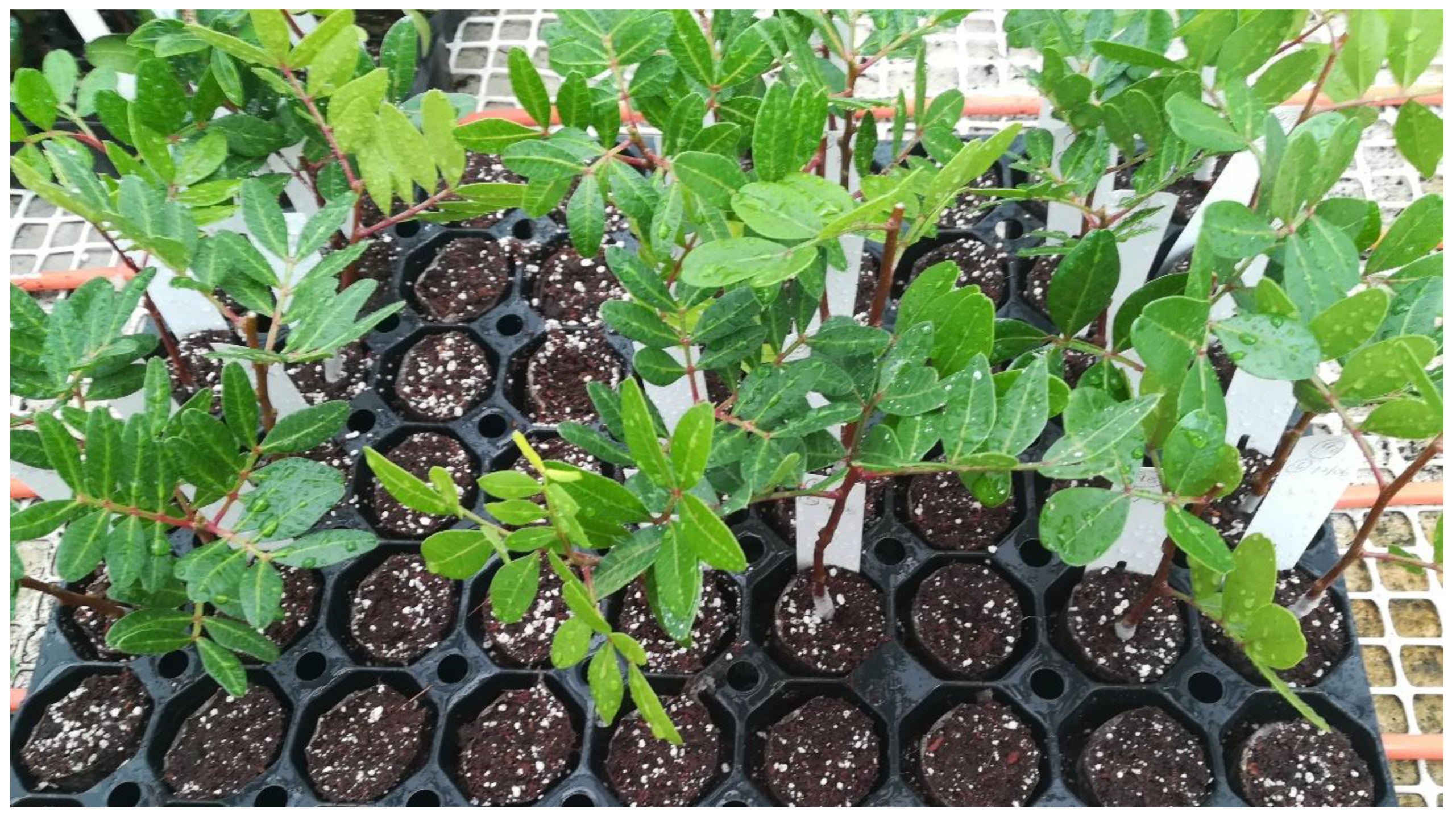
2.1. Sampling and Fungal Isolation

A total of thirty-two (32) fungal isolates were recovered from symptomatic plants in the field on Chios Island (Table 1) and in nurseries of the CMGA. The sampling period was divided into three periods (1: 12–14 May 2019; 2: 29–31 August 2019; 3–7 November 2021). Branches and twigs showing black to dark-brown discoloration of vessels and woody parts were collected from several fields and processed in the Plant Pathology Laboratory (School of Agriculture, Aristotle University of Thessaloniki, Thessaloniki, Greece), within 48 h. Other symptoms included leaf spots, defoliation, leaf, and twig blight, bud necrosis, wilting and/or apoplexy of trees, and necrotic bark tissues. Prior to the pathogen isolation process, all samples were disinfected by immersion in 50% Sodium Hypochlorite (NaOCl 5%) for 20 s, following surface disinfection with 70% ethanol for 60 s. The samples were then washed with sterile deionized water for 30 s and left to dry on a sterilized surface in a laminar flow. Small pieces of tissue were then transferred on Petri dishes with Potato Dextrose Agar (PDA, LAbM, Neogen, Lansing, MI, USA), containing 25 mg of chloramphenicol (Sigma-Aldrich, Saint Louis, MO, USA) per liter. Petri dishes were incubated in a growth chamber in the dark at 25 °C for 3–5 days, depending on the colony development. Pure colonies were obtained from single spore cultures using the serial dilution method [26] and then transferred in new PDA dishes where they were routinely kept and re-cultured every two months. Symptomatic leaves showing brown spots and shot hole symptoms, were also sampled in the field, and processed in the laboratory. Sterilization was achieved by immersion in 70% ethanol for 45 s, and 2 mm pieces of leaf tissue was transferred to the selective PDA dishes at first following growth on PDA dishes for maintenance, as mentioned above.

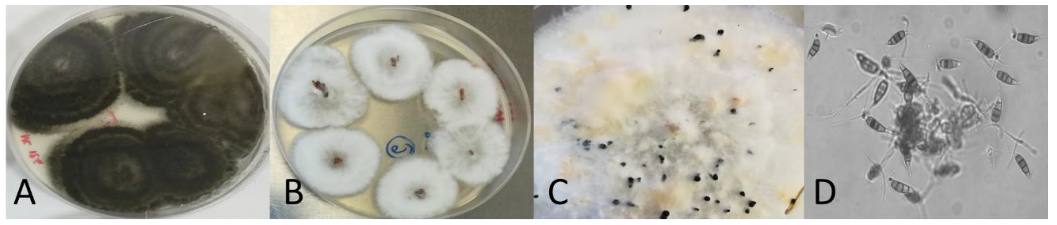
2.2. Colony Morphology and Microscopy Observation

Colony morphology of all isolates was studied macroscopically and microscopically after 5 and 10 days of incubation in the dark at 25 °C. Conidia were examined after 15 days of incubation and the formation of acervuli by some isolates was also assessed. After macroscopic and microscopic observation of the colonies, 12 isolates were identical, and the 20 remaining isolates were tested for their pathogenicity on mastic trees. Photographs were taken using a digital camera (ZEISS Axiocam ERc 5s Microscope Camera, Appleton Woods Limited, Birmingham, United Kingdom) connected to a microscope (Zeiss. Axio Lab.A1, Crespel & Deiters GmbH, Ibbenbüren, Germany).

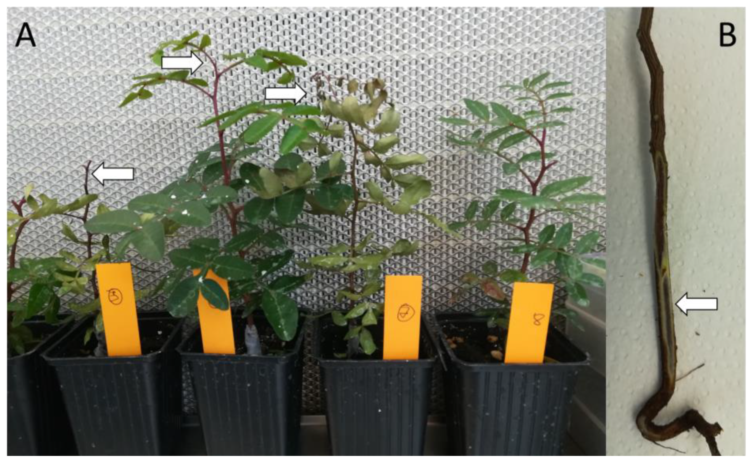
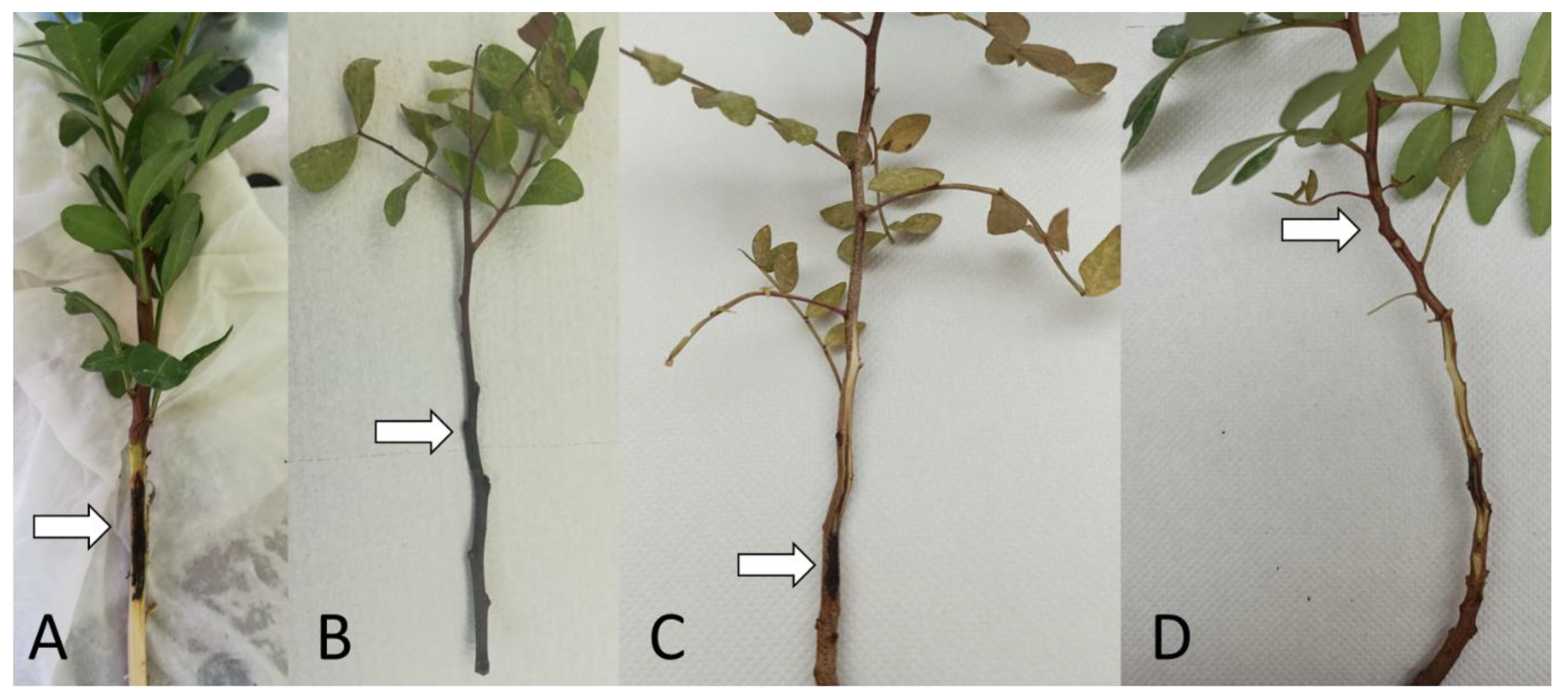
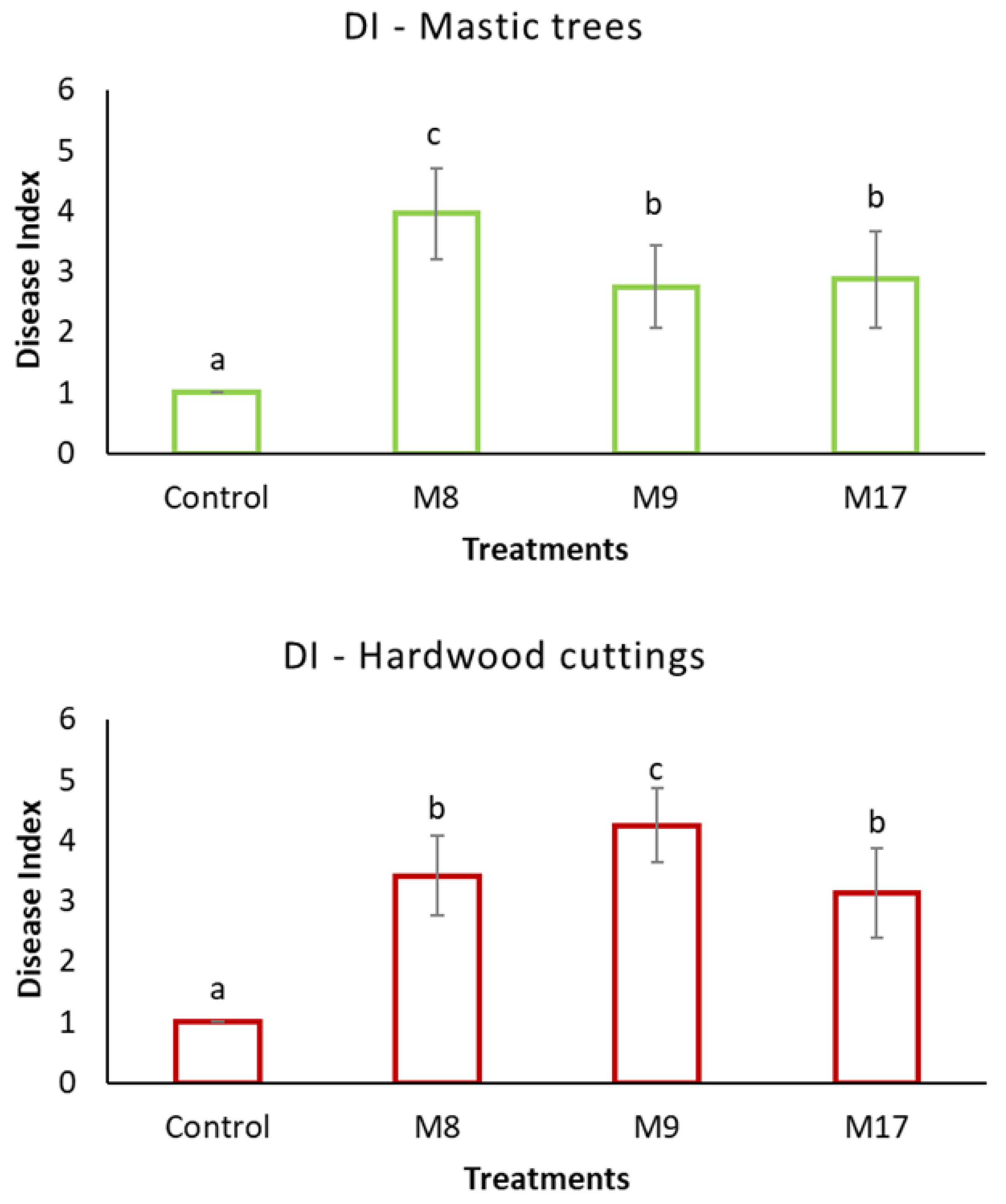
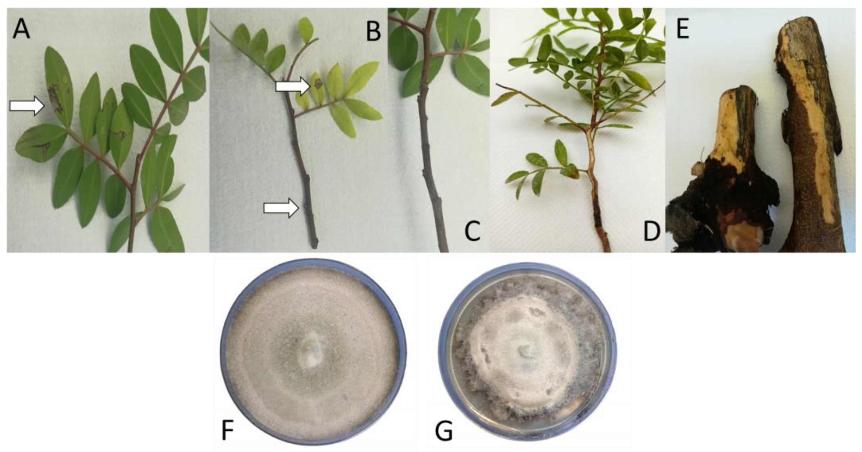
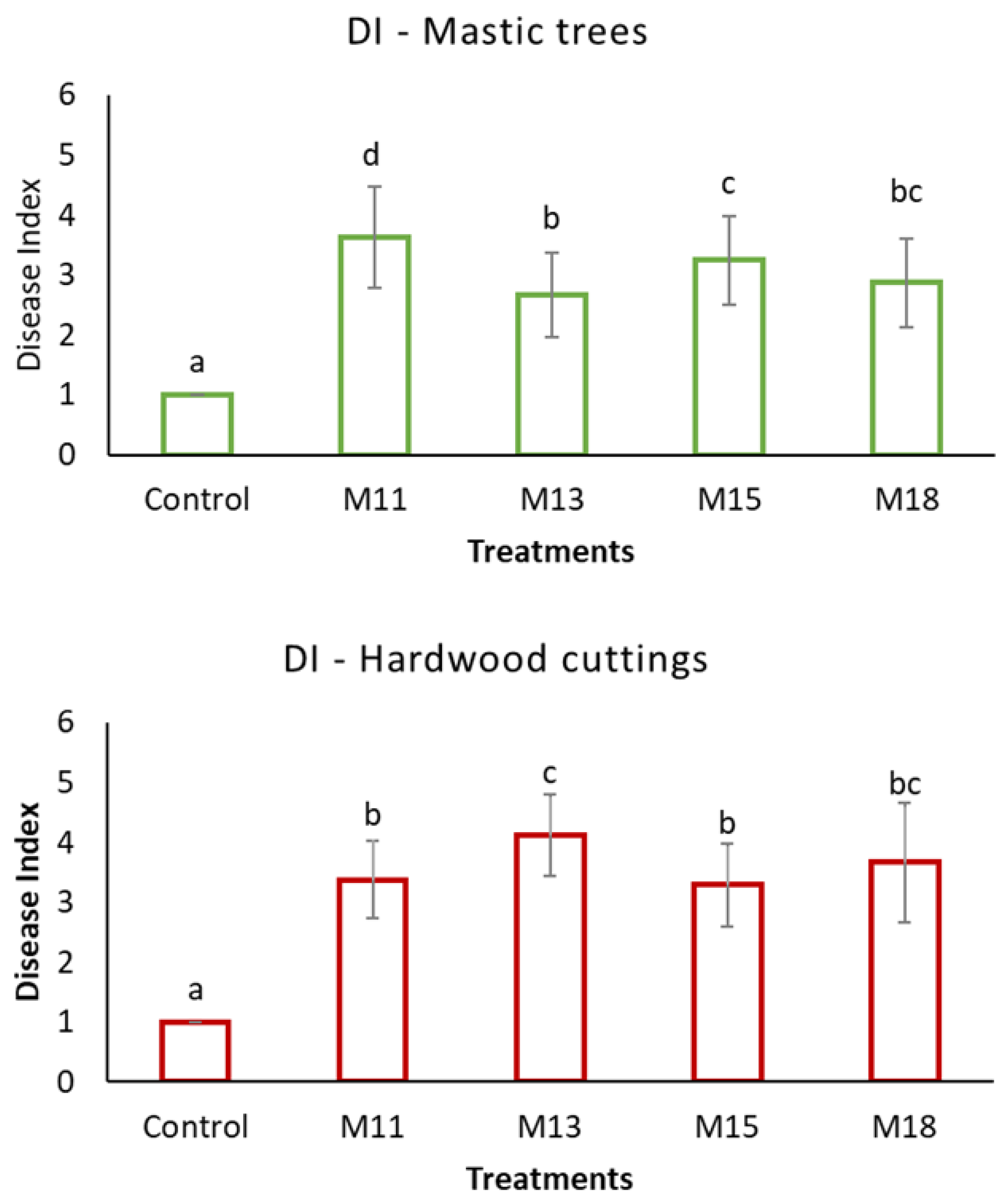
2.3. Application of Koch’s Postulates

Pathogenicity of 20 fungal isolates, sampled from woody tissues, was confirmed on two-year-old mastic trees and on hardwood cuttings used for clonal propagation of mastic trees. Colonized mycelium plugs (5 mm) were cut from the periphery of 15-day-old pure cultures of the isolates and added on previously wounded trunks, with the mycelium facing the plant tissue. The agar plugs were then covered with parafilm and sprayed with sterile distilled water to maintain humidity. Young trees and cuttings inoculated in the same way using PDA disks were kept as controls. Eight mastic trees and eight hardwood cuttings, respectively, were used per treatment and the experiment was repeated thrice. All plants were grown under controlled greenhouse conditions (20 ± 1/18 ± 1 °C day/night temperature and 60 ± 5/70 ± 5% day/night relative humidity) and inspected weekly for symptoms.
Thirty-five days post-inoculation, discoloration of the trunk, necrotic areas around the buds, and wilting of new leaves were visible on all inoculated trees and cuttings (Figure 1), while controls remained symptomless (Figure 2). Isolations from the treatments resulted in three macroscopically and microscopically distinguished fungi that were repeatedly and constantly present on the PDA dishes. Infection development was evaluated according to the following disease index scale: DI1: Healthy plants—no symptoms; 2: No symptoms on the aboveground and the root—slight discoloration of vessels, expanding to 1–3 buds that are necrotic; 3: No symptoms on the aboveground and the root–intense discoloration of vessels, expanding to 3–10 buds that are necrotic; 4: Partial wilt of the plant that is evident on the newly sprouted leaves—intense discoloration of vessels throughout the plant; 5: Total wilt of the plant—Necrosis, dieback.
The reisolated fungi were macroscopically and microscopically observed, and then molecularly identified, thus proving Koch’s postulates fulfillment.

2.4. DNA Extraction, PCR Amplification, and Sequencing

The DNA of all isolates was extracted from mycelium using the DNeasy Blood & Tissue Kit (Qiagen, GmbH, Hilden, Germany) according to the manufacturer’s protocol. Two reference genes (ITS and β-tubulin) were targeted for sequence analysis of isolates M8, M11, and M15, and regarding the other isolates, identification was achieved by sequence analysis of three (3) reference genes (ITS, Alta1, and endoPG), with the further sequence analysis of histone when the result was inconclusive. The ITS1-5.8S-ITS2 region of all single spore isolates was amplified with primers ITS1/ ITS4 [27], and the amplification of β-tubulin for isolates M8, M11, and M15 was performed using the primers Bt2a/Bt2b [28]. Regarding isolates M9, M13, M17, and M18, the endopolygalacturonase (endoPG) and the Alternaria major allergen (Alta1) genes were amplified using specific primers PG3/PG2b [29] and Alt-for/Alt-rev [30], respectively. When extra specificity was required regarding isolate M18, primers CYLH3F/CYLH3R [31] were used to amplify histone 3 (H3).
PCR conditions and reaction mixtures were those described by Testempasis et al. (2022) [32], with slight modifications. Amplification conditions were similar for all primer pairs, as follows: 94 °C for 30 s; followed by 40 cycles of 30 s at 94 °C, 30 s at 55 °C, and 30 s at 68 °C; and final extension 5 min at 68 °C. PCR products were separated by electrophoresis in 1.5% agarose gel in 1 × TAE buffer (TAE; Tris acetate EDTA) and visualized with MIDORIGreen advance (Nippon Genetics, Düren, Germany) under UV light. PCR products were purified using the QIAquick PCR Purification Kit (Qiagen, GmbH, Hilden, Germany) according to the manufacturer’s protocol. Sanger sequencing in both directions was performed using all primer pairs, DNA sequence chromatograms were edited using Geneious Prime® 2022.1.1 (Biomatters Ltd., Auckland, New Zealand) software, and all obtained sequences were compared with sequences in the National Center for Biotechnology Information database using Blastn software. Phylogenetic analysis was carried out with maximum likelihood analysis (Tamura-Nei model), which was performed at the Geneious Prime® 2022.1.1 (Biomatters Ltd., Auckland, New Zealand) software. The hierarchical clustering and tree construction were performed using the UPGMA (unweighted pair group method with arithmetic mean) model in which 1000 rapid bootstrap replicates were run. Regarding the phylogenetic tree of Neopestalotiopsis species (Figure S1), Seiridium camelliae strain SD096 was used as the outgroup and the accession numbers of all strains used are the following: Seiridium camelliae (JQ683725), Neopestalotiopsis piceana (MH855130), Neopestalotiopsis clavispora (OP783346), Neopestalotiopsis clavispora (OP895136), Neopestalotiopsis clavispora (OP895137), Neopestalotiopsis clavispora (MG729690), Neopestalotiopsis formicarum (MH860500), and Neopestalotiopsis ellipsospora (KM199343). Regarding the phylogenetic tree of Alternaria species (Figure S2), Alternaria solani was used as the outgroup and the accession numbers of all strains used are the following: Alternaria solani (KJ397978), Alternaria arborescens (BMP0582), Alternaria arborescens (KY561852), Alternaria alternata (BMP0561), Alternaria alternata (BMP0591), Alternaria alternata (BMP0653), Alternaria alternata (EGS_34_016), Alternaria alternata (EGS_34_039), Alternaria alternata (BMP0660), Alternaria tenuissima (EGS_34_015).

3. Results

Pathogenicity tests demonstrated that 11 out of 20 isolates were able to cause pathogenic symptoms in mastic tree (Table 1). Observation of colony and conidia morphology lead to the conclusion that isolates M8, M11, and M15 were identical and identified as N. clavispora. Moreover, M13 and M17 were also identical and identified as A. alternata. The typical symptoms caused by N. clavispora isolates included wilting, extended bud necrosis, discoloration of vessels, and bark cankers both on young mastic trees and on hardwood propagation cuttings (Figure 3). A. arborescens caused wilting, limited bud necrosis, vessel, and bark discoloration, on both artificially inoculated two-year-old trees and hardwood cuttings of mastic trees (Figure 4). The disease index assessment revealed that N. clavispora caused the most severe symptoms on the two-year-old trees, whereas in the case of hardwood cutting, A. arborescens provoked a higher disease index (Figure 5). Both N. clavispora and A. arborescens were constantly reisolated from all artificially inoculated trees and cuttings and in the case of N. clavispora the formation of acervuli and slimy conidial masses were evident on the PDA cultures 14 days post isolation (Figure 6). A. alternata caused wilting, bud necrosis, wood decay, bark, and vessel discoloration, on young mastic trees under controlled conditions and in the field (Figure 7). Moreover, necrotic leaf spots were caused on two-year-old trees, underlying the complex symptomatology of this plant pathogenic fungus (Figure 7).
Disease severity was observed on mastic trees and cuttings inoculated with all other isolates of N. clavispora and A. alternata as well, proving that the symptoms that were caused by those fungi were of similar intensity, respectively (Figure 8). Once again, the disease caused by isolates M11 and M15 of N. clavispora was more severe on rooted mastic trees, whereas A. alternata M13 was more virulent on hardwood cuttings (Figure 8).

4. Discussion

Following the continuous reports of mastic trees showing wilting, wood decay, leaf spots, bark, and vessel discoloration, this investigation began to define the causal agents of these symptoms. After sampling in the nursery of the CMGA and several problematic fields, some specific pathogens were consistently isolated. The fungi N. clavispora, A. arborescens, and A. alternata were identified based on culture morphology and targeted gene sequence analysis. The species identification process regarding Neopestalotiopsis genera is controversial and according to Maharachchikumbura et al. (2012) [33], who evaluated specific regions or genes for their ability to act as reliable species-defining regions, ITS region, β-tubulin, and translation elongation 1 (tef1) genes were the more accurate choice for species definition [33]. The genera of Pestalotiopsis, Pseudopestalotiopsis, and Neopestalotiopsis comprise a wide variety of species (>235) that are important plant pathogens [33,34,35]. N. clavispora is a member of the Pestalotiopsis group and, as such, is widely distributed in the tropics and recently its geographical distribution has extended in temperate ecosystems as well. Endophytic stages of N. clavispora in mangrove trees (Bruguiera sexangular, Rhizophora harrisonii, and Phoenix reclinata) are mentioned in some studies [36,37,38], showing a possible ability to switch life modes and endure unfavorable conditions, but the interest is focused on its plant pathological aspect. In detail, N. clavispora has been proven to attack and cause important symptoms on strawberry plants (Fragaria × ananassa) in Spain, Argentina, Italy, and Uruguay [39,40,41,42], on pecan (Carya illinoinensis) and macadamia (Macadamia integrifolia) in Brazil [43,44], on China rose (Rosa sinensis) and the evergreen climber Kadsurra coccinea in China [45,46], and on blueberry (Vaccinium corymbosum) in Spain [47].
El Gali and partners (2017) [23] depicted A. alternata as the dominant pathogen, causing extended brown leaf spot on mastic trees, in addition to Pestalotiopsis fici, P. guepinii, and P. palmarum, which were defined as the causal agents of leaf blight, leaf tip death, and silvery gray leaf spots, respectively, on mastic shrubs. A. alternata is an opportunistic fungus with worldwide distribution, which is often identified as the causal agent of leaf spot disease in various hosts including trees such as platan (Platanus acerifolia) in China [48], olive tree (Olea europaea) in Pakistan [49] and apricot (Prunus armeniaca) in Iraq [50], but also in shrubs such as pomegranate (Punica granatum) in Israel [51]. Reports regarding the ability of this fungus to cause wilting, wood and bud necrosis, or branch decay are very scarce and hosts include Thuja (Thuja occidentalis) in Kazakhstan [52], and olive trees and olive tree cuttings (O. europaea) in Greece [25,53]. Similarly, A. arborescens is often identified as the causal agent of leaf spots and leaf blotch on various herbaceous hosts, and more rarely on woody plants such as almond (Prunus dulcis), apple (Malus domestica), and date palm (Phoenix dactylifera) [54,55,56,57].
Subsequently, the correlation of the symptoms A. alternata and A. arborescens caused on mastic trees is of great importance since little is known about the fact that those fungi can also cause bud necrosis, vessel discoloration, and twig blight, in addition to the leaf spots they provoke.
Mastic cultivation follows organic farming strategies and given the economic value of its resin, the necessity to define the causal agents of reduced production is crucial. This study correlated the most significant symptoms observed in fields and nurseries of mastic trees with three dominant fungal pathogens. The comparison of their impact showed that the trees were significantly affected by all fungal isolates. N. clavispora caused more intense symptoms on two-year-old trees, and the Alternaria species affected hardwood cuttings of P. lentiscus var. Chia more intensively as compared to control plants and to N. clavispora. The devastating symptoms that were observed in the nursery of CMGA, in combination with the economic importance of this specific host, underlines the importance of the present study, in terms of alerting the mastic tree growers towards finding a solution and inhibiting the propagation of N. clavispora, A. arborescens, and A. alternata on the island of Chios.

Supplementary Materials

The following supporting information can be downloaded at: https://www.mdpi.com/article/10.3390/microbiolres14010010/s1, Figure S1. The phylogenetic tree was generated by Geneious Prime® 2022.1.1 (Biomatters Ltd, New Zealand) software analyzing the data set of ITS gene as downloaded from the National Center for Biotechnology Information (NCBI) database. The Tamura—Nei genetic distance model was used, and the tree was constructed according to UPGMA method. Seiridium camelliae strain SD096 was used as the outgroup and the strains isolated within this study are written in blue. The accession numbers of all strains used are the following: Seiridium camelliae (JQ683725), Neopestalotiopsis piceana (MH855130), Neopestalotiopsis clavispora (OP783346), Neopestalotiopsis clavispora (OP895136), Neopestalotiopsis clavispora (OP895137), Neopestalotiopsis clavispora (MG729690), Neopestalotiopsis formicarum (MH860500), and Neopestalotiopsis ellipsospora (KM199343); Figure S2. The phylogenetic tree was generated by Geneious Prime® 2022.1.1 (Biomatters Ltd, New Zealand) software analyzing the data set of ITS gene as downloaded from the National Center for Biotechnology Information (NCBI) database. The Tamura—Nei genetic distance model was used, and the tree was constructed according to UPGMA method. Alternaria solani was used as the outgroup and the strains isolated within this study are written in blue. The accession numbers of all strains used are the following: Alternaria solani (KJ397978), Alternaria arborescens (BMP0582), Alternaria arborescens (KY561852), Alternaria alternata (BMP0561), Alternaria alternata (BMP0591), Alternaria alternata (BMP0653), Alternaria alternata (EGS_34_016), Alternaria alternata (EGS_34_039), Alternaria alternata (BMP0660), Alternaria tenuissima (EGS_34_015).

Author Contributions

Conceptualization, N.N.K. and A.L.L.; methodology, N.N.K., S.T. and A.L.L.; software, N.N.K. and S.T.; validation, N.N.K., S.T. and A.L.L..; formal analysis, N.N.K. and S.T.; investigation, N.N.K. and A.L.L.; resources, N.N.K. and A.L.L.; data curation, N.N.K. and A.L.L.; writing—original draft preparation, N.N.K.; writing—review and editing, N.N.K., S.T. and A.L.L.; visualization, N.N.K. and A.L.L.; supervision, A.L.L.; project administration, A.L.L.; funding acquisition, A.L.L. All authors have read and agreed to the published version of the manuscript.

Funding

This research was funded by the research project «Strengthening the cultivation of mastic trees through the use of innovative molecular methods—Mast4trees» funded by the European Union and Greek national funds through the Operational Program Competitiveness, Entrepreneurship, and Innovation, under the call RESEARCH—CREATE—INNOVATE (project code: T1EDK-01133).

Institutional Review Board Statement

Not applicable.

Informed Consent Statement

Not applicable.

Data Availability Statement

Not applicable.

Acknowledgments

The authors would like to thank Stefanos Kostas, Research and Teaching Staff from the Laboratory of Floriculture, School of Agriculture Aristotle University of Thessaloniki—Greece, for the provision of mastic trees and cuttings. We are thankful to Ilias Smyrnioudis, General Director/Head of R&D at Chios Mastiha Growers Association, and his associates Tilemahos Vasilakis and Mihalis Hazakis for the help in the collection of samples and the support during the sampling periods on the island. The authors are thankful to Efstathios Hatziloukas for his valuable advice regarding the molecular identification of the pathogens.

Conflicts of Interest

The authors declare no conflict of interest. The funders had no role in the design of the study; in the collection, analyses, or interpretation of data; in the writing of the manuscript; or in the decision to publish the results.

References

  1. Browicz, K. Pistacia lentiscus cv. Chia (Anacardiaceae) on Chios Island. Plant Syst. Evol. 1987, 155, 189–195. [Google Scholar] [CrossRef]
  2. Belles, C.; Sachtouri, C. Mastiha Island; Aegeas: Chios, Greece, 2007. [Google Scholar]
  3. Paraschos, S.; Mitakou, S.; L Skaltsounis, A. Chios Gum Mastic: A Review of its Biological Activities. Curr. Med. Chem. 2012, 19, 2292–2302. [Google Scholar] [CrossRef] [PubMed]
  4. Soulaidopoulos, S.; Tsiogka, A.; Chrysohoou, C.; Lazarou, E.; Aznaouridis, K.; Doundoulakis, I.; Tyrovola, D.; Tousoulis, D.; Tsioufis, K.; Vlachopoulos, C.; et al. Overview of Chios Mastic Gum (Pistacia lentiscus) Effects on Human Health. Nutrients 2022, 14, 590. [Google Scholar] [CrossRef] [PubMed]
  5. Ljubuncic, P.; Song, H.; Cogan, U.; Azaizeh, H.; Bomzon, A. The effects of aqueous extracts prepared from the leaves of Pistacia lentiscus in experimental liver disease. J. Ethnopharmacol. 2005, 100, 198–204. [Google Scholar] [CrossRef]
  6. Koutsoudaki, C.; Krsek, M.; Rodger, A. Chemical Composition and Antibacterial Activity of the Essential Oil and the Gum of Pistacia lentiscus Var. Chia. J. Agric. Food Chem. 2005, 53, 7681–7685. [Google Scholar] [CrossRef]
  7. Magiatis, P.; Melliou, E.; Skaltsounis, A.-L.; Chinou, I.; Mitaku, S. Chemical Composition and Antimicrobial Activity of the Essential Oils of Pistacia lentiscus var. Chia. Planta Med. 1999, 65, 749–752. [Google Scholar] [CrossRef]
  8. Assimopoulou, A.; Zlatanos, S.; Papageorgiou, V. Antioxidant activity of natural resins and bioactive triterpenes in oil substrates. Food Chem. 2005, 92, 721–727. [Google Scholar] [CrossRef]
  9. Spyridopoulou, K.; Tiptiri-Kourpeti, A.; Lampri, E.; Fitsiou, E.; Vasileiadis, S.; Vamvakias, M.; Bardouki, H.; Goussia, A.; Malamou-Mitsi, V.; Panayiotidis, M.I.; et al. Dietary mastic oil extracted from Pistacia lentiscus var. Chia suppresses tumor growth in experimental colon cancer models. Sci. Rep. 2017, 7, 3782. [Google Scholar] [CrossRef] [Green Version]
  10. Pachi, V.K.; Mikropoulou, E.V.; Gkiouvetidis, P.; Siafakas, K.; Argyropoulou, A.; Angelis, A.; Mitakou, S.; Halabalaki, M. Traditional uses, phytochemistry and pharmacology of Chios mastic gum (Pistacia lentiscus var. Chia, Anacardiaceae): A review. J. Ethnopharmacol. 2020, 254, 112485. [Google Scholar] [CrossRef]
  11. Huwez, F.U.; Thirlwell, D.; Cockayne, A.; Ala’Aldeen, D.A.A. Mastic Gum Kills Helicobacter pylori. N. Engl. J. Med. 1998, 339, 1946. [Google Scholar] [CrossRef]
  12. Marone, P.; Bono, L.; Leone, E.; Bona, S.; Carretto, E.; Perversi, L. Bactericidal Activity of Pistacia lentiscus Mastic Gum Against Helicobacter pylori. J. Chemother. 2001, 13, 611–614. [Google Scholar] [CrossRef] [PubMed]
  13. Sotirios, P.; Prokopios, M.; Sofia, M.; Kalliopi, P.; Antonios, K.; Petros, M.; Andreas, M.; Dionyssios, S.; Alexios-Leandros, S. In Vitro and In Vivo Activities of Chios Mastic Gum Extracts and Constituents against Helicobacter pylori. Antimicrob. Agents Chemother. 2007, 51, 551–559. [Google Scholar] [CrossRef] [Green Version]
  14. Dabos, K.J.; Sfika, E.; Vlatta, L.J.; Giannikopoulos, G. The effect of mastic gum on Helicobacter pylori: A randomized pilot study. Phytomedicine 2010, 17, 296–299. [Google Scholar] [CrossRef] [PubMed]
  15. Miyamoto, T.; Okimoto, T.; Kuwano, M. Chemical Composition of the Essential Oil of Mastic Gum and their Antibacterial Activity Against Drug-Resistant Helicobacter pylori. Nat. Prod. Bioprospect 2014, 4, 227–231. [Google Scholar] [CrossRef] [Green Version]
  16. Shmuely, H.; Domniz, N.; Yahav, J. Non-pharmacological treatment of Helicobacter pylori. World J. Gastrointest. Pharmacol. Ther. 2016, 7, 171. [Google Scholar] [CrossRef] [PubMed]
  17. Pachi, V.K.; Mikropoulou, E.V.; Dimou, S.; Dionysopoulou, M.; Argyropoulou, A.; Diallinas, G.; Halabalaki, M. Chemical Profiling of Pistacia lentiscus var. Chia Resin and Essential Oil: Ageing Markers and Antimicrobial Activity. Processes 2021, 9, 418. [Google Scholar] [CrossRef]
  18. Rigling, M.; Fraatz, M.A.; Trögel, S.; Sun, J.; Zorn, H.; Zhang, Y. Aroma Investigation of Chios Mastic Gum (Pistacia lentiscus Variety Chia) Using Headspace Gas Chromatography Combined with Olfactory Detection and Chiral Analysis. J. Agric. Food Chem. 2019, 67, 13420–13429. [Google Scholar] [CrossRef] [PubMed]
  19. The Chios Mastiha Growers Association (CGMA)—The Association of the Mastiha Producers. Available online: https://www.gummastic.gr/en/itemlist/category/26-home-gr (accessed on 25 November 2022).
  20. European Pharmacopoeia. Herbal Drugs and Herbal Drug Preparations; European Pharmacopoeia 9.0.; Council of Europe: Strasbourg, France, 2017; p. 1430. [Google Scholar]
  21. Zografou, P.; Linos, A.; Hagidimitriou, M. Genetic diversity among different genotypes of Pistacia lentiscus var. chia (mastic tree). In Proceedings of the XIV GREMPA Meeting on Pistachios and Almonds, Athens, Greece, 1 January 2010; CIHEAM: Zaragoza, Spain, 2010. [Google Scholar]
  22. İsfendiyaroğlu, M. Propagation of Mastic Tree: From Seed to Tissue Culture. In Proceedings of the 4th International Symposium of Medicinal and Aromatic Plants, İzmir, Turkey, 2–4 October 2018. [Google Scholar]
  23. El-Gali, Z.I. Incidences of Fungal Leaf Diseases on Mastic Shrubs in Libya. Int. J. Res. 2017, 5, 22–26. [Google Scholar] [CrossRef]
  24. Göre, M.E.; Parlak, S.; Aydın, M.H. Pestalotiopsis guepinii newly reported to cause dieback on Pistacia lentiscus var. Chia in Turkey. Plant Path. 2010, 59, 1169. [Google Scholar] [CrossRef]
  25. Tziros, G.T.; Karpouzis, A.; Lagopodi, A.L. Alternaria alternata as the cause of decline and necrosis on olive tree cuttings in Greece. Australas. Plant Dis. Notes 2021, 16, 7. [Google Scholar] [CrossRef]
  26. Davis, W.H. Single Spore Isolation. Iowa Acad. Sci. 1930, 37, 151–159. Available online: https://scholarworks.uni.edu/pias/vol37/iss1/29 (accessed on 30 November 2022).
  27. White, T.J.; Bruns, T.; Lee, S.; Taylor, J. Amplification and direct sequencing of fungal ribosomal RNA genes for phylogenetics. In PCR Protocols a Guide to Methods and Applications; Academic Press: San Diego, CA, USA, 1990; pp. 315–322. [Google Scholar]
  28. Glass, N.L.; Donaldson, G.C. Development of primer sets designed for use with the PCR to amplify conserved genes from filamentous ascomycetes. Appl. Environ. Microbiol. 1995, 61, 1323–1330. [Google Scholar] [CrossRef] [PubMed] [Green Version]
  29. Andrew, M.; Peever, T.L.; Pryor, B.M. An expanded multilocus phylogeny does not resolve morphological species within the small-spored Alternaria species complex. Mycologia 2009, 101, 95–109. [Google Scholar] [CrossRef] [PubMed] [Green Version]
  30. Woudenberg, J.H.C.; Seidl, M.F.; Groenewald, J.Z.; de Vries, M.; Stielow, J.B.; Thomma, B.P.H.J.; Crous, P.W. Alternaria section Alternaria: Species, formae speciales or pathotypes? Saprobic and Phytopathogenic Dothideomycetes. Stud. Mycol. 2015, 82, 1–21. [Google Scholar] [CrossRef] [PubMed] [Green Version]
  31. Crous, P.W.; Groenewald, J.Z.; Risède, J.-M.; Simoneau, P.; Hywel-Jones, N.L. Calonectria species and their Cylindrocladium anamorphs: Species with sphaeropedunculate vesicles. Scopus 2004, 50, 415–430. Available online: https://www.scopus.com/inward/record.uri?eid=2-s2.0-20144389253&partnerID=40&md5=e6e40aed3e3969c5d7434f767b79e065 (accessed on 25 November 2022).
  32. Testempasis, S.I.; Kamou, N.N.; Papadakis, E.-N.; Menkissoglu-Spiroudi, U.; Karaoglanidis, G.S. Conventional vs. organic vineyards: Black Aspergilli population structure, mycotoxigenic capacity and mycotoxin contamination assessment in wines, using a new Q-TOF MS-MS detection method. Food Control 2022, 136, 108860. [Google Scholar] [CrossRef]
  33. Maharachchikumbura, S.S.N.; Guo, L.-D.; Cai, L.; Chukeatirote, E.; Wu, W.P.; Sun, X.; Crous, P.W.; Bhat, D.J.; McKenzie, E.H.C.; Bahkali, A.H.; et al. A multi-locus backbone tree for Pestalotiopsis, with a polyphasic characterization of 14 new species. Fungal Divers. 2012, 56, 95–129. [Google Scholar] [CrossRef]
  34. Maharachchikumbura, S.S.N.; Guo, L.-D.; Chukeatirote, E.; Bahkali, A.H.; Hyde, K.D. Pestalotiopsis—Morphology, phylogeny, biochemistry and diversity. Fungal Divers. 2011, 50, 167–187. [Google Scholar] [CrossRef]
  35. Wang, Y.; Xiong, F.; Lu, Q.; Hao, X.; Zheng, M.; Wang, L.; Li, N.; Ding, C.; Wang, X.; Yang, Y. Diversity of Pestalotiopsis-like Species Causing Gray Blight Disease of Tea Plants (Camellia sinensis) in China, Including two Novel Pestalotiopsis Species, and Analysis of Their Pathogenicity. Plant Dis. 2019, 103, 2548–2558. [Google Scholar] [CrossRef]
  36. Luo, D.-Q.; Deng, H.-Y.; Yang, X.-L.; Shi, B.-Z.; Zhang, J.-Z. Oleanane-Type Triterpenoids from the Endophytic Fungus Pestalotiopsis clavispora Isolated from the Chinese Mangrove Plant Bruguiera sexangula. Helv. Chim. Acta 2011, 94, 1041–1047. [Google Scholar] [CrossRef]
  37. Hemphill Pérez, C.F.; Daletos, G.; Liu, Z.; Lin, W.; Proksch, P. Polyketides from the Mangrove-derived fungal endophyte Pestalotiopsis clavispora. Tetrahedron Lett. 2016, 57, 2078–2083. [Google Scholar] [CrossRef]
  38. Alade, G.O.; Moody, J.O.; Bakare, A.G.; Awotona, O.R.; Adesanya, S.; Lai, D.; Debbab, A.; Proksch, P. Metabolites from endophytic fungus; Pestalotiopsis clavispora isolated from Phoenix reclinata leaf. Future J. Pharm. Sci. 2018, 4, 273–275. [Google Scholar] [CrossRef]
  39. Chamorro, M.; Aguado, A.; De los Santos, B. First report of root and crown rot caused by Pestalotiopsis clavispora (Neopestalotiopsis clavispora) on strawberry in Spain. Plant Dis. 2016, 100, 1495. [Google Scholar] [CrossRef]
  40. Obregón, V.G.; Meneguzzi, N.G.; Ibañez, J.M.; Lattar, T.E.; Kirschbaum, D.S. First Report of Neopestalotiopsis clavispora Causing Root and Crown Rot on Strawberry Plants in Argentina. Plant Dis. 2018, 102, 1856. [Google Scholar] [CrossRef]
  41. Sigillo, L.; Ruocco, M.; Gualtieri, L.; Pane, C.; Zaccardelli, M. First report of Neopestalotiopsis clavispora causing crown rot in strawberry in Italy. J. Plant Path. 2020, 102, 281. [Google Scholar] [CrossRef] [Green Version]
  42. Machín, A.; González, P.; Vicente, E.; Sánchez, M.; Estelda, C.; Ghelfi, J.; Silvera-Pérez, E. First Report of Root and Crown Rot Caused by Neopestalotiopsis clavispora on Strawberry in Uruguay. Plant Dis. 2019, 103, 2946. [Google Scholar] [CrossRef]
  43. Lazarotto, M.; Muniz, M.F.B.; Poletto, T.; Dutra, C.B.; Blume, E.; Harakawa, R.; Poletto, I. First Report of Pestalotiopsis clavispora Causing Leaf Spot of Carya illinoensis in Brazil. Plant Dis. 2012, 96, 1826. [Google Scholar] [CrossRef] [PubMed]
  44. Santos, C.C.; Domingues, J.L.; Santos, R.F.; Spósito, M.B.; Santos, A.; Novaes, Q.S. First Report of Neopestalotiopsis clavispora Causing Leaf Spot on Macadamia in Brazil. Plant Dis. 2019, 103, 1790. [Google Scholar] [CrossRef]
  45. Feng, Y.R.; Liu, B.S.; Sun, B.B. First Report of Leaf Blotch Caused by Pestalotiopsis clavispora on Rosa chinensis in China. Plant Dis. 2014, 98, 1009. [Google Scholar] [CrossRef]
  46. Xie, J.; Wei, J.G.; Huang, R.S.; Wei, J.F.; Luo, J.T.; Yang, X.H.; Yang, X.B. First Report of Ring Spot on Kadsura coccinea Caused by Neopestalotiopsis clavispora in China. Plant Dis. 2018, 102, 2032. [Google Scholar] [CrossRef]
  47. Borrero, C.; Castaño, R.; Avilés, M. First Report of Pestalotiopsis clavispora (Neopestalotiopsis clavispora) Causing Canker and Twig Dieback on Blueberry Bushes in Spain. Plant Dis. 2018, 102, 1178. [Google Scholar] [CrossRef]
  48. Qin, W.; Zhao, J.; Qiao, G.H.; Liu, J.; Tan, X. First report of leaf blight caused by Alternaria alternata on Platanus acerifolia in China. Plant Dis. 2022, 0, 1–9. [Google Scholar] [CrossRef] [PubMed]
  49. Farr, D.F.; Rossman, A.Y. Fungal Databases, U.S. National Fungus Collections, ARS, USDA. 2019. Available online: https://nt.ars-grin.gov/fungaldatabases (accessed on 25 November 2022).
  50. Hameed, Z.L.; Lahuf, A.A.; Jasim, M.T.; Mohsen, H.M.; Kadim, B.J.; Saleh, S.A.; Mohamed, A.F. First Report of Alternaria Alternata Causing Brown Leaf Spot on Apricot (Prunus Armeniaca) in Karbala Province of Iraq. In Proceedings of the Fourth International Conference for Agricultural and Sustainability Sciences, Babil, Iraq, 4–5 October 2021; Volume 910, p. 012080. [Google Scholar] [CrossRef]
  51. Ezra, D.; Shulhani, R.; Bar Ya’akov, I.; Harel-Beja, R.; Holland, D.; Shtienberg, D. Factors Affecting the Response of Pomegranate Fruit to Alternaria alternata, the Causal Agent of Heart Rot. Plant Dis. 2019, 103, 315–323. [Google Scholar] [CrossRef] [PubMed] [Green Version]
  52. Smagulova, A.; Uakhit, R.; Kiyan, V. First Record of Alternaria alternata Causing Necrosis of Thuja (Thuja occidentalis) in Kazakhstan. Plant Dis. 2022, 106, 2987. [Google Scholar] [CrossRef] [PubMed]
  53. Lagogianni, C.S.; Tjamos, E.C.; Antoniou, P.P.; Tsitsigiannis, D.I. First Report of Alternaria alternata as the Causal Agent of Alternaria Bud and Blossom Blight of Olives. Plant Dis. 2017, 101, 2151. [Google Scholar] [CrossRef]
  54. Teviotdale, B.L.; Viveros, M.; Pryor, B.; Adaskaveg, J.E. First Report of Alternaria Leaf Spot of Almond Caused by Species in the Alternaria alternata Complex in California. Plant Dis. 2001, 85, 558. [Google Scholar] [CrossRef]
  55. Wenneker, M.; Pham, K.T.K.; Woudenberg, J.H.C.; Thomma, B.P.H.J. First Report of Alternaria arborescens Species Complex Causing Leaf Blotch and Associated Premature Leaf Drop of ‘Golden Delicious’ Apple Trees in the Netherlands. Plant Dis. 2018, 102, 1654. [Google Scholar] [CrossRef]
  56. Fontaine, K.; Fourrier-Jeandel, C.; Armitage, A.D.; Boutigny, A.-L.; Crépet, M.; Caffier, V.; Gnide, D.C.; Shiller, J.; Le Cam, B.; Giraud, M.; et al. Identification and pathogenicity of Alternaria species associated with leaf blotch disease and premature defoliation in French apple orchards. PeerJ 2021, 9, e12496. [Google Scholar] [CrossRef] [PubMed]
  57. Namsi, A.; Gargouri, S.; Rabaoui, A.; Mokhtar, N.; Takrouni, M.L.; Moretti, A.; Masiello, M.; Touil, S.; Dieb, L.; Werbrouck, S.P.O. First Report of Leaf Blight Caused by Alternaria mali and A. arborescens on Date Palm (Phoenix dactylifera) in Tunisia. Plant Dis. 2019, 103, 2962. [Google Scholar] [CrossRef]
Figure 1. Discoloration of the trunk, necrotic areas around the buds and wilting of new leaves visible on all inoculated trees and cuttings 35 days post inoculation (arrows) (A). Visible dark brown discoloration of the vessels of two-year-old mastic trees artificially inoculated with Neopestalotiopsis clavispora (arrow) (B).
Microbiolres 14 00010 g001
Figure 2. Absence of disease symptoms on hardwood cuttings used for clonal propagation of mastic trees used as controls 35 days post inoculation with uncolonized PDA agar plugs.
Microbiolres 14 00010 g002
Figure 3. Disease symptoms on Pistacia lentiscus var. Chia artificially inoculated with Neopestalotiopsis clavispora: Two-year-old tree showing bark discoloration and cankers, as well as acervuli of the fungus (arrow) (A). Vessel discoloration and bud necrosis on hardwood cuttings (arrow) (Β). Wood decay and vessel discoloration (arrow) on mastic tree branches in the field (Chios Island, 38°14′07″ Ν 25°57′38″ Ε) (C). Discoloration of bark and vessels (arrows) of two-year-old trees without bud necrosis (DF). Intense wilting of young leaves, vessel discoloration and bark cankers (G). Wood decay and vessel discoloration on mastic tree branches and twigs, with defoliation symptoms in the field (Chios Island, 38°14′07″ Ν 25°57′38″ Ε) (H,I).
Microbiolres 14 00010 g003
Figure 4. Disease symptoms on Pistacia lentiscus var. Chia artificially inoculated with Alternaria arborescens: Two-year-old tree showing vessel discoloration and bud necrosis (arrow) (A). Hardwood cuttings of mastic tree showing bark discoloration (arrow) (B). Wilting, vessel discoloration and bud necrosis on young mastic trees (arrow) (C). Extended bud necrosis and vessel discoloration on two-year-old mastic trees (D).
Microbiolres 14 00010 g004
Figure 5. Severity of disease caused by Neopestalotiopsis clavispora M8, Alternaria arborescens M9, and A. alternata M17, which were artificially inoculated on two-year-old mastic trees and hardwood cuttings grown in pots under greenhouse conditions. Control plants were inoculated only with PDA plugs, without the pathogen. Disease development was assessed 35 days after inoculation. The experiment was repeated three times. Different letters indicate significant differences according to Tukey’s test at p ≤ 0.05. Error bars represent standard deviation.
Microbiolres 14 00010 g005
Figure 6. PDA culture of Alternaria arborescens constantly reisolated from artificially inoculated mastic trees with symptoms of bud necrosis and discoloration (A). Culture of Neopestalotiopsis clavispora constantly reisolated from artificially inoculated mastic trees with symptoms of brown discoloration of the vessels, bud necrosis, and wilting (B). Acervuli and conidial slimy masses formed by N. clavispora in colonies on PDA (C). Conidia of N. clavispora located in important quantities in the fungal acervuli (D).
Microbiolres 14 00010 g006
Figure 7. Disease symptoms on Pistacia lentiscus var. Chia artificially inoculated with Alternaria alternata: Two-year-old tree showing necrotic leaf spots (arrows) (A,B). Two-year-old tree showing necrotic leaf spots, bark, and vessel discoloration (arrows) (BD). Wood decay and vessel discoloration on branches in the field (Chios Island, 38°12′13″ Ν 25°59′59″ Ε) (E). PDA culture of Alternaria alternata M17 (F) and PDA culture of Alternaria alternata /A. tenuissima M18 (G).
Microbiolres 14 00010 g007
Figure 8. Severity of disease caused by Neopestalotiopsis clavispora M11 and M15, and Alternaria alternata M13 and M18 that were artificially inoculated on two-year-old mastic trees and hardwood cuttings grown in pots under greenhouse conditions. Control plants were inoculated only with PDA plugs, without the pathogen. Disease development was assessed 35 days after inoculation. The experiment was repeated three times. Different letters indicate significant differences according to Tukey’s test at p ≤ 0.05. Error bars represent standard deviation.
Microbiolres 14 00010 g008
Table 1. Locations, hosts, isolation data and accession numbers for the isolated fungi that caused symptoms on artificially inoculated mastic trees.
Isolate InformationAccession Numbers
Isolate NumberFungal SpeciesHost, TissueLocationDateITSβ-tubulinAlta-1EndoPGHistone
M8Neopestalotiopsis clavisporaCuttings, necrotized budsNursery of CMGAMay 2019OP783346OP897766n. a.n. a.n. a.
M9Alternaria arborescensCuttings, necrotized budsNursery of CMGAMay 2019OP783347n. a.OP817016OP817018n. a.
M11Neopestalotiopsis clavisporaMastic tree, wood rots on barkField, Chios Island
(38°14′07″ Ν 25°57′38″ Ε)
August 2019OP895136OP897767n. a.n. a.n. a.
M13Alternaria alternataMastic tree, necrotic twigs, and branches, necrotic budsField, Chios Island
(38°13′42″ Ν 26°00′17″ Ε)
November 2021OP895138n. a.OP897762OP897764n. a.
M15Neopestalotiopsis clavisporaMastic tree, necrotic twigs, and branches, necrotic budsField, Chios Island
(38°14′31″ Ν 26°01′01″ Ε)
November 2021OP895137OP897768n. a.n. a.n. a.
M17Alternaria alternataMastic tree, necrotic twigs, and branches with discolorations, leaves with spotsField, Chios Island
(38°12′13″ Ν 25°59′59″ Ε)
November 2021OP783348n. a.OP817017OP817019n. a.
M18Alternaria alternata/ A. tenuissimaMastic tree, necrotic twigs, and branches with discolorations, leaves with spotsField, Chios Island
(38°12′13″ Ν 25°59′59″ Ε)
November 2021OP895139n. a.OP897763OP897765OP897769
n.a. stands for not available.
Disclaimer/Publisher’s Note: The statements, opinions and data contained in all publications are solely those of the individual author(s) and contributor(s) and not of MDPI and/or the editor(s). MDPI and/or the editor(s) disclaim responsibility for any injury to people or property resulting from any ideas, methods, instructions or products referred to in the content.

