Review

Gastric Syphilis Presenting as a Nodal Inflammatory Pseudotumor Mimicking a Neoplasm: Don’t Forget the Treponema! Case Report and Scoping Review of the Literature of the Last 65 Years

1
Gastroenterology & Endoscopy Unit, Fondazione Istituto Gemelli–G. Giglio, Contrada Pietra Pollastra Pisciotto, 90015 Cefalù, Italy
2
Mini-Invasivecolorectal & Pancreatic Surgery Unit, Fondazione Istituto Gemelli–G. Giglio, Contrada Pietra Pollastra Pisciotto, 90015 Cefalù, Italy
3
Pathology Unit, Az. Osp. Ospedali Riuniti “Villa Sofia-Cervello”, Via Trabucco 180, 90146 Palermo, Italy
4
Gastroenterology Unit, National Institute of Gastroenterology-IRCCS “Saverio de Bellis”, Castellana Grotte, 70013 Bari, Italy
5
Gastroenterology and Endoscopy Unit, S. Elia-Raimondi Hospital, 93100 Caltanissetta, Italy
6
Pathology Unit, Sant’Antonio Abate Hospital, Via Cosenza 82, 91016 Casa Santa, Italy
7
Pathology Unit, Fondazione Istituto Gemelli–G. Giglio, Contrada Pietra Pollastra Pisciotto, 90015 Cefalù, Italy
8
Radiology Unit, Fondazione Istituto Gemelli–G. Giglio, Contrada Pietra Pollastra Pisciotto, 90015 Cefalù, Italy
*
Author to whom correspondence should be addressed.
Gastroenterol. Insights 2023, 14(2), 178-190; https://doi.org/10.3390/gastroent14020014
Submission received: 12 December 2022 / Revised: 13 February 2023 / Accepted: 8 March 2023 / Published: 4 April 2023
(This article belongs to the Special Issue Management and Treatment of Digestive Disorders)

Abstract

:
Despite the fact that gastric syphilis is considered rare, it is reported as a type of organic involvement that is present in a large proportion of secondary syphilis cases, even though gastritis presenting with symptoms is extremely rare. Clinical, radiological, and endoscopic findings are non-specific and frequently mimic the symptoms of gastric adenocarcinoma or lymphoma, making diagnosis difficult. Immunostaining is required for this diagnosis. We would like to emphasize the importance of being suspicious of GS when a gastric mass exhibits the histologic features of an inflammatory pseudotumor (IPT), as previously reported for nodal IPT caused by luetic infection. We described a 56-year-old man who presented to the oncology department with a 3-month history of anorexia, epigastric pain, nausea, vomiting, and weight loss, as well as an initial radiological and endoscopic suspicion of gastric adenocarcinoma, in which immune staining allowed us to diagnose GS. In addition, we conducted an updated scoping review of the scientific literature to show the clinical, laboratory, and therapeutic findings in GS patients over the last 65 years.

1. Introduction

Syphilis is a disease that is on the rise all over the world. Gastric syphilis (GS) is a very rare presentation in this setting, occurring in less than 1% of cases and typically developing in secondary syphilis [1,2]. Epigastric pain, fullness, nausea, vomiting, and weight loss are the most common symptoms of GS [1,3]. Surprisingly, up to 70% of patients have no concurrent conventional clinical symptoms of syphilis (such as genital ulcers, inguinal lymphadenopathy, etc.) [1,3]. Clinical, radiological, and endoscopic signs are non-specific and frequently mimic the symptoms of gastric adenocarcinoma or lymphoma, making diagnosis difficult. Immunostaining is required for this diagnosis. We would like to emphasize the importance of suspecting GS when a gastric mass exhibits the histologic features of an IPT, as previously reported for nodal IPT caused by luetic infection. We described the case of a 56-year-old man who presented to the oncology department with a 3-month history of anorexia, epigastric pain, nausea, vomiting, and weight loss, as well as an initial radiological and endoscopic suspicion of gastric adenocarcinoma, which was confirmed by immune staining. Furthermore, we conducted an updated review of the scientific literature, as previously executed by Mylona and colleagues [3], to show the clinical, laboratory, and therapeutic findings in GS patients over the last 65 years.

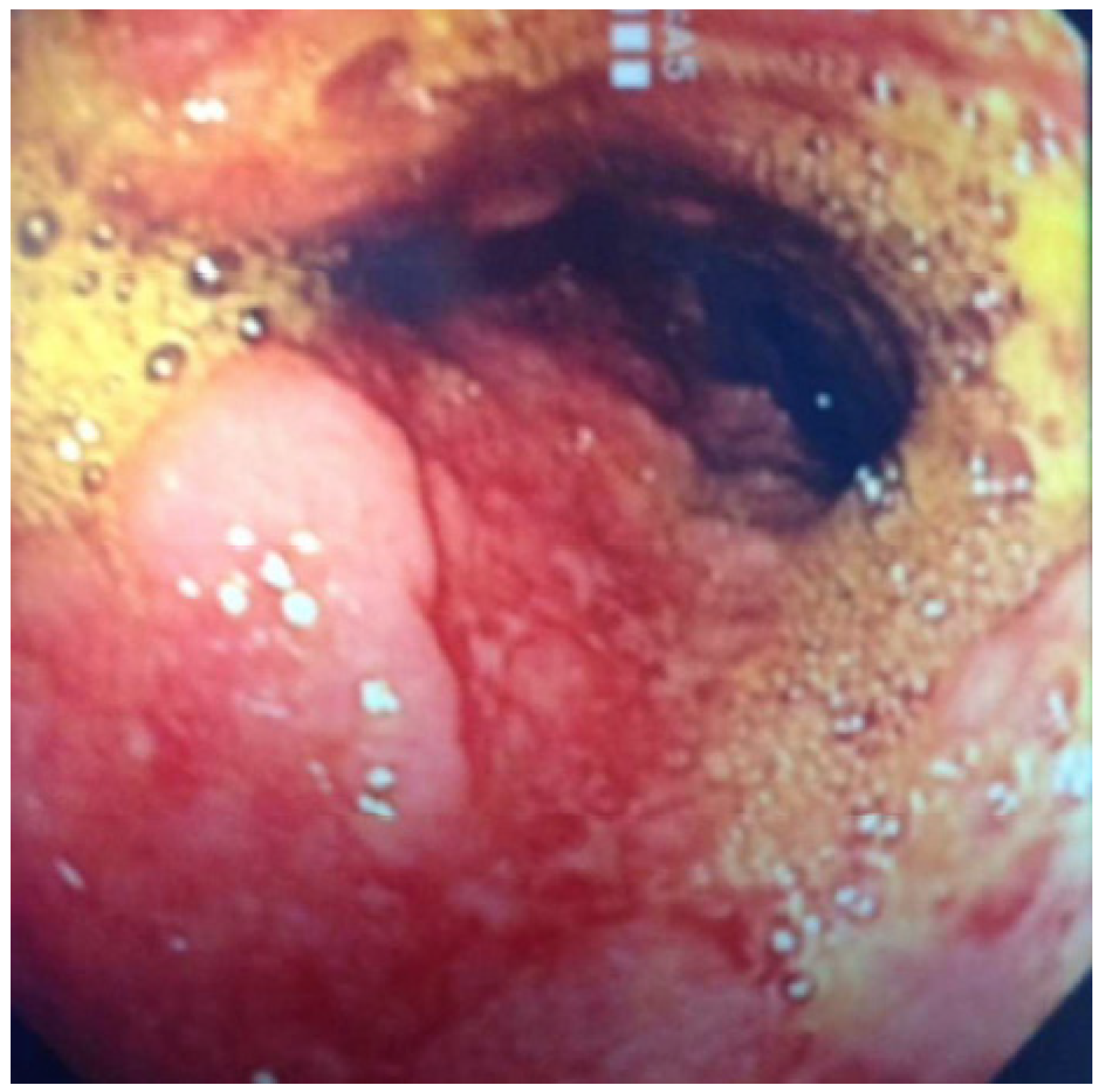
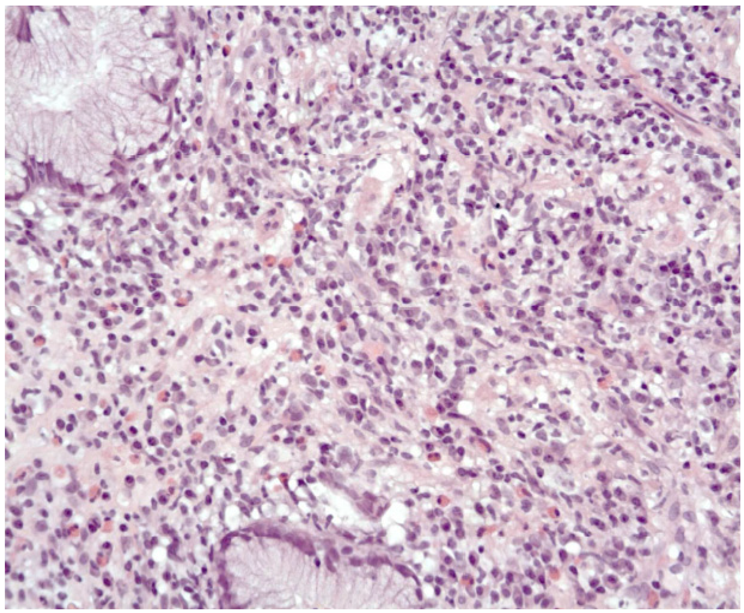
2. Case Presentation

A 56-year-old man arrived at the oncology department with a 3-month history of epigastric pain, nausea, anorexia, vomiting, and an 8-kg weight loss. He denied having ever had a fever, a skin rash, or lymphadenopathy. His previous medical history was unremarkable, and he had no personal or familial antecedents for gastrointestinal disorders, abdominal surgery, comorbidities, or previous hospitalization. A physical examination revealed significant abdominal tenderness. Anemia with a hemoglobin level of 9.1 g/dL (normal range, 12.0–16.0), a normal white blood cell (WBC) count, and normal hepatic and renal function were discovered during the initial laboratory work-up. A previous computed tomography scan revealed significant wall thickening of both the gastric corpus and antrum, raising the possibility of a gastric neoplasm. Esophagogastroduodenoscopy (EGDS) revealed a decreased expandability and an irregularly ulcerated, thickened, nodular, edematous, and friable mass from the stomach’s distal body to the antrum (Figure 1). Histopathological analysis of four gastric samples revealed marked inflammatory infiltration resembling an inflammatory pseudotumor (IPT) rich in plasmocytes and eosinophils (Figure 2 and Figure 3), but negative for Helicobacter pylori. Further testing revealed that the patient tested negative for HIV but positive for VDRL and Treponema pallidum reagents. GS was suspected based on these clinical, laboratory, radiologic, and endoscopic findings, and further investigation included immunohistochemical tests, which revealed that plasmocytes of the lamina propria were positive for Treponema pallidum, CD20, CD3, and kappa and lambda chains (Figure 4).
The patient, who had previously confirmed having promiscuous sex, was treated with 2,400,000 UI of penicillin, which resulted in a complete resolution of his clinical symptoms within one month. However, the patient refused to repeat the endoscopic follow-up to confirm ulcer healing. In addition, the patient refused to have an ileocolonoscopy to determine the presence or absence of a T. pallidum infection in the terminal ileum and the large intestine.

3. Review Methods

The current study, which was previously conducted by Mylona and colleagues [3], aims to show clinical, laboratory, and therapeutic findings in patients with GS over the last 65 years. To accomplish our goal, we searched the English medical literature from January 1957 to October 2022. We used the keywords “syphilitic gastritis”, “syphilitic gastropathy”, “gastric syphilis”, and “syphilis of the stomach” in our Medline search. The findings were limited to human studies published in English. Manual searches of reference lists from relevant papers and book chapters were also carried out to uncover any additional research that the computer-assisted technique may have missed.

3.1. Selection of the Studies Included in the Review

Two scientists (E.S. and I.M.) independently reviewed the titles and abstracts of all citations discovered during the literature search and determined their relevance to our study. All papers in which the diagnosis was supported by a combination of relevant serologies, pathologic findings, imaging tests, and Treponema pallidum detection were considered. Histology and serology assays were not included in the studies.

3.2. Data Extraction

Two reviewers extracted the data separately (E.S. and I.M.). Any disagreements were resolved through consensus. Each study collected the following information: demographic, clinical, and serological characteristics of patients (age, gender, race, previous history, and clinical characteristics of syphilis such as genital ulcer, rash, or lymphadenopathy, syphilis stage at diagnosis, serologic tests, CNS involvement, and HIV positivity), clinical characteristics of gastric involvement, radiologic and endoscopic findings, histologic findings, and T. pallidum detection.
We calculated the frequency of categorical variables based on the number of participants. For continuous variables, we calculated medians and ranges.

4. Results of the Systematic Review

4.1. Data Extraction

A search of the literature turned up 217 scholarly papers. Following a thorough examination, 52 items were identified as potentially relevant. We were eventually able to extract 64 instances of GS from 52 publications published during that time period, including our group’s example.

4.2. Demographic Data

The median age of the patients was 35 years (range, 21–72 years). A total of 42 (64%) of the 64 GS cases were male, with 27 (42.2%) being Asian patients (Table 1).

4.3. Clinical, Radiological, Endoscopical, Hystological Features and Disease-Related Outcomes
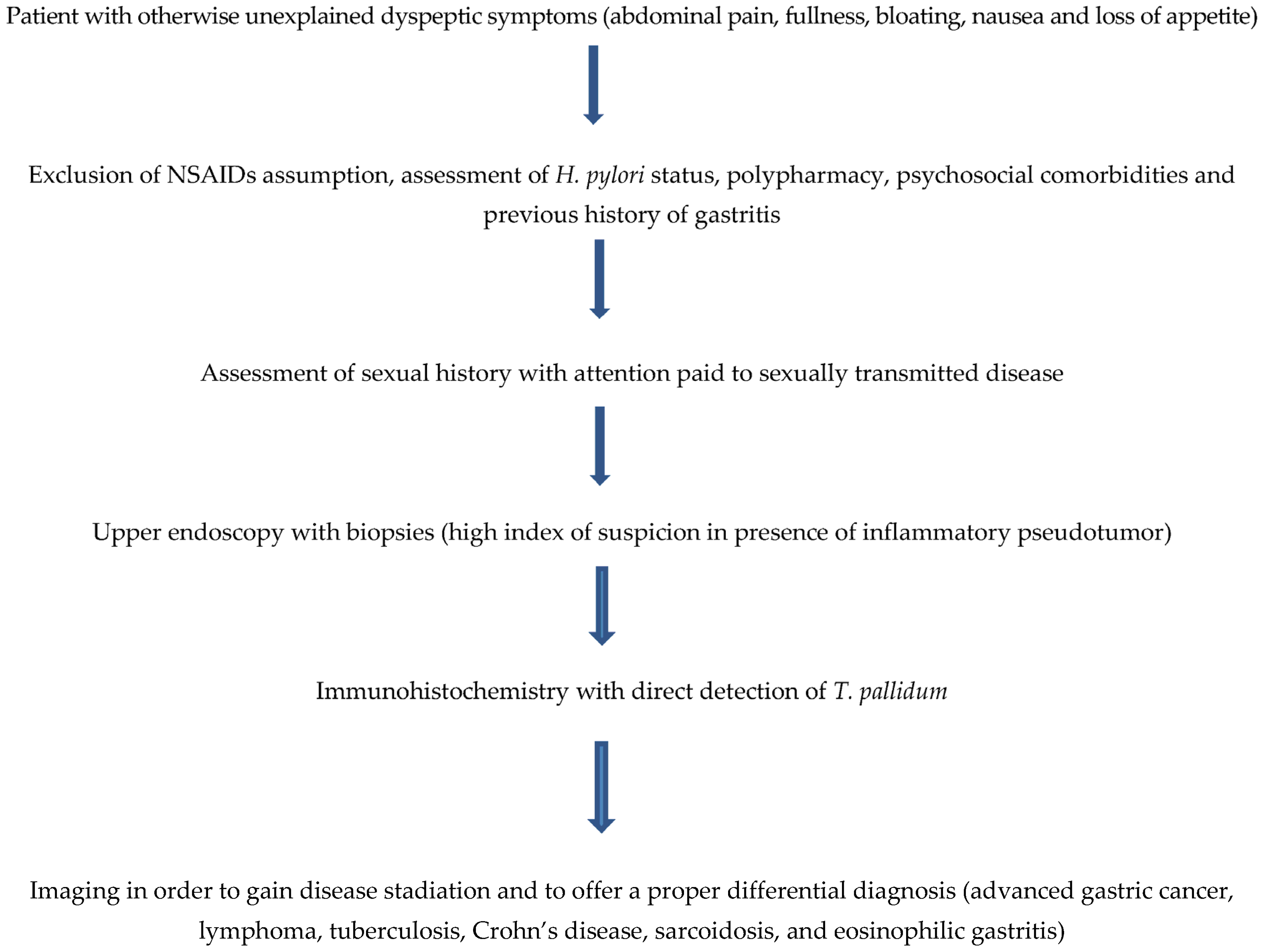
Only 7.8% of those infected had previously been diagnosed with syphilis, and 81.5% had concomitant clinical signs of the disease. Concurrent clinical symptoms included vaginal ulcer 73% of the time, rash 37.5% of the time, and lymphadenopathy 68.7% of the time (Table 1). In 15.6% of cases, more than one clinical characteristic was observed. Table 1 also shows the syphilis stage at the time of diagnosis. We also classified patients as having early or late disease, as indicated in the methodology section. There was concurrent CNS involvement in 4.6% of the cases. Except for one patient, all instances studied had serology data. In all patients, non-treponemal testing (VDRL, RPR, and/or Kolmer) was positive. FTA-abs or MHATP/TPHA assays were found to be positive in 51 of the 64 cases examined. Only 4.6% of the cases involved HIV-positive individuals.
The most common symptom at presentation in cases of stomach involvement was epigastric or abdominal pain/fullness (87.5%), followed by weight loss (46.1%), anorexia (41.8%), early satiety (12.3%), and anemia (9.3%) (Table 2).
The duration of symptoms ranged from one week to 48 months in 56 cases, with a median value of 30 days (Table 2).
In terms of radiologic findings, 31 out of the 64 patients examined had radiologic evaluations, while 49 had endoscopic examinations (Table 3).
Only one of the 31 radiologic exams found no lesion, and another was not diagnostic. In the remaining patients, the most common finding was fibrotic narrowing and rigidity of the gastric wall (35.4%), followed by hypertrophic and irregular folds (26%), a mass lesion in three cases, and one case with fibrosis and shrinking of the entire stomach leading to linitis plastica. In many cases, multiple findings coexisted.
Endoscopic examinations revealed that the majority of patients had multiple types of lesions, including large ulcers (58.4%), ulcerative gastritis (38.4%), erosions (38.4%), thickened folds (13.8%), and mass lesions (3.1%) (Table 3).
Histologic evidence of stomach lesions was found in 62 out of the 64 cases studied (Table 3).
All of them had chronic gastritis with thick plasmocytic and lymphocytic infiltrates. Shallow erosions were found in nine cases, endarteritis in seven, and atrophic gastritis in three. Surprisingly, data on Helicobacter pylori infection was available in nine studies that also documented the prevalence of such infection. In 49 cases, data on T. pallidum detection was provided. Due to the limitations of the various approaches’ sensitivity and specificity, the authors used more than one method to detect T. pallidum in the majority of cases (Figure 5).
Many of the evaluated patients (88.9%) received antibiotics (54 penicillin and 3 ceftriaxone 1 gr i.m.), and all but one had their symptoms resolved quickly (Table 4). The remaining seven patients (10.9%) were operated on due to a complication, such as a stomach rupture or blockage, or a high suspicion of an infiltrating tumor or lymphoma (six patients). Follow-up endoscopies were performed in 36 cases between 10 and 168 days after treatment initiation, and they revealed partial or near-complete healing in 26 patients and complete healing in the 10 remaining patients.

5. Discussion

GS is reported as a form of organic involvement that is thought to be present in a large proportion of cases of secondary syphilis, even though symptomatic gastritis is extremely rare [3,4,5,6,7,8,9,10,11,12,13,14,15,16,17,18,19,20,21,22,23,24,25,26,27,28,29,30,31,32,33,34,35,36,37,38,39,40,41,42]. The clinical presentation of gastric syphilis, as shown in the case report but also in a systematic search of the available scientific literature, appeared to be non-specific, characterized by dyspeptic symptoms, sometimes in conjunction with alarm symptoms such as weight loss, vomiting, or anemia. Complications such as gastric obstruction or perforation were reported infrequently. Furthermore, rare associations such as hepatomegaly mimicking liver disease [3], membranous glomerulonephritis [43], and megaloblastic anemia [44,45] were found in our literature search; finally, the HIV co-infection rate among GS cases reviewed was relatively low, as shown in the previous systematic review performed on this topic by Mylona and co-workers [3].
Even though physical examinations were performed (revealing genital ulcers in 25% of patients), clinical suspicion of syphilis was only present in 7.8% of cases. However, only a concurrently positive serologic test (reported in 79% of patients) assisted in reaching the correct diagnosis, along with a complete medical (including sexual) history, an accurate physical examination, and a high clinical suspicion [46,47,48,49,50,51,52,53,54,55,56].
The radiologic findings were mostly non-specific, with fibrotic narrowing and rigidity of the gastric wall being the most common. A mass aspect, as shown in our case, was reported in a very small percentage of patients. As a result of the non-specificity of radiological findings, an upper endoscopy with biopsy is required to obtain a specific diagnosis.
Jones and colleagues [57] provided a thorough description of the radiologic findings. In their series, radiographs revealed a nonspecific gastritis with diffusely thickened folds that may become nodular, with or without detectable ulcers, in patients with early disease.
In patients with late disease, the radiographic findings indicate that a malignant stricture typically has a sharper “shoulder” at the margins of a short isthmus rather than the more gradual transition from a long isthmus seen in GS. Therefore, this helps to distinguish between GS lesions and gastric adenocarcinomas [57].
With regard to endoscopic findings, the diagnosis of gastric syphilis remains challenging in the absence of a high index of suspicion, since the endoscopic findings reported in our search appear multiple and non-specific, ranging from a non-specific erosive/ulcerative gastritis to a linitis plastica-like aspect. In fact, gastric syphilis should be differentiated from gastric lymphoma, tuberculosis, Chron’s disease, and gastric adenocarcinoma, mainly when it resembles a mass lesion or is mimicking a linitis plastica [57].
Hyung Joo Yu and colleagues described endoscopic features in detail, demonstrating that the antrum and body were the most frequently involved sites, with multiple erosions, ulceration, nodularity, and mucosal fold hypertrophy predominating. Endoscopic findings in GS patients vary and should be distinguished from advanced gastric cancer, lymphoma, tuberculosis, Crohn’s disease, sarcoidosis, and eosinophilic gastritis. In particular, it may be difficult to distinguish malignant diseases such as advanced gastric cancer and lymphoma in some cases, making the primary diagnosis of gastric syphilis difficult. If the diagnosis is delayed, it can be treated surgically due to complications such as gastric perforation and outlet obstruction, or because there is a strong suspicion of advanced gastric cancer or lymphoma. Clinical, radiologic, and endoscopic findings in both cases of our study indicated linitis plastica. An endoscopic biopsy, however, was unable to diagnose it. Gastric syphilis was diagnosed in both cases using endoscopic mucosal resection and exploratory laparotomy [42].
There is little information available about the appearance of GS on endoscopic ultrasound (EUS).
It is widely accepted that EUS is the most accurate method for determining the local stage of gastric lymphoma. However, it is not the only method [58] as it allows sensitive detection of affected regional lymph nodes. Nonetheless, the data from the literature are limited and contradictory. They are the results of studies published between 1991 and 2002, when surgical resection of gastric lymphoma was still the standard treatment. Thus, in these studies, endosonographic staging could be compared to histopathological findings [59].
In the case reported by Shen and colleagues, ultrasonic gastroscopy revealed a slightly thickened gastric mucosa and interrupted submucosa associated with ulcerous lesions, mimicking the endosonographic appearance of a gastric lymphoma. Pathological analysis of gastric biopsies, however, revealed tissue necrosis and an inflammatory infiltrate rich in lymphocytes but no evidence of malignancy. The Warthin-Starry staining revealed a swarm of spirochetes infiltrating the lamina propria’s mucosal glands and vessels [44].
Treponema detection in affected tissue is still the only sure way to diagnose this disease. As demonstrated in our report, PCR is considered more sensitive than other techniques, such as dark-field microscopy, silver stains, and immunofluorescence microscopy.
However, a 20% false-negative rate has been primarily reported in the late stages of the disease [60]. Furthermore, histological findings alone are frequently insufficient to make a diagnosis because they are often non-specific and because of the co-infection with Helicobacter pylori, which may compete with Treponema pallidum in affecting the antrum. Typically, a diffuse gastritis with a dense plasma cell or lymphocytic infiltrate is seen in all stages of the disease, sometimes with concomitant shallow erosions, whereas a clear vasculitis, which is typical of syphilitic involvement in other sites, is rarely found in gastric biopsies, most likely because most gastric biopsies do not sample the submucosa where these vessels are located [59]. As a result, clinical, radiological, and endoscopic findings are non-specific and frequently mimic the presentation of gastric adenocarcinoma or lymphoma, making diagnosis difficult, and immune staining is required for this diagnosis. Furthermore, in the reported case, we would like to emphasize the importance of being suspicious of GS when a gastric mass reveals the presence of an IPT on histology, as previously reported for nodal IPT caused by luetic infection [61].
Dark-field microscopy is sensitive but not selective for detecting T. pallidum and necessitates the use of trained personnel. Silver impregnation techniques have a low sensitivity and specificity (33% to 71%). They also produce a variety of field artifacts but lack the precision needed to detect contaminated nontreponemal spirochetes (e.g., in oral mucosa).
Several recent studies [62,63] back up the use of immunohistochemistry (IHC) with anti-treponemal antibodies in the diagnosis of syphilis. One study that compared IHC to silver impregnation procedures found that the former was more sensitive [63]. IHC has a sensitivity of more than 90% in most cases, but lower values (71%) have been recorded in rare investigations. IHC has a sensitivity of more than 90% in most cases, but lower values (71%) have been recorded in rare investigations [64]. Sensitivity and specificity are greatest in early primary and secondary syphilis (when spirochetes are abundant), but decline as the number of microorganisms in the lesion decreases [65]. IHC can distinguish syphilis lesions from other unrelated lesions that may appear in people who have both true and false positive T. pallidum serology. Furthermore, this method detects spirochetes in the skin, with the possibility of cross-reactivity with Borrelia species [66]. Furthermore, IHC is a relatively quick and inexpensive method, with results available within 48 h.
A smaller proportion of patients with positive serologic results require treatments with penicillin or ceftriaxone. Even though some patients (10.9%) required surgical intervention due to gastric perforation, obstruction, or a strong suspicion of an infiltrating tumor or lymphoma, most of them (89.1%) experienced rapid resolution of their symptoms after treatment.
The patient’s delayed diagnosis was due to these factors: (1) Since syphilis can affect almost every system, clinical findings can be extremely diverse; (2) GS is clinically uncommon, has no distinct clinical symptoms, and is easily confused with chronic gastritis, a peptic ulcer, or functional dyspepsia. The patient was misdiagnosed with gastric cancer due to his age, the long-term course of the disease, and the severity of the symptoms of presentation; (3) The patient had not previously been diagnosed with cutaneous syphilis; (4) The patient was older and lacked knowledge about the earliest forms of syphilis, such as hard chancre erosions that heal quickly, and the patient did not pay attention to them; and (5) All clinicians are unaware of syphilis in organs other than the genitourinary system, with digestive system syphilis being especially uncommon.
Clinicians should be mindful of these points in order to reduce delayed and incorrect diagnoses. Doctors and patients should be knowledgeable about the fact that GS can cause stomach infections. GS should be considered when a patient presents with clinical symptoms such as nonspecific abdominal pain, fullness, nausea, and loss of appetite, regardless of whether the patient has had a history of chronic gastric disease in the past, and the symptoms cannot be explained by other common digestive tract diseases.
In this case, the results of the gastroscopy and biopsy did not support the diagnosis of chronic gastritis, peptic ulcer, gastric cancer, gastric lymphoma, eosinophilic gastritis, or another disease. An inflammatory pseudo-tumor was discovered during the histopathological examination, implying the possibility of an infection, possibly with T. pallidum. A complete medical history, including a history of sexually transmitted diseases, a thorough physical examination, and a high level of suspicion must be combined with the diagnosis, with care taken to protect the patient’s privacy. Patients, like the one described here, frequently conceal their sexual history, making gastric syphilis diagnosis more difficult.
The patient’s medical history, clinical, imaging, endoscopic, and histological findings, as well as endoscopic biopsy specimens stained with Warthin-Starry silver staining to visualize spirochetes, can all be used to diagnose GS. Treponema pallidum can be detected directly using direct immunofluorescent staining of gastric mucosal biopsy tissues and reverse transcription polymerase chain reaction [67], in addition to routine pathological examinations.
To our knowledge, the case reported is the second case of GS to occur in Italy, according to the scientific literature [3,17]. However, regarding the case reported by Bottari and co-workers, the patient that we managed did not have physical signs of primary syphilis previously reported nor a high diagnostic suspicion for primary syphilis (i.e., justified by the serologic diagnosis) before the endoscopic and histologic diagnosis, which was, in the case here reported, essential for the management of such a patient. Furthermore, a limitation of our study is the absence of an endoscopic follow-up, which is mandatory to show the healing of gastric lesions.
Since GS is a prominent mimicker of other gastric disorders and syphilis is a multisystemic disease in general, most clinicians are unaware of its existence. However, as syphilis rates rise, GS may become a more common therapeutic issue with which we should become familiar. As a result, GS should be considered in the differential diagnosis of all individuals who present with stomach symptoms and atypical endoscopic lesions but no conclusive diagnosis of another gastric condition. Furthermore, the absence of primary or secondary luetic lesions does not rule out the possibility of GS.
Patients with peptic ulcer disease who are resistant to antiulcer medication, regardless of the presence of Helicobacter pylori, and whose histopathologic examination suggests syphilis, especially if an IPT is found, should be investigated. Not only infectious disease specialists and dermatologists should be aware of this rare entity, but so should clinical gastroenterologists, endoscopists, radiologists, and pathologists, in order to secure a specific diagnosis that could save the patient from the disease’s most severe complications.

Author Contributions

Conceptualization, E.S. (Emanuele Sinagra) and I.M.; methodology, D.R., M.S., S.D.G., F.R., G.C., R.A. and G.P.; data curation, M.M. (Marta Marasà), S.T., M.M. (Marcello Maida) and G.S.; writing—original draft preparation, E.S. (Endrit Shahini) and I.M.; editing, D.R.; supervision, C.R., A.G.R. and D.R. All authors have read and agreed to the published version of the manuscript.

Funding

This research received no external funding.

Institutional Review Board Statement

Ethical review and approval were waived for this study due to the fact that this was only a retrospective case report, as per internal policy.

Informed Consent Statement

Informed consent was obtained from all subjects involved in the study.

Acknowledgments

All the authors thank the Hospital G. Giglio for the support provided to all the authors.

Conflicts of Interest

The authors declare no conflict of interest.

References

  1. Okamoto, K.; Hatakeyama, S.; Umezawa, M.; Hayashi, S. Gastric syphilis: The great imitator in the stomach. IDCases 2018, 12, 97–98. [Google Scholar] [CrossRef] [PubMed]
  2. Atten, M.J.; Attar, B.M.; Teopengco, E.; Nadimpalli, V. Gastric syphilis: A disease with multiple manifestations. Am. J. Gastroenterol. 1994, 89, 2227–2229. [Google Scholar] [PubMed]
  3. Mylona, E.E.; Baraboutis, I.G.; Papastamopoulos, V.; Tsagalou, E.P.; Vryonis, E.; Samarkos, M. Gastric syphilis: A systematic review of published cases of the last 50 years. Sex. Transm. Dis. 2010, 37, 177–183. [Google Scholar] [CrossRef] [PubMed]
  4. Cooley, R.N.; Childers, J.H. Acquired syphilis of the stomach: Report of two cases. Gastroenterology 1960, 39, 201–207. [Google Scholar] [CrossRef] [PubMed]
  5. Madding, G.F.; Baer, L.S.; Kennedy, P.A. Gastric syphilis: A casereport. Ann. Surg. 1963, 159, 271–274. [Google Scholar] [CrossRef]
  6. Mitchell, R.M.; Bradlow, S.P. Acute erosive gastritis due to early syphilis. Ann. Intern. Med. 1964, 61, 933–938. [Google Scholar] [CrossRef]
  7. Prolla, J.C.; Kobayashi, S.; Yoshii, Y.; Yamaoka, Y.; Kasugai, T. Diagnostic cytology of the stomach in gastric syphilis: Report of two cases. Acta Cytol. 1970, 14, 333–337. [Google Scholar]
  8. Sachar, D.B.; Klein, R.S.; Swerdlow, F.; Bottone, E.; Khilnani, M.T.; Waye, J.D.; Wisniewski, M. Erosive syphilitic gastritis: Dark-field and immunofluorescent diagnosis from biopsy specimen. Ann. Intern. Med. 1974, 80, 512–515. [Google Scholar] [CrossRef]
  9. Butz, W.C.; Watts, J.C.; Rosales-Wuintana, S.; Hicklin, M.D. Erosive gastritis as a manifestation of secondary syphilis. Am. J. Clin. Pathol. 1975, 63, 895–900. [Google Scholar] [CrossRef] [Green Version]
  10. Reisman, T.N.; Leverett, F.L.; Hudson, J.R.; Kalser, M.H. Syphilitic gastropathy. Am. J. Dig. Dis. 1975, 20, 588–593. [Google Scholar] [CrossRef]
  11. Vaughan, W.P.; Straus, F.H., II; Paloyan, D. Squamous carcinoma of the stomach after luetic linitisplastica. Gastroenterology 1977, 72 Pt 1, 945–948. [Google Scholar] [CrossRef]
  12. Morin, M.E.; Tan, A. Diffuse enlargement of gastric folds as a manifestation of secondary syphilis. Am. J. Gastroenterol. 1980, 74, 170–172. [Google Scholar]
  13. Reid, A.C.; Behan, P.O. Subacute Wernicke’s encephalopathy due to gastric syphilis. Br. J. Vener. Dis. 1981, 57, 309–311. [Google Scholar] [CrossRef] [PubMed]
  14. Lichtenstein, J.E. Case: Syphilitic gastritis. Gastrointest. Radiol. 1981, 6, 371–374. [Google Scholar] [PubMed]
  15. Beckman, J.W.; Schuman, B.M. Antral gastritis and ulceration in a patient with secondary syphilis. Gastrointest. Endosc. 1986, 32, 355–356. [Google Scholar] [CrossRef]
  16. Besses, C.; Sans-Sabrafen, J.; Badia, X.; Rodríguez-Méndez, F.; Salord, J.C.; Armengol, J.R. Ulceroinfiltrative syphilitic gastropathy: Silver stain diagnosis from biopsy specimen. Am. J. Gastroenterol. 1987, 82, 773–774. [Google Scholar]
  17. Bottari, M.; Melina, D.; Napoli, P.; Pallio, S.; Puglisi, A.; Villari, D. Gastric lesions in secondary syphilis. Gastrointest. Endosc. 1988, 34, 437–439. [Google Scholar] [CrossRef] [PubMed]
  18. Chung, K.Y.; Lee, M.G.; Chon, C.Y.; Lee, J.B. Syphilitic gastritis: Demonstration of Treponema pallidum with the use of fluorescent treponemal antibody absorption complement and immunoperoxidase stains. J. Am. Acad. Dermatol. 1989, 21 Pt 1, 183–185. [Google Scholar] [CrossRef]
  19. Anai, H.; Okada, Y.; Okubo, K.; Okamura, T.; Sakaguchi, Y.; Maehara, Y.; Sugimachi, K.; Nakamura, K. Gastric syphilis simulating linitisplastica type of gastric cancer. Gastrointest. Endosc. 1990, 36, 624–626. [Google Scholar] [CrossRef]
  20. Shy, S.W.; Lai, Y.S.; Lee, W.H.; Tseng, H.H. Ulceronodular gastritis insecondary syphilis. J. Infect. 1991, 22, 277–279. [Google Scholar] [CrossRef]
  21. Rank, E.L.; Goldenberg, S.A.; Hasson, J.; Cartun, R.W.; Grey, N. Treponema pallidum and Helicobacter pylori recovered in a case of chronic active gastritis. Am. J. Clin. Pathol. 1992, 97, 116–120. [Google Scholar] [CrossRef]
  22. Winters, H.A.; Notar-Francesco, V.; Bromberg, K.; Rawstrom, S.A.; Vetrano, J.; Prego, V.; Kuan, J.; Raufman, J.P. Gastric syphilis: Five recent cases and a review of the literature. Ann. Intern. Med. 1992, 116, 314–319. [Google Scholar] [CrossRef] [PubMed]
  23. Kasmin, F.; Reddy, S.; Mathur-Wagh, U.; Sarlin, J.; Goldman, A.; Antosofsky, H.; Strutynsky, N. Syphilitic gastritis in an HIV-infected individual. Am. J. Gastroenterol. 1992, 87, 1820–1822. [Google Scholar] [PubMed]
  24. Fyfe, B.; Poppiti, R.J.; Lubin, J.; Robinson, M.J. Gastric syphilis. Primary diagnosis by gastric biopsy: Report of four cases. Arch. Pathol. Lab. Med. 1993, 117, 820–823. [Google Scholar]
  25. Abdu, R.A.; Carter, K.; Pomidor, W.J. Gastric syphilis mimicking linitisplastica. Arch. Surg. 1993, 128, 103–104. [Google Scholar] [CrossRef]
  26. Greenstein, D.B.; Wilcox, C.M.; Schwartz, D.A. Gastric syphilis: Report of seven cases and review of the literature. J. Clin. Gastroenterol. 1994, 18, 4–9. [Google Scholar] [CrossRef]
  27. Smith, M.B.; Levin, T.N. Gastric syphilis: An unusual endoscopic appearance. Gastrointest. Endosc. 1992, 38, 94–96. [Google Scholar] [CrossRef]
  28. Sinagra, E.; Raimondo, D.; Gallo, E.; Stella, M.; Cottone, M.; Orlando, A.; Rossi, F.; Orlando, E.; Messina, M.; Tomasello, G.; et al. Could JC virus provoke metastasis in colon cancer? World J. Gastroenterol. 2014, 42, 15745–15749. [Google Scholar] [CrossRef] [PubMed] [Green Version]
  29. Long, B.W.; Johnston, J.H.; Wetzel, W.; Flowers, R.H.; Haick, A. Gastric syphilis: Endoscopic and histological features mimicking lymphoma. Am. J. Gastroenterol. 1995, 90, 1504–1507. [Google Scholar] [PubMed]
  30. Inagaki, H.; Kawai, T.; Miyata, M.; Nagaya, S.; Tateyama, H.; Eimoto, T. Gastric syphilis: Polymerasechain reaction detection of treponemal DNA in pseudolymphomatous lesions. Hum. Pathol. 1996, 27, 761–765. [Google Scholar] [CrossRef]
  31. Kolb, J.C.; Woodward, L.A. Gastric syphilis. Am. J. Emerg. Med. 1997, 15, 164–166. [Google Scholar] [CrossRef]
  32. Ishimaru, T.; Mizuno, Y.; Shiga, H.; Nagayama, I.; Furukawa, M. Patient with primary tonsillar and gastric syphilis. J. Laryngol. Otol. 1997, 111, 766–768. [Google Scholar] [CrossRef] [PubMed]
  33. Yoshida, K.; Tada, S.; Ueno, N.; Owan, T.; Suko, H.; Kamio, T.; Matsumoto, T. Gastric syphilis. Gastrointest. Endosc. 2003, 58, 908–909. [Google Scholar] [CrossRef] [PubMed]
  34. Guerrero, A.F.; Straight, T.M.; Eastone, J.; Spooner, K. Gastric syphilis in an HIV-infected patient. AIDS Patient Care STDS 2005, 19, 281–285. [Google Scholar] [CrossRef] [PubMed]
  35. Massironi, S.; Carmagnola, S.; Penagini, R.; Conte, D. Gastric involvement in a patient with secondary syphilis. Dig. Liver Dis. 2005, 37, 368–371. [Google Scholar] [CrossRef]
  36. Chen, C.Y.; Chi, K.H.; George, R.W.; Cox, D.L.; Srivastava, A.; Rui Silva, M.; Carneiro, F.; Lauwers, G.Y.; Ballard, R.C. Diagnosis of gastricsyphilis by direct immunofluorescence staining and real-time PCR testing. J. Clin. Microbiol. 2006, 44, 3452–3456. [Google Scholar] [CrossRef] [Green Version]
  37. Choi, Y.-L.; Han, J.J.; Lee, D.K.; Cho, M.H.; Kwon, G.Y.; Ko, Y.H.; Park, C.K.; Ahn, G. Gastric syphilis mimicking adenocarcinoma. J. Korean Med. Sci. 2006, 21, 559–562. [Google Scholar] [CrossRef] [Green Version]
  38. Cai, J.; Ji, D.; Guan, J.L. Gastric syphilis. IDCases 2017, 8, 87–88. [Google Scholar] [CrossRef]
  39. Itoh, N.; Katano, H.; Nakayama, S.I.; Kurai, H. GastricSyphilis. Intern. Med. 2017, 56, 1753. [Google Scholar] [CrossRef] [Green Version]
  40. Lan, Y.M.; Yang, S.W.; Dai, M.G.; Ye, B.; He, F.Y. Gastric syphilis mimicking gastric cancer: A case report. World J. Clin. Cases 2021, 9, 7798–7804. [Google Scholar] [CrossRef]
  41. Amato, A.; Sinagra, E.; Celsa, C.; Enea, M.; Buda, A.; Vieceli, F.; Scaramella, L.; Belletrutti, P.; Fugazza, A.; Cammà, C.; et al. Efficacy of lumen-apposing metal stents or self-expandable metal stents for endoscopic ultrasound-guided choledochoduodenostomy: A systematic review and meta-analysis. Endoscopy. 2021, 10, 1037–1047. [Google Scholar] [CrossRef] [PubMed]
  42. Yu, H.J.; Kim, S.J.; Oh, H.H.; Im, C.M.; Han, B.; Myung, E.; Yun, S.J.; Lee, K.H.; Joo, Y.E. Case report of gastric syphilis in Korea: Clinical features, pathology, management, and prognosis. Medicine 2021, 100, e28212. [Google Scholar] [CrossRef] [PubMed]
  43. Roh, M.; Sohn, J.H.; Kim, T.Y.; Kim, S.J.; Kim, J.S.; Chung, S.J.; Pyo, J.Y.; Oh, Y.H. Gastric Syphilis and Membranous Glomerulonephritis. Clin. Endosc. 2015, 48, 256–259. [Google Scholar] [CrossRef] [Green Version]
  44. Shen, Y.; Nie, L.; Zhang, M.; Tang, B.; Qin, Z.; Meng, K.; Lu, Y. Gastric syphilis mimicking lymphoma. Endoscopy 2015, 47 (Suppl. 1), uctn:E170-1. [Google Scholar] [CrossRef] [Green Version]
  45. Lacerda, P.N.; Campos, L.M.; de Ré, M.R.; Miot, H.A. Cutaneous hyperpigmentation and megaloblastic anemia as manifestations of gastric syphilis. Int. J. Dermatol. 2021, 60, e356–e358. [Google Scholar] [CrossRef]
  46. Guimarães, T.F.; Novis, C.F.; Bottino, C.B.; D’Acri, A.M.; Lima, R.B.; Martins, C.J. Gastric syphilis–Case report. An. Bras. Dermatol. 2016, 91, 670–672. [Google Scholar] [CrossRef] [PubMed] [Green Version]
  47. Adachi, E.; Koibuchi, T.; Yotsuyanagi, H. Gastric Syphilis in a Human Immunodeficiency Virus-Infected Patient. JMA J. 2019, 2, 93–94. [Google Scholar] [CrossRef] [PubMed]
  48. Souza VarellaFrazão, M.; GuimarãesVilaça, T.; OlavoAragão Andrade Carneiro, F.; Toma, K.; Eliane Reina-Forster, C.; Ryoka Baba, E.; Cheng, S.; Ferreira de Souza, T.; Guimarães Hourneaux de Moura, E.; Sakai, P. Endoscopic aspects of gastric syphilis. Case Rep. Med. 2012, 2012, 646525. [Google Scholar] [CrossRef] [Green Version]
  49. Navea, C.; von Mühlenbrock, C.; Cabello, N.; Echeverría, M.; Jiménez, A.; Poniachik, J. Syphilis, unusual cause of abdominal pain. Gastroenterol. Hepatol. 2018, 41, 565–566. [Google Scholar] [CrossRef]
  50. Lai, K.; Pinto-Sander, N.; Richardson, D.; Wei, S.; Zeng, K. Syphilis gastritis: A case report. Int. J. STD AIDS 2018, 29, 723–725. [Google Scholar] [CrossRef]
  51. Osman, M.; Hasan, S.; Azher, Q.; Elbedawi, M.; Bachuwa, G. Syphilitic gastritis: A rare presentation of secondary syphilis. BMJ Case Rep. 2018, 2018, bcr2017223868. [Google Scholar] [CrossRef] [PubMed] [Green Version]
  52. Shinn, B.; Kistler, C.; Dhanekula, R.K.; Civan, J. Syphilis, The Great Mimicker, Presents As a Rare Case of Concurrent Hepatitis and Gastroparesis. ACG Case Rep. J. 2019, 6, e00067. [Google Scholar] [CrossRef]
  53. Adachi, K. Syphilitic gastritis mimicking gastric neoplasms. Dig. Liver Dis. 2011, 43, 748. [Google Scholar] [CrossRef] [PubMed]
  54. Kim, Y.C.; Chung, Y.E.; Lim, J.S. An uncommon cause of ulceroinfiltrative gastric wall thickening in a young patient. Gastroenterology 2012, 143, e6–e7. [Google Scholar] [CrossRef] [PubMed]
  55. Sun, L.; Zheng, N.; Yang, Y.; Zhang, H.N. Syphilitic meningomyelitis presenting with visceral crisis: A case report. Medicine 2018, 97, e11661. [Google Scholar] [CrossRef]
  56. Maimone, S.; Saffioti, F.; Filomia, R.; Caccamo, G.; Saitta, C.; Pallio, S.; Consolo, P.; Sabatini, S.; Sitajolo, K.; Franzè, M.S.; et al. Elective endoscopic variceal ligation is not a risk factor for bacterial infection in patients with liver cirrhosis. Dig. Liver Dis. 2018, 50, 366–369. [Google Scholar] [CrossRef]
  57. Sinagra, E.; Aragona, E.; Romano, C.; Maisano, S.; Orlando, A.; Virdone, R.; Tesè, L.; Modesto, I.; Criscuoli, V.; Cottone, M. The role of portal vein thrombosis in the clinical course of inflammatory bowel diseases: Report on three cases and review of the literature. Gastroenterol. Res. Pract. 2012, 2012, 916428. [Google Scholar] [CrossRef] [Green Version]
  58. Melita, G.; Pallio, S.; Tortora, A.; Crinò, S.F.; Macrì, A.; Dionigi, G. Diagnostic and Interventional Role of Endoscopic Ultrasonography for the Management of Pancreatic Neuroendocrine Neoplasms. J. Clin. Med. 2021, 10, 2638. [Google Scholar] [CrossRef]
  59. Janssen, J. The impact of EUS in primary gastric lymphoma. Best Pract. Res. Clin. Gastroenterol. 2009, 23, 671–678. [Google Scholar] [CrossRef]
  60. Facchetti, F.; Incardona, P.; Lonardi, S.; Fisogni, S.; Legrenzi, L.; Chioda, C.; Ponzoni, M.; Chiodera, P.L. Nodal inflammatory pseudotumor caused by lueticinfection. Am. J. Surg. Pathol. 2009, 33, 447–453. [Google Scholar] [CrossRef]
  61. Hernández, C.; Fúnez, R.; Repiso, B.; Frieyro, M. Usefulness of immunohistochemial staining with antitrepenomal antibodies in the diagnosis of syphilis. Actas Dermosifiliogr. 2013, 104, 926–928, English, Spanish. [Google Scholar] [CrossRef]
  62. Phelps, R.G.; Knispel, J.; Tu, E.S.; Cernainu, G.; Saruk, M. Immunoperoxidase technique for detecting spirochetes in tissue sections: Comparison with other methods. Int. J. Dermatol. 2000, 39, 609–613. [Google Scholar] [CrossRef] [PubMed]
  63. Martín-Ezquerra, G.; Fernandez-Casado, A.; Barco, D.; Jucglà, A.; Juanpere-Rodero, N.; Manresa, J.M.; de Almeida, L.M.; Rodríguez-Peralto, J.L.; Kutzner, H.; Cerroni, L.; et al. Treponema pallidum distribution patterns in mucocutaneous lesions of primary and secondary syphilis: An immunohistochemical and ultrastructural study. Hum. Pathol. 2009, 40, 624–630. [Google Scholar] [CrossRef]
  64. Quatresooz, P.; Piérard, G.E. Skin homing of Treponema pallidum in early syphilis: An immunohistochemical study. Appl. Immunohistochem. Mol. Morphol. 2009, 17, 47–50. [Google Scholar] [CrossRef] [PubMed]
  65. Müller, H.; Eisendle, K.; Bräuninger, W.; Kutzner, H.; Cerroni, L.; Zelger, B. Comparative analysis of immunohistochemistry, polymerase chain reaction and focus-floating microscopy for the detection of Treponema pallidum in mucocutaneous lesions of primary, secondary and tertiary syphilis. Br. J. Dermatol. 2011, 165, 50–60. [Google Scholar] [CrossRef] [PubMed]
  66. Tammaro, L.; Buda, A.; Di Paolo, M.C.; Zullo, A.; Hassan, C.; Riccio, E.; Vassallo, R.; Caserta, L.; Anderloni, A.; Natali, A. T-Score Validation Study Group; T-Score Validation Study Group. A simplified clinical risk score predicts the need for early endoscopy in non-variceal upper gastrointestinal bleeding. Dig. Liver. Dis. 2014, 9, 783–787. [Google Scholar] [CrossRef]
  67. Almeida, M.C.D.; Cordeiro, A.M.R.; Cunha-Oliveira, A.; Barros, D.M.S.; Santos, D.G.S.M.; Lima, T.S.; Valentim, R.A.M. Syphilis response policies and their assessments: A scoping review. Front. Public Health 2022, 10, 1002245. [Google Scholar] [CrossRef]
Figure 1. Endoscopic view of the gastric syphilis mimicking a gastric adenocarcinoma. The image was created through Olympus EvisExera II, 165 series (Tokyo, Japan), in which narrow-band imaging was not available.
Gastroent 14 00014 g001
Figure 2. Hematoxylin-eosin staining showing gastric mucosa with an inflammatory infiltrate consisting mainly of eosinophils and plasma cells (magnification 200×, Patox Software, Tesi Group 2014).
Gastroent 14 00014 g002
Figure 3. Immunohistochemical staining, through antibodies against treponema (polyclonal antibodies Rabbit DAKO), showing several elements of Treponema palladium within the glandular epithelium (magnification 200×, Patox Software, Tesi Group, 2014).
Gastroent 14 00014 g003
Figure 4. Polymerase chain reaction in agarose gel with evidence of positivity for Treponema pallidum in the second band starting from the left (Patox Software, Tesi Group 2014).
Gastroent 14 00014 g004
Figure 5. Flow chart of the diagnostic algorithm necessary to obtain a proper diagnosis of gastric syphilis.
Gastroent 14 00014 g005
Table 1. Demographic, clinical, and serological characteristics of syphilis in patients with gastric syphilis.
CharacteristicNo. Patients: 64
Age
Median (range)35 (21–72)
Sex
Male42 (64%)
Female23 (36%)
Race
Black race15 (23.4%)
Asian27 (42.2%)
White22 (34.4%)
Prior syphilis diagnosis6 (7.8%)
Concurrent clinical findings of syphilis52 (81.5%)
Genital ulcer13 (25%)
Rash22 (42.3%)
Lymphadenpathy6 (11.5%)
Syphilis classification at diagnosis
Early disease38/64 (59.4%)
Primary3/64 (4.6%)
Secondary32/64 (50%)
Early latent3/64 (4.6%)
Late disease25/64 (39.1%)
Late latent21/64 (32.8%)
Tertiary4/64 (6.2%)
Congenital1/64 (1.5%)
CNS involvement2 (4.6%)
Positive serology
FTA-abs51/64 (79%)
HIV EIA positivity3 (4.6%)
Table 2. Clinical characteristics of gastric involvement.
CharacteristicNo. Patients: 64
Duration of gastric symptoms (days)
Mean
Median (range)30 (6–2520)
Symptoms
Epigastric/abdominal pain or fullness56 (87.5%)
Anorexia27 (41.8%)
Nausea/vomiting17 (26.5%)
Early satiety8 (12.3%)
Weight loss (kg)30 (46.1%)
Laboratory Findings
Anemia6 (9.3%)
Table 3. Radiologic, endoscopic, histologic findings, and T. pallidum detection in patients with gastric syphilis.
CharacteristicNo. Patients
Radiologic findings31/64 (48.4%)
Fibrotic narrowing and rigitidy11 (35.4%)
Hypertrophic and irregular folds6 (19.4%)
Mucosal nodules9/31 (29.0%)
Mass1/31 (3.2%)
Linitis plastica1/31 (3.2%)
Endoscopic findings49/64 (76.5%)
Multiple ulcerations/ulcerative gastritis38/49 (77.5%)
Erosions25/49 (51.0%)
Large ulcer28/49 (57.1%)
Thickened folds7/49 (14.2%)
Mass1/49 (2.0%)
Histologic Findings
Chronic gastritis with dense plasmocytic and/or lymphocytic infiltrate49/49 (100%)
Shallow erosions9/49 (18.3%)
Atrophic gastritis3/49 (6.1%)
Proliferative endoarteritis7/49 (14.2%)
T. pallidum detection49/49 (100%)
Table 4. Treatment and reevaluation data of patients with gastric syphilis.
CharacteristicNo. Patients
Gastrectomy7/64 (10.9%)
Penicilline therapy
Penicillin54/64 (84.3%)
Ceftriaxone3/64 (4.6%)
Clinical response to therapy56/57 (98.2%)
Endoscopic response to therapy
Partial or near complete26/36 (72.2%)
Complete10/36 (27.7%)
Disclaimer/Publisher’s Note: The statements, opinions and data contained in all publications are solely those of the individual author(s) and contributor(s) and not of MDPI and/or the editor(s). MDPI and/or the editor(s) disclaim responsibility for any injury to people or property resulting from any ideas, methods, instructions or products referred to in the content.

