Review

Scientific Rationale for the Treatment of Cognitive Deficits from Long COVID

by
Arman Fesharaki Zadeh
1,2,*,
Amy F. T. Arnsten
3 and
Min Wang
3,*
1
Departments of Neurology, Yale University School of Medicine, New Haven, CT 06510, USA
2
Departments of Psychiatry, Yale University School of Medicine, New Haven, CT 06510, USA
3
Departments of Neuroscience, Yale University School of Medicine, New Haven, CT 06510, USA
*
Authors to whom correspondence should be addressed.
Neurol. Int. 2023, 15(2), 725-742; https://doi.org/10.3390/neurolint15020045
Submission received: 23 March 2023 / Revised: 26 April 2023 / Accepted: 11 May 2023 / Published: 31 May 2023

Abstract

:
Sustained cognitive deficits are a common and debilitating feature of “long COVID”, but currently there are no FDA-approved treatments. The cognitive functions of the dorsolateral prefrontal cortex (dlPFC) are the most consistently afflicted by long COVID, including deficits in working memory, motivation, and executive functioning. COVID-19 infection greatly increases kynurenic acid (KYNA) and glutamate carboxypeptidase II (GCPII) in brain, both of which can be particularly deleterious to PFC function. KYNA blocks both NMDA and nicotinic-alpha-7 receptors, the two receptors required for dlPFC neurotransmission, and GCPII reduces mGluR3 regulation of cAMP-calcium-potassium channel signaling, which weakens dlPFC network connectivity and reduces dlPFC neuronal firing. Two agents approved for other indications may be helpful in restoring dlPFC physiology: the antioxidant N-acetyl cysteine inhibits the production of KYNA, and the α2A-adrenoceptor agonist guanfacine regulates cAMP-calcium-potassium channel signaling in dlPFC and is also anti-inflammatory. Thus, these agents may be helpful in treating the cognitive symptoms of long COVID.

1. Introduction

Many patients are experiencing sustained cognitive deficits as a consequence of COVID-19 infection, a phenomenon often referred to colloquially as “brain fog”, as part of long COVID. The World Health Organization states that “approximately 10–20% of people experience a variety of mid- and long-term effects after they recover from their initial illness. These mid- and long-term effects are collectively known as post-COVID-19 condition or “long COVID.” Accumulating data indicate that the cognitive functions of the prefrontal cortex (PFC), including working memory and the executive functions, are the most consistently impaired in long COVID. These cognitive deficits can be sufficiently debilitating to interfere with the ability to work and/or care for families. Although there is an urgent need for treatment, there are currently no FDA-approved treatments for long COVID cognitive deficits.
The current review describes strategies for pharmacological treatment of long COVID cognitive deficits using medications already approved for human use, but for other indications. As described in detail below, these strategies are based on (1) the accumulating evidence that long COVID consistently impairs the functions of the PFC, (2) that COVID-19 infection increases kynurenine and cAMP-calcium signaling in brain, both of which may be especially damaging to PFC function given the unique molecular needs of PFC circuits, and (3) previous studies showing that N-acetyl-cysteine (NAC) and the α2A-adrenoceptor (α2A-AR) agonist, guanfacine, can reduce the effects of kynurenine and excessive cAMP-calcium signaling, respectively, and have been shown to protect the PFC from non-COVID stressors in both animal models and human subjects. Thus, these agents may be helpful in treating the cognitive symptoms of long COVID.

2. Search Strategy and Selection Criteria

The references for this review were identified through searches of PubMed with the search terms “kynurenine” or “kynurenic acid”, “long-COVID”, “cognitive”, “GCPII”, “N-acetyl cysteine”, “guanfacine”, and “prefrontal cortex”, with combined search terms of “cognitive”, “long-COVID”, and “meta-analysis” to help create Table 1. Articles were also identified through searches of the authors’ own files. Only papers published in English were reviewed. The final reference list was generated on the basis of originality and relevance to the scope of this review, with an emphasis on review articles for summaries of PFC functions.

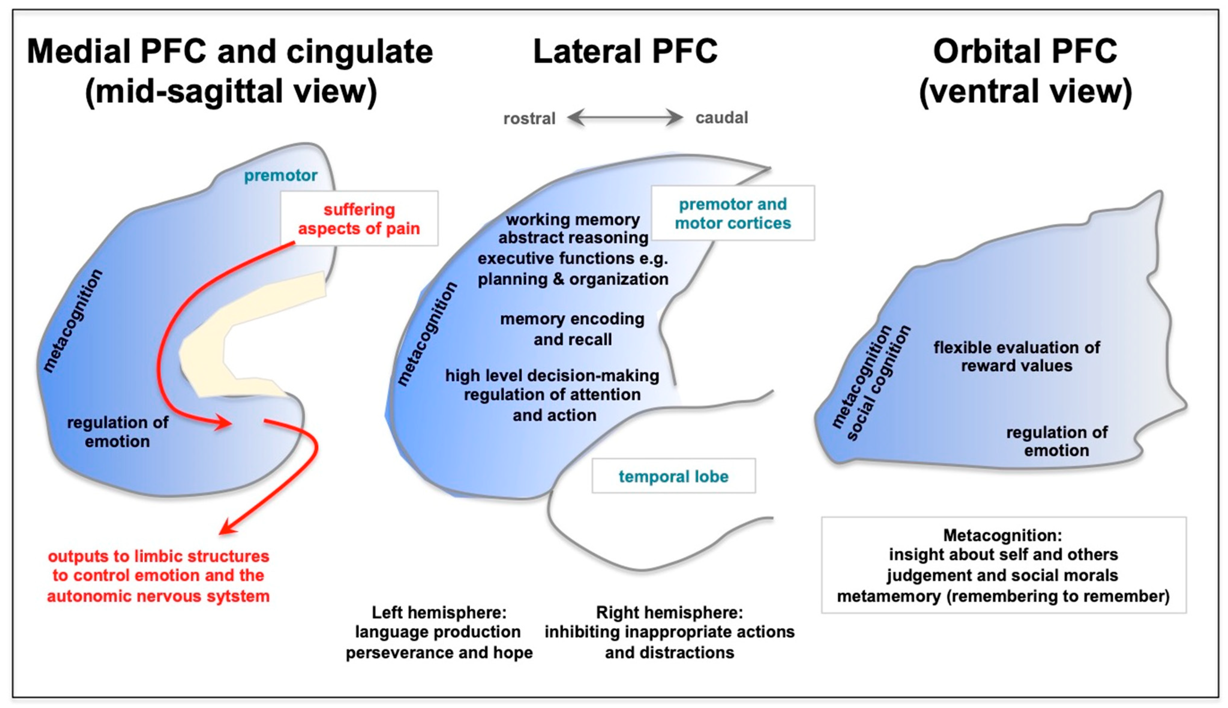
3. Long COVID Preferentially Afflicts the Cognitive Functioning of the Prefrontal Cortex

The PFC is a recently evolved part of the brain, situated rostral to the motor cortices in the frontal lobe, which expands enormously in primates [1,2,3,4,5]. The dorsolateral PFC (dlPFC) has been the focus of intensive research in monkeys and humans, as this area has the remarkable ability to represent information in the absence of sensory stimulation, the neural foundation for abstract thought and cognitive control. As summarized in Figure 1, this foundational mental capacity underlies a wide variety of functions, including abstract reasoning, working memory, and the executive functions, which include top-down control of attention, action, and emotion [1,6,7,8,9,10,11,12,13,14,15,16]. These abilities allow us to focus, divide or sustain our attention, e.g., as needed to multi-task, to inhibit inappropriate actions and impulses, and to plan and organize for the near and farther future. PFC circuits are needed for high-order, flexible decision-making, and the most rostral areas of the PFC mediate meta-cognitive functions such as insight and judgment about ourselves and others [1,17]. The rostal and lateral PFC regions project to the ventral and medial PFC to regulate emotion and motivation, allowing us to persist through difficulties, to regulate angry or aggressive impulses, and to have optimism about our future [18]. Lesions to the PFC impair these functions in humans and monkeys, including recent data showing that symptoms of PTSD and depression correlate with synapse loss in the dlPFC [19].
Many studies are finding that long COVID especially afflicts the functions of the PFC, in what patients often describe as “brain fog”. There are consistent impairments in working memory, abstract reasoning, and the executive abilities, interfering with top-down control of attention (e.g., impaired concentration and multi-tasking), as well as regulation of actions and emotional state [20,21,22,23,24,25]. As described in Table 1, meta-analyses and reviews of this rapidly emerging field have emphasized the consistent impairment in executive functions [23,26,27,28,29,30,31], with the relative sparing of memory recognition, which, in the context of impaired encoding and recall, is consistent with impaired PFC function [21]. Becker and colleagues describe this constellation of deficits as having “considerable implications for occupational, psychological, and functional outcomes.” [21]. The functional impairments in executive functioning are consistent with brain imaging studies, which show frontotemporal hypoperfusion in patients hospitalized with COVID-19 [20]. Long COVID is also commonly associated with symptoms of depression and anxiety [23], which may involve loss of PFC top-down regulation of emotion [19,32]. While there is some controversy regarding the relationships of severity of illness, age, or sex with the degree or frequency of cognitive impairments, e.g., deficits greater in young [33] vs. aged [34], there is general agreement that impairments in recall memory, working memory, attention regulation, and other executive functions are common (Table 1). It is clear that cognitive deficits can be problematic even after even mild illness [28,34,35] and that cognitive deficits may be related to inflammatory markers in plasma such as cytokines [36] and kynurenine [37]. There is also recent evidence that severe COVID infection can cause increased calcium dysregulation, kynurenic acid expression, and tau hyperphosphorylation in brain, suggesting that it may increase risk of future Alzheimer’s disease [38].
Why are the functions of the PFC so afflicted in long COVID, while other functions (e.g., visuospatial abilities, sensory perception, with the exception of olfaction) are so resilient? New data from animal studies indicate that the PFC is particularly vulnerable to the effects of stress and inflammation [39,40], which may help to explain why PFC deficits are such a common component of long COVID mental changes.
Figure 1. A schematic depiction of the functions of the prefrontal cortex (PFC), as illustrated on the medial, lateral, and ventral surfaces. There is a general topography, where representation of the external world is performed by lateral PFC of and the internal world by the ventral and medial PFC. The ventral (orbital) PFC flexibly represents our internal state, e.g., whether something is rewarding. The most caudal aspects of the medial surface also contain the cingulate cortices, which can activate emotional responses and are a key aspect of the suffering aspects of pain that are overactive in depression. Conversely, metacognitive functions such as insight about ourselves and others are localized most rostrally in the frontal pole. The lateral PFC generates many higher cognitive operations, including working memory, abstract thought, the executive functions, regulation of attention and action, and flexible decision-making. The PFC also assists with memory encoding and recall. The rostral and lateral PFC provides thoughtful “top-down” control of emotion through connections into the medial PFC. As described in the text, many of these PFC functions are preferentially afflicted in long COVID.
Neurolint 15 00045 g001
Table 1. Examples of recent reviews that describe PFC deficits associated with long COVID.
CitationSummary
Vanderlind et al. 2021 [23]“Thirty-three studies met inclusion/exclusion criteria for review. Emerging findings link COVID-19 to cognitive deficits, particularly attention, executive function, and memory. Psychiatric symptoms occur at high rates in COVID-19 survivors, including anxiety, depression, fatigue, sleep disruption, and to a lesser extent posttraumatic stress.”
Perrottelli et al. 2022 [26]“The available evidence revealed the presence of impairment in executive functions, speed of processing, attention and memory in subjects recovered from COVID-19.”
Zawilska and Kuczyńska. 2022 [27]“Fatigue and cognitive dysfunction, such as concentration problems, short-term memory deficits, general memory loss, a specific decline in attention, language and praxis abilities, encoding and verbal fluency, impairment of executive functions, and psychomotor coordination, are amongst the most common and debilitating features of neuropsychatric symptoms of post COVID syndrome.”
Newhouse et al. 2022 [41]“Many of these symptoms [42] are neuropsychiatric, such as inattention, impaired memory, and executive dysfunction; these are often colloquially termed “brain fog”.”
Bertuccelli et al. 2022 [28]“Memory, attention, and executive functions appeared to be the most affected domains. Delayed recall and learning were the most impaired domains of memory. Among the executive functions, abstraction, inhibition, set shifting, and sustained and selective attention were most commonly impaired.”
Houben and Bonnechère. 2022 [29]Meta-analysis showed impairments in attention, executive functioning, and verbal memory (recall)
Zeng et al. 2023 [30]“Individuals with severe infection suffered more from PTSD, sleep disturbance, cognitive deficits, concentration impairment, and gustatory dysfunction. Survivors with mild infection had high burden of anxiety and memory impairment after recovery.”
Ceban et al. 2022 [31]“The proportion of individuals exhibiting cognitive impairment was 0.22 (95% CI, 0.17, 0.28; p < 0.001; n = 13,232; I2 = 98.0). Moreover, narrative synthesis revealed elevations in proinflammatory markers and considerable functional impairment in a subset of individuals.”
Many studies are finding that long COVID especially afflicts the functions of the PFC, in what patients often describe as “brain fog”. There are consistent impairments in working memory, abstract reasoning, and the executive abilities, interfering with top-down control of attention (e.g., impaired concentration and multi-tasking), as well as regulation of actions and emotional state [20,21,22,23,24,25]. Meta-analyses have emphasized the consistent impairment in executive functions [23], with the relative sparing of memory recognition, which in the context of impaired encoding and recall is consistent with impaired PFC function [21]. Becker and colleagues describe this constellation of deficits as having “considerable implications for occupational, psychological, and functional outcomes.” [21]. The functional impairments in executive functioning are consistent with brain imaging studies, which show frontotemporal hypoperfusion in patients hospitalized with COVID-19 [20]. Long COVID is also commonly associated with symptoms of depression and anxiety [23], which may involve loss of PFC top-down regulation of emotion [19]. Emerging data from animal studies indicate that the PFC is particularly vulnerable to the effects of stress and inflammation, which may help to explain why PFC deficits are such a common component of long COVID mental changes.

4. The Prefrontal Cortex Is Especially Vulnerable to Physiological and Psychological Stressors

PFC dysfunction occurs with multiple physiological and psychological stressors. Deficits in working memory and executive dysfunction are a major component of the sustained cognitive deficits following traumatic brain injury (TBI) in both human patients [43,44,45,46] and rodents [47,48]. The studies in rodents showed that there are multiple chemical and structural changes in the rat PFC, even though the concussive site was to the posterior cortex and not the PFC itself [47,48]. Similar patterns were seen with sustained hypoxia, which impairs working memory and the executive functions in both humans [49] and rodents [50], the latter studies showing pronounced chemical and structural changes in the PFC [50,51].
PFC cognitive deficits are also produced by psychological stressors, particularly when the subject feels that they have no control over the stressor [52]. Research in both humans and animals has shown that exposure to even an acute, uncontrollable stress rapidly impairs PFC working memory abilities and reduces the activity of the dlPFC [53]. With chronic exposure to uncontrollable psychological stress, there are additional architectural changes, with loss of dendrites and spines (and thus neural connections) from PFC neurons which correlate with impaired working memory and attentional control [39,54,55]. Brain imaging of human subjects also shows weaker dlPFC connectivity with chronic stress exposure [54] as well as loss of PFC gray matter [56]. Exposure to a traumatic psychological stressor can induce symptoms of PTSD, which are related to reduced dlPFC synapses [19].
As reviewed below, data from animal studies suggest that psychological and physiological stressors activate similar molecular signaling pathways that may be especially deleterious in the PFC due to its unusual molecular dependencies.

5. Unique Neurotransmission and Neuromodulation Renders PFC Circuits Especially Vulnerable to Stress and Inflammation

dlPFC circuits have special circuits and molecular properties that allow them to generate and sustain neuronal firing without any sensory stimulation. The circuits have extensive, local, recurrent excitation, whereby the neurons excite each other in order to keep firing without needing external stimulation [6]. This recurrent excitation relies on unusual neurotransmission and neuromodulation, summarized in Figure 2, with the molecular machinery to magnify calcium [53]. Although the calcium is necessary to maintain neuronal firing, if the levels are too high, they open nearby K+ channels to functionally disconnect circuits, which markedly reduces neuronal firing and impairs cognition [53]. With sustained high levels of calcium, inflammatory mechanisms are evoked that lead to loss of spines and dendrites. These properties render PFC neurons very vulnerable to atrophy under conditions of inflammation [40], where molecular changes induced by COVID-19 would be especially deleterious to the dlPFC.
Unusual neurotransmission—The persistent firing of dlPFC neurons during working memory differs from typical glutamate neurotransmission in its great dependence on NMDA receptors (NMDAR) rather than AMPA receptors (AMPAR). Classic glutamate neurotransmission depends heavily on AMPAR, where glutamate stimulation of AMPAR fluxes Na+ ions to depolarize the synaptic membrane. This depolarization ejects Mg2+ from the NMDAR pore, permitting NMDAR neurotransmission [53]. As NMDARs flux both Na+ and Ca2+, they can be particularly important for neuroplasticity. AMPAR neurotransmission has a rapid on- and offset, and thus is especially appropriate for encoding sensory events [58].
In contrast to traditional glutamate synapses, the recurrent excitation in dlPFC circuits has little reliance on AMPAR, but it relies heavily on NMDAR neurotransmission, including NMDAR with GluN2B subunits, which close slowly and flux high levels of calcium [57]. The permissive role normally played by AMPAR is instead played by acetylcholine, including stimulation of nicotinic α7 receptors (nic-α7R), which reside within the glutamate synapse in dlPFC circuits [58]. The reliance on acetylcholine makes dlPFC especially dependent on arousal state as acetylcholine is released during waking and not during deep sleep [58]. As described below, this dependence on NMDAR and nic-α7R makes dlPFC circuits particularly vulnerable to blockade by kynurenic acid inflammatory signaling.
Unusual neuromodulation—In most neurons, increased cAMP-calcium signaling strengthens neuronal function and synaptic connections, e.g., as occurs in hippocampus for long-term increases in synaptic strength [53]. However, in layer III dlPFC, there is an enrichment of potassium (K+) channels on spines that are opened by cAMP-PKA-calcium signaling, which weakens rather than strengthens dlPFC connectivity and function [53]. As stress and inflammation increase cAMP-calcium signaling, the concentration of K+ channels opened by cAMP-calcium signaling on layer III dlPFC spines makes these neurons particularly vulnerable to stress and inflammation [53].
As described above, dlPFC dendritic spines contain the cAMP-PKA molecular machinery to magnify calcium signaling near the PSD to support persistent neuronal firing [53]. As shown in Figure 2, there is evidence of extensive feedforward, cAMP-PKA-calcium signaling, whereby cAMP-PKA increases calcium release from the smooth endoplasmic reticulum (SER), and increased calcium drives more cAMP signaling. Moderate levels of cAMP-calcium signaling are needed to support sustained neuronal firing needed for working memory and top-down control. However, high levels of cAMP-PKA-calcium signaling reduce firing by opening nearby K+ channels (e.g., HCN-Slack, KCNQ2) on spines (Figure 2). Low levels provide negative feedback to prevent seizures, but high levels of K+ channel opening, as occurs with stress exposure, markedly reduce neuronal firing and impair working memory and top-down control [53]. This occurs with stress exposure when there are high levels of catecholamine release in PFC [62,63], which engage low-affinity receptors (e.g., α1-AR, Figure 3) that increase cAMP-calcium-K+ signaling and impair working memory [53]. Sustained elevations in cAMP-PKA-calcium-K+ signaling, i.e., continued weakening of synaptic connections, lead to removal of spines and dendrites, involving calcium-induced inflammatory mechanisms (Figure 3) [39].
Under healthy conditions, cAMP-PKA drive on calcium-K+ channel signaling is regulated by α2A-adrenoceptors (α2A-AR) [59] and type 3 metabotropic glutamate receptors (mGluR3s) [61] on spines, which inhibit cAMP production, strengthen connectivity and enhance neuronal firing (Figure 2). mGluR3 signaling expands in primates and is particularly important for human cognition [64]. mGluR3 are stimulated by both glutamate and NAAG (N-acetylaspartylglutamate), which is co-released with glutamate and is selective for mGluR3. As summarized in Figure 3, NAAG is catabolized by GCPII (glutamate carboxypeptidase II), which is increased under conditions of inflammation, reducing mGluR3 regulation of cAMP-calcium-K+ channel signaling, which decreases dlPFC neuronal firing and impairs working memory [61,65].

6. Stress and Inflammatory Signaling in Brain—Similar Pathways Activated by Psychological and Physiological Stressors, Including Activation by COVID-19

Emerging data suggest that stress and inflammation activate many of the same intracellular signaling pathways, and that they may have interactive effects, e.g., whereby inflammation reduces regulation of the stress response, which in turn causes more inflammation [53]. These vicious cycles may trap the brain in a detrimental state, requiring treatment to restore normal function.
As described above, rodent studies of psychological stressors, such as restraint stress or conditioned fear, increase catecholamine release in the PFC, which activates feedforward cAMP-calcium signaling to open K+ channels, functionally disconnecting dlPFC circuits and rapidly impairing PFC cognitive abilities (Figure 3). With chronic psychological stress in rodents, sustained elevations in cAMP-PKA-calcium-K+ signaling leads to removal of spines and dendrites, which correlates with impaired PFC function [55,66]. Similar findings can be seen in humans, where chronic psychological stress is associated with reductions in PFC gray matter [56] and reduced dlPFC functional connectivity [54]. The loss of spines likely involves calcium overload of mitochondria, initiating complement inflammatory signaling to microglia to remove the weakened spine (Figure 3) [39].
Although psychological and physiological stressors are remarkably different (e.g., fear of losing your job vs. an hypoxic event), they appear to activate many of the same molecular signaling pathways in PFC and these are worsened/mimicked by inflammation, which dysregulate stress signaling pathways. Data from animal and human studies show that both psychological and physiological stressors increase catecholamine release and/or induce high levels of cAMP-PKA-calcium signaling in PFC. For example, TBI to the posterior cortex, such as psychological stress, increases catecholamine release, α1-AR expression, and PKA-calcium signaling in the rodent PFC and causes working memory deficits, even though the injury occurred distant to the PFC [47,67,68,69]. Similarly, hypoxia increases catecholamine release and calcium dysregulation in the rodent PFC and impairs working memory [50,51]. Both conditions, similarly to chronic psychological stress [55,66], cause loss of dendrites and spines in PFC, eroding PFC connectivity [48,51].
A recent study of brains of patients who died from COVID-19 found similar changes to those reported with psychological and other physiological stressors, with marked increases in cAMP-PKA signaling and calcium dysregulation [38]. This study also showed elevations in GCPII and in kynurenine-kynurenic acid signaling [38], which may particularly impair PFC function. As patients who died from COVID-19 also had significant hypoxia, it is important to note that hypoxia on its own induces calcium dysregulation and spine loss in the rat PFC, as described above. Thus, we cannot know if the brain changes in patients who died from COVID-19 were caused by the viral infection, hypoxia, or both. The finding of sustained PFC cognitive deficits in patients with long COVID who did not have hypoxic episodes suggests that at least some of the brain changes are due to an inflammatory response to the virus, independent of hypoxia, e.g., due to increases in systemic kynurenine production.

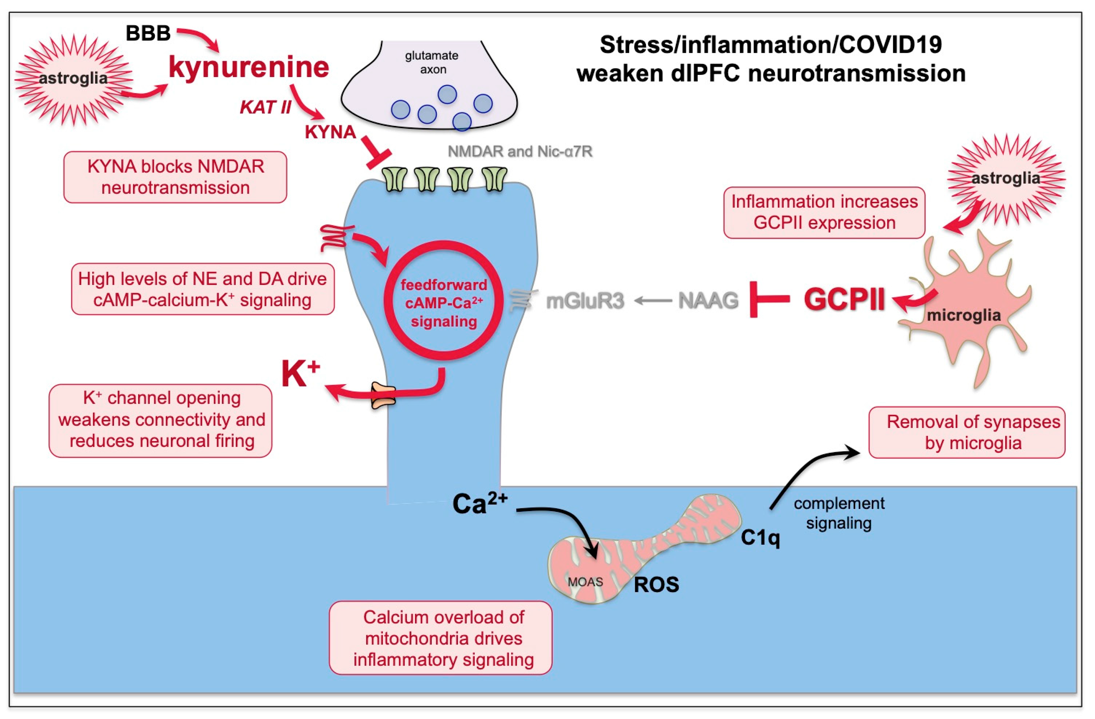
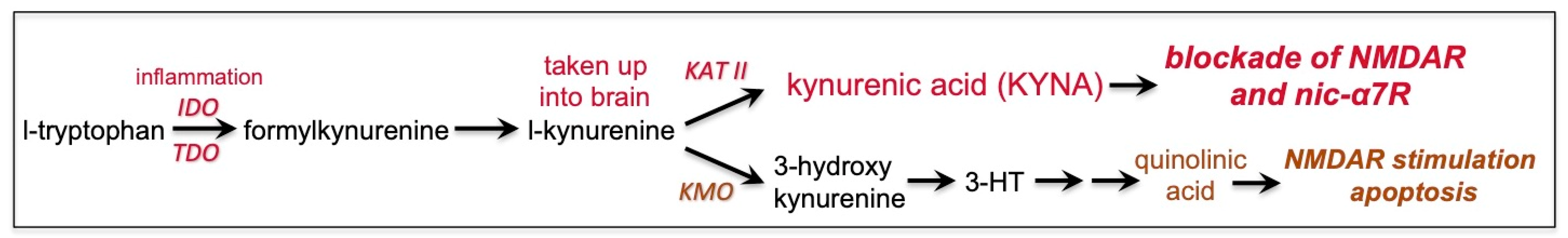
7. Kynurenic Acid Signaling Is Increased by Inflammation, Including by COVID-19, and Can Reduce NMDAR and nic-α7R Neurotransmission

Kynurenine is metabolized from tryptophan under conditions of inflammation (Figure 3 and Figure 4). Kynurenine is synthesized locally in brain by astrocytes and is also actively taken up from the blood into the brain, where it can be further metabolized by KAT II (kynurenine aminotransferase II) into kynurenic acid (KYNA), or by KMO (kynurenine 3-monooxygenase) to produce quinolinic acid [70]. KYNA blocks both NMDAR and nic-α7R, while quinolinic acid stimulates NMDA R [70]. Researchers examining the mechanisms underlying excitotoxicity have shown that under conditions of high glutamate release, e.g., during a stroke or traumatic brain injury, or when the brain is sliced for in vitro recordings, quinolinic acid stimulation of NMDAR contributes to toxic actions and neuronal death, while KYNA blockade of NMDAR is protective, preventing cell death [71]. Thus, KYNA is often included in the bath during in vitro slice recordings.
However, researchers have also examined the effects of KYNA under non-excitotoxic conditions, e.g., in chronic neuroinflammatory disorders when there is normal or even reduced glutamate release but elevated kynurenine signaling. Under these conditions, KYNA blockade of NMDAR is detrimental as it impairs cognition [72]. For example, schizophrenia is associated with lower or normal levels of glutamate in the PFC [73] and with increased neuroinflammation [74], including elevated KYNA expression in the dlPFC that correlates with impaired dlPFC function [75]. As dlPFC neurotransmission depends heavily on NMDAR and nic-α7R [57,58], KYNA blockade of both of these receptors would be particularly detrimental to dlPFC function.
It is likely that increased KYNA expression in the PFC contributes to the cognitive deficits of long COVID-19. There are extensive data showing that COVID19 infection increases kynurenine levels in plasma that correlate with cytokine levels and are particularly high in men [42,76,77,78,79]. Similar findings have been seen in macaques infected with SARS-CoV-2 [80]. Post-mortem evaluations of brains of patients who died from COVID infection show very large elevations in KYNA expression [38], demonstrating its kynurenine conversion to KYNA in brain. In patients with long COVID, plasma kynurenine levels remain high [81]. Most pertinent to the current review, cognitive deficits in long COVID highly correlate with plasma kynurenine levels [37], as do symptoms of anxiety and stress, which are also signs of PFC dysfunction [82].
Studies in animals support the hypothesis that elevated KYNA in the PFC impairs cognitive functioning. Treatments that increase KYNA in the PFC impair cognitive function in rats [83,84]. Conversely, treatments that reduce KYNA expression in brain improve cognitive performance in monkeys, rats, and mice [85,86]. Our preliminary data show that iontophoresis of KYNA markedly reduces dlPFC neuronal firing in monkeys performing a working memory task, consistent with this hypothesis (M. Wang, unpublished). Thus, agents that reduce KYNA production may be especially helpful in restoring dlPFC cognitive functioning in patients with long COVID.

8. GCPII Expression Is Increased by Inflammation, Including by COVID-19, and Reduces mGluR3 Regulation of cAMP-Calcium Signaling

As summarized in Figure 3, the large production of GCPII under inflammatory conditions destroys NAAG and reduces mGluR3 signaling. Elevated GCPII has been seen in multiple inflammatory conditions [87,88,89], including elevated GCPII in rodent brain following hypoxia from stroke [90]. Elevated GCPII is associated with impaired cognition in animal models, where GCPII inhibition can restore cognitive abilities [91,92]. Most pertinent to the current review, there are very large increases in GCPII expression in the brains of patients who died from COVID-19 [38]. Elevations in GCPII would be particularly harmful to the proper functioning of the primate dlPFC, as mGluR3 signaling is needed to regulate feedforward cAMP-calcium-K+ signaling in dlPFC spines (Figure 3). Thus, elevated GCPII signaling may have contributed to the large increases in cAMP-PKA-calcium signaling in the brains of patients who died from COVID-19 [38].
In summary, the data suggest that KYNA blockade of NMDAR and nic-α7R neurotransmission, as well as GCPII dysregulated cAMP-calcium signaling, may especially weaken dlPFC circuit firing by (1) reducing neurotransmission and (2) increasing cAMP-PKA-calcium opening of K+ channels, functionally disconnecting dlPFC circuits (Figure 3). Sustained elevations in feedforward calcium signaling would also lead to loss of spines and dendrites through calcium overload of nearby mitochondria and the initiation of inflammatory signals to microglia to engulf the afflicted spine (Figure 3). Thus, treatments that inhibit the production of KYNA and/or that restore regulation of cAMP-calcium signaling in dlPFC may be effective in treating the PFC cognitive symptoms of long COVID.

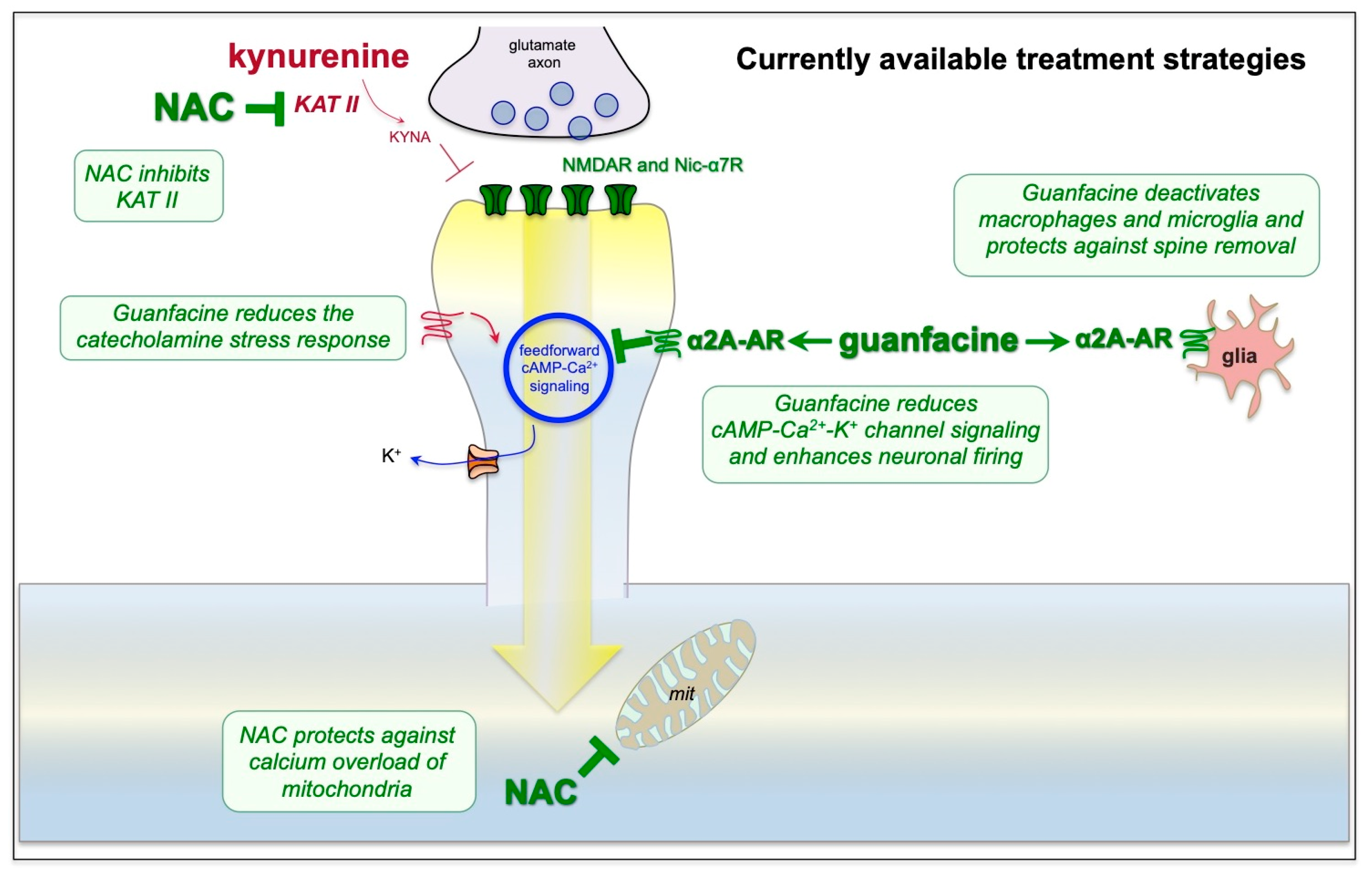
9. Strategies for Treatment Based on the Neuroscience

Specific inhibitors of kynurenine [85] or GCPII [93] signaling are currently under development, but are not yet available for human use. The following describes two compounds that are FDA-approved for other indications that may be useful in normalizing dlPFC physiology and restoring cognitive function by reducing the production of KYNA and increasing the regulation of cAMP-calcium signaling in dlPFC.
N-acetylcysteine (NAC)—NAC is FDA-approved for the treatment of acetaminophen overdose [94], which has antioxidant properties, e.g., by increasing endogenous glutathione levels [94] and protecting mitochondria from calcium overload [95]. Animal models of TBI have shown that NAC reduces neuropathology [95,96], and NAC is now in widespread, off-label use to treat TBI in patients, with few side effects [97,98]. NAC has also been proposed as an adjunct treatment for acute COVID-19 infection based on its antioxidant properties [99]. However, it has recently been discovered that NAC also inhibits KAT-II, the enzyme that converts kynurenine to kynurenic acid. NAC treatment significantly reduced KYNA levels in the rodent PFC [100] and chronic NAC treatment reduced KYNA and protected cognitive function in mice treated with kynurenine [86]. Thus, its ability to inhibit KAT II, in addition to its antioxidant properties, may be helpful in restoring NMDAR and nic-α7R neurotransmission in the dlPFC of patients with PFC cognitive deficits due to long COVID (Figure 5).
Guanfacine—Guanfacine is a selective α2A-AR agonist that was originally FDA-approved for the treatment of hypertension in 1986 and more recently FDA-approved for the treatment of Attention Deficit Hyperactivity Disorder (ADHD) in 2009 [101]. ADHD is characterized by weak regulation of attention and impulse control, involving impaired development and/or functioning of the PFC [102]. The extended release formulation of guanfacine (Intuniv) is often used to treat ADHD in children [101] and younger adults [103], given the faster drug metabolism in younger patients. As summarized in Figure 5, guanfacine’s beneficial actions likely involve both strengthening of dlPFC neuronal network connections and reducing neuroinflammation. Guanfacine’s ability to strengthen dlPFC function has been studied at the cellular level, where α2A-ARs are localized post-synaptically on dlPFC spines near the HCN channels that weaken network connectivity [59]. Local or systemic administration of guanfacine enhances dlPFC neuronal firing and cognitive functions by inhibiting cAMP-calcium-K+ channel signaling, and thus strengthening network connectivity and PFC activity [104]. Guanfacine and other α2A-AR agonists are also anti-inflammatory, deactivating both microglia [105,106] and macrophages [107]. Guanfacine has been shown to enhance and/or protect PFC structure and function in a variety of animal models, including rescuing PFC cognitive functioning and spine density from the deleterious effects of chronic psychological stress [55] or hypoxia [50,51]. Guanfacine has also been used off-label in human subjects to treat cognitive deficits caused by TBI [44,108], hypoxia [109], and psychological stress [110], and it has established its safety over decades of use. It is currently being tested for the treatment of delirium (https://clinicaltrials.gov: NCT04742673 and NCT04578886), a condition that would benefit from both anti-inflammatory and PFC-strengthening actions.
Given the established safety of both NAC and guanfacine and the urgent need for treatment of cognitive impairment in long COVID, these agents may provide interim relief while waiting for FDA-approved therapies. Our preliminary, open-label findings [111] indicate improvement in cognitive functioning in patients with long COVID following treatment with a combined daily treatment with both NAC (600 mg) and guanfacine (1 mg, escalating to 2–3 mg as needed). Placebo-controlled trials are needed to establish efficacy, but the scientific rationale for their use, outlined here, suggests they may have immediate utility in patients with urgent need.

10. Outstanding Questions and Summary

The current pandemic has created a sudden and urgent need for treatments that may alleviate the sustained cognitive deficits caused by COVID-19 infection. Understanding how COVID-19 affects the brain is an arena where much still needs to be learned, especially with regard to those with less severe infection who nonetheless retain debilitating cognitive deficits. This is particularly challenging in living individuals, where in vivo brain imaging of inflammation is very limited, and analyses often rely on distal measures such as CSF or plasma measures of inflammation.
There is also an outstanding need for new treatments aimed at this indication, such as GCPII inhibitors approved for human use, which will likely require many years for successful development. There is also a need for placebo-controlled trials of existing compounds approved for other indications, such as NAC and guanfacine, as described in this review. However, it takes many years to acquire funding and to conduct sufficiently large trials to have confident results. As the cognitive deficits of long COVID are often sufficiently severe to impede work and home life, rational strategies for treatment are needed to help those whose lives have been upended by the ongoing pandemic.
In summary, the cognitive deficits of long COVID particularly involve deficits in dlPFC function. The unusual neurotransmission and neuromodulation of dlPFC circuits make them especially vulnerable to inflammation, including KYNA blockade of NMDAR and nic-α7R, the receptors needed for dlPFC neurotransmission, and the inflammatory factors such as GCPII that lessen regulation of magnified calcium signaling, thus increasing calcium’s toxic actions. Compounds such as NAC and guanfacine, which block the production of KYNA and increase the regulation of cAMP-calcium signaling, respectively, may be helpful in restoring dlPFC physiology and function in the absence of FDA-approved treatments for this indication.

Author Contributions

A.F.Z., A.F.T.A. and M.W. wrote/revised this review based on their expertise in the clinical and basic neuroscience, respectively. All authors have read and agreed to the published version of the manuscript.

Funding

A.F.Z. is supported by 1P30AG066508-01. A.F.T.A. and M.W. are supported by NIH AG061190 and Neuronex NSF 2015276.

Institutional Review Board Statement

Not applicable.

Informed Consent Statement

Not applicable.

Data Availability Statement

Not applicable.

Acknowledgments

The authors acknowledge that there were no additional external funding for the work presented.

Conflicts of Interest

The authors declare no conflict of interest.

References

  1. Szczepanski, S.M.; Knight, R.T. Insights into human behavior from lesions to the prefrontal cortex. Neuron 2014, 83, 1002–1018. [Google Scholar] [CrossRef] [PubMed] [Green Version]
  2. Preuss, T.M.; Wise, S.P. Evolution of prefrontal cortex. Neuropsychopharmacology 2022, 47, 3–19. [Google Scholar] [CrossRef] [PubMed]
  3. Alexander, L.; Wood, C.M.; Roberts, A.C. The ventromedial prefrontal cortex and emotion regulation: Lost in translation? J. Physiol. 2023, 601, 37–50. [Google Scholar] [CrossRef] [PubMed]
  4. Elston, G.N.; Benavides-Piccione, R.; Elston, A.; Zietsch, B.; Defelipe, J.; Manger, P.; Casagrande, V.; Kaas, J.H. Specializations of the granular prefrontal cortex of primates: Implications for cognitive processing. Anat. Rec. A Discov. Mol. Cell. Evol. Biol. 2006, 288, 26–35. [Google Scholar] [CrossRef]
  5. Defelipe, J. The evolution of the brain, the human nature of cortical circuits, and intellectual creativity. Front. Neuroanat. 2011, 5, 29. [Google Scholar] [CrossRef] [Green Version]
  6. Goldman-Rakic, P. Cellular Basis of Working Memory. Neuron 1995, 14, 477–485. [Google Scholar]
  7. Levy, R.; Dubois, B. Apathy and the functional anatomy of the prefrontal cortex-basal ganglia circuits. Cereb. Cortex 2006, 16, 916–928. [Google Scholar] [CrossRef] [Green Version]
  8. Friedman, N.P.; Robbins, T.W. The role of prefrontal cortex in cognitive control and executive function. Neuropsychopharmacology 2022, 47, 72–89. [Google Scholar] [CrossRef]
  9. Robbins, T.W. Dissociating executive functions of the prefrontal cortex. Philos. Trans. R. Soc. Lond. Ser. B Biol. Sci. 1996, 351, 1463–1470. [Google Scholar] [CrossRef]
  10. Kimberg, D.Y.; Farah, M.J. A unified account of cognitive impairments following frontal lobe damage: The role of working memory in complex, organized behavior. J. Exp. Psychol. Gen. 1993, 122, 411–428. [Google Scholar] [CrossRef]
  11. Menon, V.; D’Esposito, M. The role of PFC networks in cognitive control and executive function. Neuropsychopharmacology 2022, 47, 90–103. [Google Scholar] [CrossRef]
  12. Dolcos, F.; McCarthy, G. Brain systems mediating cognitive interference by emotional distraction. J. Neurosci. 2006, 26, 2072–2079. [Google Scholar] [CrossRef] [Green Version]
  13. Dias, E.C.; McGinnis, T.; Smiley, J.F.; Foxe, J.J.; Schroeder, C.E.; Javitt, D.C. Changing plans: Neural correlates of executive control in monkey and human frontal cortex. Exp. Brain Res. 2006, 174, 279–291. [Google Scholar] [CrossRef] [PubMed]
  14. Fuster, J.M. Prefrontal neurons in networks of executive memory. Brain Res. Bull. 2000, 52, 331–336. [Google Scholar] [CrossRef]
  15. Aron, A.R. The neural basis of inhibition in cognitive control. Neuroscientist 2007, 13, 214–228. [Google Scholar] [CrossRef] [PubMed]
  16. Ungerleider, L.G.; Courtney, S.M.; Haxby, J.V. A neural system for human visual working memory. Proc. Natl. Acad. Sci. USA 1998, 95, 883–890. [Google Scholar] [CrossRef] [PubMed] [Green Version]
  17. Tsujimoto, S.; Genovesio, A.; Wise, S.P. Frontal pole cortex: Encoding ends at the end of the endbrain. Trends Cogn. Sci. 2011, 15, 169–176. [Google Scholar] [CrossRef]
  18. Joyce, M.K.P.; Marshall, L.G.; Banik, S.L.; Wang, J.; Xiao, D.; Bunce, J.G.; Barbas, H. Pathways for Memory, Cognition and Emotional Context: Hippocampal, Subgenual Area 25, and Amygdalar Axons Show Unique Interactions in the Primate Thalamic Reuniens Nucleus. J. Neurosci. 2022, 42, 1068–1089. [Google Scholar] [CrossRef]
  19. Holmes, S.E.; Scheinost, D.; Finnema, S.J.; Naganawa, M.; Davis, M.T.; DellaGioia, N.; Nabulsi, N.; Matuskey, D.; Angarita, G.A.; Pietrzak, R.H.; et al. Lower synaptic density is associated with depression severity and network alterations. Nat. Commun. 2019, 10, 1529. [Google Scholar] [CrossRef] [Green Version]
  20. Helms, J.; Kremer, S.; Merdji, H.; Clere-Jehl, R.; Schenck, M.; Kummerlen, C.; Collange, O.; Boulay, C.; Fafi-Kremer, S.; Ohana, M.; et al. Neurologic Features in Severe SARS-CoV-2 Infection. N. Engl. J. Med. 2020, 382, 2268–2270. [Google Scholar] [CrossRef]
  21. Becker, J.H.; Lin, J.J.; Doernberg, M.; Stone, K.; Navis, A.; Festa, J.R.; Wisnivesky, J.P. Assessment of Cognitive Function in Patients After COVID-19 Infection. JAMA Netw. Open. 2021, 4, e2130645. [Google Scholar] [CrossRef]
  22. Hellmuth, J.; Barnett, T.A.; Asken, B.M.; Kelly, J.D.; Torres, L.; Stephens, M.L.; Greenhouse, B.; Martin, J.N.; Chow, F.C.; Deeks, S.G.; et al. Persistent COVID-19-associated neurocognitive symptoms in non-hospitalized patients. J. Neurovirol. 2021, 27, 191–195. [Google Scholar] [CrossRef]
  23. Vanderlind, W.M.; Rabinovitz, B.B.; Miao, I.Y.; Oberlin, L.E.; Bueno-Castellano, C.; Fridman, C.; Jaywant, A.; Kanellopoulos, D. A systematic review of neuropsychological and psychiatric sequalae of COVID-19: Implications for treatment. Curr. Opin. Psychiatry 2021, 34, 420–433. [Google Scholar] [CrossRef] [PubMed]
  24. Hampshire, A.; Trender, W.; Chamberlain, S.R.; Jolly, A.E.; Grant, J.E.; Patrick, F.; Mazibuko, N.; Williams, S.C.; Barnby, J.M.; Hellyer, P.; et al. Cognitive deficits in people who have recovered from COVID-19. EClinicalMedicine 2021, 39, 101044. [Google Scholar] [CrossRef]
  25. Spudich, S.; Nath, A. Nervous system consequences of COVID-19. Science 2022, 375, 267–269. [Google Scholar] [CrossRef]
  26. Perrottelli, A.; Sansone, N.; Giordano, G.M.; Caporusso, E.; Giuliani, L.; Melillo, A.; Pezzella, P.; Bucci, P.; Mucci, A.; Galderisi, S. Cognitive Impairment after Post-Acute COVID-19 Infection: A Systematic Review of the Literature. J. Pers. Med. 2022, 12, 2070. [Google Scholar] [CrossRef]
  27. Zawilska, J.B.; Kuczyńska, K. Psychiatric and neurological complications of long COVID. J. Psychiatr. Res. 2022, 156, 349–360. [Google Scholar] [CrossRef] [PubMed]
  28. Bertuccelli, M.; Ciringione, L.; Rubega, M.; Bisiacchi, P.; Masiero, S.; Del Felice, A. Cognitive impairment in people with previous COVID-19 infection: A scoping review. Cortex 2022, 154, 212–230. [Google Scholar] [CrossRef] [PubMed]
  29. Houben, S.; Bonnechère, B. The Impact of COVID-19 Infection on Cognitive Function and the Implication for Rehabilitation: A Systematic Review and Meta-Analysis. Int. J. Environ. Res. Public Health 2022, 19, 7748. [Google Scholar] [CrossRef] [PubMed]
  30. Zeng, N.; Zhao, Y.-M.; Yan, W.; Li, C.; Lu, Q.-D.; Liu, L.; Ni, S.-Y.; Mei, H.; Yuan, K.; Shi, L.; et al. A systematic review and meta-analysis of long term physical and mental sequelae of COVID-19 pandemic: Call for research priority and action. Mol. Psychiatry 2023, 28, 423–433. [Google Scholar] [CrossRef]
  31. Ceban, F.; Ling, S.; Lui, L.M.W.; Lee, Y.; Gill, H.; Teopiz, K.M.; Rodrigues, N.B.; Subramaniapillai, M.; Di Vincenzo, J.D.; Cao, B.; et al. Fatigue and cognitive impairment in Post-COVID-19 Syndrome: A systematic review and meta-analysis. Brain Behav. Immun. 2022, 101, 93–135. [Google Scholar] [CrossRef]
  32. Arnsten, A.F.T.; Joyce, M.K.; Roberts, A.C. The Aversive Lens: Stress effects on the prefrontal-cingulate cortical pathways that regulate emotion. Neurosci. Biobehav. Rev. 2022, 145, 105000. [Google Scholar] [CrossRef]
  33. Herrera, E.; Pérez-Sánchez, M.D.C.; San Miguel-Abella, R.; Barrenechea, A.; Blanco, C.; Solares, L.; González, L.; Iza, C.; Castro, I.; Nicolás, E.; et al. Cognitive impairment in young adults with post COVID-19 syndrome. Sci. Rep. 2023, 13, 6378. [Google Scholar] [CrossRef] [PubMed]
  34. Gamberini, G.; Masuccio, F.G.; Cerrato, M.; Strazzacappa, M.; Ferraro, D.; Solaro, C. Previously independent patients with mild-symptomatic COVID-19 are at high risk of developing cognitive impairment but not depression or anxiety. J. Affect. Disord. 2023, 324, 645–651. [Google Scholar] [CrossRef]
  35. Taruffi, L.; Muccioli, L.; Mitolo, M.; Ferri, L.; Descovich, C.; Mazzoni, S.; Michelucci, R.; Lodi, R.; Liguori, R.; Cortelli, P.; et al. Neurological Manifestations of Long COVID: A Single-Center One-Year Experience. Neuropsychiatr. Dis. Treat. 2023, 19, 311–319. [Google Scholar] [CrossRef] [PubMed]
  36. He, D.; Yuan, M.; Dang, W.; Bai, L.; Yang, R.; Wang, J.; Ma, Y.; Liu, B.; Liu, S.; Zhang, S.; et al. Long term neuropsychiatric consequences in COVID-19 survivors: Cognitive impairment and inflammatory underpinnings fifteen months after discharge. Asian J. Psychiatr. 2023, 80, 103409. [Google Scholar] [CrossRef] [PubMed]
  37. Cysique, L.A.; Jakabek, D.; Bracken, S.G.; Allen-Davidian, Y.; Heng, B.; Chow, S.; Dehhaghi, M.; Staats Pires, A.; Darley, D.R.; Byrne, A.; et al. Post-acute COVID-19 cognitive impairment and decline uniquely associate with kynurenine pathway activation: A longitudinal observational study. medRxiv 2022. [Google Scholar] [CrossRef]
  38. Reiken, S.; Sittenfeld, L.; Dridi, H.; Liu, Y.; Liu, X.; Marks, A.R. Alzheimer’s-like signaling in brains of COVID-19 patients. Alzheimer’s Dement. 2022, 18, 955–965. [Google Scholar] [CrossRef]
  39. Woo, E.; Sansing, L.H.; Arnsten, A.F.T.; Datta, D. Chronic Stress Weakens Connectivity in the Prefrontal Cortex: Architectural and Molecular Changes. Chronic Stress 2021, 5, 24705470211029254. [Google Scholar] [CrossRef]
  40. Arnsten, A.F.T.; Woo, E.; Yang, S.; Wang, M.; Datta, D. Unusual Molecular Regulation of Dorsolateral Prefrontal Cortex Layer III Synapses Increases Vulnerability to Genetic and Environmental Insults in Schizophrenia. Biol. Psychiatry 2022. ahead of print. [Google Scholar] [CrossRef]
  41. Newhouse, A.; Kritzer, M.D.; Eryilmaz, H.; Praschan, N.; Camprodon, J.A.; Fricchione, G.; Chemali, Z. Neurocircuitry Hypothesis and Clinical Experience in Treating Neuropsychiatric Symptoms of Postacute Sequelae of Severe Acute Respiratory Syndrome Coronavirus 2. J. Acad. Consult. Liaison Psychiatry 2022, 63, 619–627. [Google Scholar] [CrossRef]
  42. Bizjak, D.A.; Stangl, M.; Börner, N.; Bösch, F.; Durner, J.; Drunin, G.; Buhl, J.L.; Abendroth, D. Kynurenine serves as useful biomarker in acute, Long- and Post-COVID-19 diagnostics. Front. Immunol. 2022, 13, 1004545. [Google Scholar] [CrossRef] [PubMed]
  43. McDonald, B.; Flashman, L.; Saykin, A.J. Executive dysfunction following traumatic brain injury: Neural substrates and treatment strategies. NeuroRehabilitation 2002, 17, 33–44. [Google Scholar] [CrossRef] [Green Version]
  44. McAllister, T.W.; Flashman, L.A.; Sparling, M.B.; Saykin, A.J. Working memory deficits after traumatic brain injury: Catecholaminergic mechanisms and prospects for treatment—A review. Brain Inj. 2004, 18, 331–350. [Google Scholar] [CrossRef] [PubMed]
  45. Graner, J.; Oakes, T.R.; French, L.M.; Riedy, G. Functional MRI in the investigation of blast-related traumatic brain injury. Front. Neurol. 2013, 4, 16. [Google Scholar] [CrossRef] [PubMed] [Green Version]
  46. Ozga, J.E.; Povroznik, J.M.; Engler-Chiurazzi, E.B.; Vonder Haar, C. Executive (dys)function after traumatic brain injury: Special considerations for behavioral pharmacology. Behav. Pharmacol. 2018, 29, 617–637. [Google Scholar] [CrossRef]
  47. Hoskison, M.M.; Moore, A.N.; Hu, B.; Orsi, S.A.; Kobori, N.; Dash, P.K. Persistent working memory dysfunction following traumatic brain injury: Evidence for a time-dependent mechanism. Neuroscience 2009, 159, 483–491. [Google Scholar] [CrossRef] [Green Version]
  48. Zhao, J.; Huynh, J.; Hylin, M.J.; O’Malley, J.J.; Perez, A.; Moore, A.N.; Dash, P.K. Mild Traumatic Brain Injury Reduces Spine Density of Projection Neurons in the Medial Prefrontal Cortex and Impairs Extinction of Contextual Fear Memory. J. Neurotrauma 2018, 35, 149–156. [Google Scholar] [CrossRef]
  49. Yan, X.; Zhang, J.; Shi, J.; Gong, Q.; Weng, X. Cerebral and functional adaptation with chronic hypoxia exposure: A multi-modal MRI study. Brain Res. 2010, 1348, 21–29. [Google Scholar] [CrossRef]
  50. Kauser, H.; Sahu, S.; Kumar, S.; Panjwani, U. Guanfacine is an effective countermeasure for hypobaric hypoxia-induced cognitive decline. Neuroscience 2013, 254, 110–119. [Google Scholar] [CrossRef]
  51. Kauser, H.; Sahu, S.; Panjwani, U. Guanfacine promotes neuronal survival in medial prefrontal cortex under hypobaric hypoxia. Brain Res. 2016, 1636, 152–160. [Google Scholar] [CrossRef] [PubMed]
  52. Maier, S.F.; Amat, J.; Baratta, M.V.; Paul, E.; Watkins, L.R. Behavioral control, the medial prefrontal cortex, and resilience. Dialogues Clin. Neurosci. 2006, 8, 397–406. [Google Scholar] [CrossRef]
  53. Arnsten, A.F.T.; Datta, D.; Wang, M. The Genie in the Bottle-Magnified calcium signaling in dorsolateral prefrontal cortex. Mol. Psychiatry 2021, 26, 3684–3700. [Google Scholar] [CrossRef] [PubMed]
  54. Liston, C.; McEwen, B.S.; Casey, B.J. Psychosocial stress reversibly disrupts prefrontal processing and attentional control. Proc. Nat. Acad. Sci. USA 2009, 106, 912–917. [Google Scholar] [CrossRef] [PubMed] [Green Version]
  55. Hains, A.B.; Yabe, Y.; Arnsten, A.F.T. Chronic stimulation of alpha-2A-adrenoceptors with guanfacine protects rodent prefrontal cortex dendritic spines and cognition from the effects of chronic stress. Neurobiol. Stress 2015, 2, 1–9. [Google Scholar] [CrossRef] [PubMed] [Green Version]
  56. Ansell, E.B.; Rando, K.; Tuit, K.; Guarnaccia, J.; Sinha, R. Cumulative adversity and smaller gray matter volume in medial prefrontal, anterior cingulate, and insula regions. Biol. Psychiatry 2012, 72, 57–64. [Google Scholar] [CrossRef] [Green Version]
  57. Wang, M.; Yang, Y.; Wang, C.J.; Gamo, N.J.; Jin, L.E.; Mazer, J.A.; Morrison, J.H.; Wang, X.-J.; Arnsten, A.F. NMDA receptors subserve working memory persistent neuronal firing In dorsolateral prefrontal cortex. Neuron 2013, 77, 736–749. [Google Scholar] [CrossRef] [Green Version]
  58. Yang, Y.; Paspalas, C.D.; Jin, L.E.; Picciotto, M.R.; Arnsten, A.F.T.; Wang, M. Nicotinic α7 receptors enhance NMDA cognitive circuits in dorsolateral prefrontal cortex. Proc. Nat. Acad. Sci. USA 2013, 110, 12078–12083. [Google Scholar] [CrossRef] [Green Version]
  59. Wang, M.; Ramos, B.; Paspalas, C.; Shu, Y.; Simen, A.; Duque, A.; Vijayraghavan, S.; Brennan, A.; Dudley, A.G.; Nou, E.; et al. Alpha2A-adrenoceptor stimulation strengthens working memory networks by inhibiting cAMP-HCN channel signaling in prefrontal cortex. Cell 2007, 129, 397–410. [Google Scholar] [CrossRef] [Green Version]
  60. Vornov, J.J.; Hollinger, K.R.; Jackson, P.F.; Wozniak, K.M.; Farah, M.H.; Majer, P.; Rais, R.; Slusher, B.S. Still NAAG’ing After All These Years: The Continuing Pursuit of GCPII Inhibitors. Adv. Pharmacol. 2016, 76, 215–255. [Google Scholar]
  61. Jin, L.E.; Wang, M.; Galvin, V.C.; Lightbourne, T.C.; Conn, P.J.; Arnsten, A.F.T.; Paspalas, C.D. mGluR2 vs. mGluR3 in Primate Prefrontal Cortex: Postsynaptic mGluR3 Strengthen Cognitive Networks. Cereb. Cortex 2018, 28, 974–987. [Google Scholar] [CrossRef] [Green Version]
  62. Finlay, J.M.; Zigmond, M.J.; Abercrombie, E.D. Increased dopamine and norepinephrine release in medial prefrontal cortex induced by acute and chronic stress: Effects of diazepam. Neuroscience 1995, 64, 619–628. [Google Scholar] [CrossRef]
  63. Goldstein, L.E.; Rasmusson, A.M.; Bunney, S.B.; Roth, R.H. Role of the amygdala in the coordination of behavioral, neuroendocrine and prefrontal cortical monoamine responses to psychological stress in the rat. J. Neurosci. 1996, 16, 4787–4798. [Google Scholar] [CrossRef] [PubMed] [Green Version]
  64. Zink, C.; Barker, P.; Sawa, A.; Weinberger, D.; Wang, A.; Quillian, H.; Ulrich, W.; Chen, Q.; Jaffe, A.; Kleinman, J.; et al. Missense Mutation in FOLH1 is Associated with Decreased NAAG Levels and Impaired Working Memory Circuitry and Cognition. Am. J. Psychiatry 2020, 177, 1129–1139. [Google Scholar] [CrossRef] [PubMed]
  65. Yang, S.; Datta, D.; Woo, E.; Duque, A.; Morozov, Y.M.; Arellano, J.; Slusher, B.S.; Wang, M.; Arnsten, A.F.T. Inhibition of glutamate-carboxypeptidase-II in dorsolateral prefrontal cortex: Potential therapeutic target for neuroinflammatory cognitive disorders. Mol. Psychiatry 2022, 27, 4252–4263. [Google Scholar] [CrossRef]
  66. Liston, C.; Miller, M.M.; Goldwater, D.S.; Radley, J.J.; Rocher, A.B.; Hof, P.R.; Morrison, J.H.; McEwen, B.S. Stress-induced alterations in prefrontal cortical dendritic morphology predict selective impairments in perceptual attentional set-shifting. J. Neurosci. 2006, 26, 7870–7874. [Google Scholar] [CrossRef] [PubMed] [Green Version]
  67. Kobori, N.; Clifton, G.L.; Dash, P.K. Enhanced catecholamine synthesis in the prefrontal cortex after traumatic brain injury: Implications for prefrontal dysfunction. J. Neurotrauma 2006, 23, 1094–1102. [Google Scholar] [CrossRef]
  68. Kobori, N.; Hu, B.; Dash, P.K. Altered adrenergic receptor signaling following traumatic brain injury contributes to working memory dysfunction. Neuroscience 2011, 172, 293–302. [Google Scholar] [CrossRef] [Green Version]
  69. Kobori, N.; Moore, A.N.; Dash, P.K. Altered regulation of protein kinase a activity in the medial prefrontal cortex of normal and brain-injured animals actively engaged in a working memory task. J. Neurotrauma 2015, 32, 139–148. [Google Scholar] [CrossRef] [Green Version]
  70. Schwarcz, R.; Bruno, J.P.; Muchowski, P.J.; Wu, H.Q. Kynurenines in the mammalian brain: When physiology meets pathology. Nat. Rev. Neurosci. 2012, 13, 465–477. [Google Scholar] [CrossRef]
  71. Yan, E.B.; Frugier, T.; Lim, C.K.; Heng, B.; Sundaram, G.; Tan, M.; Rosenfeld, J.V.; Walker, D.W.; Guillemin, G.J.; Morganti-Kossmann, M.C. Activation of the kynurenine pathway and increased production of the excitotoxin quinolinic acid following traumatic brain injury in humans. J. Neuroinflamm. 2015, 12, 110. [Google Scholar] [CrossRef] [Green Version]
  72. Szalardy, L.; Zadori, D.; Toldi, J.; Fulop, F.; Klivenyi, P.; Vecsei, L. Manipulating kynurenic acid levels in the brain—On the edge between neuroprotection and cognitive dysfunction. Curr. Top. Med. Chem. 2012, 12, 1797–1806. [Google Scholar] [CrossRef]
  73. Godlewska, B.R.; Minichino, A.; Emir, U.; Angelescu, I.; Lennox, B.; Micunovic, M.; Howes, O.; Cowen, P.J. Brain glutamate concentration in men with early psychosis: A magnetic resonance spectroscopy case-control study at 7 T. Transl. Psychiatry 2021, 11, 367. [Google Scholar] [CrossRef] [PubMed]
  74. Cannon, T.D.; Chung, Y.; He, G.; Sun, D.; Jacobson, A.; van Erp, T.G.; McEwen, S.; Addington, J.; Bearden, C.E.; Cadenhead, K.; et al. Progressive reduction in cortical thickness as psychosis develops: A multisite longitudinal neuroimaging study of youth at elevated clinical risk. Biol. Psychiatry 2014, 77, 147–157. [Google Scholar] [CrossRef] [PubMed] [Green Version]
  75. Kindler, J.; Lim, C.K.; Weickert, C.S.; Boerrigter, D.; Galletly, C.; Liu, D.; Jacobs, K.R.; Balzan, R.; Bruggemann, J.; O’Donnell, M.; et al. Dysregulation of kynurenine metabolism is related to proinflammatory cytokines, attention, and prefrontal cortex volume in schizophrenia. Mol. Psychiatry 2020, 25, 2860–2872. [Google Scholar] [CrossRef] [PubMed] [Green Version]
  76. Thomas, T.; Stefanoni, D.; Reisz, J.A.; Nemkov, T.; Bertolone, L.; Francis, R.O.; Hudson, K.E.; Zimring, J.C.; Hansen, K.C.; Hod, E.A.; et al. COVID-19 infection alters kynurenine and fatty acid metabolism, correlating with IL-6 levels and renal status. JCI Insight 2020, 5, e140327. [Google Scholar] [CrossRef]
  77. Cai, Y.; Kim, D.J.; Takahashi, T.; Broadhurst, D.I.; Yan, H.; Ma, S.; Rattray, N.J.W.; Casanovas-Massana, A.; Israelow, B.; Klein, J.; et al. Kynurenic acid may underlie sex-specific immune responses to COVID-19. Sci. Signal. 2021, 14, eabf8483. [Google Scholar] [CrossRef]
  78. Lawler, N.G.; Gray, N.; Kimhofer, T.; Boughton, B.; Gay, M.; Yang, R.; Morillon, A.C.; Chin, S.T.; Ryan, M.; Begum, S.; et al. Systemic Perturbations in Amine and Kynurenine Metabolism Associated with Acute SARS-CoV-2 Infection and Inflammatory Cytokine Responses. J. Proteome Res. 2021, 20, 2796–2811. [Google Scholar] [CrossRef]
  79. Almulla, A.F.; Supasitthumrong, T.; Amrapala, A.; Tunvirachaisakul, C.; Jaleel, A.K.A.; Oxenkrug, G.; Al-Hakeim, H.K.; Maes, M. The Tryptophan Catabolite or Kynurenine Pathway in Alzheimer’s Disease: A Systematic Review and Meta-Analysis. J. Alzheimer’s Dis. 2022, 88, 1325–1339. [Google Scholar] [CrossRef]
  80. Ma, Q.; Ma, W.; Song, T.Z.; Wu, Z.; Liu, Z.; Hu, Z.; Han, J.B.; Xu, L.; Zeng, B.; Wang, B.; et al. Single-nucleus transcriptomic profiling of multiple organs in a rhesus macaque model of SARS-CoV-2 infection. Zool. Res. 2022, 43, 1041–1062. [Google Scholar] [CrossRef]
  81. Holmes, E.; Wist, J.; Masuda, R.; Lodge, S.; Nitschke, P.; Kimhofer, T.; Loo, R.L.; Begum, S.; Boughton, B.; Yang, R.; et al. Incomplete Systemic Recovery and Metabolic Phenoreversion in Post-Acute-Phase Nonhospitalized COVID-19 Patients: Implications for Assessment of Post-Acute COVID-19 Syndrome. J. Proteome Res. 2021, 20, 3315–3329. [Google Scholar] [CrossRef] [PubMed]
  82. Kucukkarapinar, M.; Yay-Pence, A.; Yildiz, Y.; Buyukkoruk, M.; Yaz-Aydin, G.; Deveci-Bulut, T.S.; Gulbahar, O.; Senol, E.; Candansayar, S. Psychological outcomes of COVID-19 survivors at sixth months after diagnose: The role of kynurenine pathway metabolites in depression, anxiety, and stress. J. Neural Transm. 2022, 129, 1077–1089. [Google Scholar] [CrossRef] [PubMed]
  83. Chess, A.C.; Simoni, M.K.; Alling, T.E.; Bucci, D.J. Elevations of endogenous kynurenic acid produce spatial working memory deficits. Schizophr. Bull. 2007, 33, 797–804. [Google Scholar] [CrossRef] [PubMed] [Green Version]
  84. Alexander, K.S.; Wu, H.Q.; Schwarcz, R.; Bruno, J.P. Acute elevations of brain kynurenic acid impair cognitive flexibility: Normalization by the alpha7 positive modulator galantamine. Psychopharmacology 2012, 220, 627–637. [Google Scholar] [CrossRef] [PubMed] [Green Version]
  85. Kozak, R.; Campbell, B.M.; Strick, C.A.; Horner, W.; Hoffmann, W.E.; Kiss, T.; Chapin, D.S.; McGinnis, D.; Abbott, A.L.; Roberts, B.M.; et al. Reduction of brain kynurenic acid improves cognitive function. J. Neurosci. 2014, 34, 10592–10602. [Google Scholar] [CrossRef] [Green Version]
  86. Blanco Ayala, T.B.; Ramírez Ortega, D.R.; Ovalle Rodríguez, P.O.; Pineda, B.; Pérez de la Cruz, G.P.; González Esquivel, D.G.; Schwarcz, R.; Sathyasaikumar, K.V.; Jiménez Anguiano, A.J.; Pérez de la Cruz, V.P. Subchronic N-acetylcysteine Treatment Decreases Brain Kynurenic Acid Levels and Improves Cognitive Performance in Mice. Antioxidants 2021, 10, 147. [Google Scholar] [CrossRef]
  87. Zhang, W.; Murakawa, Y.; Wozniak, K.M.; Slusher, B.; Sima, A.A. The preventive and therapeutic effects of GCPII (NAALADase) inhibition on painful and sensory diabetic neuropathy. J. Neurol. Sci. 2006, 247, 217–223. [Google Scholar] [CrossRef]
  88. Rahn, K.A.; Watkins, C.C.; Alt, J.; Rais, R.; Stathis, M.; Grishkan, I.; Crainiceau, C.M.; Pomper, M.G.; Rojas, C.; Pletnikov, M.V.; et al. Inhibition of glutamate carboxypeptidase II (GCPII) activity as a treatment for cognitive impairment in multiple sclerosis. Proc. Natl. Acad. Sci. USA 2012, 109, 20101–20106. [Google Scholar] [CrossRef] [Green Version]
  89. Rais, R.; Jiang, W.; Zhai, H.; Wozniak, K.M.; Stathis, M.; Hollinger, K.R.; Thomas, A.G.; Rojas, C.; Vornov, J.J.; Marohn, M.; et al. FOLH1/GCPII is elevated in IBD patients, and its inhibition ameliorates murine IBD abnormalities. JCI Insight 2016, 1, e88634. [Google Scholar] [CrossRef] [Green Version]
  90. Arteaga Cabeza, O.; Zhang, Z.; Smith Khoury, E.; Sheldon, R.A.; Sharma, A.; Zhang, F.; Slusher, B.S.; Kannan, R.M.; Kannan, S.; Ferriero, D.M. Neuroprotective effects of a dendrimer-based glutamate carboxypeptidase inhibitor on superoxide dismutase transgenic mice after neonatal hypoxic-ischemic brain injury. Neurobiol. Dis. 2021, 148, 105201. [Google Scholar] [CrossRef]
  91. Neale, J.H.; Olszewski, R. A role for N-acetylaspartylglutamate (NAAG) and mGluR3 in cognition. Neurobiol. Learn. Mem. 2019, 158, 9–13. [Google Scholar] [CrossRef] [PubMed]
  92. Datta, D.; Leslie, S.N.; Woo, E.; Amancharla, N.; Elmansy, A.; Lepe, M.; Mecca, A.P.; Slusher, B.S.; Nairn, A.C.; Arnsten, A.F.T. Glutamate Carboxypeptidase II in aging rat prefrontal cortex impairs working memory performance. Front. Aging Neurosci. 2021, 13, 760270. [Google Scholar] [CrossRef] [PubMed]
  93. Hollinger, K.R.; Sharma, A.; Tallon, C.; Lovell, L.; Thomas, A.G.; Zhu, X.; Wiseman, R.; Wu, Y.; Kambhampati, S.P.; Liaw, K.; et al. Dendrimer-2PMPA selectively blocks upregulated microglial GCPII activity and improves cognition in a mouse model of multiple sclerosis. Nanotheranostics 2022, 6, 126–142. [Google Scholar] [CrossRef] [PubMed]
  94. Dodd, S.; Dean, O.; Copolov, D.L.; Malhi, G.S.; Berk, M. N-acetylcysteine for antioxidant therapy: Pharmacology and clinical utility. Expert. Opin. Biol. Ther. 2008, 8, 1955–1962. [Google Scholar] [CrossRef] [PubMed]
  95. Xiong, Y.; Peterson, P.L.; Lee, C.P. Effect of N-acetylcysteine on mitochondrial function following traumatic brain injury in rats. J. Neurotrauma 1999, 16, 1067–1082. [Google Scholar] [CrossRef]
  96. Whitney, K.; Nikulina, E.; Rahman, S.N.; Alexis, A.; Bergold, P.J. Delayed dosing of minocycline plus N-acetylcysteine reduces neurodegeneration in distal brain regions and restores spatial memory after experimental traumatic brain injury. Exp. Neurol. 2021, 45, 113816. [Google Scholar] [CrossRef]
  97. Bhatti, J.; Nascimento, B.; Akhtar, U.; Rhind, S.G.; Tien, H.; Nathens, A.; da Luz, L.T. Systematic Review of Human and Animal Studies Examining the Efficacy and Safety of N-Acetylcysteine (NAC) and N-Acetylcysteine Amide (NACA) in Traumatic Brain Injury: Impact on Neurofunctional Outcome and Biomarkers of Oxidative Stress and Inflammation. Front. Neurol. 2018, 8, 744. [Google Scholar] [CrossRef] [Green Version]
  98. Mcpherson, R.; Mangram, A.J.; Barletta, J.F.; Dzandu, J.K. N-Acetylcysteine Is Associated with Reduction Of Post Concussive Symptoms In Elderly Patients: A Pilot Study. J. Trauma Acute Care Surg. 2022. ahead of print. [Google Scholar] [CrossRef]
  99. Dominari, A.; Hathaway, I.D.; Kapasi, A.; Paul, T.; Makkar, S.S.; Castaneda, V.; Gara, S.; Singh, B.M.; Agadi, K.; Butt, M.; et al. Bottom-up analysis of emergent properties of N-acetylcysteine as an adjuvant therapy for COVID-19. World J. Virol. 2021, 10, 34–52. [Google Scholar] [CrossRef]
  100. Blanco-Ayala, T.; Sathyasaikumar, K.V.; Uys, J.D.; Pérez-de-la-Cruz, V.; Pidugu, L.S.; Schwarcz, R. N-Acetylcysteine Inhibits Kynurenine Aminotransferase II. Neuroscience 2020, 444, 160–169. [Google Scholar] [CrossRef]
  101. Biederman, J.; Melmed, R.D.; Patel, A.; McBurnett, K.; Konow, J.; Lyne, A.; Scherer, N.; SPD503 Study Group. A randomized, double-blind, placebo-controlled study of guanfacine extended release in children and adolescents with attention-deficit/hyperactivity disorder. Pediatrics 2008, 121, e73–e84. [Google Scholar] [CrossRef] [PubMed]
  102. Rubia, K.; Alegría, A.A.; Brinson, H. Brain abnormalities in attention-deficit hyperactivity disorder: A review. Rev. Neurol. 2014, 58 (Suppl. S1), S3–S16. [Google Scholar] [PubMed]
  103. Ota, T.; Yamamuro, K.; Okazaki, K.; Kishimoto, T. Evaluating Guanfacine Hydrochloride in the Treatment of Attention Deficit Hyperactivity Disorder (ADHD) in Adult Patients: Design, Development and Place in Therapy. Drug Des. Dev. Ther. 2021, 15, 1965–1969. [Google Scholar] [CrossRef] [PubMed]
  104. Arnsten, A.F.T.; Wang, M. The Evolutionary Expansion of mGluR3-NAAG-GCPII Signaling: Relevance to Human Intelligence and Cognitive Disorders. Am. J. Psychiatry 2020, 177, 1103–1106. [Google Scholar] [CrossRef] [PubMed]
  105. Gyoneva, S.; Traynelis, S.F. Norepinephrine modulates the motility of resting and activated microglia via different adrenergic receptors. J. Biol. Chem. 2013, 288, 15291–15302. [Google Scholar] [CrossRef] [PubMed] [Green Version]
  106. Zhang, Y.; Tan, S.L.; Du, J.; Chen, Y.; Jia, J.; Feng, J.G.; Liu, K.X.; Zhou, J. Dexmedetomidine alleviates neuroinflammation, restores sleep disorders and neurobehavioral abnormalities in rats with minimal hepatic encephalopathy. Int. Immunopharmacol. 2021, 96, 107795. [Google Scholar] [CrossRef] [PubMed]
  107. Fujimoto, M.; Higuchi, H.; Honda-Wakasugi, Y.; Miyake, S.; Nishioka, Y.; Yabuki-Kawase, A.; Maeda, S.; Miyawaki, T. Dexmedetomidine inhibits LPS-induced inflammatory responses through peroxisome proliferator-activated receptor gamma (PPARγ) activation following binding to α2 adrenoceptors. Eur. J. Pharmacol. 2021, 892, 173733. [Google Scholar] [CrossRef]
  108. McAllister, T.W.; McDonald, B.C.; Flashman, L.A.; Ferrell, R.B.; Tosteson, T.D.; Yanofsky, N.N.; Grove, M.R.; Saykin, A.J. Alpha-2 adrenergic challenge with guanfacine one month after mild traumatic brain injury: Altered working memory and BOLD response. Int. J. Psychophysiol. 2011, 82, 107–114. [Google Scholar] [CrossRef] [Green Version]
  109. Miyoshi, T.; Mizushima, C.; Noborio, Y.; Kimoto, Y.; Nakaharu, Y.; Shimamoto, S. Efficacy of combination therapy with gabapentin and guanfacine for paroxysmal sympathetic hyperactivity following hypoxic encephalopathy: A case report. J. Int. Med. Res. 2021, 49, 3000605211009721. [Google Scholar] [CrossRef]
  110. Connor, D.F.; Grasso, D.J.; Slivinsky, M.D.; Pearson, G.S.; Banga, A. An open-label study of guanfacine extended release for traumatic stress related symptoms in children and adolescents. J. Child Adolesc. Psychopharmacol. 2013, 23, 244–251. [Google Scholar] [CrossRef] [Green Version]
  111. Fesharaki-Zadeh, A.; Lowe, N.; Arnsten, A.F. Clinical experience with the α2A-adrenoceptor agonist, guanfacine, and N-acetylcysteine for the treatment of cognitive deficits in “Long-COVID19”. Neuroimmunol. Rep. 2023, 3, 100154. [Google Scholar] [CrossRef]
Figure 2. The recently evolved neuronal circuits in the primate dlPFC have unusual neurotransmission and neuromodulation for healthy cognitive functioning. In contrast to typical glutamate circuits which rely heavily on AMPAR stimulation, dlPFC neurotransmission relies on NMDAR and nic-α7R with little contribution from AMPAR [57,58]. NMDAR opening can only occur in a depolarized synaptic membrane, which may be sustained by both nic-α7R stimulation and by higher calcium levels near the synapse. Layer III dlPFC spines express the molecular machinery to magnify calcium signaling near the synapse, where cAMP-PKA signaling is positioned to increase internal calcium release from the SER (smooth endoplasmic reticulum) through ryanodine receptors (RyR) and IP3 receptors (IP3R) [53]. Calcium in turn increases cAMP production, thus producing feedforward signaling that must be tightly regulated by α2A-AR and mGluR3, as described below. (The feedforward signaling is represented by the blue circle.) Moderate levels of cAMP-calcium signaling are necessary for dlPFC neuronal firing during working memory, but higher levels can reduce firing by opening nearby potassium (K+) channels. Under nonstress conditions, there are moderate levels of norepinephrine (NE) and dopamine release. Moderate levels of NE engage high-affinity α2A-AR (rather than lower-affinity α1-AR or β-AR), which are localized on dendritic spines [59]. mGluR3 are also on spines and are stimulated by both glutamate and NAAG (N-acetylaspartylglutamate), which is co-released with glutamate and is selective for mGluR3 [60]. α2A-AR and mGluR3 signaling regulate feedforward cAMP-calcium signaling, reducing cAMP-calcium opening of nearby K+ channels [53,61]. This strengthens network connectivity and increases the neuronal firing needed for higher cognition. mit = mitochondrion.
Neurolint 15 00045 g002
Figure 3. The effects of stress and inflammation on dlPFC networks. Uncontrollable stress and/or inflammation weaken dlPFC network connections in multiple ways. (1) Kynurenine is metabolized from tryptophan under inflammatory conditions in astrocytes and is also actively taken up from the blood through the blood brain barrier (BBB). In the brain, kynurenine can be further metabolized by KAT II to KYNA, which blocks both NMDA and nic-α7R, the two receptors needed for dlPFC neurotransmission. Thus, KYNA impairs PFC function. (2) Both psychological and physiological stressors cause high levels of catecholamine release in the PFC, which engage low-affinity α1-AR or β-AR as well as high levels of D1R stimulation, driving cAMP-calcium opening of large numbers of K+ channels, which weakens connectivity and reduces neuronal firing. (3) Microglial and astrocytic production of GCPII with inflammation catabolizes NAAG, reducing mGluR3 regulation of cAMP-calcium signaling, thus decreasing neuronal firing. (4) Sustained high levels of cytosolic calcium and calcium release from the SER with chronic stress/inflammation also cause calcium overload of mitochondria, leading to complement C1q signaling to microglia to remove the spine. COVID-19 infection is known to increase both KYNA and GCPII in brain and to increase cAMP-PKA-calcium signaling (see text). MOAS = mitochondria on a string, an abnormal mitochondrial phenotype consistent with calcium overload and an inflammatory state. ROS = reactive oxygen species.
Neurolint 15 00045 g003
Figure 4. A summary of kynurenine signaling, which is increased by COVID-19 infection. Kynurenine is metabolized from tryptophan by the immune system and by astrocytes under inflammatory conditions. It is actively taken up into the brain, where it can be further metabolized to KYNA, which blocks NMDAR and nic-α7R or to quinolinic acid, which stimulates NMDAR.
Neurolint 15 00045 g004
Figure 5. Potential treatments for the cognitive deficits of long COVID can normalize dlPFC physiology and thus may be helpful in treating the cognitive symptoms of long COVID. NAC may normalize dlPFC neurotransmission by blocking the production of KYNA, while the α2A-AR agonist, guanfacine, may restore regulation of cAMP-calcium-K+ signaling by stimulating α2A-AR on spines. NAC is also known to have antioxidant properties that would protect mitochondria, and α2A-AR agonists deactivate microglia and macrophages and thus have general anti-inflammatory properties.
Neurolint 15 00045 g005
Disclaimer/Publisher’s Note: The statements, opinions and data contained in all publications are solely those of the individual author(s) and contributor(s) and not of MDPI and/or the editor(s). MDPI and/or the editor(s) disclaim responsibility for any injury to people or property resulting from any ideas, methods, instructions or products referred to in the content.

