Next Issue
Volume 15, June
Previous Issue
Volume 14, December


Neurol. Int., Volume 15, Issue 1 (March 2023) – 32 articles

Cover Story (view full-size image): 
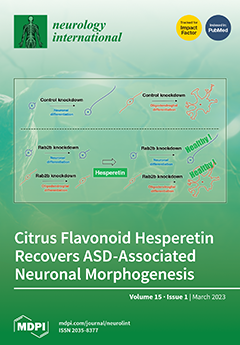
The rab2b gene is associated with autism spectrum disorder (ASD), although it remains unclear how it can cause ASD at the molecular and cellular levels. The important question of how cellular phenotypes of ASD can be recovered remains to be clarified. Here, this paper not only shows that Rab2b knockdown inhibits neuronal cell morphological differentiation, but also that its knockdown inhibits oligodendroglial cell morphological differentiation. Furthermore, hesperetin, a citrus flavonoid, recovers defective differentiation of neuronal and oligodendroglial cell differentiation. The underlying molecular mechanism involves the activation of classical mitogen-activated protein kinse (MAPK). Hesperetin may be considered as a chemical candidate for recovering ASD-associated neuronal and oligodendroglial cell morphogenesis. View this paper
  • Issues are regarded as officially published after their release is announced to the table of contents alert mailing list.
  • You may sign up for e-mail alerts to receive table of contents of newly released issues.
  • PDF is the official format for papers published in both, html and pdf forms. To view the papers in pdf format, click on the "PDF Full-text" link, and use the free Adobe Reader to open them.
Order results
Result details
Select all
Export citation of selected articles as:
15 pages, 2695 KiB  
Article
A Pilot Investigation of Visual Pathways in Patients with Mild Traumatic Brain Injury
by Paul Harris and Mark H. Myers
Neurol. Int. 2023, 15(1), 534-548; https://doi.org/10.3390/neurolint15010032 - 20 Mar 2023
Cited by 1 | Viewed by 1499
Abstract
In this study, we examined visual processing within primary visual areas (V1) in normal and visually impaired individuals who exhibit significant visual symptomology due to sports-related mild traumatic brain injury (mTBI). Five spatial frequency stimuli were applied to the right, left and both [...] Read more.
In this study, we examined visual processing within primary visual areas (V1) in normal and visually impaired individuals who exhibit significant visual symptomology due to sports-related mild traumatic brain injury (mTBI). Five spatial frequency stimuli were applied to the right, left and both eyes in order to assess the visual processing of patients with sports-related mild traumatic brain injuries who exhibited visual abnormalities, i.e., photophobia, blurriness, etc., and controls. The measurement of the left/right eye and binocular integration was accomplished via the quantification of the spectral power and visual event-related potentials. The principal results have shown that the power spectral density (PSD) measurements display a distinct loss in the alpha band-width range, which corresponded to more instances of medium-sized receptive field loss. Medium-size receptive field loss may correspond to parvocellular (p-cell) processing deprecation. Our major conclusion provides a new measurement, using PSD analysis to assess mTBI conditions from primary V1 areas. The statistical analysis demonstrated significant differences between the mTBI and control cohort in the Visual Evoked Potentials (VEP) amplitude responses and PSD measurements. Additionally, the PSD measurements were able to assess the improvement in the mTBI primary visual areas over time through rehabilitation. Full article
(This article belongs to the Special Issue Recent Advances in Traumatic Brain Injury)
Show Figures

Figure 1

16 pages, 1277 KiB  
Review
Chronic Administration of Melatonin: Physiological and Clinical Considerations
by Donald Givler, Amy Givler, Patrick M. Luther, Danielle M. Wenger, Shahab Ahmadzadeh, Sahar Shekoohi, Amber N. Edinoff, Bradley K. Dorius, Carlo Jean Baptiste, Elyse M. Cornett, Adam M. Kaye and Alan D. Kaye
Neurol. Int. 2023, 15(1), 518-533; https://doi.org/10.3390/neurolint15010031 - 15 Mar 2023
Cited by 2 | Viewed by 7654
Abstract
Background: Exogenous melatonin is commonly used to treat insomnia, other sleep problems, and numerous medical illnesses, including Alzheimer’s disease, autism spectrum disorder, and mild cognitive impairment in adults and children. There is evolving information regarding issues with the use of chronic melatonin. Methods: [...] Read more.
Background: Exogenous melatonin is commonly used to treat insomnia, other sleep problems, and numerous medical illnesses, including Alzheimer’s disease, autism spectrum disorder, and mild cognitive impairment in adults and children. There is evolving information regarding issues with the use of chronic melatonin. Methods: The present investigation was a narrative review. Results: Melatonin usage has risen dramatically in recent years. Many countries only allow melatonin prescriptions. In the United States (U.S.), it is classified as a dietary supplement accessible over the counter and can be derived from animals, microorganisms, or, most commonly, made synthetically. No regulatory agency oversees its manufacturing or sale in the U.S. melatonin concentration of marketed preparations varies widely between product labels and manufacturers. Melatonin’s ability to induce sleep is detectable. However, it is modest for most people. Sleep length appears to be less important in sustained-release preparations. The optimal dosage is unknown, and routinely used amounts vary substantially. Melatonin’s short-term negative effects are minimal, resolve at medicine cessation, and do not usually prevent usage overall. Much research on long-term melatonin administration has found no difference between exogenous melatonin and placebo in terms of long-term negative effects. Conclusion: Melatonin at low to moderate dosages (approximately 5–6 mg daily or less) appears safe. Long-term usage appears to benefit certain patient populations, such as those with autism spectrum disorder. Studies investigating potential benefits in reducing cognitive decline and increased longevity are ongoing. However, it is widely agreed that the long-term effects of taking exogenous melatonin have been insufficiently studied and warrant additional investigation. Full article
Show Figures

Figure 1

10 pages, 1510 KiB  
Article
Clinical Features of Acute Ischemic Stroke Patients with Hypoesthesia as an Initial Symptom
by Takayoshi Akimoto, Katsuhiko Ogawa, Makoto Hara, Satoko Ninomiya, Masaki Ishihara, Akihiko Morita, Satoshi Kamei and Hideto Nakajima
Neurol. Int. 2023, 15(1), 508-517; https://doi.org/10.3390/neurolint15010030 - 15 Mar 2023
Viewed by 1646
Abstract
This study aimed to evaluate the clinical characteristics of acute ischemic stroke (AIS) patients who experienced hypoesthesia as the initial symptom. We retrospectively analyzed the medical records of 176 hospitalized AIS patients who met our inclusion and exclusion criteria and evaluated their clinical [...] Read more.
This study aimed to evaluate the clinical characteristics of acute ischemic stroke (AIS) patients who experienced hypoesthesia as the initial symptom. We retrospectively analyzed the medical records of 176 hospitalized AIS patients who met our inclusion and exclusion criteria and evaluated their clinical features and MRI findings. Among this cohort, 20 (11%) patients presented with hypoesthesia as the initial symptom. MRI scans of these 20 patients identified lesions in the thalamus or pontine tegmentum in 14 and brain lesions at other sites in 6. The 20 hypoesthesia patients had higher systolic (p = 0.031) and diastolic blood pressure (p = 0.037) on admission, and a higher rate of small-vessel occlusion (p < 0.001) than patients without hypoesthesia. The patients with hypoesthesia had a significantly shorter average hospital stay (p = 0.007) but did not differ significantly from those without hypoesthesia in National Institutes of Health Stroke Scale scores on admission (p = 0.182) or the modified Rankin Scale scores for neurologic disability on discharge (p = 0.319). In the patients with acute onset hypoesthesia, high blood pressure, and neurological deficits were more likely to be due to AIS than other causes. Since most of the lesions in AIS patients with hypoesthesia as the initial symptom were found to be small, we recommend performing MRI scans with such patients to confirm AIS. Full article
(This article belongs to the Special Issue Stroke: From Pathophysiology to Therapy)
Show Figures

Figure 1

11 pages, 457 KiB  
Review
Sleep and Chronobiology as a Key to Understand Cluster Headache
by Laura Pilati, Angelo Torrente, Paolo Alonge, Lavinia Vassallo, Simona Maccora, Andrea Gagliardo, Antonia Pignolo, Salvatore Iacono, Salvatore Ferlisi, Vincenzo Di Stefano, Cecilia Camarda and Filippo Brighina
Neurol. Int. 2023, 15(1), 497-507; https://doi.org/10.3390/neurolint15010029 - 15 Mar 2023
Cited by 5 | Viewed by 1971
Abstract
The cluster headache is a primary headache characterized by attacks of unilateral pain associated with ipsilateral cranial autonomic features. These attacks recur in clusters during the years alternating with periods of complete remission, and their onset is often during the night. This annual [...] Read more.
The cluster headache is a primary headache characterized by attacks of unilateral pain associated with ipsilateral cranial autonomic features. These attacks recur in clusters during the years alternating with periods of complete remission, and their onset is often during the night. This annual and nocturnal periodicity hides a strong and mysterious link among CH, sleep, chronobiology and circadian rhythm. Behind this relationship, there may be the influence of genetic components or of anatomical structures such as the hypothalamus, which are both involved in regulating the biological clock and contributing even to the periodicity of cluster headaches. The bidirectional relationship manifests itself also with the presence of sleep disturbances in patients affected by cluster headaches. What if the key to studying the physiopathology of such disease could rely on the mechanisms of chronobiology? The purpose of this review is to analyze this link in order to interpret the pathophysiology of cluster headaches and the possible therapeutic implications. Full article
Show Figures

Figure 1

82 pages, 628 KiB  
Conference Report
Abstracts of the Fifth Brainstorming Research Assembly for Young Neuroscientists (BraYn), Italy, 28–30 September 2022
by Giovanni Ferrara
Neurol. Int. 2023, 15(1), 415-496; https://doi.org/10.3390/neurolint15010028 - 14 Mar 2023
Viewed by 2073
Abstract
On behalf of the BraYn Association Ets, we are pleased to present the Abstracts of the Fifth Brainstorming Research Assembly for Young Neuroscientists, which was held in Rome, Italy from 28–30 September 2022. We congratulate all the presenters on their research work and [...] Read more.
On behalf of the BraYn Association Ets, we are pleased to present the Abstracts of the Fifth Brainstorming Research Assembly for Young Neuroscientists, which was held in Rome, Italy from 28–30 September 2022. We congratulate all the presenters on their research work and contribution. Full article
10 pages, 1018 KiB  
Article
Parameters Associated with the Required Drug Dose of Intravenous Immunoglobulin in Stable Chronic Inflammatory Demyelinating Polyradiculoneuropathy
by Ludger Feyen, Christina Schaub, Julian Zimmermann and Louisa Nitsch
Neurol. Int. 2023, 15(1), 405-414; https://doi.org/10.3390/neurolint15010027 - 10 Mar 2023
Cited by 1 | Viewed by 1642
Abstract
Background: Intravenous immunoglobulin (IVIg) is efficient and one of very few treatment options for patients with chronic inflammatory demyelinating polyradiculoneuropathy (CIDP). However, finding the optimal dose of IVIg for individual CIDP patients remains challenging. The dose of IVIg needs to be adjusted individually. [...] Read more.
Background: Intravenous immunoglobulin (IVIg) is efficient and one of very few treatment options for patients with chronic inflammatory demyelinating polyradiculoneuropathy (CIDP). However, finding the optimal dose of IVIg for individual CIDP patients remains challenging. The dose of IVIg needs to be adjusted individually. Considering the high healthcare costs of IVIg therapy, the overtreatment of some patients seen in placebo studies and the shortage of IVIg we recently experienced, as well as identifying factors associated with the required dose of IVIg in maintenance treatment, is extremely important. Thus, in this retrospective study, we analyze characteristics of patients with stable CIDP, which are associated with the required drug dose. Methods: 32 patients with stable CIDP treated with IVIg between July 2021 and July 2022 were identified from our database and included in this retrospective study. Patients’ characteristics were registered, and parameters were identified that were associated with the IVIg dose. Results: Age, cerebrospinal fluid protein elevation, disease duration, delay between symptom onset/diagnosis, Inflammatory Neuropathy Cause and Treatment (INCAT) score, and Medical Research Council Sum Score (MRC SS) were significantly associated with the required drug dose. In addition, an association of age, sex, elevated CSF protein, time interval between symptom onset and diagnosis, and the MRC SS with the required IVIg dose could be demonstrated in the multivariable regression analysis. Conclusions: Our model, which is based on routine parameters that are simple to address in the clinical practice, can be useful in adjusting the IVIg dose in patients with stable CIDP. Full article
Show Figures

Figure 1

13 pages, 805 KiB  
Review
The Role of Human Microbiota in Myasthenia Gravis: A Narrative Review
by Giuseppe Schirò, Salvatore Iacono and Carmela Rita Balistreri
Neurol. Int. 2023, 15(1), 392-404; https://doi.org/10.3390/neurolint15010026 - 10 Mar 2023
Cited by 2 | Viewed by 3297
Abstract
Myasthenia gravis (MG) is an autoimmune neuromuscular disease characterized by fluctuating weakness of the skeletal muscles. Although antibodies against the neuromuscular junction components are recognized, the MG pathogenesis remains unclear, even if with a well-known multifactorial character. However, the perturbations of human microbiota [...] Read more.
Myasthenia gravis (MG) is an autoimmune neuromuscular disease characterized by fluctuating weakness of the skeletal muscles. Although antibodies against the neuromuscular junction components are recognized, the MG pathogenesis remains unclear, even if with a well-known multifactorial character. However, the perturbations of human microbiota have been recently suggested to contribute to MG pathogenesis and clinical course. Accordingly, some products derived from commensal flora have been demonstrated to have anti-inflammatory effects, while other have been shown to possess pro-inflammatory properties. In addition, patients with MG when compared with age-matched controls showed a distinctive composition in the oral and gut microbiota, with a typical increase in Streptococcus and Bacteroides and a reduction in Clostridia as well as short-chain fatty acid reduction. Moreover, restoring the gut microbiota perturbation has been evidenced after the administration of probiotics followed by an improvement of symptoms in MG cases. To highlight the role of the oral and gut microbiota in MG pathogenesis and clinical course, here, the current evidence has been summarized and reviewed. Full article
Show Figures

Figure 1

21 pages, 7322 KiB  
Article
Hesperetin Ameliorates Inhibition of Neuronal and Oligodendroglial Cell Differentiation Phenotypes Induced by Knockdown of Rab2b, an Autism Spectrum Disorder-Associated Gene Product
by Yukino Kato, Remina Shirai, Katsuya Ohbuchi, Hiroaki Oizumi, Masahiro Yamamoto, Wakana Miyata, Tomoki Iguchi, Yoshihiro Mimaki, Yuki Miyamoto and Junji Yamauchi
Neurol. Int. 2023, 15(1), 371-391; https://doi.org/10.3390/neurolint15010025 - 10 Mar 2023
Cited by 3 | Viewed by 1725
Abstract
Autism spectrum disorder (ASD) is a central nervous system (CNS) neurodevelopmental disorder that includes autism, pervasive developmental disorder, and Asperger’s syndrome. ASD is characterized by repetitive behaviors and social communication deficits. ASD is thought to be a multifactorial disorder with a range of [...] Read more.
Autism spectrum disorder (ASD) is a central nervous system (CNS) neurodevelopmental disorder that includes autism, pervasive developmental disorder, and Asperger’s syndrome. ASD is characterized by repetitive behaviors and social communication deficits. ASD is thought to be a multifactorial disorder with a range of genetic and environmental factors/candidates. Among such factors is the rab2b gene, although it remains unclear how Rab2b itself is related to the CNS neuronal and glial developmental disorganization observed in ASD patients. Rab2 subfamily members regulate intracellular vesicle transport between the endoplasmic reticulum and the Golgi body. To the best of our knowledge, we are the first to report that Rab2b positively regulates neuronal and glial cell morphological differentiation. Knockdown of Rab2b inhibited morphological changes in N1E-115 cells, which are often used as the neuronal cell differentiation model. These changes were accomplished with decreased expression levels of marker proteins in neuronal cells. Similar results were obtained for FBD-102b cells, which are used as the model of oligodendroglial cell morphological differentiation. In contrast, knockdown of Rab2a, which is another Rab2 family member not known to be associated with ASD, affected only oligodendroglial and not neuronal morphological changes. In contrast, treatment with hesperetin, a citrus flavonoid with various cellular protective effects, in cells recovered the defective morphological changes induced by Rab2b knockdown. These results suggest that knockdown of Rab2b inhibits differentiation in neuronal and glial cells and may be associated with pathological cellular phenotypes in ASD and that hesperetin can recover their phenotypes at the in vitro level at least. Full article
Show Figures

Figure 1

9 pages, 3564 KiB  
Case Report
Two Patients with Spontaneous Spinal Epidural Hematoma Carrying a Good Prognosis without Surgical Operations
by Katsuhiko Ogawa, Takayoshi Akimoto, Makoto Hara, Midori Fujishiro, Hiroshi Uei and Hideto Nakajima
Neurol. Int. 2023, 15(1), 362-370; https://doi.org/10.3390/neurolint15010024 - 07 Mar 2023
Cited by 3 | Viewed by 2741
Abstract
(1) Introduction: Spontaneous spinal epidural hematoma (SSEH) points to hematoma within the epidural space of the spinal cord without traumatic or iatrogenic causes. (2) Case Reports: One patient showed paraplegia, numbness of both legs with acute onset, acute myelopathic signs, subsequent to back [...] Read more.
(1) Introduction: Spontaneous spinal epidural hematoma (SSEH) points to hematoma within the epidural space of the spinal cord without traumatic or iatrogenic causes. (2) Case Reports: One patient showed paraplegia, numbness of both legs with acute onset, acute myelopathic signs, subsequent to back pain. Magnetic resonance imaging (MRI) showed hematoma in the posterior part of the thoracic spinal cord. Another patient showed acute numbness in the shoulder, upper part of the back, and the upper extremity on the right side after pain in the back, shoulder, and neck on the right side. Sagittal computed tomography (CT) images of the cervical bone showed a high-density area behind the spinal cord between C4 and C7. MRI analysis showed hematoma in the right diagonally posterior part of the cervical spinal cord. These 2 patients lacked traumatic or iatrogenic events, and their symptoms abated without surgical operation. (3) Conclusions: The location of hematoma correlated with symptoms in each patient. SSEH is rare but should be taken into account in patients with myelopathy or radiculopathy with acute onset subsequent to back pain. The usefulness of emergent CT scans of the spinal cord prior to MRI analysis was shown in the diagnosis of SSEH. Full article
Show Figures

Figure 1

10 pages, 287 KiB  
Review
Ketamine Evolving Clinical Roles and Potential Effects with Cognitive, Motor and Driving Ability
by Amber N. Edinoff, Saveen Sall, Colby B. Koontz, Ajah K. Williams, DeMarcus Drumgo, Aya Mouhaffel, Elyse M. Cornett, Kevin S. Murnane and Alan D. Kaye
Neurol. Int. 2023, 15(1), 352-361; https://doi.org/10.3390/neurolint15010023 - 03 Mar 2023
Viewed by 1907
Abstract
While driving under the influence of drugs, drivers are more likely to be involved in and cause more accidents than drivers who do not drive under the influence. Ketamine is derived from phencyclidine and acts as a noncompetitive antagonist and allosteric modulator of [...] Read more.
While driving under the influence of drugs, drivers are more likely to be involved in and cause more accidents than drivers who do not drive under the influence. Ketamine is derived from phencyclidine and acts as a noncompetitive antagonist and allosteric modulator of N-methyl-D-aspartate receptors. Ketamine has been used to treat a variety of psychiatric disorders, with the most notable being treatment-resistant depression. With the rise of at-home ketamine treatment companies, the safety of unsupervised administration remains under evaluation. A study with ketamine and a ketamine-like medication, rapasitnel, showed that those who were given ketamine experienced more sleepiness and had decreased self-reported motivation and confidence in their driving abilities. Moreover, there seem to be significant differences in the acute versus persistent effects of ketamine, as well as the anesthetic versus subanesthetic doses, both in terms of effects and outcomes. These divergent effects complicate the clinical uses of ketamine, specifically involving driving, drowsiness, and cognitive abilities. This review aims to describe not only the various clinical uses of ketamine but also the potentially detrimental effects of driving under the influence, which should be understood to help with counseling the patients who use these substances, both for their well-being and to protect public safety. Full article
(This article belongs to the Collection Advances in Neurodegenerative Diseases)
13 pages, 2442 KiB  
Article
Protein Metabolism Changes and Alterations in Behavior of Trace Amine-Associated Receptor 1 Knockout Mice Fed a High-Fructose Diet
by Sergey A. Apryatin, Ilya S. Zhukov, Ekaterina A. Zolotoverkhaya, Saveliy R. Kuvarzin, Temirkan A. Khunagov, Sanelya V. Ushmugina and Victor M. Klimenko
Neurol. Int. 2023, 15(1), 339-351; https://doi.org/10.3390/neurolint15010022 - 28 Feb 2023
Cited by 1 | Viewed by 1639
Abstract
Trace amines and their receptors are a family of G protein-coupled receptors widely distributed in the central nervous system and periphery. The trace amine-associated receptor 1 (TAAR1) plays a significant role as a therapeutic target for schizophrenia, depression, diabetes, and obesity. In this [...] Read more.
Trace amines and their receptors are a family of G protein-coupled receptors widely distributed in the central nervous system and periphery. The trace amine-associated receptor 1 (TAAR1) plays a significant role as a therapeutic target for schizophrenia, depression, diabetes, and obesity. In this study, TAAR1 knockout mice and WT groups were tested in conditions of a high-fructose diet. The consumption of a high-fructose diet may be due to the influence on the metabolism processes by dopamine in the brain, neuromotor function, and level of anxiety of TAAR1 knockout mice. During a comparative analysis of behavioral, biochemical, and morphological parameters, significant differences were found between liver and biochemical parameters, the regulation of protein metabolism (AST/ALT ratio, creatine kinase activity, urea), and alterations in behavior. An elevated plus maze analysis showed the influence of fructose and genetic factors on the level of anxiety. A new marker of the grooming microstructure (depression ratio) was tested, which showed high efficiency as a marker of depression-like behavioral changes and a possible association with dopamine-dependent regulation of protein metabolism. These results confirm a possible association of the TAAR1 gene knockout with an increase in catabolic reaction levels by AST/ALT-dependent and possible dopamine-mediated protein metabolism regulation and depression-like behavior. Full article
Show Figures

Graphical abstract

14 pages, 310 KiB  
Review
Transcranial Stimulation for the Treatment of Stimulant Use Disorder
by Amber N. Edinoff, Saveen Sall, T. Dean Roberts, Henry H. Tomlinson, Lenise G. Soileau III, Eric D. Jackson, Kevin S. Murnane, Danielle M. Wenger, Elyse M. Cornett, Jaime Toms, Deepak Kumbhare, Adam M. Kaye and Alan D. Kaye
Neurol. Int. 2023, 15(1), 325-338; https://doi.org/10.3390/neurolint15010021 - 27 Feb 2023
Viewed by 2102
Abstract
The increasing prevalence of stimulant use disorder (StUD) involving methamphetamine and cocaine has been a growing healthcare concern in the United States. Cocaine usage is associated with atherosclerosis, systolic and diastolic dysfunction, and arrhythmias. Furthermore, approximately one of every four MIs is cocaine-induced [...] Read more.
The increasing prevalence of stimulant use disorder (StUD) involving methamphetamine and cocaine has been a growing healthcare concern in the United States. Cocaine usage is associated with atherosclerosis, systolic and diastolic dysfunction, and arrhythmias. Furthermore, approximately one of every four MIs is cocaine-induced among patients aged 18 to 45. Methamphetamine use has been associated with nerve terminal damage in the dopaminergic system resulting in impaired motor function, cognitive decline, and co-morbid psychiatric disorders. Current treatment options for StUD are extremely limited, and there are currently no FDA-approved pharmacotherapies. Behavioral interventions are considered first-line treatment; however, in a recent meta-analysis comparing behavioral treatment options for cocaine, contingency management programs provided the only significant reduction in use. Current evidence points to the potential of various neuromodulation techniques as the next best modality in treating StUD. The most promising evidence thus far has been transcranial magnetic stimulation which several studies have shown to reduce risk factors associated with relapse. Another more invasive neuromodulation technique being studied is deep-brain stimulation, which has shown promising results in its ability to modulate reward circuits to treat addiction. Results showing the impact of transcranial magnetic stimulation (TMS) in the treatment of StUD are limited by the lack of studies conducted and the limited understanding of the neurological involvement driving addiction-based diseases such as StUD. Future studies should seek to provide data on consumption-reducing effects rather than craving evaluations. Full article
(This article belongs to the Collection Advances in Neurodegenerative Diseases)
7 pages, 836 KiB  
Case Report
Fremanezumab and Non-High-Dose Galcanezumab for Comorbid Cluster Headache in Patients with Migraine: Three Cases
by Kenta Kashiwagi, Masahito Katsuki, Shin Kawamura, Senju Tachikawa, Atsuko Ono and Akihito Koh
Neurol. Int. 2023, 15(1), 318-324; https://doi.org/10.3390/neurolint15010020 - 24 Feb 2023
Cited by 2 | Viewed by 2061
Abstract
A new treatment option for cluster headache (CH) prevention is needed. Monoclonal antibodies (mABs) against calcitonin gene-related peptide (CGRP) ligands are used as a preventative treatment for migraine. Considering the CGRP’s role in the CH attack’s ignition and upkeep, fremanezumab and galcanezumab have [...] Read more.
A new treatment option for cluster headache (CH) prevention is needed. Monoclonal antibodies (mABs) against calcitonin gene-related peptide (CGRP) ligands are used as a preventative treatment for migraine. Considering the CGRP’s role in the CH attack’s ignition and upkeep, fremanezumab and galcanezumab have been evaluated for CH preventative treatment. However, only high-dose (300 mg) galcanezumab has been approved for episodic CH prevention. We herein report three cases of migraine and comorbid CH with previous failures of preventive treatments. Two cases were treated with fremanezumab and one with non-high-dose galcanezumab. All three cases showed good results, not only for migraine, but also for CH attacks. This report suggests the efficacy of CGRP-mABs for CH prevention. Our cases differed from cases in the phase 3 trials of CGRP-mABs for CH prevention in two ways: first, our patients had both migraine and comorbid CH, and second, we used a combination of CGRP-mABs with preventative drugs, such as verapamil and/or prednisolone, to treat CH. Future accumulation of real-world data may prove the efficacy of CGRP-mABs for CH prevention. Full article
(This article belongs to the Collection Brain Health Initiative: Advocacy in Global Neurology)
Show Figures

Figure 1

17 pages, 833 KiB  
Systematic Review
Monocyte to HDL and Neutrophil to HDL Ratios as Potential Ischemic Stroke Prognostic Biomarkers
by Aimilios Gkantzios, Dimitrios Tsiptsios, Vaia Karapepera, Stella Karatzetzou, Stratis Kiamelidis, Pinelopi Vlotinou, Erasmia Giannakou, Evangeli Karampina, Katerina Paschalidou, Nikolaos Kourkoutsakis, Nikolaos Papanas, Nikolaos Aggelousis and Konstantinos Vadikolias
Neurol. Int. 2023, 15(1), 301-317; https://doi.org/10.3390/neurolint15010019 - 20 Feb 2023
Cited by 5 | Viewed by 1915
Abstract
Ischemic stroke (IS) exhibits significant heterogeneity in terms of etiology and pathophysiology. Several recent studies highlight the significance of inflammation in the onset and progression of IS. White blood cell subtypes, such as neutrophils and monocytes, participate in the inflammatory response in various [...] Read more.
Ischemic stroke (IS) exhibits significant heterogeneity in terms of etiology and pathophysiology. Several recent studies highlight the significance of inflammation in the onset and progression of IS. White blood cell subtypes, such as neutrophils and monocytes, participate in the inflammatory response in various ways. On the other hand, high-density lipoproteins (HDL) exhibit substantial anti-inflammatory and antioxidant actions. Consequently, novel inflammatory blood biomarkers have emerged, such as neutrophil to HDL ratio (NHR) and monocyte to HDL ratio (MHR). Literature research of two databases (MEDLINE and Scopus) was conducted to identify all relevant studies published between 1 January 2012 and 30 November 2022 dealing with NHR and MHR as biomarkers for IS prognosis. Only full-text articles published in the English language were included. Thirteen articles have been traced and are included in the present review. Our findings highlight the utility of NHR and MHR as novel stroke prognostic biomarkers, the widespread application, and the calculation of which, along with their inexpensive cost, make their clinical application extremely promising. Full article
(This article belongs to the Collection Biomarkers in Stroke Prognosis)
Show Figures

Figure 1

16 pages, 1771 KiB  
Review
Preclinical Research on Focused Ultrasound-Mediated Blood–Brain Barrier Opening for Neurological Disorders: A Review
by Chanho Kong and Won Seok Chang
Neurol. Int. 2023, 15(1), 285-300; https://doi.org/10.3390/neurolint15010018 - 14 Feb 2023
Cited by 5 | Viewed by 2593
Abstract
Several therapeutic agents for neurological disorders are usually not delivered to the brain owing to the presence of the blood–brain barrier (BBB), a special structure present in the central nervous system (CNS). Focused ultrasound (FUS) combined with microbubbles can reversibly and temporarily open [...] Read more.
Several therapeutic agents for neurological disorders are usually not delivered to the brain owing to the presence of the blood–brain barrier (BBB), a special structure present in the central nervous system (CNS). Focused ultrasound (FUS) combined with microbubbles can reversibly and temporarily open the BBB, enabling the application of various therapeutic agents in patients with neurological disorders. In the past 20 years, many preclinical studies on drug delivery through FUS-mediated BBB opening have been conducted, and the use of this method in clinical applications has recently gained popularity. As the clinical application of FUS-mediated BBB opening expands, it is crucial to understand the molecular and cellular effects of FUS-induced microenvironmental changes in the brain so that the efficacy of treatment can be ensured, and new treatment strategies established. This review describes the latest research trends in FUS-mediated BBB opening, including the biological effects and applications in representative neurological disorders, and suggests future directions. Full article
(This article belongs to the Collection Advances in Neurodegenerative Diseases)
Show Figures

Figure 1

12 pages, 1127 KiB  
Article
Migraine Disability Improvement during Treatment with Galcanezumab in Patients with Chronic and High Frequency Episodic Migraine
by Francesca Schiano di Cola, Marco Bolchini, Salvatore Caratozzolo, Giulia Ceccardi, Matteo Cortinovis, Paolo Liberini, Renata Rao and Alessandro Padovani
Neurol. Int. 2023, 15(1), 273-284; https://doi.org/10.3390/neurolint15010017 - 13 Feb 2023
Cited by 1 | Viewed by 1478
Abstract
Background: The aim of the present study was to assess the migraine outcome, in particular migraine disability, in chronic (CM) and high frequency episodic migraine (HFEM) patients in treatment with galcanezumab. Methods: The present study was conducted at the Headache Centre of Spedali [...] Read more.
Background: The aim of the present study was to assess the migraine outcome, in particular migraine disability, in chronic (CM) and high frequency episodic migraine (HFEM) patients in treatment with galcanezumab. Methods: The present study was conducted at the Headache Centre of Spedali Civili of Brescia. Patients were treated with galcanezumab 120 mg monthly. Clinical and demographical information were collected at the baseline (T0). Data about outcome, analgesics consumption and disability (MIDAS and HIT-6 scores) were collected quarterly. Results: Fifty-four consecutive patients were enrolled. Thirty-seven patients had a diagnosis of CM, 17 of HFEM. During treatment, patients reported a significant reduction in terms of mean headache/migraine days (p < 0.001), the attacks’ pain intensity (p = 0.001) and monthly consumed analgesics (p < 0.001). The MIDAS and HIT-6 scores also documented a significant improvement (p < 0.001). At the baseline, all patients documented a severe degree of disability (MIDAS score ≥ 21). Following six months of treatment, only 29.2% of patients still documented a MIDAS score ≥ 21, with one third of patients documenting little or no disability. A > 50% MIDAS reduction, compared to baseline, was observed in up to 94.6% of patients, following the first three months of treatment. A similar outcome was found for HIT-6 scores. A significant positive correlation was found between headache days and MIDAS at T3 and T6 (T6 > T3), but not baseline. Discussion: Monthly prophylactic treatment with galcanezumab was found to be effective in both CM and HFEM, especially in reducing migraine burden and disability. Full article
(This article belongs to the Collection Advances in Neurodegenerative Diseases)
Show Figures

Figure 1

35 pages, 1045 KiB  
Systematic Review
Leukoaraiosis as a Predictor of Depression and Cognitive Impairment among Stroke Survivors: A Systematic Review
by Eftychia Tziaka, Foteini Christidi, Dimitrios Tsiptsios, Anastasia Sousanidou, Stella Karatzetzou, Anna Tsiakiri, Triantafyllos K. Doskas, Konstantinos Tsamakis, Nikolaos Retzepis, Christos Konstantinidis, Christos Kokkotis, Aspasia Serdari, Nikolaos Aggelousis and Konstantinos Vadikolias
Neurol. Int. 2023, 15(1), 238-272; https://doi.org/10.3390/neurolint15010016 - 13 Feb 2023
Cited by 7 | Viewed by 2018
Abstract
Stroke survivors are at increased risk of developing depression and cognitive decline. Thus, it is crucial for both clinicians and stroke survivors to be provided with timely and accurate prognostication of post-stroke depression (PSD) and post-stroke dementia (PSDem). Several biomarkers regarding stroke patients’ [...] Read more.
Stroke survivors are at increased risk of developing depression and cognitive decline. Thus, it is crucial for both clinicians and stroke survivors to be provided with timely and accurate prognostication of post-stroke depression (PSD) and post-stroke dementia (PSDem). Several biomarkers regarding stroke patients’ propensity to develop PSD and PSDem have been implemented so far, leukoaraiosis (LA) being among them. The purpose of the present study was to review all available work published within the last decade dealing with pre-existing LA as a predictor of depression (PSD) and cognitive dysfunction (cognitive impairment or PSDem) in stroke patients. A literature search of two databases (MEDLINE and Scopus) was conducted to identify all relevant studies published between 1 January 2012 and 25 June 2022 that dealt with the clinical utility of preexisting LA as a prognostic indicator of PSD and PSDem/cognitive impairment. Only full-text articles published in the English language were included. Thirty-four articles were traced and are included in the present review. LA burden, serving as a surrogate marker of “brain frailty” among stroke patients, appears to be able to offer significant information about the possibility of developing PSD or cognitive dysfunction. Determining the extent of pre-existing white matter abnormalities can properly guide decision making in acute stroke settings, as a greater degree of such lesioning is usually coupled with neuropsychiatric aftermaths, such as PSD and PSDem. Full article
(This article belongs to the Collection Biomarkers in Stroke Prognosis)
Show Figures

Figure 1

13 pages, 602 KiB  
Article
Prediction of Poor Outcome after Successful Thrombectomy in Patients with Severe Acute Ischemic Stroke: A Pilot Retrospective Study
by Burak B. Ozkara, Mert Karabacak, Apoorva Kotha, Alperen Aslan, Omar Hamam, Namratha Edpuganti, Meisam Hoseinyazdi, Richard Wang, Brian C. Cristiano and Vivek S. Yedavalli
Neurol. Int. 2023, 15(1), 225-237; https://doi.org/10.3390/neurolint15010015 - 03 Feb 2023
Cited by 1 | Viewed by 2092
Abstract
Several baseline hematologic and metabolic laboratory parameters have been linked to acute ischemic stroke (AIS) clinical outcomes in patients who successfully recanalized. However, no study has directly investigated these relationships within the severe stroke subgroup. The goal of this study is to identify [...] Read more.
Several baseline hematologic and metabolic laboratory parameters have been linked to acute ischemic stroke (AIS) clinical outcomes in patients who successfully recanalized. However, no study has directly investigated these relationships within the severe stroke subgroup. The goal of this study is to identify potential predictive clinical, lab, and radiographic biomarkers in patients who present with severe AIS due to large vessel occlusion and have been successfully treated with mechanical thrombectomy. This single-center, retrospective study included patients who experienced AIS secondary to large vessel occlusion with an initial NIHSS score ≥ 21 and were recanalized successfully with mechanical thrombectomy. Retrospectively, demographic, clinical, and radiologic data from electronic medical records were extracted, and laboratory baseline parameters were obtained from emergency department records. The clinical outcome was defined as the modified Rankin Scale (mRS) score at 90 days, which was dichotomized into favorable functional outcome (mRS 0–3) or unfavorable functional outcome (mRS 4–6). Multivariate logistic regression was used to build predictive models. A total of 53 patients were included. There were 26 patients in the favorable outcome group and 27 in the unfavorable outcome group. Age and platelet count (PC) were found to be predictors of unfavorable outcomes in the multivariate logistic regression analysis. The areas under the receiver operating characteristic (ROC) curve of models 1 (age only model), 2 (PC only model), and 3 (age and PC model) were 0.71, 0.68, and 0.79, respectively. This is the first study to reveal that elevated PC is an independent predictor of unfavorable outcomes in this specialized group. Full article
(This article belongs to the Collection Biomarkers in Stroke Prognosis)
Show Figures

Figure 1

37 pages, 642 KiB  
Systematic Review
Exploring the Impact of Cerebral Microbleeds on Stroke Management
by Anastasia Sousanidou, Dimitrios Tsiptsios, Foteini Christidi, Stella Karatzetzou, Christos Kokkotis, Aimilios Gkantzios, Chrisostomos Bairaktaris, Vaia Karapepera, Paschalina Bebeletsi, Ioanna Karagiannakidou, Marinos Marinidis, Nikolaos Aggelousis and Konstantinos Vadikolias
Neurol. Int. 2023, 15(1), 188-224; https://doi.org/10.3390/neurolint15010014 - 01 Feb 2023
Cited by 3 | Viewed by 1953
Abstract
Stroke constitutes a major cause of functional disability and mortality, with increasing prevalence. Thus, the timely and accurate prognosis of stroke outcomes based on clinical or radiological markers is vital for both physicians and stroke survivors. Among radiological markers, cerebral microbleeds (CMBs) constitute [...] Read more.
Stroke constitutes a major cause of functional disability and mortality, with increasing prevalence. Thus, the timely and accurate prognosis of stroke outcomes based on clinical or radiological markers is vital for both physicians and stroke survivors. Among radiological markers, cerebral microbleeds (CMBs) constitute markers of blood leakage from pathologically fragile small vessels. In the present review, we evaluated whether CMBs affect ischemic and hemorrhagic stroke outcomes and explored the fundamental question of whether CMBs may shift the risk–benefit balance away from reperfusion therapy or antithrombotic use in acute ischemic stroke patients. A literature review of two databases (MEDLINE and Scopus) was conducted to identify all the relevant studies published between 1 January 2012 and 9 November 2022. Only full-text articles published in the English language were included. Forty-one articles were traced and included in the present review. Our findings highlight the utility of CMB assessments, not only in the prognostication of hemorrhagic complications of reperfusion therapy, but also in forecasting hemorrhagic and ischemic stroke patients’ functional outcomes, thus indicating that a biomarker-based approach may aid in the provision of counseling for patients and families, improve the selection of more appropriate medical therapies, and contribute to a more accurate choice of patients for reperfusion therapy. Full article
(This article belongs to the Collection Biomarkers in Stroke Prognosis)
Show Figures

Figure 1

26 pages, 419 KiB  
Review
Thwarting Alzheimer’s Disease through Healthy Lifestyle Habits: Hope for the Future
by Vijaya Laxmi Govindugari, Sowmya Golla, S. Deepak Mohan Reddy, Alisha Chunduri, Lakshmayya S. V. Nunna, Jahanavi Madasu, Vishwanutha Shamshabad, Mounica Bandela and Vidyani Suryadevara
Neurol. Int. 2023, 15(1), 162-187; https://doi.org/10.3390/neurolint15010013 - 28 Jan 2023
Cited by 4 | Viewed by 2711
Abstract
Alzheimer’s disease (AD) is a neurodegenerative disorder that slowly disintegrates memory and thinking skills. Age is known to be the major risk factor in AD, but there are several nonmodifiable and modifiable causes. The nonmodifiable risk factors such as family history, high cholesterol, [...] Read more.
Alzheimer’s disease (AD) is a neurodegenerative disorder that slowly disintegrates memory and thinking skills. Age is known to be the major risk factor in AD, but there are several nonmodifiable and modifiable causes. The nonmodifiable risk factors such as family history, high cholesterol, head injuries, gender, pollution, and genetic aberrations are reported to expediate disease progression. The modifiable risk factors of AD that may help prevent or delay the onset of AD in liable people, which this review focuses on, includes lifestyle, diet, substance use, lack of physical and mental activity, social life, sleep, among other causes. We also discuss how mitigating underlying conditions such as hearing loss and cardiovascular complications could be beneficial in preventing cognitive decline. As the current medications can only treat the manifestations of AD and not the underlying process, healthy lifestyle choices associated with modifiable factors is the best alternative strategy to combat the disease. Full article
(This article belongs to the Collection Advances in Neurodegenerative Diseases)
Show Figures

Graphical abstract

22 pages, 735 KiB  
Review
Narrative Review Concerning the Clinical Spectrum of Ophthalmological Impairments in Parkinson’s Disease
by Alina Zorina Stuparu, Sanda Jurja, Alexandru Floris Stuparu and Any Axelerad
Neurol. Int. 2023, 15(1), 140-161; https://doi.org/10.3390/neurolint15010012 - 26 Jan 2023
Cited by 2 | Viewed by 1651
Abstract
Ophthalmic non-motor impairments are common in Parkinson’s disease patients, from the onset of the neurodegenerative disease and even prior to the development of motor symptoms. This is a very crucial component of the potential for early detection of this disease, even in its [...] Read more.
Ophthalmic non-motor impairments are common in Parkinson’s disease patients, from the onset of the neurodegenerative disease and even prior to the development of motor symptoms. This is a very crucial component of the potential for early detection of this disease, even in its earliest stages. Since the ophthalmological disease is extensive and impacts all extraocular and intraocular components of the optical analyzer, a competent assessment of it would be beneficial for the patients. Because the retina is an extension of the nervous system and has the same embryonic genesis as the central nervous system, it is helpful to investigate the retinal changes in Parkinson’s disease in order to hypothesize insights that may also be applicable to the brain. As a consequence, the detection of these symptoms and signs may improve the medical evaluation of PD and predict the illness’ prognosis. Another valuable aspect of this pathology is the fact that the ophthalmological damage contributes significantly to the decrease in the quality of life of patients with Parkinson’s disease. We provide an overview of the most significant ophthalmologic impairments associated with Parkinson’s disease. These results certainly constitute a large number of the prevalent visual impairments experienced by PD patients. Full article
(This article belongs to the Collection Advances in Neurodegenerative Diseases)
Show Figures

Figure 1

16 pages, 985 KiB  
Review
Unexplored Roles of Erythrocytes in Atherothrombotic Stroke
by Charalampos Papadopoulos, Konstantinos Anagnostopoulos, Dimitrios Tsiptsios, Stella Karatzetzou, Eirini Liaptsi, Irene Zacharo Lazaridou, Christos Kokkotis, Evangelia Makri, Maria Ioannidou, Nikolaos Aggelousis and Konstantinos Vadikolias
Neurol. Int. 2023, 15(1), 124-139; https://doi.org/10.3390/neurolint15010011 - 23 Jan 2023
Cited by 1 | Viewed by 2661
Abstract
Stroke constitutes the second highest cause of morbidity and mortality worldwide while also impacting the world economy, triggering substantial financial burden in national health systems. High levels of blood glucose, homocysteine, and cholesterol are causative factors for atherothrombosis. These molecules induce erythrocyte dysfunction, [...] Read more.
Stroke constitutes the second highest cause of morbidity and mortality worldwide while also impacting the world economy, triggering substantial financial burden in national health systems. High levels of blood glucose, homocysteine, and cholesterol are causative factors for atherothrombosis. These molecules induce erythrocyte dysfunction, which can culminate in atherosclerosis, thrombosis, thrombus stabilization, and post-stroke hypoxia. Glucose, toxic lipids, and homocysteine result in erythrocyte oxidative stress. This leads to phosphatidylserine exposure, promoting phagocytosis. Phagocytosis by endothelial cells, intraplaque macrophages, and vascular smooth muscle cells contribute to the expansion of the atherosclerotic plaque. In addition, oxidative stress-induced erythrocytes and endothelial cell arginase upregulation limit the pool for nitric oxide synthesis, leading to endothelial activation. Increased arginase activity may also lead to the formation of polyamines, which limit the deformability of red blood cells, hence facilitating erythrophagocytosis. Erythrocytes can also participate in the activation of platelets through the release of ADP and ATP and the activation of death receptors and pro-thrombin. Damaged erythrocytes can also associate with neutrophil extracellular traps and subsequently activate T lymphocytes. In addition, reduced levels of CD47 protein in the surface of red blood cells can also lead to erythrophagocytosis and a reduced association with fibrinogen. In the ischemic tissue, impaired erythrocyte 2,3 biphosphoglycerate, because of obesity or aging, can also favor hypoxic brain inflammation, while the release of damage molecules can lead to further erythrocyte dysfunction and death. Full article
(This article belongs to the Special Issue Stroke: From Pathophysiology to Therapy)
Show Figures

Figure 1

3 pages, 174 KiB  
Editorial
Acknowledgment to the Reviewers of Neurology International in 2022
by Neurology International Editorial Office
Neurol. Int. 2023, 15(1), 121-123; https://doi.org/10.3390/neurolint15010010 - 19 Jan 2023
Viewed by 808
Abstract
High-quality academic publishing is built on rigorous peer review [...] Full article
21 pages, 5956 KiB  
Article
Inflammation and Treatment-Resistant Depression from Clinical to Animal Study: A Possible Link?
by Lara F. Almutabagani, Raghad A. Almanqour, Jawza F. Alsabhan, Abdulaziz M. Alhossan, Maha A. Alamin, Haya M. Alrajeh, Asma S. Alonazi, Ahmed M. El-Malky and Nouf M. Alrasheed
Neurol. Int. 2023, 15(1), 100-120; https://doi.org/10.3390/neurolint15010009 - 16 Jan 2023
Cited by 7 | Viewed by 2123
Abstract
The aim of this study was to investigate the relationship between treatment-resistant depression (TRD) and inflammation in humans and experimental models. For the human study, a retrospective cohort study was conducted with 206 participants; half were on antidepressants for major depressive disorder. The [...] Read more.
The aim of this study was to investigate the relationship between treatment-resistant depression (TRD) and inflammation in humans and experimental models. For the human study, a retrospective cohort study was conducted with 206 participants; half were on antidepressants for major depressive disorder. The patients were divided into healthy and depressed groups. Inflammation was assessed based on the values of the main inflammatory biomarkers (CRP, WBC and ESR). For the animal experiments, 35 adult male Wistar rats were assigned to stressed and non-stressed groups. Inflammation and stress were induced using lipopolysaccharide and chronic unpredictable mild stress. A 10 mg/kg intraperitoneal injection of fluoxetine (FLX), a known antidepressant, was simultaneously administered daily for 4 weeks. Behavioral tests were performed. The plasma levels of inflammatory and stress biomarkers were measured and were significantly higher in the stressed and non-responsive groups in both studies. This study provides evidence of the link between inflammation and TRD. We further observed a possible link via the Phosphorylated Janus Kinase 2 and Phosphorylated Signal Transducer and Activator of Transcription 3 (P-JAK2/P-STAT3) signaling pathway and found that chronic stress and high inflammation hinder the antidepressant effects of FLX. Thus, non-response to antidepressants could be mitigated by treating inflammation to improve the antidepressant effect in patients with TRD. Full article
(This article belongs to the Collection Advances in Neurodegenerative Diseases)
Show Figures

Graphical abstract

17 pages, 475 KiB  
Systematic Review
Copeptin Implementation on Stroke Prognosis
by Stella Karatzetzou, Dimitrios Tsiptsios, Anastasia Sousanidou, Styliani Fotiadou, Foteini Christidi, Christos Kokkotis, Aimilios Gkantzios, Eleftherios Stefas, Pinelopi Vlotinou, Antonia Kaltsatou, Nikolaos Aggelousis and Konstantinos Vadikolias
Neurol. Int. 2023, 15(1), 83-99; https://doi.org/10.3390/neurolint15010008 - 16 Jan 2023
Cited by 5 | Viewed by 1520
Abstract
Predicting functional outcome following stroke is considered to be of key importance in an attempt to optimize overall stroke care. Although clinical prognostic tools have been widely implemented, optimal blood biomarkers might be able to yield additional information regarding each stroke survivor’s propensity [...] Read more.
Predicting functional outcome following stroke is considered to be of key importance in an attempt to optimize overall stroke care. Although clinical prognostic tools have been widely implemented, optimal blood biomarkers might be able to yield additional information regarding each stroke survivor’s propensity for recovery. Copeptin seems to have interesting prognostic potential poststroke. The present review aims to explore the prognostic significance of copeptin in stroke patients. Literature research of two databases (MEDLINE and Scopus) was conducted to trace all relevant studies published between 16 February 2012 and 16 February 2022 that focused on the utility of copeptin as a prognostic marker in acute stroke setting. 25 studies have been identified and included in the present review. The predictive ability of copeptin regarding both functional outcome and mortality appears to be in the range of established clinical variables, thus highlighting the added value of copeptin evaluation in stroke management. Apart from acute ischemic stroke, the discriminatory accuracy of the biomarker was also demonstrated among patients with transient ischemic attack, intracerebral hemorrhage, and subarachnoid hemorrhage. Overall, copeptin represents a powerful prognostic tool, the clinical implementation of which is expected to significantly facilitate the individualized management of stroke patients. Full article
(This article belongs to the Collection Biomarkers in Stroke Prognosis)
Show Figures

Figure 1

5 pages, 217 KiB  
Case Report
Post-Traumatic Trigeminal Neuropathic Pain after Dental Implant Surgery and the Injustice Experience Questionnaire
by Souichirou Tadokoro, Keita Takizawa, Kana Ozasa, Akiko Okada-ogawa, Yasuhide Kaneko, Jumi Nakata and Noboru Noma
Neurol. Int. 2023, 15(1), 78-82; https://doi.org/10.3390/neurolint15010007 - 10 Jan 2023
Viewed by 2727
Abstract
Painful post-traumatic trigeminal neuropathy (PTTN) is a known complication of dental implant therapy. Patients with PTTN develop sensory abnormalities in the orofacial region, which may be a psychosocial aspect, and dentists should assess somatosensory testing and psychosocial factors. The patients were assessed using [...] Read more.
Painful post-traumatic trigeminal neuropathy (PTTN) is a known complication of dental implant therapy. Patients with PTTN develop sensory abnormalities in the orofacial region, which may be a psychosocial aspect, and dentists should assess somatosensory testing and psychosocial factors. The patients were assessed using quantitative sensory testing (QST). A 64-year-old female presented with allodynia of the left lower lip that occurred after a surgical implant procedure. Persistent pain started 4 months after the placement of two dental implants in the mandible. Sensory testing of these areas revealed warm hyposensitivity and mechanical hypersensitivity of the mandibular region. We also assessed PTTN-related perceived injustice using the Injustice Experience Questionnaire. The patient refused medication therapy such as pregabalin; therefore, autogenic training was adopted as an alternative management strategy. We conclude that for expensive dental procedures, such as implant placement, sufficient consensus should be obtained preoperatively before proceeding with surgery. Full article
9 pages, 924 KiB  
Article
Post-Stroke Pneumonia in Real-World Practice: Background, Microbiological Examination, and Treatment
by Takayoshi Akimoto, Makoto Hara, Masaki Ishihara, Katsuhiko Ogawa and Hideto Nakajima
Neurol. Int. 2023, 15(1), 69-77; https://doi.org/10.3390/neurolint15010006 - 09 Jan 2023
Cited by 2 | Viewed by 1658
Abstract
Post-stroke pneumonia (PSP) has an impact on acute ischemic stroke (AIS). Although predictive scores for PSP have been developed, it is occasionally difficult to predict. Clarifying how PSP was treated after its onset in clinical practice is important. Admitted patients with AIS over [...] Read more.
Post-stroke pneumonia (PSP) has an impact on acute ischemic stroke (AIS). Although predictive scores for PSP have been developed, it is occasionally difficult to predict. Clarifying how PSP was treated after its onset in clinical practice is important. Admitted patients with AIS over a 2-year period were retrospectively reviewed. Of 281 patients with AIS, 24 (8.5%) developed PSP. The integer-based pneumonia risk score was higher in patients with PSP. The onset of PSP was frequently seen up to the 4th day of hospitalization. Of patients with PSP, sputum examination yielded Geckler 4 or 5 in only 8.3%. Angiotensin-converting enzyme inhibitor (ACE-I) was more frequently administered to patients with PSP; however, all these cases were started with ACE-I following PSP onset. Nasogastric tubes (NGTs) were inserted in 16 of the patients with PSP, of whom 11 were inserted following PSP onset. Multivariate analysis showed that PSP onset was a poor prognostic factor independent of the female sex, urinary tract infection, and National Institutes of Health Stroke Scale. PSP treatment would benefit from the administration of antimicrobials and ACE-I, as well as NGT insertion. To select effective agents for PSP and evaluate the indications for NGT insertion, further case studies are needed. Full article
(This article belongs to the Special Issue Stroke: From Pathophysiology to Therapy)
Show Figures

Figure 1

14 pages, 3204 KiB  
Article
Magnetic Resonance Imaging (MRI) Findings in COVID-19 Associated Encephalitis
by Manoj Tanwar, Aparna Singhal, Mohammadreza Alizadeh and Houman Sotoudeh
Neurol. Int. 2023, 15(1), 55-68; https://doi.org/10.3390/neurolint15010005 - 05 Jan 2023
Viewed by 2324
Abstract
We conducted this study to investigate the scope of the MRI neuroimaging manifestations in COVID-19-associated encephalitis. From January 2020 to September 2021, patients with clinical diagnosis of COVID-19-associated encephalitis, as well as concomitant abnormal imaging findings on brain MRI, were included. Two board-certified [...] Read more.
We conducted this study to investigate the scope of the MRI neuroimaging manifestations in COVID-19-associated encephalitis. From January 2020 to September 2021, patients with clinical diagnosis of COVID-19-associated encephalitis, as well as concomitant abnormal imaging findings on brain MRI, were included. Two board-certified neuro-radiologists reviewed these selected brain MR images, and further discerned the abnormal imaging findings. 39 patients with the clinical diagnosis of encephalitis as well as abnormal MRI findings were included. Most (87%) of these patients were managed in ICU, and 79% had to be intubated-ventilated. 15 (38%) patients died from the disease, while the rest were discharged from the hospital. On MRI, FLAIR hyperintensities in the insular cortex were the most common finding, seen in 38% of the patients. Micro-hemorrhages on the SWI images were equally common, also seen in 38% patients. FLAIR hyperintensities in the medial temporal lobes were seen in 30%, while FLAIR hyperintensities in the posterior fossa were evident in 20%. FLAIR hyperintensities in basal ganglia and thalami were seen in 15%. Confluent FLAIR hyperintensities in deep and periventricular white matter, not explained by microvascular angiopathy, were detected in 7% of cases. Cortical-based FLAIR hyperintensities in 7%, and FLAIR hyperintensity in the splenium of the corpus callosum in 7% of patients. Finally, isolated FLAIR hyperintensity around the third ventricle was noted in 2% of patients. Full article
(This article belongs to the Special Issue COVID-19, Neuroinflammation and Therapeutics)
Show Figures

Figure 1

15 pages, 2352 KiB  
Article
miR-21, miR-93, miR-191, miR-let-7b, and miR-499 Expression Level in Plasma and Cerebrospinal Fluid in Patients with Prolonged Disorders of Consciousness
by Tatiana A. Petrova, Sergey A. Kondratyev, Anna A. Kostareva, Roman V. Rutkovskiy, Irina A. Savvina and Ekaterina A. Kondratyeva
Neurol. Int. 2023, 15(1), 40-54; https://doi.org/10.3390/neurolint15010004 - 29 Dec 2022
Cited by 4 | Viewed by 1653
Abstract
In recent decades, significant progress has been achieved in understanding the mechanisms of disturbance and restoration of consciousness in patients after severe brain damage resulting in prolonged disorders of consciousness (pDOC). MicroRNAs (miRs) may be potential candidates as possible biomarkers for the classification [...] Read more.
In recent decades, significant progress has been achieved in understanding the mechanisms of disturbance and restoration of consciousness in patients after severe brain damage resulting in prolonged disorders of consciousness (pDOC). MicroRNAs (miRs) may be potential candidates as possible biomarkers for the classification of disease subtypes, and prognosis in patients with pDOC. The aim of the study was to analyze miRs expression levels (hsa-miR-21-5p, hsa-miR-93-5p, hsa-miR-191-5p, mmu-miR-499-5p, hsa-let-7b-5p) by a real-time polymerase chain reaction in plasma and cerebrospinal fluid (CSF) from patients with pDOC and to identify a potential biomarker for dividing patients into groups according to disease severity. We analyzed the levels of investigated miRs in pDOC patients, divided by etiology, CRSI, and the total group compared with controls. Our results showed that dividing patients with pDOC into groups according to the etiology of the disease resulted in the most significant differences in the levels of miR-93, -21, and -191 in CSF and plasma samples between groups of patients. Among the analyzed miRs, we did not find a marker that would help to distinguish VS/UWS patient groups from MCS. Examining of miRs as possible prognostic markers in patients with pDOC, the starting point seems to be the cause that led to the development of the disease. Full article
(This article belongs to the Special Issue Recent Advances in Traumatic Brain Injury)
Show Figures

Graphical abstract

16 pages, 1275 KiB  
Review
New Insights into Risk Genes and Their Candidates in Multiple Sclerosis
by Remina Shirai and Junji Yamauchi
Neurol. Int. 2023, 15(1), 24-39; https://doi.org/10.3390/neurolint15010003 - 29 Dec 2022
Cited by 1 | Viewed by 2187
Abstract
Oligodendrocytes are central nervous system glial cells that wrap neuronal axons with their differentiated myelin membranes as biological insulators. There has recently been an emerging concept that multiple sclerosis could be triggered and promoted by various risk genes that appear likely to contribute [...] Read more.
Oligodendrocytes are central nervous system glial cells that wrap neuronal axons with their differentiated myelin membranes as biological insulators. There has recently been an emerging concept that multiple sclerosis could be triggered and promoted by various risk genes that appear likely to contribute to the degeneration of oligodendrocytes. Despite the known involvement of vitamin D, immunity, and inflammatory cytokines in disease progression, the common causes and key genetic mechanisms remain unknown. Herein, we focus on recently identified risk factors and risk genes in the background of multiple sclerosis and discuss their relationships. Full article
(This article belongs to the Special Issue New Insights into Genetic Neurological Diseases)
Show Figures

Figure 1

Previous Issue
Next Issue
Back to TopTop