


Pharmaceutics, Volume 9, Issue 3 (September 2017) – 15 articles
Cover Story (view full-size image):
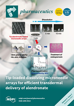
To improve the transdermal bioavailability and safety of alendronate (ALN), a nitrogen-containing bisphosphonate, we developed self-dissolving microneedle arrays, in which ALN is loaded only at the tip portion of micron-scale needles by a dip-coating method (ALN(TIP)-MN). The tip portions of MN completely dissolved in the rat skin within 5 min after application in vivo. After application of ALN(TIP)-MN in mice, the bioavailability of ALN was approximately 96 %. In addition, the decrease in growth plate was effectively suppressed by this efficient delivery of ALN in a rat model of osteoporosis. Furthermore, no skin irritation was observed after application of ALN(TIP)-MN. View the paper.
- Issues are regarded as officially published after their release is announced to the table of contents alert mailing list.
- You may sign up for e-mail alerts to receive table of contents of newly released issues.
- PDF is the official format for papers published in both, html and pdf forms. To view the papers in pdf format, click on the "PDF Full-text" link, and use the free Adobe Reader to open them.
Previous Issue
Next Issue