

Dendrimers: Advancements and Potential Applications in Cancer Diagnosis and Treatment—An Overview

 , , and
Abstract
:1. Introduction
1.1. Generalities
1.2. Methods
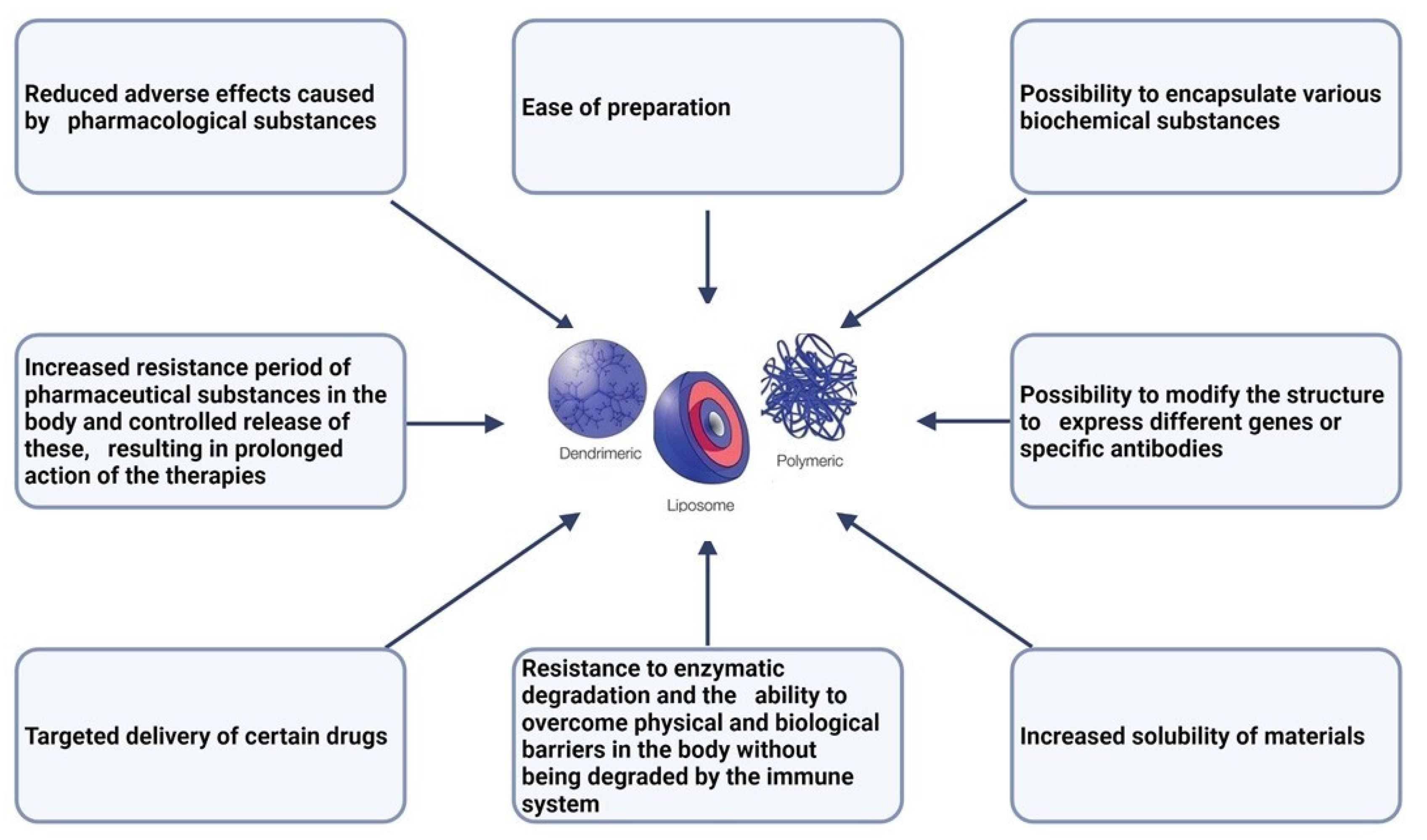
1.3. Nanoparticles: Classification and Characterization
2. Dendrimers: Polymer-Based Nanoparticles
3. The Use of Dendrimers in Cancer Diagnosis
4. Targeting and Treatment
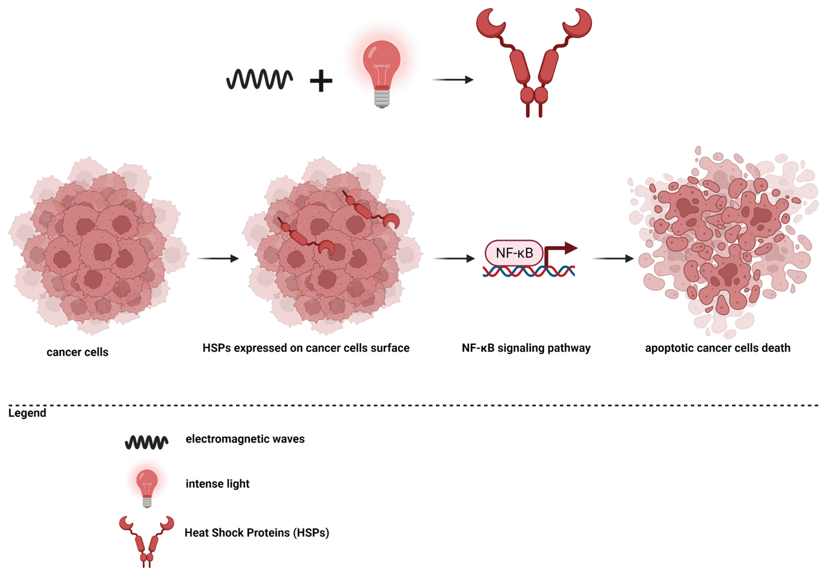
5. Photothermal Therapy
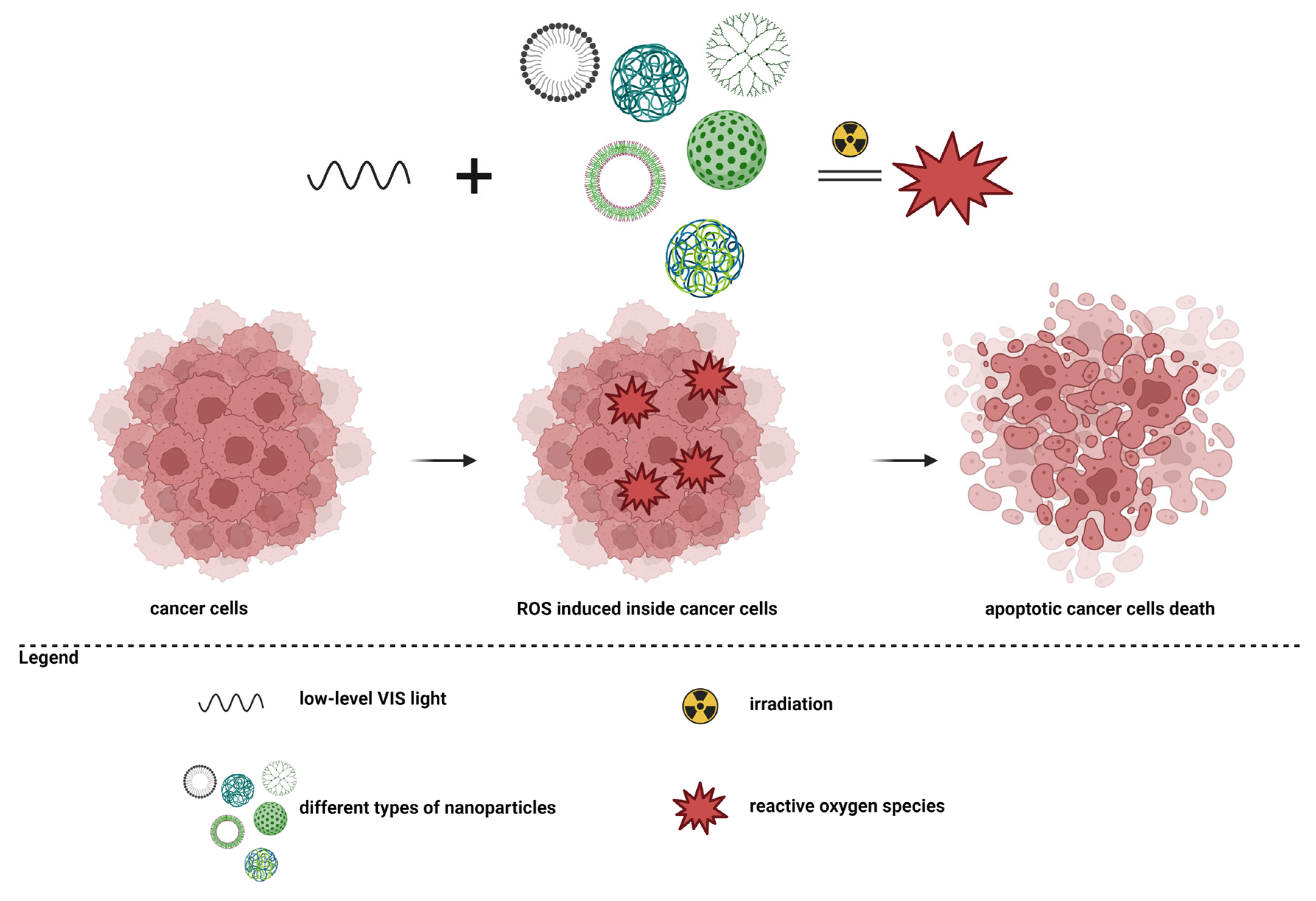
6. Photodynamic Therapy
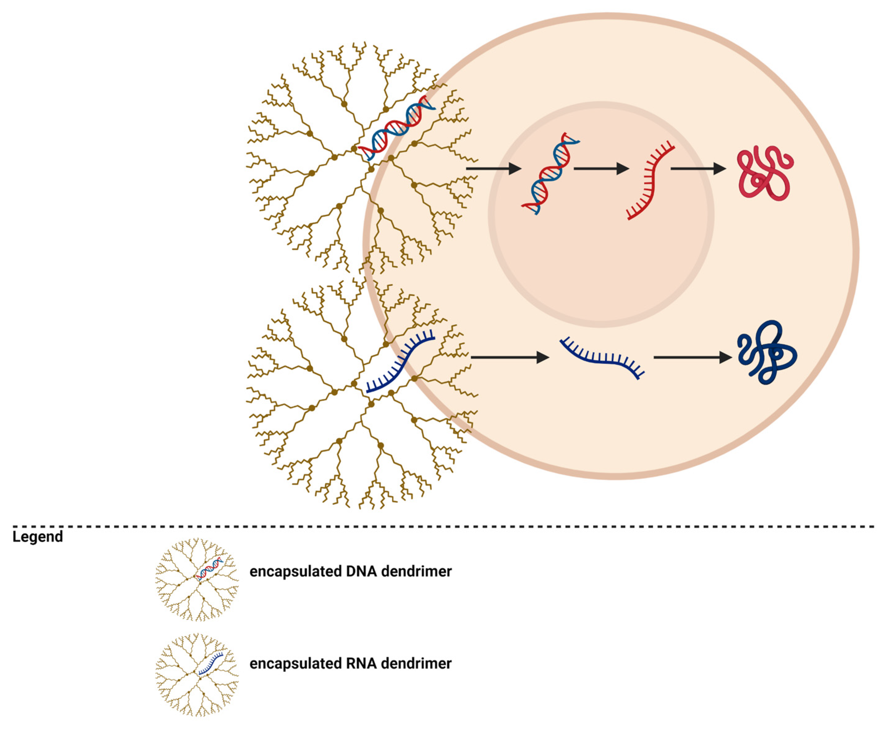
7. Gene Transfection
8. Conclusions
9. Future Perspectives
Author Contributions
Funding
Institutional Review Board Statement
Informed Consent Statement
Data Availability Statement
Conflicts of Interest
References
- Bober, Z.; Bartusik-Aebisher, D.; Aebisher, D. Application of Dendrimers in Anticancer Diagnostics and Therapy. Molecules 2022, 27, 3237. [Google Scholar] [CrossRef] [PubMed]
- Sheikh, A.; Md, S.; Kesharwani, P. RGD Engineered Dendrimer Nanotherapeutic as an Emerging Targeted Approach in Cancer Therapy. J. Controll. Release 2021, 340, 221–242. [Google Scholar] [CrossRef] [PubMed]
- What Is Cancer? NIH 2021. Available online: https://www.cancer.gov/about-cancer/understanding/what-is-cancer (accessed on 28 April 2023).
- Cancer. World Health Organization 2022. Available online: https://www.who.int/news-room/fact-sheets/detail/cancer (accessed on 28 April 2023).
- Debela, D.T.; Muzazu, S.G.; Heraro, K.D.; Ndalama, M.T.; Mesele, B.W.; Haile, D.C.; Kitui, S.K.; Manyazewal, T. New Approaches and Procedures for Cancer Treatment: Current Perspectives. SAGE Open Med. 2021, 9, 20503121211034370. [Google Scholar] [CrossRef] [PubMed]
- Galmarini, C.M. Why We Do What We Do. A Brief Analysis of Cancer Therapies. EXCLI J. 2020, 19, 1401. [Google Scholar]
- Baskar, R.; Lee, K.A.; Yeo, R.; Yeoh, K.-W. Cancer and Radiation Therapy: Current Advances and Future Directions. Int. J. Med. Sci. 2012, 9, 193. [Google Scholar] [CrossRef] [PubMed]
- Arruebo, M.; Vilaboa, N.; Sáez-Gutierrez, B.; Lambea, J.; Tres, A.; Valladares, M.; González-Fernández, Á. Assessment of the Evolution of Cancer Treatment Therapies. Cancers 2011, 3, 3279–3330. [Google Scholar] [CrossRef] [PubMed]
- Schirrmacher, V. From Chemotherapy to Biological Therapy: A Review of Novel Concepts to Reduce the Side Effects of Systemic Cancer Treatment. Int. J. Oncol. 2019, 54, 407–419. [Google Scholar] [CrossRef] [PubMed]
- MacDonald, V. Chemotherapy: Managing Side Effects and Safe Handling. Can. Vet. J. 2009, 50, 665. [Google Scholar]
- Turvey, B.E.; Crowder, S. Anabolic Steroid Abuse in Public Safety Personnel: A Forensic Manual; Academic Press: Cambridge, MA, USA, 2015; ISBN 0-12-802877-7. [Google Scholar]
- Sawyers, C. Targeted Cancer Therapy. Nature 2004, 432, 294–297. [Google Scholar] [CrossRef]
- Wilkes, G.M. Targeted Therapy: Attacking Cancer with Molecular and Immunological Targeted Agents. Asia-Pac. J. Oncol. Nurs. 2018, 5, 137–155. [Google Scholar] [CrossRef]
- Crintea, A.; Dutu, A.G.; Samasca, G.; Florian, I.A.; Lupan, I.; Craciun, A.M. The Nanosystems Involved in Treating Lung Cancer. Life 2021, 11, 682. [Google Scholar] [CrossRef] [PubMed]
- Jin, C.; Wang, K.; Oppong-Gyebi, A.; Hu, J. Application of Nanotechnology in Cancer Diagnosis and Therapy-a Mini-Review. Int. J. Med. Sci. 2020, 17, 2964. [Google Scholar] [CrossRef]
- Sanna, V.; Pala, N.; Sechi, M. Targeted Therapy Using Nanotechnology: Focus on Cancer. Int. J. Nanomed. 2014, 9, 467. [Google Scholar]
- Mosleh-Shirazi, S.; Abbasi, M.; Vaez, A.; Shafiee, M.; Kasaee, S.R.; Amani, A.M.; Hatam, S. Nanotechnology Advances in the Detection and Treatment of Cancer: An Overview. Nanotheranostics 2022, 6, 400–423. [Google Scholar] [CrossRef] [PubMed]
- Sahoo, B.M.; Kumar, B.V.; Patra, C.N.; Panda, J.R.; Mohanta, B.C.; Palei, N.N. Nanotechnology: A Novel Approach for Drug Development in Health Care System. Curr. Nanomater. 2020, 5, 12–25. [Google Scholar] [CrossRef]
- Murthy, S.K. Nanoparticles in Modern Medicine: State of the Art and Future Challenges. Int. J. Nanomed. 2007, 2, 129–141. [Google Scholar]
- Riehemann, K.; Schneider, S.W.; Luger, T.A.; Godin, B.; Ferrari, M.; Fuchs, H. Nanomedicine—Challenge and Perspectives. Angew. Chem. Int. Ed. 2009, 48, 872–897. [Google Scholar] [CrossRef]
- Théry, C.; Witwer, K.W.; Aikawa, E.; Alcaraz, M.J.; Anderson, J.D.; Andriantsitohaina, R.; Antoniou, A.; Arab, T.; Archer, F.; Atkin-Smith, G.K. Minimal Information for Studies of Extracellular Vesicles 2018 (MISEV2018): A Position Statement of the International Society for Extracellular Vesicles and Update of the MISEV2014 Guidelines. J. Extracell. Vesicles 2018, 7, 1535750. [Google Scholar] [CrossRef]
- Gautam, A.; van Veggel, F.C. Synthesis of Nanoparticles, Their Biocompatibility, and Toxicity Behavior for Biomedical Applications. J. Mater. Chem. B 2013, 1, 5186–5200. [Google Scholar] [CrossRef]
- Zhao, C.-Y.; Cheng, R.; Yang, Z.; Tian, Z.-M. Nanotechnology for Cancer Therapy Based on Chemotherapy. Molecules 2018, 23, 826. [Google Scholar] [CrossRef]
- Demetzos, C.; Pippa, N. Advanced Drug Delivery Nanosystems (ADDnSs): A Mini-Review. Drug Deliv. 2014, 21, 250–257. [Google Scholar] [CrossRef] [PubMed]
- Sim, S.; Wong, N.K. Nanotechnology and Its Use in Imaging and Drug Delivery. Biomed. Rep. 2021, 14, 1–9. [Google Scholar] [CrossRef] [PubMed]
- Chapman, S.; Dobrovolskaia, M.; Farahani, K.; Goodwin, A.; Joshi, A.; Lee, H.; Meade, T.; Pomper, M.; Ptak, K.; Rao, J. Nanoparticles for Cancer Imaging: The Good, the Bad, and the Promise. Nano Today 2013, 8, 454–460. [Google Scholar] [CrossRef]
- Ross, J.S. Targeted Therapy for Cancer: Integrating Diagnostics and Therapeutics. Am. J. Cancer 2004, 3, 205–214. [Google Scholar] [CrossRef]
- Mundekkad, D.; Cho, W.C. Nanoparticles in Clinical Translation for Cancer Therapy. Int. J. Mol. Sci. 2022, 23, 1685. [Google Scholar] [CrossRef] [PubMed]
- Patras, L.; Banciu, M. Intercellular Crosstalk via Extracellular Vesicles in Tumor Milieu as Emerging Therapies for Cancer Progression. Curr. Pharm. Des. 2019, 25, 1980–2006. [Google Scholar] [CrossRef]
- Kooijmans, S.A.; Vader, P.; van Dommelen, S.M.; van Solinge, W.W.; Schiffelers, R.M. Exosome Mimetics: A Novel Class of Drug Delivery Systems. Int. J. Nanomed. 2012, 7, 1525–1541. [Google Scholar]
- Waheed, S.; Li, Z.; Zhang, F.; Chiarini, A.; Armato, U.; Wu, J. Engineering Nano-Drug Biointerface to Overcome Biological Barriers toward Precision Drug Delivery. J. Nanobiotechnol. 2022, 20, 395. [Google Scholar] [CrossRef]
- Saleh, T.A. Nanomaterials: Classification, Properties, and Environmental Toxicities. Environ. Technol. Innov. 2020, 20, 101067. [Google Scholar] [CrossRef]
- Mishra, S.; Banode, K.; Belgamwar, V. Nanotechnology: A Tool for Targeted Drug Delivery. In Nanotechnology Applied to Pharmaceutical Technology; Springer International Publishing: Cham, Switzerland, 2017; pp. 116–123. [Google Scholar]
- Chis, A.A.; Dobrea, C.; Morgovan, C.; Arseniu, A.M.; Rus, L.L.; Butuca, A.; Juncan, A.M.; Totan, M.; Vonica-Tincu, A.L.; Cormos, G. Applications and Limitations of Dendrimers in Biomedicine. Molecules 2020, 25, 3982. [Google Scholar] [CrossRef]
- Abdelkader, H.; Alani, A.W.; Alany, R.G. Recent Advances in Non-Ionic Surfactant Vesicles (Niosomes): Self-Assembly, Fabrication, Characterization, Drug Delivery Applications and Limitations. Drug Deliv. 2014, 21, 87–100. [Google Scholar] [CrossRef] [PubMed]
- Crintea, A.; Dutu, A.G.; Sovrea, A.; Constantin, A.-M.; Samasca, G.; Masalar, A.L.; Ifju, B.; Linga, E.; Neamti, L.; Tranca, R.A. Nanocarriers for Drug Delivery: An Overview with Emphasis on Vitamin D and K Transportation. Nanomaterials 2022, 12, 1376. [Google Scholar] [CrossRef] [PubMed]
- Begines, B.; Ortiz, T.; Pérez-Aranda, M.; Martínez, G.; Merinero, M.; Argüelles-Arias, F.; Alcudia, A. Polymeric Nanoparticles for Drug Delivery: Recent Developments and Future Prospects. Nanomaterials 2020, 10, 1403. [Google Scholar] [CrossRef] [PubMed]
- Lim, K.M.; Lee, J.H. Electrical Conductivity and Compressive Strength of Cement Paste with Multiwalled Carbon Nanotubes and Graphene Nanoplatelets. Appl. Sci. 2022, 12, 1160. [Google Scholar] [CrossRef]
- Bozzuto, G.; Molinari, A. Liposomes as Nanomedical Devices. Int. J. Nanomed. 2015, 10, 975. [Google Scholar] [CrossRef] [PubMed]
- Sercombe, L.; Veerati, T.; Moheimani, F.; Wu, S.Y.; Sood, A.K.; Hua, S. Advances and Challenges of Liposome Assisted Drug Delivery. Front. Pharmacol. 2015, 6, 286. [Google Scholar] [CrossRef]
- Nakhaei, P.; Margiana, R.; Bokov, D.O.; Abdelbasset, W.K.; Jadidi Kouhbanani, M.A.; Varma, R.S.; Marofi, F.; Jarahian, M.; Beheshtkhoo, N. Liposomes: Structure, Biomedical Applications, and Stability Parameters with Emphasis on Cholesterol. Front. Bioeng. Biotechnol. 2021, 9, 748. [Google Scholar] [CrossRef]
- Nsairat, H.; Khater, D.; Sayed, U.; Odeh, F.; Al Bawab, A.; Alshaer, W. Liposomes: Structure, Composition, Types, and Clinical Applications. Heliyon 2022, 8, e09394. [Google Scholar] [CrossRef]
- Petros, R.A.; DeSimone, J.M. Strategies in the Design of Nanoparticles for Therapeutic Applications. Nat. Rev. Drug Discov. 2010, 9, 615–627. [Google Scholar] [CrossRef]
- Anik, M.I.; Mahmud, N.; Al Masud, A.; Hasan, M. Gold Nanoparticles (GNPs) in Biomedical and Clinical Applications: A Review. Nano Sel. 2022, 3, 792–828. [Google Scholar] [CrossRef]
- Hu, X.; Zhang, Y.; Ding, T.; Liu, J.; Zhao, H. Multifunctional Gold Nanoparticles: A Novel Nanomaterial for Various Medical Applications and Biological Activities. Front. Bioeng. Biotechnol. 2020, 8, 990. [Google Scholar] [CrossRef]
- Al-Tikriti, Y.; Hansson, P. Drug-Induced Phase Separation in Polyelectrolyte Microgels. Gels 2021, 8, 4. [Google Scholar] [CrossRef]
- Hanafy, N.A.; El-Kemary, M.; Leporatti, S. Micelles Structure Development as a Strategy to Improve Smart Cancer Therapy. Cancers 2018, 10, 238. [Google Scholar] [CrossRef]
- Puri, A.; Loomis, K.; Smith, B.; Lee, J.-H.; Yavlovich, A.; Heldman, E.; Blumenthal, R. Lipid-Based Nanoparticles as Pharmaceutical Drug Carriers: From Concepts to Clinic. Crit. Rev. Ther. Drug Carr. Syst. 2009, 26, 13–18. [Google Scholar] [CrossRef] [PubMed]
- Yadav, N.; Khatak, S.; Sara, U.S. Solid Lipid Nanoparticles-a Review. Int. J. Appl. Pharm 2013, 5, 8–18. [Google Scholar]
- Tomalia, D.A.; Fréchet, J.M. Discovery of Dendrimers and Dendritic Polymers: A Brief Historical Perspective. J. Polym. Sci. Part A: Polym. Chem. 2002, 40, 2719–2728. [Google Scholar] [CrossRef]
- Klajnert, B.; Bryszewska, M. Dendrimers: Properties and Applications. Acta Biochim. Pol. 2001, 48, 199–208. [Google Scholar] [CrossRef]
- Caminade, A.-M. Dendrimers, an Emerging Opportunity in Personalized Medicine? J. Pers. Med. 2022, 12, 1334. [Google Scholar] [CrossRef]
- Newkome, G.R.; Shreiner, C.D. Poly (Amidoamine), Polypropylenimine, and Related Dendrimers and Dendrons Possessing Different 1→ 2 Branching Motifs: An Overview of the Divergent Procedures. Polymer 2008, 49, 1–173. [Google Scholar] [CrossRef]
- Munavalli, B.B.; Naik, S.R.; Torvi, A.I.; Kariduraganavar, M.Y. Dendrimers. In Functional Polymers; Springer: Berlin/Heidelberg, Germany, 2019; pp. 289–345. [Google Scholar]
- Santos, A.; Veiga, F.; Figueiras, A. Dendrimers as Pharmaceutical Excipients: Synthesis, Properties, Toxicity and Biomedical Applications. Materials 2019, 13, 65. [Google Scholar] [CrossRef]
- Singh, S.; Sharma, V. Dendrimers: A Class of Polymer in the Nanotechnology for Drug Delivery. In Nanomedicine for Drug Delivery and Therapeutics; Scrivener Publishing: Beverly, MA, USA, 2013; pp. 373–405. [Google Scholar]
- Boas, U.; Heegaard, P.M. Dendrimers in Drug Research. Chem. Soc. Rev. 2004, 33, 43–63. [Google Scholar] [CrossRef] [PubMed]
- Bharali, D.J.; Khalil, M.; Gurbuz, M.; Simone, T.M.; Mousa, S.A. Nanoparticles and Cancer Therapy: A Concise Review with Emphasis on Dendrimers. Int. J. Nanomed. 2009, 4, 1–7. [Google Scholar]
- Mittal, P.; Saharan, A.; Verma, R.; Altalbawy, F.; Alfaidi, M.A.; Batiha, G.E.-S.; Akter, W.; Gautam, R.K.; Uddin, M.; Rahman, M. Dendrimers: A New Race of Pharmaceutical Nanocarriers. BioMed Res. Int. 2021, 2021, 8844030. [Google Scholar] [CrossRef] [PubMed]
- Kharwade, R.; More, S.; Warokar, A.; Agrawal, P.; Mahajan, N. Starburst Pamam Dendrimers: Synthetic Approaches, Surface Modifications, and Biomedical Applications. Arab. J. Chem. 2020, 13, 6009–6039. [Google Scholar] [CrossRef]
- Gillani, S.S.; Munawar, M.A.; Khan, K.M.; Chaudhary, J.A. Synthesis, Characterization and Applications of Poly-Aliphatic Amine Dendrimers and Dendrons. J. Iran. Chem. Soc. 2020, 17, 2717–2736. [Google Scholar] [CrossRef]
- Zenze, M.; Daniels, A.; Singh, M. Dendrimers as Modifiers of Inorganic Nanoparticles for Therapeutic Delivery in Cancer. Pharmaceutics 2023, 15, 398. [Google Scholar] [CrossRef]
- Fischer, M.; Vögtle, F. Dendrimers: From Design to Application—A Progress Report. Angew. Chem. Int. Ed. 1999, 38, 884–905. [Google Scholar] [CrossRef]
- Caminati, G.; Turro, N.J.; Tomalia, D.A. Photophysical Investigation of Starburst Dendrimers and Their Interactions with Anionic and Cationic Surfactants. J. Am. Chem. Soc. 1990, 112, 8515–8522. [Google Scholar] [CrossRef]
- Sato, K.; Anzai, J. Dendrimers in Layer-by-Layer Assemblies: Synthesis and Applications. Molecules 2013, 18, 8440–8460. [Google Scholar] [CrossRef]
- Abbasi, E.; Aval, S.F.; Akbarzadeh, A.; Milani, M.; Nasrabadi, H.T.; Joo, S.W.; Hanifehpour, Y.; Nejati-Koshki, K.; Pashaei-Asl, R. Dendrimers: Synthesis, Applications, and Properties. Nanoscale Res. Lett. 2014, 9, 1–10. [Google Scholar] [CrossRef]
- Hawker, C.J.; Frechet, J.M. Preparation of Polymers with Controlled Molecular Architecture. A New Convergent Approach to Dendritic Macromolecules. J. Am. Chem. Soc. 1990, 112, 7638–7647. [Google Scholar] [CrossRef]
- Huang, A.Y.-T.; Kao, C.-L.; Selvaraj, A.; Peng, L. Solid-Phase Dendrimer Synthesis: A Promising Approach to Transform Dendrimer Construction. Mater. Today Chem. 2023, 27, 101285. [Google Scholar] [CrossRef]
- Zangabad, P.S.; Karimi, M.; Mehdizadeh, F.; Malekzad, H.; Ghasemi, A.; Bahrami, S.; Zare, H.; Moghoofei, M.; Hekmatmanesh, A.; Hamblin, M.R. Nanocaged Platforms: Modification, Drug Delivery and Nanotoxicity. Opening Synthetic Cages to Release the Tiger. Nanoscale 2017, 9, 1356–1392. [Google Scholar] [CrossRef]
- Svenson, S.; Tomalia, D.A. Dendrimers in Biomedical Applications—Reflections on the Field. Adv. Drug Deliv. Rev. 2012, 64, 102–115. [Google Scholar] [CrossRef]
- Shende, P.; Govardhane, S. Strategies-Based Intrathecal Targeted Drug Delivery System for Effective Therapy, Modeling, and Controlled Release Action. In Modeling and Control of Drug Delivery Systems; Elsevier: Amsterdam, The Netherlands, 2021; pp. 213–225. [Google Scholar]
- Martinho, N.; Florindo, H.; Silva, L.; Brocchini, S.; Zloh, M.; Barata, T. Molecular Modeling to Study Dendrimers for Biomedical Applications. Molecules 2014, 19, 20424–20467. [Google Scholar] [CrossRef] [PubMed]
- Ardabevskaia, S.; Milenin, S. From Dendrimers to Megamers: The State-of-the-Art. INEOS Open 2021, 4, 176–188. [Google Scholar] [CrossRef]
- Madaan, K.; Kumar, S.; Poonia, N.; Lather, V.; Pandita, D. Dendrimers in Drug Delivery and Targeting: Drug-Dendrimer Interactions and Toxicity Issues. J. Pharm. Bioallied Sci. 2014, 6, 139. [Google Scholar]
- Wang, J.; Li, B.; Qiu, L.; Qiao, X.; Yang, H. Dendrimer-Based Drug Delivery Systems: History, Challenges, and Latest Developments. J. Biol. Eng. 2022, 16, 1–12. [Google Scholar] [CrossRef]
- Choudhary, S.; Gupta, L.; Rani, S.; Dave, K.; Gupta, U. Impact of Dendrimers on Solubility of Hydrophobic Drug Molecules. Front. Pharmacol. 2017, 8, 261. [Google Scholar] [CrossRef]
- Kumari, S.; Sharma, N.; Sahi, S.V. Advances in Cancer Therapeutics: Conventional Thermal Therapy to Nanotechnology-Based Photothermal Therapy. Pharmaceutics 2021, 13, 1174. [Google Scholar] [CrossRef]
- Glazer, E.S.; Curley, S.A. Non-Invasive Radiofrequency Ablation of Malignancies Mediated by Quantum Dots, Gold Nanoparticles and Carbon Nanotubes. Ther. Deliv. 2011, 2, 1325–1330. [Google Scholar] [CrossRef] [PubMed]
- Curto, S.; Taj-Eldin, M.; Fairchild, D.; Prakash, P. Microwave Ablation at 915 MHz vs 2.45 GHz: A Theoretical and Experimental Investigation. Med. Phys. 2015, 42, 6152–6161. [Google Scholar] [CrossRef] [PubMed]
- Sanhai, W.R.; Sakamoto, J.H.; Canady, R.; Ferrari, M. Seven Challenges for Nanomedicine. Nat. Nanotechnol. 2008, 3, 242–244. [Google Scholar] [CrossRef] [PubMed]
- Huang, Y.-F.; Sefah, K.; Bamrungsap, S.; Chang, H.-T.; Tan, W. Selective Photothermal Therapy for Mixed Cancer Cells Using Aptamer-Conjugated Nanorods. Langmuir 2008, 24, 11860–11865. [Google Scholar] [CrossRef] [PubMed]
- Jain, K.; Kesharwani, P.; Gupta, U.; Jain, N. Dendrimer Toxicity: Let’s Meet the Challenge. Int. J. Pharm. 2010, 394, 122–142. [Google Scholar] [CrossRef]
- Hamidi, A.; Sharifi, S.; Davara, S.; Ghasemi, S.; Omidi, Y.; Rashidi, M.-R. Novel Aldehyde-Terminated Dendrimers; Synthesis and Cytotoxicity Assay. Bioimpacts 2012, 2, 97–103. [Google Scholar]
- Bhadra, D.; Bhadra, S.; Jain, S.; Jain, N. A PEGylated Dendritic Nanoparticulate Carrier of Fluorouracil. Int. J. Pharm. 2003, 257, 111–124. [Google Scholar] [CrossRef]
- Asthana, A.; Chauhan, A.S.; Diwan, P.V.; Jain, N.K. Poly (Amidoamine)(PAMAM) Dendritic Nanostructures for Controlled Sitespecific Delivery of Acidic Anti-Inflammatory Active Ingredient. Aaps Pharmscitech 2005, 6, E536–E542. [Google Scholar] [CrossRef]
- Agashe, H.B.; Dutta, T.; Garg, M.; Jain, N. Investigations on the Toxicological Profile of Functionalized Fifth-generation Poly (Propylene Imine) Dendrimer. J. Pharm. Pharmacol. 2006, 58, 1491–1498. [Google Scholar] [CrossRef]
- Roberts, J.C.; Bhalgat, M.K.; Zera, R.T. Preliminary Biological Evaluation of Polyamidoamine (PAMAM) StarburstTM Dendrimers. J. Biomed. Mater. Res. Off. J. Soc. Biomater. Jpn. Soc. Biomater. 1996, 30, 53–65. [Google Scholar] [CrossRef]
- Wolinsky, J.B.; Grinstaff, M.W. Therapeutic and Diagnostic Applications of Dendrimers for Cancer Treatment. Adv. Drug Deliv. Rev. 2008, 60, 1037–1055. [Google Scholar] [CrossRef] [PubMed]
- Fass, L. Imaging and Cancer: A Review. Mol. Oncol. 2008, 2, 115–152. [Google Scholar] [CrossRef]
- Hussain, T.; Nguyen, Q.T. Molecular Imaging for Cancer Diagnosis and Surgery. Adv. Drug Deliv. Rev. 2014, 66, 90–100. [Google Scholar] [CrossRef] [PubMed]
- Frangioni, J.V. New Technologies for Human Cancer Imaging. J. Clin. Oncol. 2008, 26, 4012. [Google Scholar] [CrossRef] [PubMed]
- Murphy, D.; Aghayev, A.; Steigner, M. Vascular CT and MRI: A Practical Guide to Imaging Protocols. Insights Imaging 2018, 9, 215–236. [Google Scholar] [CrossRef]
- Carnevale, L.; Lembo, G. Innovative MRI Techniques in Neuroimaging Approaches for Cerebrovascular Diseases and Vascular Cognitive Impairment. Int. J. Mol. Sci. 2019, 20, 2656. [Google Scholar] [CrossRef]
- Longmire, M.R.; Ogawa, M.; Choyke, P.L.; Kobayashi, H. Dendrimers as High Relaxivity MR Contrast Agents. Wiley Interdiscip. Rev.: Nanomed. Nanobiotechnol. 2014, 6, 155–162. [Google Scholar] [CrossRef]
- Sampathkumar, S.; Yarema, K.J. Dendrimers in Cancer Treatment and Diagnosis. Nanotechnol. Life Sci. Online 2007, 7, 1–43. [Google Scholar]
- Wahsner, J.; Gale, E.M.; Rodríguez-Rodríguez, A.; Caravan, P. Chemistry of MRI Contrast Agents: Current Challenges and New Frontiers. Chem. Rev. 2018, 119, 957–1057. [Google Scholar] [CrossRef]
- Bourne, M.W.; Margerun, L.; Hylton, N.; Campion, B.; Lai, J.; Derugin, N.; Higgins, C.B. Evaluation of the Effects of Intravascular MR Contrast Media (Gadolinium Dendrimer) on 3D Time of Flight Magnetic Resonance Angiography of the Body. J. Magn. Reson. Imaging 1996, 6, 305–310. [Google Scholar] [CrossRef]
- Kobayashi, H.; Reijnders, K.; English, S.; Yordanov, A.T.; Milenic, D.E.; Sowers, A.L.; Citrin, D.; Krishna, M.C.; Waldmann, T.A.; Mitchell, J.B. Application of a Macromolecular Contrast Agent for Detection of Alterations of Tumor Vessel Permeability Induced by Radiation. Clin. Cancer Res. 2004, 10, 7712–7720. [Google Scholar] [CrossRef] [PubMed]
- Nune, S.K.; Gunda, P.; Majeti, B.K.; Thallapally, P.K.; Forrest, M.L. Advances in Lymphatic Imaging and Drug Delivery. Adv. Drug Deliv. Rev. 2011, 63, 876–885. [Google Scholar] [CrossRef] [PubMed]
- Kobayashi, H.; Brechbiel, M.W. Dendrimer-Based Macromolecular MRI Contrast Agents: Characteristics and Application. Mol. Imaging 2003, 2, 15353500200303100. [Google Scholar] [CrossRef] [PubMed]
- Abedi-Gaballu, F.; Dehghan, G.; Ghaffari, M.; Yekta, R.; Abbaspour-Ravasjani, S.; Baradaran, B.; Dolatabadi, J.E.N.; Hamblin, M.R. PAMAM Dendrimers as Efficient Drug and Gene Delivery Nanosystems for Cancer Therapy. Appl. Mater. Today 2018, 12, 177–190. [Google Scholar] [CrossRef]
- Kesharwani, P.; Iyer, A.K. Recent Advances in Dendrimer-Based Nanovectors for Tumor-Targeted Drug and Gene Delivery. Drug Discov. Today 2015, 20, 536–547. [Google Scholar] [CrossRef]
- Cai, X.; Hu, J.; Xiao, J.; Cheng, Y. Dendrimer and Cancer: A Patent Review (2006–Present). Expert Opin. Ther. Pat. 2013, 23, 515–529. [Google Scholar] [CrossRef]
- Palena, P.D.; Barto, R.R.; Borders, T.L.; Stuart, J.A. Nano-Getter Device. U.S. Patent No. 8,236,243, 2012. [Google Scholar]
- Lohcharoenkal, W.; Abbas, Z.; Rojanasakul, Y. Advances in Nanotechnology-Based Biosensing of Immunoregulatory Cytokines. Biosensors 2021, 11, 364. [Google Scholar] [CrossRef]
- Jain, K.K.; Jain, K.K. Nanomolecular Diagnostics. In The Handbook of Nanomedicine; Spinger: Berlin/Heidelberg, Germany, 2017; pp. 133–200. [Google Scholar]
- Hong, S.; Eddington, D.; Myung, J.H.; Launiere, C. Methods and Devices for Capturing Circulating Tumor Cells. U.S. Patent Application No. 13/265,916, 2012. [Google Scholar]
- Zhao, Y.; Liu, S.; Li, Y.; Jiang, W.; Chang, Y.; Pan, S.; Fang, X.; Wang, Y.A.; Wang, J. Synthesis and Grafting of Folate–PEG–PAMAM Conjugates onto Quantum Dots for Selective Targeting of Folate-Receptor-Positive Tumor Cells. J. Colloid Interface Sci. 2010, 350, 44–50. [Google Scholar] [CrossRef]
- Wei, F.; Liao, W. Probe Immobilization and Signal Amplification for Polymer-Based Biosensor. U.S. Patent No. 9,127,304, 8 September 2015. [Google Scholar]
- Palmerston Mendes, L.; Pan, J.; Torchilin, V.P. Dendrimers as Nanocarriers for Nucleic Acid and Drug Delivery in Cancer Therapy. Molecules 2017, 22, 1401. [Google Scholar] [CrossRef]
- Ong, K.; Jenkins, A.; Cheng, R.; Tomalia, D.; Durst, H.; Jensen, J.; Emanuel, P.; Swim, C.; Yin, R. Dendrimer Enhanced Immunosensors for Biological Detection. Anal. Chim. Acta 2001, 444, 143–148. [Google Scholar] [CrossRef]
- Sánchez, A.; Villalonga, A.; Martínez-García, G.; Parrado, C.; Villalonga, R. Dendrimers as Soft Nanomaterials for Electrochemical Immunosensors. Nanomaterials 2019, 9, 1745. [Google Scholar] [CrossRef] [PubMed]
- Çevik, E.; Bahar, Ö.; Şenel, M.; Abasıyanık, M.F. Construction of Novel Electrochemical Immunosensor for Detection of Prostate Specific Antigen Using Ferrocene-PAMAM Dendrimers. Biosens. Bioelectron. 2016, 86, 1074–1079. [Google Scholar] [CrossRef] [PubMed]
- Singh, J.; Jain, K.; Mehra, N.K.; Jain, N. Dendrimers in Anticancer Drug Delivery: Mechanism of Interaction of Drug and Dendrimers. Artif. Cells Nanomed. Biotechnol. 2016, 44, 1626–1634. [Google Scholar] [CrossRef] [PubMed]
- Mohapatra, S.; Majhi, S.R.; Behera, D.J.; Kar, R.K. Dendrimers: A Potential Carrier for Cancer Therapy. High Technol. Lett. 2022, 28, 11. [Google Scholar]
- ud Din, F.; Aman, W.; Ullah, I.; Qureshi, O.S.; Mustapha, O.; Shafique, S.; Zeb, A. Effective Use of Nanocarriers as Drug Delivery Systems for the Treatment of Selected Tumors. Int. J. Nanomed. 2017, 12, 7291. [Google Scholar] [CrossRef] [PubMed]
- Allen, T.M.; Cullis, P.R. Drug Delivery Systems: Entering the Mainstream. Science 2004, 303, 1818–1822. [Google Scholar] [CrossRef]
- Baker, J.R., Jr.; Shukla, R.; Thomas, T.P. Dendrimer Based Compositions and Methods of Using the Same. Patent No.:US-20090088376-A1/2009-04-02, 2009. Available online: https://worldwide.espacenet.com/publicationDetails/biblio?CC=US&NR=2009088376&KC=&FT=E&locale=en_EP (accessed on 28 April 2023).
- Amjad, M.W. Dendrimers in Anticancer Targeted Drug Delivery: Accomplishments, Challenges and Directions for Future. Pharm. Pharmacol. 2021, 9, 4–16. [Google Scholar] [CrossRef]
- Svenson, S.; Chauhan, A.S. Peham Dendrimers as Excipients. Patent No.:WO-2008030591-A2/2008-03-13, 2009. Available online: https://patents.google.com/patent/WO2008030591A2/en?oq=WO-2008030591-A2+%2f+2008-03-13 (accessed on 28 April 2023).
- Rizvi, S.A.; Saleh, A.M. Applications of Nanoparticle Systems in Drug Delivery Technology. Saudi Pharm. J. 2018, 26, 64–70. [Google Scholar] [CrossRef]
- Vieira Gonzaga, R.; da Silva Santos, S.; Da Silva, J.V.; Campos Prieto, D.; Feliciano Savino, D.; Giarolla, J.; Igne Ferreira, E. Targeting Groups Employed in Selective Dendrons and Dendrimers. Pharmaceutics 2018, 10, 219. [Google Scholar] [CrossRef]
- Attia, M.F.; Anton, N.; Wallyn, J.; Omran, Z.; Vandamme, T.F. An Overview of Active and Passive Targeting Strategies to Improve the Nanocarriers Efficiency to Tumour Sites. J. Pharm. Pharmacol. 2019, 71, 1185–1198. [Google Scholar] [CrossRef]
- Subhan, M.A.; Yalamarty, S.S.K.; Filipczak, N.; Parveen, F.; Torchilin, V.P. Recent Advances in Tumor Targeting via EPR Effect for Cancer Treatment. J. Pers. Med. 2021, 11, 571. [Google Scholar] [CrossRef] [PubMed]
- Iyer, A.K.; Khaled, G.; Fang, J.; Maeda, H. Exploiting the Enhanced Permeability and Retention Effect for Tumor Targeting. Drug Discov. Today 2006, 11, 812–818. [Google Scholar] [CrossRef] [PubMed]
- Danhier, F.; Feron, O.; Préat, V. To Exploit the Tumor Microenvironment: Passive and Active Tumor Targeting of Nanocarriers for Anti-Cancer Drug Delivery. J. Control. Release 2010, 148, 135–146. [Google Scholar] [CrossRef] [PubMed]
- Shukla, T.; Upmanyu, N.; Pandey, S.P.; Sudheesh, M. Site-Specific Drug Delivery, Targeting, and Gene Therapy. In Nanoarchitectonics in Biomedicine; Elsevier: Amsterdam, The Netherlands, 2019; pp. 473–505. [Google Scholar]
- Cheng, Y.; Xu, Z.; Ma, M.; Xu, T. Dendrimers as Drug Carriers: Applications in Different Routes of Drug Administration. J. Pharm. Sci. 2008, 97, 123–143. [Google Scholar] [CrossRef]
- Malik, N.; Evagorou, E.G.; Duncan, R. Dendrimer-Platinate: A Novel Approach to Cancer Chemotherapy. Anti-Cancer Drugs 1999, 10, 767–776. [Google Scholar] [CrossRef]
- Vu, M.T.; Bach, L.G.; Nguyen, D.C.; Ho, M.N.; Nguyen, N.H.; Tran, N.Q.; Nguyen, D.H.; Nguyen, C.K.; Hoang Thi, T.T. Modified Carboxyl-Terminated PAMAM Dendrimers as Great Cytocompatible Nano-Based Drug Delivery System. Int. J. Mol. Sci. 2019, 20, 2016. [Google Scholar] [CrossRef]
- Balogh, L.; Swanson, D.R.; Tomalia, D.A.; Hagnauer, G.L.; McManus, A.T. Dendrimer− Silver Complexes and Nanocomposites as Antimicrobial Agents. Nano Lett. 2001, 1, 18–21. [Google Scholar] [CrossRef]
- Fox, L.J.; Richardson, R.M.; Briscoe, W.H. PAMAM Dendrimer-Cell Membrane Interactions. Adv. Colloid Interface Sci. 2018, 257, 1–18. [Google Scholar] [CrossRef]
- Kojima, C.; Kono, K.; Maruyama, K.; Takagishi, T. Synthesis of Polyamidoamine Dendrimers Having Poly (Ethylene Glycol) Grafts and Their Ability to Encapsulate Anticancer Drugs. Bioconjugate Chem. 2000, 11, 910–917. [Google Scholar] [CrossRef]
- Murphy, D.E.; de Jong, O.G.; Brouwer, M.; Wood, M.J.; Lavieu, G.; Schiffelers, R.M.; Vader, P. Extracellular Vesicle-Based Therapeutics: Natural versus Engineered Targeting and Trafficking. Exp. Mol. Med. 2019, 51, 1–12. [Google Scholar] [CrossRef]
- Suk, J.S.; Xu, Q.; Kim, N.; Hanes, J.; Ensign, L.M. PEGylation as a Strategy for Improving Nanoparticle-Based Drug and Gene Delivery. Adv. Drug Deliv. Rev. 2016, 99, 28–51. [Google Scholar] [CrossRef] [PubMed]
- Yang, Q.; Lai, S.K. Anti-PEG Immunity: Emergence, Characteristics, and Unaddressed Questions. Wiley Interdiscip. Rev. Nanomed. Nanobiotech. 2015, 7, 655–677. [Google Scholar] [CrossRef] [PubMed]
- Qi, R.; Gao, Y.; Tang, Y.; He, R.-R.; Liu, T.-L.; He, Y.; Sun, S.; Li, B.-Y.; Li, Y.-B.; Liu, G. PEG-Conjugated PAMAM Dendrimers Mediate Efficient Intramuscular Gene Expression. AAPS J. 2009, 11, 395–405. [Google Scholar] [CrossRef] [PubMed]
- WIENER, E.C.; KONDA, S.; SHADRON, A.; BRECHBIEL, M.; Gansow, O. Targeting Dendrimer-Chelates to Tumors and Tumor Cells Expressing the High-Affinity Folate Receptor. Investig. Radiol. 1997, 32, 748–754. [Google Scholar] [CrossRef] [PubMed]
- Choi, Y.; Baker, J.R., Jr. Targeting Cancer Cells with DNA-Assembled Dendrimers: A Mix-and-Match Strategy for Cancer. Cell Cycle 2005, 4, 669–671. [Google Scholar] [CrossRef] [PubMed]
- Zwicke, G.L.; Ali Mansoori, G.; Jeffery, C.J. Utilizing the Folate Receptor for Active Targeting of Cancer Nanotherapeutics. Nano Rev. 2012, 3, 18496. [Google Scholar] [CrossRef]
- Patri, A.K.; Kukowska-Latallo, J.F.; Baker, J.R., Jr. Targeted Drug Delivery with Dendrimers: Comparison of the Release Kinetics of Covalently Conjugated Drug and Non-Covalent Drug Inclusion Complex. Adv. Drug Deliv. Rev. 2005, 57, 2203–2214. [Google Scholar] [CrossRef]
- Majoros, I.J.; Williams, C.R.; Becker, A.; Baker Jr, J.R. Methotrexate Delivery via Folate Targeted Dendrimer-based Nanotherapeutic Platform. Wiley Interdiscip. Rev. Nanomed. Nanobiotech. 2009, 1, 502–510. [Google Scholar] [CrossRef]
- Daftarian, P.; Kaifer, A.E.; Li, W.; Blomberg, B.B.; Frasca, D.; Roth, F.; Chowdhury, R.; Berg, E.A.; Fishman, J.B.; Al Sayegh, H.A. Peptide-Conjugated PAMAM Dendrimer as a Universal DNA Vaccine Platform to Target Antigen-Presenting CellsPPD as a Universal Platform for Genetic Vaccine. Cancer Res. 2011, 71, 7452–7462. [Google Scholar] [CrossRef]
- Graham-Gurysh, E.G.; Carpenter, B.W.; Beck, W.A.; Varma, D.M.; Vincent, B.G.; Bachelder, E.M.; Ainslie, K.M. Delivery Strategies for Cancer Vaccines and Immunoadjuvants. In Systemic Drug Delivery Strategies; Elsevier: Amsterdam, The Netherlands, 2022; pp. 359–408. [Google Scholar]
- Yang, H. Targeted Nanosystems: Advances in Targeted Dendrimers for Cancer Therapy. Nanomed. Nanotechnol. Biol. Med. 2016, 12, 309–316. [Google Scholar] [CrossRef]
- Quinteros, D.A.; Bermúdez, J.M.; Ravetti, S.; Cid, A.; Allemandi, D.A.; Palma, S.D. Therapeutic Use of Monoclonal Antibodies: General Aspects and Challenges for Drug Delivery. In Nanostructures for Drug Delivery; Elsevier: Amsterdam, The Netherlands, 2017; pp. 807–833. [Google Scholar]
- Patri, A.K.; Myc, A.; Beals, J.; Thomas, T.P.; Bander, N.H.; Baker, J.R. Synthesis and in Vitro Testing of J591 Antibody− Dendrimer Conjugates for Targeted Prostate Cancer Therapy. Bioconjugate Chem. 2004, 15, 1174–1181. [Google Scholar] [CrossRef]
- Sheikh, A.; Kesharwani, P. An Insight into Aptamer Engineered Dendrimer for Cancer Therapy. Eur. Polym. J. 2021, 159, 110746. [Google Scholar] [CrossRef]
- Sheikh, A.; Kaur, H.; Abourehab, M.A.; Alam, M.S.; Kesharwani, P. Aptamer-Functionalized Dendrimers for Targeted Cancer Therapy. Aptamers Eng. Nanocarriers Cancer Ther. 2023, 2023, 255–275. [Google Scholar]
- Taghdisi, S.M.; Danesh, N.M.; Ramezani, M.; Lavaee, P.; Jalalian, S.H.; Robati, R.Y.; Abnous, K. Double Targeting and Aptamer-Assisted Controlled Release Delivery of Epirubicin to Cancer Cells by Aptamers-Based Dendrimer in Vitro and in Vivo. Eur. J. Pharm. Biopharm. 2016, 102, 152–158. [Google Scholar] [CrossRef] [PubMed]
- Mignani, S.; Shi, X.; Ceña, V.; Majoral, J.-P. Dendrimer–and Polymeric Nanoparticle–Aptamer Bioconjugates as Nonviral Delivery Systems: A New Approach in Medicine. Drug Discov. Today 2020, 25, 1065–1073. [Google Scholar] [CrossRef]
- Emran, T.B.; Shahriar, A.; Mahmud, A.R.; Rahman, T.; Abir, M.H.; Faijanur-Rob-Siddiquee, M.; Ahmed, H.; Rahman, N.; Nainu, F.; Wahyudin, E. Multidrug Resistance in Cancer: Understanding Molecular Mechanisms, Immunoprevention, and Therapeutic Approaches. Front. Oncol. 2022, 12, 891652. [Google Scholar] [CrossRef]
- Lee, C.Y. Dendritic Nano-Antioxidants. Patent No.: US 8,895,032 B2, 2014. Available online: https://patents.google.com/patent/US8895032B2/en (accessed on 28 April 2023).
- Carneiro, B.A.; El-Deiry, W.S. Targeting Apoptosis in Cancer Therapy. Nat. Rev. Clin. Oncol. 2020, 17, 395–417. [Google Scholar] [CrossRef]
- Barrett, T.; Ravizzini, G.; Choyke, P.L.; Kobayashi, H. Dendrimers Application Related to Bioimaging. IEEE Eng. Med. Biol. Mag. Q. Mag. Eng. Med. Biol. Soc. 2009, 28, 12. [Google Scholar] [CrossRef]
- Kobayashi, H.; Choyke, P. Methods for Tumor Treatment Using Dendrimer Conjugates. U.S. Patent Application No. 11/371,780, 2006. [Google Scholar]
- Opina, A.C.; Wong, K.J.; Griffiths, G.L.; Turkbey, B.I.; Bernardo, M.; Nakajima, T.; Kobayashi, H.; Choyke, P.L.; Vasalatiy, O. Preparation and Long-Term Biodistribution Studies of a PAMAM Dendrimer G5–Gd-BnDOTA Conjugate for Lymphatic Imaging. Nanomedicine 2015, 10, 1423–1437. [Google Scholar] [CrossRef]
- Jamsranjav, E.; Ito, A.; Kato, Y.; Tatebe, Y.; Takase, N.; Yoshida, S. DNA Strand Breaks Induced by Fast and Thermal Neutrons from YAYOI Research Reactor in the Presence and Absence of Boric Acid. Radiat. Res. 2019, 191, 483–489. [Google Scholar] [CrossRef]
- Tjarks, W.; Tiwari, R.; Byun, Y.; Narayanasamy, S.; Barth, R.F. Carboranyl Thymidine Analogues for Neutron Capture Therapy. Chem. Commun. 2007, 47, 4978–4991. [Google Scholar] [CrossRef] [PubMed]
- Ho, S.L.; Yue, H.; Tegafaw, T.; Ahmad, M.Y.; Liu, S.; Nam, S.-W.; Chang, Y.; Lee, G.H. Gadolinium Neutron Capture Therapy (GdNCT) Agents from Molecular to Nano: Current Status and Perspectives. ACS Omega 2022, 7, 2533–2553. [Google Scholar] [CrossRef] [PubMed]
- Lepock, J.R. Cellular Effects of Hyperthermia: Relevance to the Minimum Dose for Thermal Damage. Int. J. Hyperth. 2003, 19, 252–266. [Google Scholar] [CrossRef] [PubMed]
- Wust, P.; Hildebrandt, B.; Sreenivasa, G.; Rau, B.; Gellermann, J.; Riess, H.; Felix, R.; Schlag, P. Hyperthermia in Combined Treatment of Cancer. Lancet Oncol. 2002, 3, 487–497. [Google Scholar] [CrossRef] [PubMed]
- Van der Zee, J. Heating the Patient: A Promising Approach? Ann. Oncol. 2002, 13, 1173–1184. [Google Scholar] [CrossRef] [PubMed]
- Tang, X.; Cao, F.; Ma, W.; Tang, Y.; Aljahdali, B.; Alasir, M.; Dibart, S. Cancer Cells Resist Hyperthermia Due to Its Obstructed Activation of Caspase 3. Rep. Pract. Oncol. Radiother. 2020, 25, 323–326. [Google Scholar] [CrossRef] [PubMed]
- Sapareto, S.A.; Dewey, W.C. Thermal Dose Determination in Cancer Therapy. Int. J. Radiat. Oncol. Biol. Phys. 1984, 10, 787–800. [Google Scholar] [CrossRef] [PubMed]
- Roy, S.; Kumaravel, S.; Sharma, A.; Duran, C.L.; Bayless, K.J.; Chakraborty, S. Hypoxic Tumor Microenvironment: Implications for Cancer Therapy. Exp. Biol. Med. 2020, 245, 1073–1086. [Google Scholar] [CrossRef] [PubMed]
- Siemann, D.W.; Horsman, M.R. Modulation of the Tumor Vasculature and Oxygenation to Improve Therapy. Pharmacol. Ther. 2015, 153, 107–124. [Google Scholar] [CrossRef]
- Behrouzkia, Z.; Joveini, Z.; Keshavarzi, B.; Eyvazzadeh, N.; Aghdam, R.Z. Hyperthermia: How Can It Be Used? Oman Med. J. 2016, 31, 89. [Google Scholar] [CrossRef]
- Jeevanandam, J.; Barhoum, A.; Chan, Y.S.; Dufresne, A.; Danquah, M.K. Review on Nanoparticles and Nanostructured Materials: History, Sources, Toxicity and Regulations. Beilstein J. Nanotechnol. 2018, 9, 1050–1074. [Google Scholar] [CrossRef] [PubMed]
- Peeken, J.C.; Vaupel, P.; Combs, S.E. Integrating Hyperthermia into Modern Radiation Oncology: What Evidence Is Necessary? Front. Oncol. 2017, 7, 132. [Google Scholar] [CrossRef] [PubMed]
- Xu, P.; Liang, F. Nanomaterial-Based Tumor Photothermal Immunotherapy. Int. J. Nanomed. 2020, 15, 9159–9180. [Google Scholar] [CrossRef] [PubMed]
- Salimi, M.; Mosca, S.; Gardner, B.; Palombo, F.; Matousek, P.; Stone, N. Nanoparticle-Mediated Photothermal Therapy Limitation in Clinical Applications Regarding Pain Management. Nanomaterials 2022, 12, 922. [Google Scholar] [CrossRef] [PubMed]
- Sagar, V.; Nair, M. Near-Infrared Biophotonics-Based Nanodrug Release Systems and Their Potential Application for Neuro-Disorders. Expert Opin. Drug Deliv. 2018, 15, 137–152. [Google Scholar] [CrossRef] [PubMed]
- Siddique, S.; Chow, J.C. Recent Advances in Functionalized Nanoparticles in Cancer Theranostics. Nanomaterials 2022, 12, 2826. [Google Scholar] [CrossRef]
- Aliannezhadi, M.; Minbashi, M.; Tuchin, V.V. Effect of Laser Intensity and Exposure Time on Photothermal Therapy with Nanoparticles Heated by a 793-Nm Diode Laser and Tissue Optical Clearing. Quantum Electron. 2018, 48, 559. [Google Scholar] [CrossRef]
- Sheng, W.; He, S.; Seare, W.J.; Almutairi, A. Review of the Progress toward Achieving Heat Confinement—The Holy Grail of Photothermal Therapy. J. Biomed. Opt. 2017, 22, 080901. [Google Scholar] [CrossRef]
- B Chithrani, D. Nanoparticles for Improved Therapeutics and Imaging in Cancer Therapy. Recent Pat. Nanotechnol. 2010, 4, 171–180. [Google Scholar] [CrossRef]
- Li, X.; Takeda, K.; Yuba, E.; Harada, A.; Kono, K. Preparation of PEG-Modified PAMAM Dendrimers Having a Gold Nanorod Core and Their Application to Photothermal Therapy. J. Mater. Chem. B 2014, 2, 4167–4176. [Google Scholar] [CrossRef]
- Mei, S.; Xu, X.; Priestley, R.D.; Lu, Y. Polydopamine-Based Nanoreactors: Synthesis and Applications in Bioscience and Energy Materials. Chem. Sci. 2020, 11, 12269–12281. [Google Scholar] [CrossRef] [PubMed]
- Grześkowiak, B.F.; Maziukiewicz, D.; Kozłowska, A.; Kertmen, A.; Coy, E.; Mrówczyński, R. Polyamidoamine Dendrimers Decorated Multifunctional Polydopamine Nanoparticles for Targeted Chemo-and Photothermal Therapy of Liver Cancer Model. Int. J. Mol. Sci. 2021, 22, 738. [Google Scholar] [CrossRef]
- Correia, J.H.; Rodrigues, J.A.; Pimenta, S.; Dong, T.; Yang, Z. Photodynamic Therapy Review: Principles, Photosensitizers, Applications, and Future Directions. Pharmaceutics 2021, 13, 1332. [Google Scholar] [CrossRef] [PubMed]
- Triesscheijn, M.; Baas, P.; Schellens, J.H.; Stewart, F.A. Photodynamic Therapy in Oncology. Oncologist 2006, 11, 1034–1044. [Google Scholar] [CrossRef] [PubMed]
- Stratton, M.R.; Campbell, P.J.; Futreal, P.A. The Cancer Genome. Nature 2009, 458, 719–724. [Google Scholar] [CrossRef] [PubMed]
- Soenen, S.J.; Manshian, B.; Montenegro, J.M.; Amin, F.; Meermann, B.; Thiron, T.; Cornelissen, M.; Vanhaecke, F.; Doak, S.; Parak, W.J. Cytotoxic Effects of Gold Nanoparticles: A Multiparametric Study. ACS Nano 2012, 6, 5767–5783. [Google Scholar] [CrossRef]
- Zhang, Z.-J.; Wang, K.-P.; Mo, J.-G.; Xiong, L.; Wen, Y. Photodynamic Therapy Regulates Fate of Cancer Stem Cells through Reactive Oxygen Species. World J. Stem Cells 2020, 12, 562. [Google Scholar] [CrossRef]
- Ostańska, E.; Aebisher, D.; Bartusik-Aebisher, D. The Potential of Photodynamic Therapy in Current Breast Cancer Treatment Methodologies. Biomed. Pharmacother. 2021, 137, 111302. [Google Scholar] [CrossRef]
- Castano, A.P.; Mroz, P.; Hamblin, M.R. Photodynamic Therapy and Anti-Tumour Immunity. Nat. Rev. Cancer 2006, 6, 535–545. [Google Scholar] [CrossRef]
- Tan, L.; Shen, X.; He, Z.; Lu, Y. The Role of Photodynamic Therapy in Triggering Cell Death and Facilitating Antitumor Immunology. Front. Oncol. 2022, 12, 863107. [Google Scholar] [CrossRef]
- Sharma, S.K.; Mroz, P.; Dai, T.; Huang, Y.; Denis, T.G.S.; Hamblin, M.R. Photodynamic Therapy for Cancer and for Infections: What Is the Difference? Isr. J. Chem. 2012, 52, 691–705. [Google Scholar] [CrossRef]
- Avci, P.; Erdem, S.S.; Hamblin, M.R. Photodynamic Therapy: One Step Ahead with Self-Assembled Nanoparticles. J. Biomed. Nanotechnol. 2014, 10, 1937–1952. [Google Scholar] [CrossRef] [PubMed]
- Nishiyama, N.; Nakagishi, Y.; Morimoto, Y.; Lai, P.-S.; Miyazaki, K.; Urano, K.; Horie, S.; Kumagai, M.; Fukushima, S.; Cheng, Y. Enhanced Photodynamic Cancer Treatment by Supramolecular Nanocarriers Charged with Dendrimer Phthalocyanine. J. Controll. Release 2009, 133, 245–251. [Google Scholar] [CrossRef] [PubMed]
- Lu, H.-L.; Syu, W.-J.; Nishiyama, N.; Kataoka, K.; Lai, P.-S. Dendrimer Phthalocyanine-Encapsulated Polymeric Micelle-Mediated Photochemical Internalization Extends the Efficacy of Photodynamic Therapy and Overcomes Drug-Resistance in Vivo. J. Controll. Release 2011, 155, 458–464. [Google Scholar] [CrossRef] [PubMed]
- Dougherty, T.J.; Gomer, C.J.; Henderson, B.W.; Jori, G.; Kessel, D.; Korbelik, M.; Moan, J.; Peng, Q. Photodynamic Therapy. J. Natl. Cancer Inst. 1998, 90, 889–905. [Google Scholar] [CrossRef]
- Kojima, C.; Toi, Y.; Harada, A.; Kono, K. Preparation of Poly (Ethylene Glycol)-Attached Dendrimers Encapsulating Photosensitizers for Application to Photodynamic Therapy. Bioconjugate Chem. 2007, 18, 663–670. [Google Scholar] [CrossRef]
- Chis, A.A.; Dobrea, C.M.; Rus, L.-L.; Frum, A.; Morgovan, C.; Butuca, A.; Totan, M.; Juncan, A.M.; Gligor, F.G.; Arseniu, A.M. Dendrimers as Non-Viral Vectors in Gene-Directed Enzyme Prodrug Therapy. Molecules 2021, 26, 5976. [Google Scholar] [CrossRef]
- Kanvinde, S.; Kulkarni, T.; Deodhar, S.; Bhattacharya, D.; Dasgupta, A. Non-Viral Vectors for Delivery of Nucleic Acid Therapies for Cancer. BioTech 2022, 11, 6. [Google Scholar] [CrossRef]
- Shah, D.S.; Sakthivel, T.; Toth, I.; Florence, A.T.; Wilderspin, A.F. DNA Transfection and Transfected Cell Viability Using Amphipathic Asymmetric Dendrimers. Int. J. Pharm. 2000, 208, 41–48. [Google Scholar] [CrossRef]
- Richardson, S.C.; Pattrick, N.G.; Stella Man, Y.; Ferruti, P.; Duncan, R. Poly (Amidoamine) s as Potential Nonviral Vectors: Ability to Form Interpolyelectrolyte Complexes and to Mediate Transfection in Vitro. Biomacromolecules 2001, 2, 1023–1028. [Google Scholar] [CrossRef]
- Biswas, S.; Torchilin, V.P. Dendrimers for SiRNA Delivery. Pharmaceuticals 2013, 6, 161–183. [Google Scholar] [CrossRef] [PubMed]
- Singh, I.; Rehni, A.; Kalra, R.; Joshi, G.; Kumar, M. Dendrimers and Their Pharmaceutical Applications–a Review. Die Pharm. Int. J. Pharm. Sci. 2008, 63, 491–496. [Google Scholar]
- Tripathy, S.; Das, M.K. Dendrimers and Their Applications as Novel Drug Delivery Carriers. J. Appl. Pharm. Sci. 2013, 3, 142–149. [Google Scholar]
- Kukowska-Latallo, J.F.; Bielinska, A.U.; Johnson, J.; Spindler, R.; Tomalia, D.A.; Baker Jr, J. Efficient Transfer of Genetic Material into Mammalian Cells Using Starburst Polyamidoamine Dendrimers. Proc. Natl. Acad. Sci. USA 1996, 93, 4897–4902. [Google Scholar] [CrossRef]
- Hanahan, D.; Folkman, J. Patterns and Emerging Mechanisms of the Angiogenic Switch during Tumorigenesis. Cell 1996, 86, 353–364. [Google Scholar] [CrossRef]
- Shcharbin, D.; Klajnert, B.; Bryszewska, M. Dendrimers in Gene Transfection. Biochemistry 2009, 74, 1070–1079. [Google Scholar] [CrossRef]
- Teleanu, R.I.; Chircov, C.; Grumezescu, A.M.; Teleanu, D.M. Tumor Angiogenesis and Anti-Angiogenic Strategies for Cancer Treatment. J. Clin. Med. 2019, 9, 84. [Google Scholar] [CrossRef]
- El-Kenawi, A.E.; El-Remessy, A.B. Angiogenesis Inhibitors in Cancer Therapy: Mechanistic Perspective on Classification and Treatment Rationales. Br. J. Pharmacol. 2013, 170, 712–729. [Google Scholar] [CrossRef]
- Comunanza, V.; Bussolino, F. Therapy for Cancer: Strategy of Combining Anti-Angiogenic and Target Therapies. Front. Cell Dev. Biol. 2017, 5, 101. [Google Scholar] [CrossRef]
- Vincent, L.; Varet, J.; Pille, J.; Bompais, H.; Opolon, P.; Maksimenko, A.; Malvy, C.; Mirshahi, M.; Lu, H.; Vannier, J. Efficacy of Dendrimer-mediated Angiostatin and TIMP-2 Gene Delivery on Inhibition of Tumor Growth and Angiogenesis: In Vitro and in Vivo Studies. Int. J. Cancer 2003, 105, 419–429. [Google Scholar] [CrossRef]
- Arenas, I.A.; Xu, Y.; Lopez-Jaramillo, P.; Davidge, S.T. Angiotensin II-Induced MMP-2 Release from Endothelial Cells Is Mediated by TNF-α. Am. J. Physiol. Cell Physiol. 2004, 286, C779–C784. [Google Scholar] [CrossRef] [PubMed]
- Arima, H.; Kihara, F.; Hirayama, F.; Uekama, K. Enhancement of Gene Expression by Polyamidoamine Dendrimer Conjugates with α-, β-, and γ-Cyclodextrins. Bioconjugate Chem. 2001, 12, 476–484. [Google Scholar] [CrossRef] [PubMed]
- Han, S.; Mahato, R.I.; Sung, Y.K.; Kim, S.W. Development of Biomaterials for Gene Therapy. Mol. Ther. 2000, 2, 302–317. [Google Scholar] [CrossRef] [PubMed]
- Tack, F.; Janssen, H.M.; Meijer, E.W.; Janicot, M.M.F.; Brewster, M.E. Modified Poly (Propylene-Imine) Dendrimers and Their Use as Transfection Agents for Anionic Bioactive Factors. U.S. Pat. Appl. 2010, 11, 576. [Google Scholar]
- Luo, D.; Li, Y. Dendrimer-like Modular Delivery Vector. U.S. Pat. Appl. 2012, 13, 95. [Google Scholar]


| Type of Particle | Characteristics and Properties | Strengths | Weaknesses |
|---|---|---|---|
| Polymeric nanoparticles |
|
|
|
| Carbon nanotubes |
|
|
|
| Liposomes |
|
|
|
| Gold nanoparticles |
|
|
|
| Micelles |
|
|
|
| Solid lipid nanoparticles |
|
|
|
| Dendrimers |
|
|
|
| Type of Dendrimer | Characteristics |
|---|---|
| Chiral dendrimers |
|
| PAMAM dendrimers |
|
| PPI dendrimers |
|
| Tacto dendrimers |
|
| Hybrid dendrimers |
|
| Peptide dendrimers |
|
| Glicodendrimers |
|
| Advantages of Dendrimers | Advantages of Using Dendrimers in Cancer Treatment |
|---|---|
| Ability to synthesize molecules with desired characteristics based on the intended purpose | |
| High endocytosis capacity | |
| High capacity for drug encapsulation | |
| Can be administered orally, intravenously, or in combination | |
| Biocompatible and biodegradable | |
| Ability to improve the solubility of hydrophobic drugs | |
| Delivery and controlled release of drugs | |
| Possibility of conjugation with different molecules that reduce toxicity | Possibility of attaching specific ligands to target tumor tissue and reducing cytotoxicity towards healthy cells |
| Globular structure→ small hydrodynamic volume | |
| Monodisperse architecture | |
| Multivalent surface | Possibility of covalent conjugation with several different anticancer molecules |
| Possibility of monitoring the effectiveness of the treatment | |
| Treatment administration avoiding the possibility of developing drug resistance | |
| [14,34,51,55,57,58,70,74,75] | [76,77,78,79,80,81] |
Disclaimer/Publisher’s Note: The statements, opinions and data contained in all publications are solely those of the individual author(s) and contributor(s) and not of MDPI and/or the editor(s). MDPI and/or the editor(s) disclaim responsibility for any injury to people or property resulting from any ideas, methods, instructions or products referred to in the content. |
© 2023 by the authors. Licensee MDPI, Basel, Switzerland. This article is an open access article distributed under the terms and conditions of the Creative Commons Attribution (CC BY) license (https://creativecommons.org/licenses/by/4.0/).
Share and Cite
Crintea, A.; Motofelea, A.C.; Șovrea, A.S.; Constantin, A.-M.; Crivii, C.-B.; Carpa, R.; Duțu, A.G. Dendrimers: Advancements and Potential Applications in Cancer Diagnosis and Treatment—An Overview. Pharmaceutics 2023, 15, 1406. https://doi.org/10.3390/pharmaceutics15051406
Crintea A, Motofelea AC, Șovrea AS, Constantin A-M, Crivii C-B, Carpa R, Duțu AG. Dendrimers: Advancements and Potential Applications in Cancer Diagnosis and Treatment—An Overview. Pharmaceutics. 2023; 15(5):1406. https://doi.org/10.3390/pharmaceutics15051406
Chicago/Turabian StyleCrintea, Andreea, Alexandru Cătălin Motofelea, Alina Simona Șovrea, Anne-Marie Constantin, Carmen-Bianca Crivii, Rahela Carpa, and Alina Gabriela Duțu. 2023. "Dendrimers: Advancements and Potential Applications in Cancer Diagnosis and Treatment—An Overview" Pharmaceutics 15, no. 5: 1406. https://doi.org/10.3390/pharmaceutics15051406