Article

An In Vitro and In Silico Characterization of Salvia sclarea L. Methanolic Extracts as Spasmolytic Agents

by
Milica Randjelović
1,*,
Suzana Branković
2,
Miloš Jovanović
1,
Nemanja Kitić
3,
Slavoljub Živanović
4,
Tatjana Mihajilov-Krstev
5,
Bojana Miladinović
1,
Milica Milutinović
1 and
Dušanka Kitić
1,*
1
Department of Pharmacy, Faculty of Medicine, University of Niš, Blvd. Dr Zorana Djindjića 81, 18000 Niš, Serbia
2
Department of Physiology, Faculty of Medicine, University of Niš, Blvd. Dr Zorana Djindjića 81, 18000 Niš, Serbia
3
Faculty of Medicine, University of Niš, Blvd. Dr Zorana Djindjića 81, 18000 Niš, Serbia
4
Research Center for Biomedicine, Faculty of Medicine, University of Niš, Blvd. Dr Zorana Djindjića 81, 18000 Niš, Serbia
5
Department of Biology and Ecology, Faculty of Science and Mathematics, University of Nis, Višegradska 33, 18000 Niš, Serbia
*
Authors to whom correspondence should be addressed.
Pharmaceutics 2023, 15(5), 1376; https://doi.org/10.3390/pharmaceutics15051376
Submission received: 3 April 2023 / Revised: 24 April 2023 / Accepted: 25 April 2023 / Published: 29 April 2023

Abstract

:
The use of medicinal plant species and their products is widespread in the field of gastrointestinal and respiratory diseases. This study aimed to evaluate the traditional use of Salvia sclarea L., clary sage, finding the possible mechanisms of its spasmolytic and bronchodilator actions in in vitro conditions supported by molecular docking analysis, along with the antimicrobial effects. Four dry extracts were prepared from the aerial parts of S. sclarea, using absolute or 80% (v/v) methanol by the method of a single-stage maceration or an ultrasound-assisted extraction. Characterization of the bioactive compounds by high-performance liquid chromatography indicated the presence of significant amounts of polyphenolics, with rosmarinic acid as the prevalent one. The spontaneous ileal contractions were best inhibited by the extract prepared with 80% methanol and maceration. The same extract was superior in the carbachol- and KCl-induced tracheal smooth muscle contractions, being the strongest bronchodilator agent. The most powerful relaxation of KCl-induced ileal contractions was achieved with the extract made of absolute methanol by maceration, while the 80% methanolic extract made with the ultrasound method generated the best spasmolytic effects in the acetylcholine-induced ileal contractions. Docking analysis suggested that apigenin-7-O-glucoside and luteolin-7-O-glucoside exhibited the highest binding affinity to voltage-gated calcium channels. Gram (+) bacteria were more susceptible to the effects of the extracts, particularly Staphylococcus aureus, in contrast to Gram (−) bacteria and Candida albicans. This is the first study to point out the influence of S. sclarea methanolic extracts on the gastrointestinal and respiratory spasm reduction, paving the way for their potential place in complementary medicine.

1. Introduction

New products harnessing natural sources are rapidly increasing in the market. Following that, studies showing the efficacy and importance of medicinal plant species are being conducted in many countries around the world and cover a wide range of developmental stages [1]. A modern term, coined “reverse pharmacology”, describes a strategy of developing new herbal medicines in modern phytotherapy, by reverse engineering traditional herbal medicine. It involves the study of active compounds based on the traditional use of herbal medicines or formulations; thus, most research on these species relates to the confirmation of their traditional use [2].
The use of medicinal plant species and their products is very widespread in the field of gastrointestinal diseases. The most common indications of herbal drugs and preparations include dyspepsia, constipation, gastro-oesophageal reflux, irritable bowel syndrome, esophagitis, gastritis, and chronic gastrointestinal infections (dysbiosis). They also show properties that are useful in allergies, and with intolerance, peptic ulcers, inflammatory diseases, ulcerative colitis, Crohn’s disease, diverticulitis, as well as anorexia. Aromatic drugs (e.g., from the Lamiaceae and Apiaceae families) are used as excellent spasmolytic and carminative agents and are often used in the cases of colics, flatulence, slow digestion problems, chronic inflammations, and infections [3]. In addition, they are known as natural remedies to respiratory problems due to their broncholytic, secretomotor and spasmolytic effects, and as supportive cough therapy agents [4,5]. A concomitant symptom of many respiratory tract disorders is bronchoconstriction or bronchospasm, which is an abnormal contraction of the smooth muscle airway, thought to result from an intrinsic abnormality in the airway myocytes [6]. Herbal preparations are often used in the treatment or co-treatment of many respiratory diseases, most commonly in inflammation of the upper respiratory organs, and can be helpful in chronic bronchitis and tracheitis, asthma, and chronic obstructive pulmonary disease [3].
Aromatic plants that express significant pharmacological effects from the Lamiaceae family are used in phytomedicine worldwide [7]. Numerous species of the Lamiaceae family belong to the genus of sage, Salvia L., whose range of traditional uses in medicine is extremely broad. They are most often used as carminatives and antispasmodics, but also as antiseptic, insecticidal, and anti-inflammatory agents [8]. Salvia sclarea L., clary sage, is mostly used in a dry or fresh form as a stomachic for digestive problems thanks to its antispasmodic effect and distinctly aromatic properties. Its usage in traditional medicine is also applied as a mean of relieving respiratory problems, as emmenagogue, hypoglycemic, and a hemostatic agent, in the treatment of menstrual discomfort, gingivitis, polyarthritis, rheumatism, etc. [9,10,11,12]. According to the instructions of Hager’s manual (1994) the aerial parts of S. sclarea are used per os for digestive and menstrual problems, general weakness, catarrh, headache, spasms, and kidney problems, and topically for inflammatory wounds. For a per os administration, 4–5 spoons of the drug are used that have to be previously boiled with 0.5 L of water for several minutes. For the treatment of gingivitis, other inflammatory processes in the oral cavity, and for cleaning wounds, the S. sclarea tea is prepared in the form of a decoction (50 g of the drug and 1 l of water) by boiling for 1–2 min. Additionally, the manual recommends the combination of S. sclarea with vinegar and honey for purulent nail infections [13]. Previous studies confirmed the effectiveness of the essential oil and extracts of S. sclarea concerning their antimicrobial, anti-inflammatory, antioxidant, cytotoxic, anticholinesterase, and antidiabetic activities [14,15,16,17,18]. Various secondary metabolites, such as phenolic acids, flavonoids, and terpenes, present in S. sclarea, are responsible for these effects [19,20]. Therefore, the aim of this work was to confirm the spasmolytic activities of clary sage methanolic extracts in in vitro conditions relating to the effects of their phytocompounds determined by a docking analysis, along with a study of their antimicrobial effects. The microorganisms selected for testing are the most common causative agents of gastrointestinal and respiratory infections. Spasms in these organ systems are often associated with the infections [21,22], so the spasmolytic activity studies were complemented with the antimicrobial activity test.

2. Materials and Methods

2.1. Plant Material and Extraction

Aerial parts of S. sclarea were collected in the surrounding area of Niš, Malča (Serbia), during the flowering period. The plant material was identified by prof. Dr. Bojan Zlatkovic, from the Department of Biology and Ecology, Faculty of Science, University of Niš. A voucher specimen was deposited in the Herbarium of the Institute of Botany and Botanical Garden “Jevremovac” of the Faculty of Biology, University of Belgrade under collector number 17077.
The plant material was dried in a dark, cool and well-ventilated place and ground to powder on a mechanical mill. The powdered plant material was extracted with absolute and 80% (v/v) methanol using the ultrasonic technique and the single-stage maceration in the 1:10 ratio. The method of ultrasonic extraction involved the extraction of the drug with a solvent in an ultrasonic bath for 20 min (extracts MU and M80U). The extraction was conducted at room temperature in an ultrasonic bath: Elmasonic S 40 H (220–240 V, 340 W, 37 Hz) (Elma Schmidbauer GmbH, Singen, Germany). The extraction process of the single-stage maceration lasted five days with shaking conducted twice a day, after which the macerate was separated from the rest of the drug by filtering and pressing. The resulting macerate was kept in a cold place for another two days [23] (extracts MM and M80M). The final four extracts were obtained after the filtration and total evaporation of the used solvents in a rotary vacuum evaporator at 40 °C (IKA-Werke GmbH & Co. KG, Staufen, Germany). The extracts were stored in well-closed glass vials at a temperature of up to 4 °C until analysis.

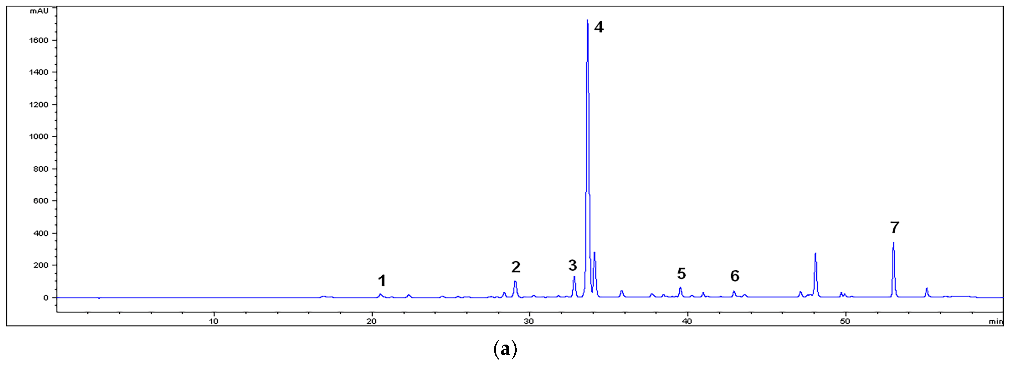
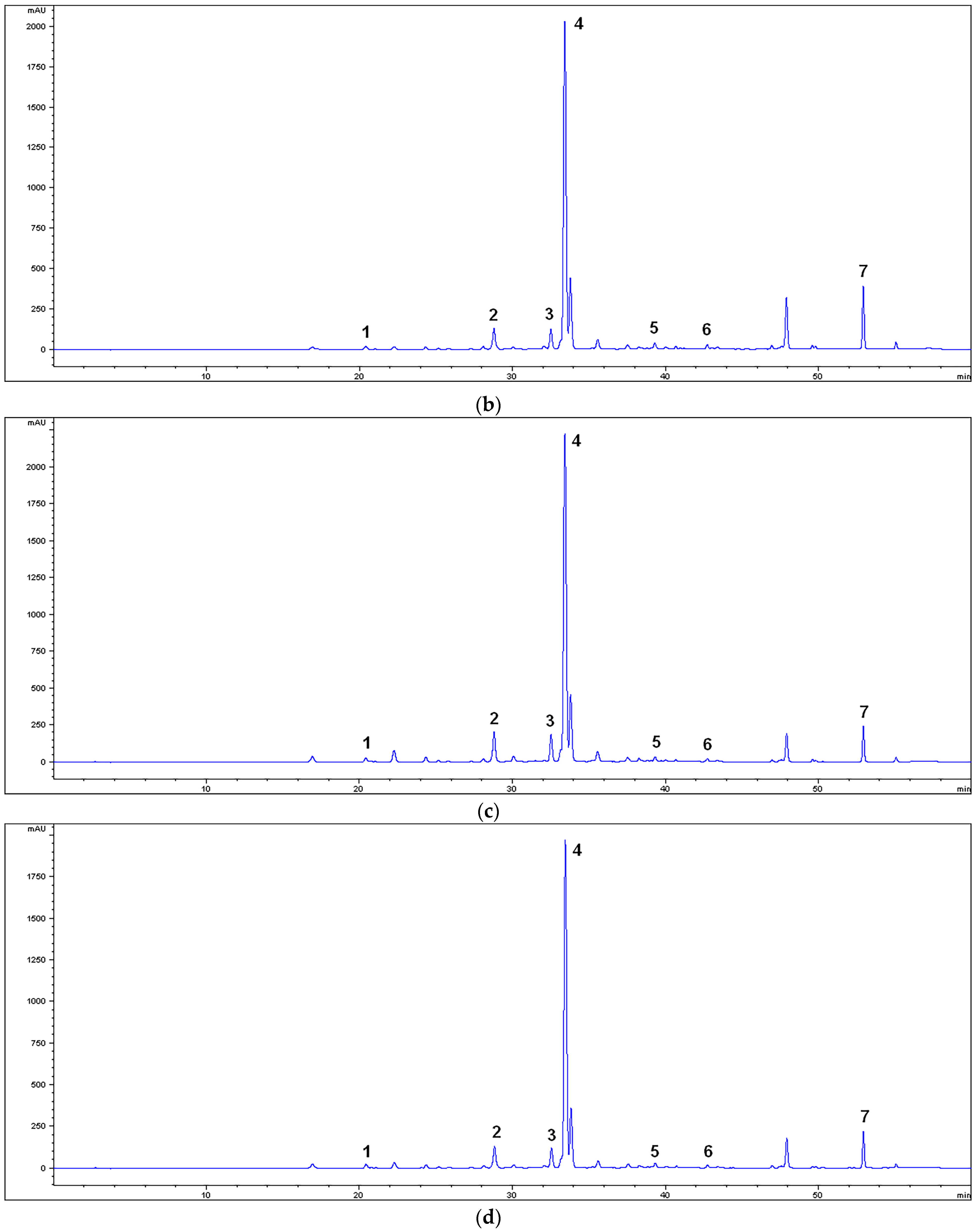
2.2. HPLC Characterization of the Extracts

The extracts were chemically characterized on an Agilent 1200 HPLC system (Agilent Technologies, Palo Alto, CA, USA) with a diode array detector. Purospher STAR RP-18e (150 × 4.6 mm) with the particle size of 5 µm (Merck, Darmstadt, Germany) was used as the analytical column. The extracts were primarily dissolved in ultrapure methanol, HPLC grade (10 mg/mL), and filtered through a 0.45 µm microfilter. The extract solutions were injected at a volume of 10 µL. The mobile phase consisted of a mixture of 0.1% aqueous trifluoroacetic acid (A) and acetonitrile (B) with a linear gradient: 0–3 min 5–5% B, 3–32 min 5–28% B, 32–44 min 25–50% B, 44–52 min 50–80% B, 52–54 min 80–90% B, 54–59 min 90–5% B, and 59–60 min 5% B. The flow in the column was 0.7 mL/min and the operating temperature was maintained at 30 °C. Phytocompounds were identified and quantified on the basis of UV-Vis signal response compared to standards. Their quantities in the extracts were expressed as µg/mg [24].

2.3. Effects of the Extracts on Ileum and Trachea Contractions

2.3.1. Experimental Animals

All experimental procedures were performed in accordance with the European Directive 2010/63/EU for animal experiments, with the special approval of the Veterinary Directorate of the Ministry of Agriculture and Environmental Protection of the Republic of Serbia (decision number 323-07-00073/2017-05/04). Male Wistar albino rats, which weighed 200–250 g and were 10–12 weeks of age, and were bred in the vivarium of the Faculty of Medicine, University of Niš, were used for all of the experimental series. A week before the experimentations, the animals were separated and housed in stainless steel cages under standard conditions (room temperature 20–24 °C, with a 12 h light/dark regime). The animals had free access to food and water, except for the last 24 h before the experiments, when they were deprived of food.

2.3.2. Isolation and Placement of Ileum and Trachea

After anaesthesia, the thorax and the aorta of the rats were dissected and the ileum and trachea were isolated and cleaned off the mesentery and connective tissue, respectively. The ileal and tracheal fragments were placed in a 20 mL tissue bath, containing Tyrode’s or Krebs’ solution, respectively, and maintained at 37 °C with a constant introduction of a mixture of oxygen (95%) and carbon dioxide (5%). Tyrode’s solution contained NaCl (150 mM), KCl (2.7 mM), MgCl2 (2 mM), NaHCO3 (12 mM), NaH2PO4 (0.4 mM), CaCl2 (1.8 mM), and glucose (5.5 mM). Kreb’s solution consisted of NaCl (137 mM), KCl (2.81 mM), CaCl2 (1.8 mM), MgCl2 (0.1 mM), NaH2PO4 (0.417 mM), NaHCO3 (11.9 mM), and glucose (11.10 mM). The fragments were stretched and stabilized for at least 30 min (ileum) or 60 min (trachea) before starting experiments [21]. The changes in the contractility of the organs were recorded using the system Transducer-TSZ-04-E (Experimetria Ltd., Budapest, Hungary). The data were analyzed using the SPEL Advanced ISOSYS Data Acquisition System software.

2.3.3. Experimental Design with Ileum

The first experimental series analyzed the effects of S. sclarea methanolic extracts on spontaneous contractions of the isolated rat ileum. The extract solutions were added in cumulative doses in the range from 0.005 to 1.5 mg/mL after the period of adaptation, forming a concentration–response curve. The result of the spasmolytic effect of each extract concentration was expressed as a percentage in relation to the initial spontaneous activity of the isolated ileum (% of inhibition of ileal contractility). Papaverine was used as a positive control with the concentrations of 0.01–3 μg/mL [25].
The second experimental series examined the effects of the extracts on the contractions induced by a high concentration of potassium ions by adding KCl solution (80 mM) after the adaptation period. The tonic contractions of the ileum were followed by the addition of the cumulative doses of each extract solution (0.005–1.5 mg/mL) at 15 min intervals. The relaxations of the ileum smooth muscles, pre-contracted with potassium ions, were expressed as the percentage of the control response mediated by potassium ions. The same procedure was carried out with a calcium channel antagonist, verapamil, with the concentrations range of 0.015–1.5 μg/mL [25].
In the last series, contractions of ileum smooth muscles were stimulated by the cumulative addition of acetylcholine solutions in concentrations of 5, 15, 50, 150, 500, and 1500 nM after a period of adaptation. A control curve of the dose-dependent contractions was constructed according to the obtained results. The ileum segments were washed with Tyrode’s solution until stable spontaneous contractions were established again. Each extract was added at a concentration of 0.5 mg/mL and 1.5 mg/mL to the bath, and after 5 min, a series with the same acetylcholine concentrations was repeated. New curves of the dose-dependent acetylcholine contractions were constructed. The spasmolytic effects of the S. sclarea extracts were presented through a series of curves showing the contractile effect of acetylcholine (%) in the presence of the extracts compared with the effect of acetylcholine without them. The same procedure was repeated with atropine (140 nM), a non-selective muscarinic receptor blocker [25].

2.3.4. Experimental Design with Rat Trachea

The first series of experiments studied the effects of the S. sclarea methanolic extracts on tracheal contractions caused by carbachol. Carbachol was added at a concentration of 1 μM after spontaneous contractions were established. The extracts were cumulatively added to the organ bath (0.005–1.5 mg/mL). The spasmolytic effects of the extracts were presented as an inhibition percentage of the contractile action of carbachol. Atropine was used as a positive control (0.41–100 μg/mL) [18].
The second set of experiments studied the spasmolytic effects of the extracts on contractions induced by a high concentration of potassium ions (KCl, 80 mM) after the period of stabilization. The extracts were added at the same cumulative concentration. The relaxations of tracheal spasms by the extracts were expressed as a percentage of the contractile action inhibition carried out by the potassium ions. Verapamil was used as a positive control (0.41–100 μg/mL) [18].

2.4. Molecular Docking Analysis

Simulation of molecular docking of the ligands to the target protein was performed using the AutoDock Vina software. Dominant phytochemicals, previously determined in S. sclarea extracts, and verapamil, as a standard drug, were considered as ligands. The structures of ligands were retrieved from PubChem (https://pubchem.ncbi.nlm.nih.gov, accessed on 20 January 2022) with the following CID numbers: 5281792 rosmarinic acid, 689043 caffeic acid, 5280443 apigenin, 5280445 luteolin, 161271 salvigenin, 12304093 apigenin-7-O-glucoside, 45933934 luteolin-7-O-glucoside, and 2520 verapamil (standard drug). Guided by the results of in vitro studies that indicate that the spasmolytic activity of S. sclarea extract is mainly mediated via calcium-mediated signaling pathways, voltage-gated calcium channels belonging to this signal pathway were selected as the target. The three-dimensional crystal structure of the voltage-gated calcium channel subunit beta2a (PDB: 1T0J) used as the target protein was obtained in PDB format from the RCSB Protein Data Bank (https://www.rcsb.org, accessed on 20 January 2022). The target protein and ligands were prepared in a dockable PDBQT format using AutoDock Tools. The coordinates of the grid box center were adjusted in the binding pocket of the standard drug (x: −3.348, y: 3.044, and z: −12.198), while the box dimensions were 20 × 20 × 20 Å. To compare the in silico performance, the binding affinities of the selected ligands with the target molecule were calculated and scored according to their affinity scores calculated through the binding free energy (kcal/mol). The molecular interactions of docked complex ligand–target protein were determined using the AutoDock Vina analysis and visualized with the BIOVIA Discovery Studio Visualizer.

2.5. Evaluation of Antimicrobial Activity of the Extracts

The antimicrobial activity of the S. sclarea methanolic extracts was estimated with laboratory control strains from the American Type Culture Collection (ATCC). Gram (+) bacteria used in the evaluation were: Staphylococcus aureus ATCC 6538, Enterococcus faecalis ATCC 9433, Streptococcus pneumoniae ATCC 6301, Streptococcus pyogenes ATCC19615, Bacillus cereus ATCC 11778, and Lysteria monocytogenes ATCC 15313. The following bacteria were selected for the testing of the effects of the extracts against Gram (−) representatives: Pseudomonas aeruginosa ATCC 9027, Proteus mirabilis ATCC 12453, Salmonela enteritidis ATCC 13076, Escherichia coli ATCC 8739, Enterobacter aerogenes ATCC 13048, and Klebsiella pneumoniae ATCC 10031. A fungus (yeast), Candida albicans ATCC 24433, was used for assessing antifungal activity. The antimicrobial activity of the extracts was evaluated using the microdilution method according to the CLSI (Clinical and Laboratory Standards Institute) [26].
Overnight broth cultures of the tested bacteria and the yeast were used for the preparation of the suspensions, which were adjusted to 0.5 McFarland standard turbidity (corresponded to 1.5 × 10⁸ CFU (colony forming units)/mL for the bacteria and 1.5 × 10⁷ CFU/mL for the yeast). Primarily, the extracts were dissolved in a sterile 10% aqueous solution of dimethyl sulfoxide. Serial double dilutions of the extracts, in the range from 0.1 to 100 mg/mL, were prepared in microtiter plates (96-well) with an inoculated nutrient broth. The final volume of the wells was 100 µL and the final concentrations were 2 × 10⁶ for the bacteria and 2 × 10⁵ for the yeast. The incubation of the microtiter plates was performed at 37 °C for 24 h for the bacteria or at 25 °C for 48 h for the yeast. Microbial growth was detected by a 0.5% aqueous solution of 2,3,5-triphenyl tetrazolium chloride (20 µL) added to each well [27]. The minimal concentration of the extract with no visible growth of microorganisms was defined as the minimum inhibitory concentration (MIC). The minimum bactericidal/fungicidal concentration (MBC/MFC) was defined as the minimal concentration of the extract that killed 99.9% of the tested bacteria or the yeast. For the determination of the MBC/MFC of the extract, the broth was taken from each well with no visible growth and inoculated into an agar (Mueller–Hinton agar at 37 °C for 24 h for the bacteria or Sabouraud dextrose agar 25 °C for 48 h for the yeast). Sterile 10% aqueous DMSO solution was used as a negative control. Chloramphenicol, streptomycin, and nystatin (0.008–16 μg/mL) were used as the controls.

2.6. Statistical Analysis

The final results are expressed as mean values of three or six parallel measurements ± the standard deviations (chemical composition or spasmolytic analyses, respectively), except for the antimicrobial activity. The EC50 values, which presented the concentrations causing 50% of maximal response, were obtained by a regression analysis. Student’s t-test or one-way ANOVA with Duncan’s post hoc test were used for the determination of significant statistical differences between/among the means (p < 0.05 or p < 0.01). Statistical analyses were carried out using the SPSS 20.0 statistical package (SPSS, Inc., Chicago, IL, USA).

3. Results

3.1. Chemical Characterization of the Extracts

The yields of the extractions were 12.75% for the MM extract, 11.93% for MU, 19.65% for M80M, and 13.30% for M80U. The HPLC analysis of the extracts indicated the presence of phenolic acids as well as flavon type flavonoid aglycons and flavon type flavonoid heterosides (Figure 1). Table 1 displays the content of the individual compounds. Rosmarinic acid was predominant in all of the extracts in the range of 171.99 ± 1.88–197.48 ± 2.00 µg/mg. The M80M extract contained the highest quantity of phenolic acids and flavonoid heterosides, while aglycons were mostly present in the extracts prepared with absolute methanol. Dominant aglycon in the extracts was salvigenin, followed by luteolin and apigenin.
Based on the obtained values of the content of phytocompounds in the extracts, the maceration method generally proved to be more effective. This difference is especially noticeable between the extracts prepared with 80% methanol.

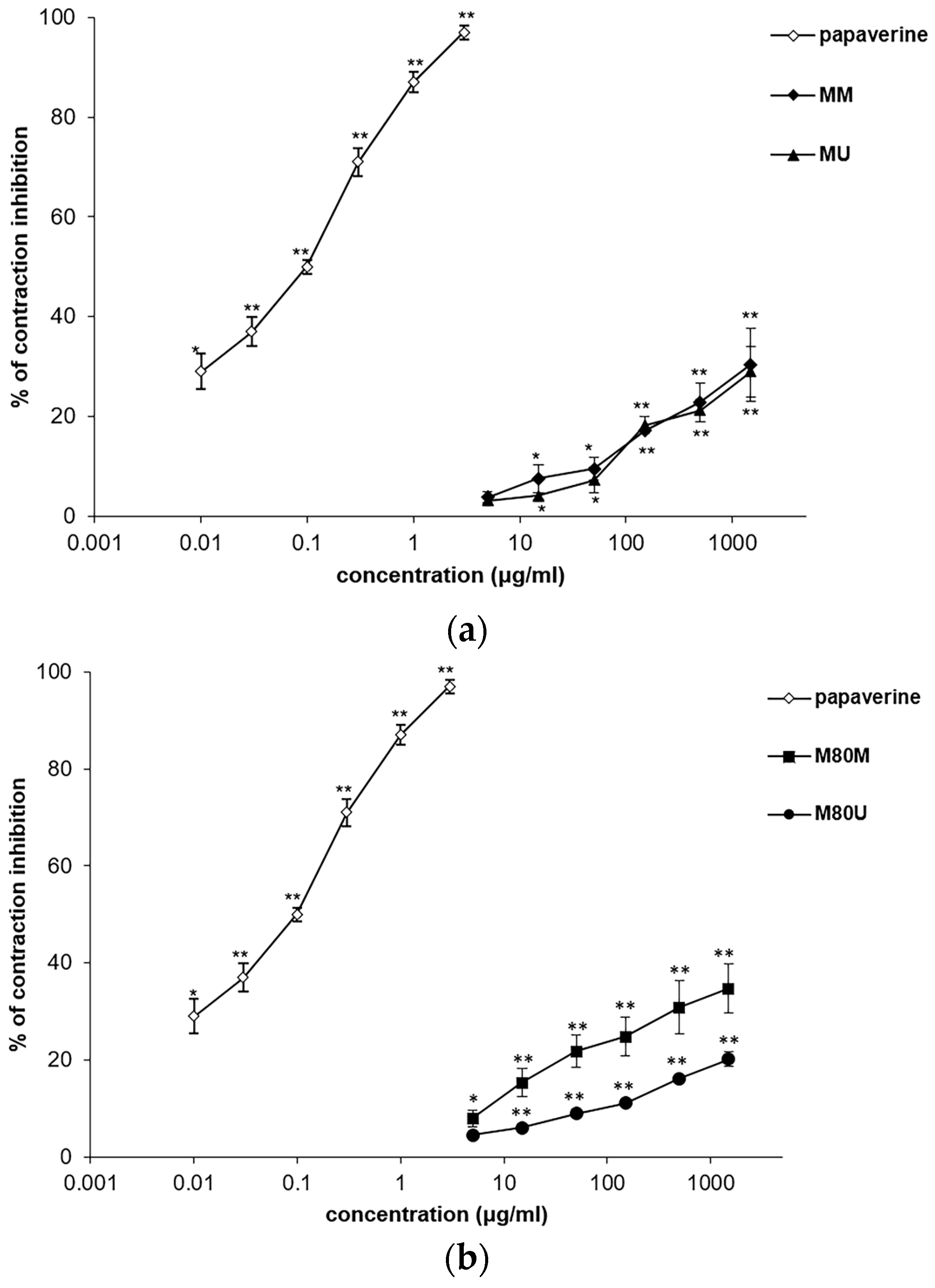
3.2. Spasmolytic Effects of the Extracts on Spontaneous Ileum Contractions

The S. sclarea methanolic extracts exhibited the significant, dose-dependent, and spasmolytic effect of the ileum smooth muscle in the first experimental series by reducing the spontaneous contractions. The lowest EC50 value was determined for the extract M80M. In addition, this extract, MM, and MU acted in a similar manner with the narrow range of EC50 from 2.44 ± 0.10 to 2.69 ± 0.22 mg/mL and the inhibitions of maximal concentrations (1.5 mg/mL) from 28.96 ± 1.86 to 34.73 ± 1.20%. The M80U extract was less effective (Table 2; Figure 2). The maximal concentration of papaverine (0.003 mg/mL), used as a positive control, was able to reduce 97% of all spontaneous ileum contractions.

3.3. Spasmolytic Effects of the Extracts on KCl-Induced Ileum Contractions

The extracts had an inhibitory activity on the contractions induced by the application of KCl solution (80 mM). Ileum smooth muscle relaxation was dose-dependent, with the range of EC50 values from 3.69 ± 0.30 to 5.76 ± 0.34 mg/mL (Table 2; Figure 3). The best activity was achieved after the addition of the MM extract, whose maximal concentration of 1.5 mg/mL was able to reduce contraction to 74.71 ± 2.29%. Verapamil was used as a control in this study, whereby a maximum concentration of 0.0015 mg/mL reduced the contraction to 5%.

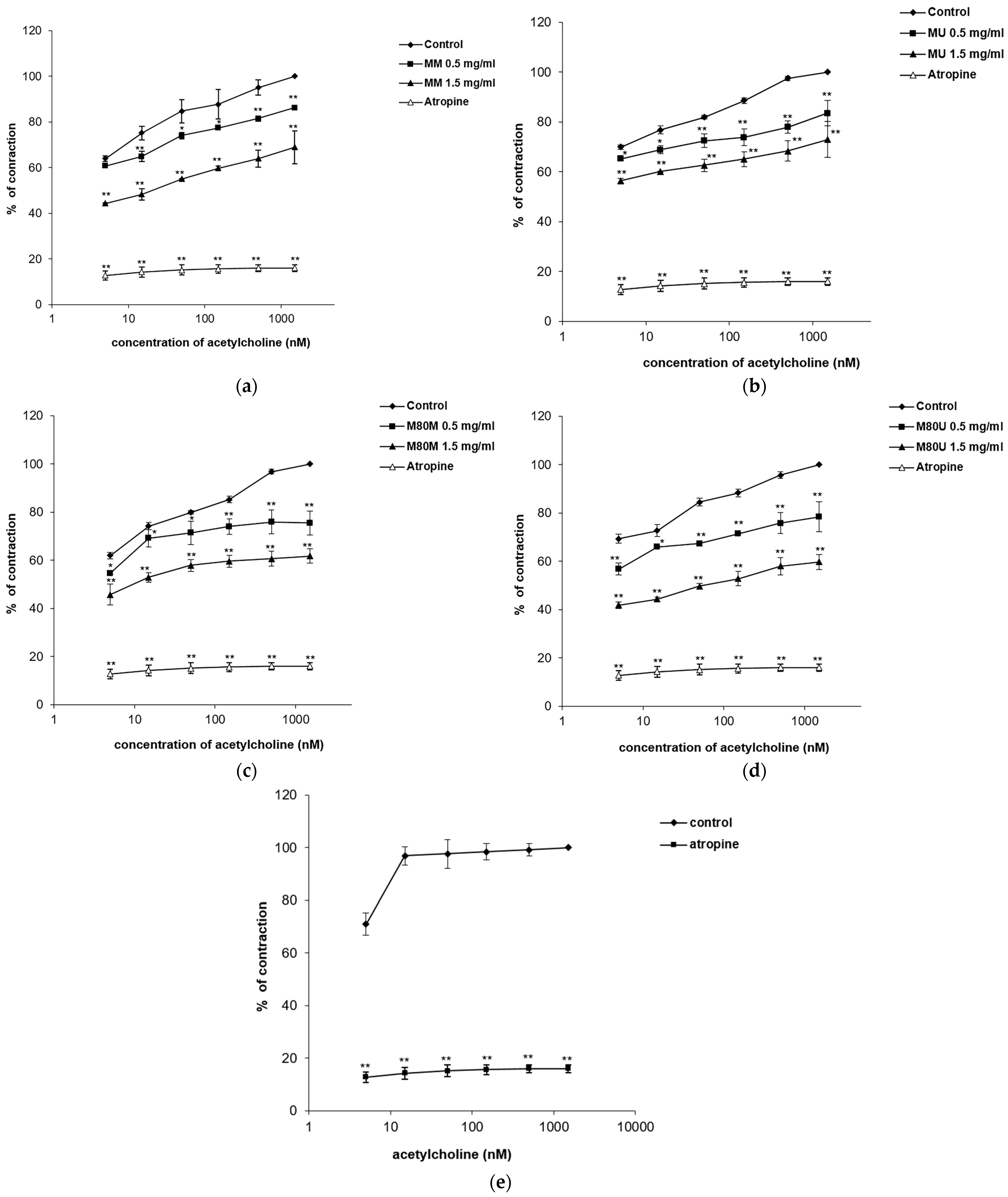
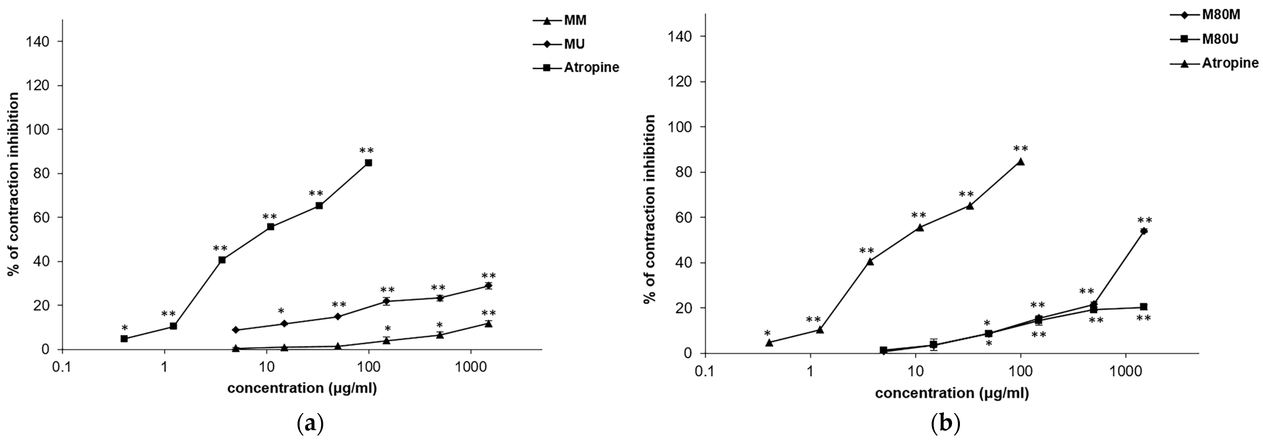
3.4. Spasmolytic Effects of the Extracts on Acetylcholine-Induced Ileum Contractions

The tested extracts were able to reduce ileum contractions induced by cumulative doses of acetylcholine with statistical significance (p < 0.01). The control EC50 values of acetylcholine were modified and increased after the addition of all the extract concentrations (0.5 and 1.5 mg/mL) (Table 3; Figure 4). The M80U extract stood out in this series of experiments modifying the baseline EC50 value of acetylcholine (0.17 ± 0.00 nM) two times after the application of 0.5 mg/mL. An even greater increase in the EC50 value of acetylcholine was observed after applying a dose of 1.5 mg/mL (62.15 ± 3.22 nM and contractions reduction from baseline 100% to 59.78 ± 3.10%). The effects of the other methanolic extracts were significantly lower: M80M > MM > MU. Atropine, a muscarinic receptor antagonist, was used as a positive control in the concentration of 140 nM, modifying the EC50 value of acetylcholine from 0.10 ± 0.00 nM to 18,261.96 ± 958.32 nM and reducing acetylcholine-induced ileum contractions to 16.02%.

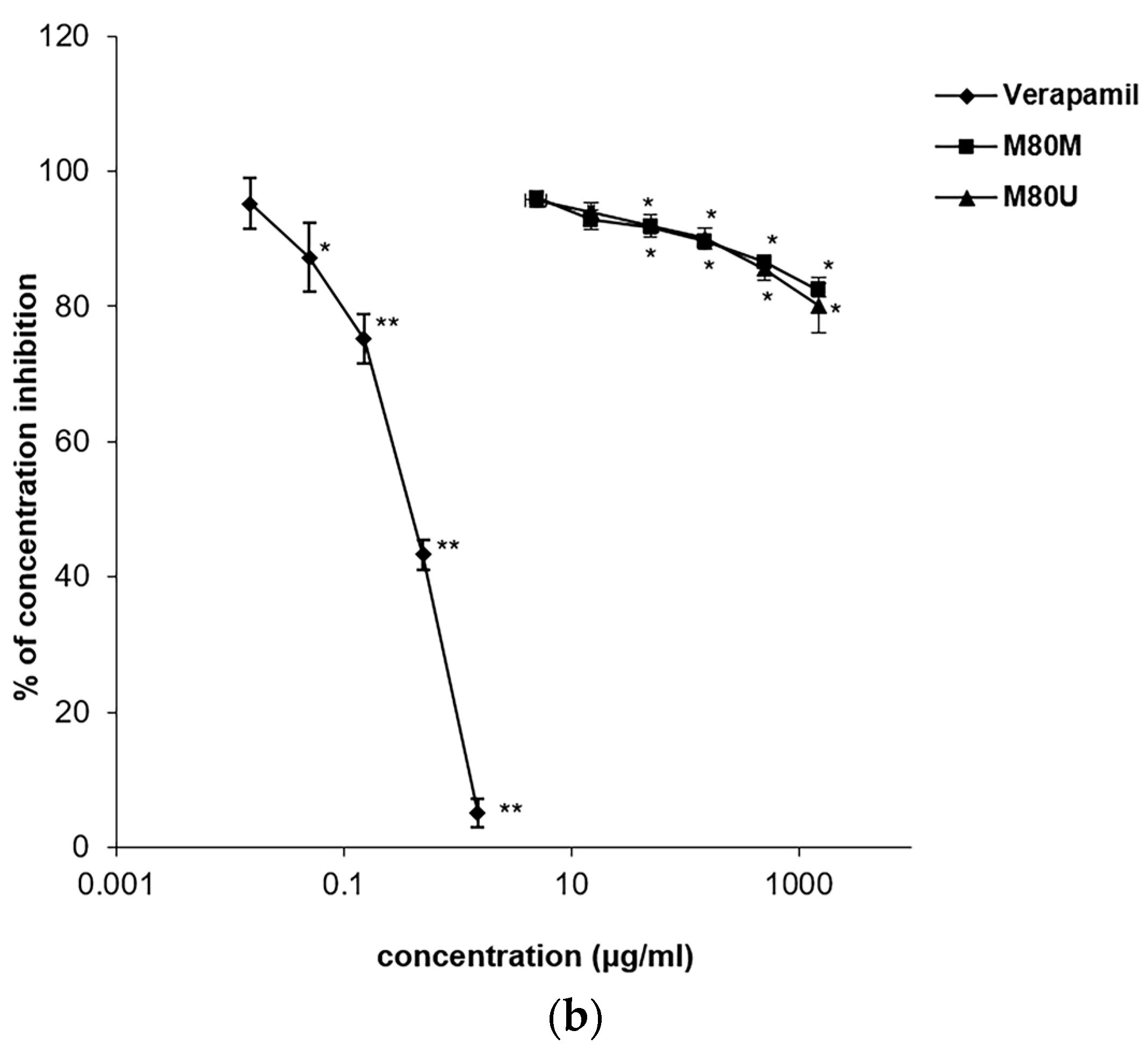
3.5. Spasmolytic Effects of the Extracts on KCl-Induced Tracheal Contractions

After a single dose of KCl (80 nM) all tested extracts expressed inhibitory effects on tracheal rat smooth muscle contractions in a dose-dependent regime (Table 4). Samples were characterized as moderate spasmolytic agents, whereby extracts prepared by the maceration method were stronger, especially M80M. A maximal concentration of 1.5 mg/mL of this extract inhibited the contractions by 15.37 ± 0.81 and 17.22 ± 0.99%, respectively (Figure 5). Verapamil inhibited 74.23 ± 1.20% of the contractions at the maximum concentration (100 μg/mL) with an EC50 value of 15.23 ± 0.08 μg/mL.

3.6. Spasmolytic Effects of the Extracts on Carbachol-Induced Tracheal Contractions

Methanolic S. sclarea extracts had a relaxing effect on the tracheal smooth muscle contractions induced by a single dose of carbachol (1 μM) (Table 4; Figure 6). The M80M extract stood out in in its series with the lowest EC50 (1.36 ± 0.01 mg/mL) and an inhibition of 54.09 ± 1.66% achieved with a maximum concentration of 1.5 mg/mL. Atropine, used as a control, inhibited 84.89 ± 2.00% of the tracheal contractions with an EC50 value of 9.78 ± 0.00 μg/mL.

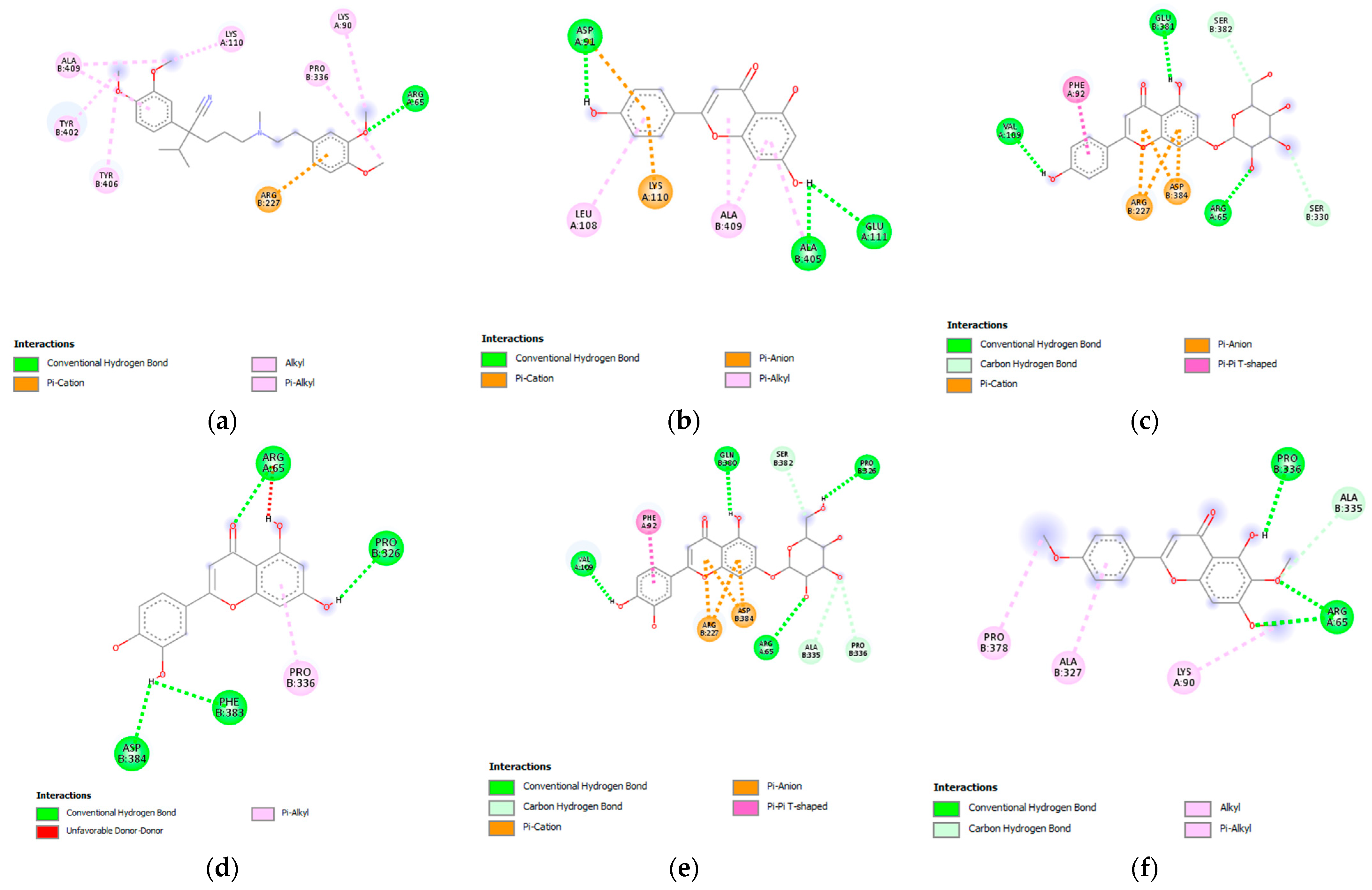
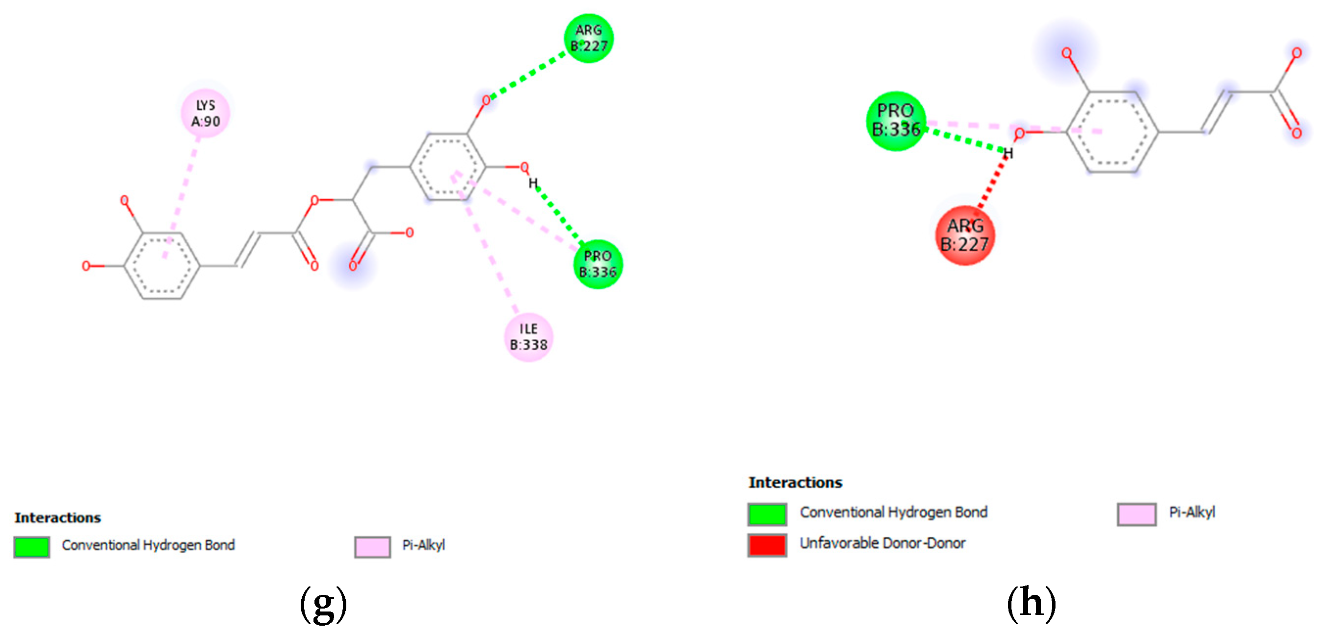
3.7. Molecular Docking Analysis

The results of the molecular docking analysis with the binding affinities and residues of the amino acids involved in the ligand–target binding, including their interatomic distance (Å), are listed in Table 5. The results show that the docking scores of all tested ligands are negative, with the binding energies in the range from −5.8 to −8.6 kcal/mol. This suggests that ligands are able to bind to the target protein. The docking score of the tested ligands with voltage-gated calcium channels is as follows: apigenin-7-O-glucoside = luteolin-7-O-glucoside, salvigenin, luteolin, apigenin, rosmarinic acid = verapamil, and caffeic acid. The most prominent apigenin-7-O-glucoside and luteolin-7-O-glucoside both had a binding score of −8.6 kcal/mol. For a deeper analysis of the interaction profile, two-dimensional structures of the binding pocket of the target protein in a complex with tested ligands were constructed (Figure 7). The analysis of the interaction profile shows that in the case of both mentioned compounds binding via conventional hydrogen bonds to the amino acid residue, Arg65 is involved, similar to the binding of the standard drug verapamil to its binding pocket. In the case of apigenin-7-O-glucoside, the length of this bond was 2.21 Å; while in the case of luteolin-7-O-glucoside, it was slightly longer. Apart from this hydrogen bond interaction, apigenin-7-O-glucoside forms conventional hydrogen bonds with the residues of Val109 and Glu381, while luteolin-7-O-glucoside forms conventional hydrogen bonds with Pro326, Val109, and Gln380. Such a developed hydrogen bond network contributes greatly to the binding affinity value. Regarding the hydrophobic/electrostatic interactions, both apigenin- and luteolin-7-O-glucoside achieved a π-cation interaction via Arg227 such as verapamil. Beyond, both apigenin- and luteolin-7-O-glucoside showed a poorer network of hydrophobic interactions compared to verapamil (Table 5).

3.8. Antimicrobial Activity of the Extracts

According to the obtained results of the MIC and MBC/MFC values, the extracts are moderate antimicrobial agents (Table 6). The effects were more prominent toward Gram (+) bacteria compared to Gram (−). In addition, their bactericidal effects were almost minor with the MBC values of 100 or >100 mg/mL, with the exception of the M80M extract, which had somewhat stronger effects on P. aeruginosa (50 mg/mL). The MU extract, prepared with absolute methanol and the ultrasound method, showed the best antimicrobial effectiveness, particularly toward S. aureus, B. cereus, and L. monocytogenes, with the MIC values of 6.25, 12.5, and 25 mg/mL, respectively. Its bacteriostatic effects against Gram (−) bacteria were mostly expressed toward P. aeruginosa and E. aerogenes with the MIC values of 50 mg/mL. The effects on the yeast, C. albicans, were of no great importance because the MIC and MFC values of the extracts were 100 and/or >100 mg/mL. The DMSO aqueous solution showed no activity against the investigated microbial strains. MICs/MBCs values of the used positive controls were expectedly much lower (chloramphenicol 0.06–7.81/0.12–15.61 µg/mL, streptomycin 0.16–0.6/0.16–0.6 µg/mL, and nystatine 3.91–7.81 µg/mL).

4. Discussion

Many factors of plant extraction have important roles in its efficiency, primarily the type of solvent and method of extraction. The most commonly used methods include conventional techniques, such as maceration, percolation, infusion, decoction, or hot continuous extraction. However, in the last three decades, various new, alternative techniques, such as ultrasonic and microwave extraction, or supercritical fluid extraction, were developed [28]. We used the ultrasound-assisted extraction because it is a technique that generally achieves high reproducibility in a short period of time and a high yield of bioactive compounds. Additionally, this technique is characterized by its simplicity, a low temperature during processing, and a reduced consumption of solvents and energy [29]. On the other hand, the disadvantage of this extraction technique is the risk of possible free radicals formation when the ultrasound energy exceeds 20 kHz [29,30]. The ultrasound method was shown to be very good in terms of the extraction yield in previous studies, although some studies show it to be weaker than Soxhlet extraction, maceration, or microwave extraction [31,32]. Another extraction technique used is single maceration. It is a traditional method that involves the extraction of plant material at room temperature for a minimum of three days [30]. Its advantage is reflected in the use of a cold solvent, which reduces the possibility of the decomposition of active compounds. The content of phytocompounds in the extracts indicates that the maceration method was more effective. The extract prepared with this method, using a more polar solvent (80% methanol), contained the highest quantity of phenolic acids and flavonoid heterosides. In addition, absolute methanol extracted higher amounts of flavonoid aglycones, compared to 80% methanol, due to its lower polarity.
Functional gastrointestinal disorders include a number of morphological and physiological disorders characterized by impaired intestinal motility, visceral hypersensitivity, changes in mucosal function, immune system, altered microbiota, and nervous system processes [33]. The term “functional” generally refers to disturbances in the neuromuscular function of the affected part of the gastrointestinal tract that induce discomfort [33,34]. The Salvia species was traditionally used in the therapy of various gastrointestinal complaints [35]. In addition, the European Medicinal Agency published a monograph on S. officinalis leaf, indicating its use as traditional herbal medicine in mild dyspeptic complaints, such as heartburn and bloating [36]. Previous studies confirmed that other Salvia species could be potential therapeutic agents in functional gastrointestinal disorders as well [37,38,39,40]. In this study, methanolic S. sclarea extracts exhibited inhibitory effects to a lesser or greater extent in all experimental models performed on the isolated rat ileum. The contractions of smooth ileum muscles are the result of the increased concentration of free calcium ions in the cytoplasm, which is achieved through voltage-dependent L-type channels or by the release from the intracellular depot [41]. Spontaneous contractions of the smooth ileal muscle were effectively reduced with EC50 ranging from 2.44 ± 0.10 mg/mL to 4.59 ± 0.33 mg/mL. The weakest activity was achieved with the M80U extract, which was characterized by the lowest concentration of rosmarinic acid, flavonoids aglycons, and flavonoids heterosides. The other three extracts exhibited similarly in spasmolytic activity. Furthermore, the extracts were able to reduce the contractions stimulated by a high single dose of the KCl solution (80 mM). Smooth muscle contractility is triggered by the depolarization of the smooth muscle membrane, which is caused by high concentrations of potassium ions opening the voltage-dependent L-type channels, causing extracellular calcium ions to enter the cell and cause the contraction [42]. Therefore, the inhibitory effects of the tested extracts on the potassium ion-induced contractions can be explained by the blocking of voltage-dependent L-type calcium channels and the opening of potassium channels. The extract prepared with absolute methanol and maceration turned out to be the best, having the highest amount of apigenin and luteolin. The third series of experiments conducted on the rat ileum showed the inhibitory effects of S. sclarea methanolic extracts on contractions induced by acetylcholine, suggesting that the effects of the extracts are mediated by the action on the muscarinic receptors. It is known that the gastrointestinal tract has an abundance of M2 and M3 muscarinic receptor subtypes, whereby the M2 subtype is more prevalent. The acetylcholine neurotransmitter causes the contractions of the isolated ileum primarily through the M3 receptor pathway, which involves the hydrolysis of phosphoinositol and the mobilization of intracellular calcium ions [43], as well as the opening of voltage-dependent calcium L-type channels [44]. The tested extracts, in both concentrations (0.5 and 1.5 mg/mL), were able to reduce the smooth muscle contractions induced by cumulative doses of acetylcholine. While it was the weakest in reducing spontaneous and KCl-induced contractions, the M80U extract was the strongest in this series of experiments. This extract modified the baseline EC50 value of acetylcholine two times after the application of the first dose and over three hundred times after applying the dose of 1.5 mg/mL. The effects of the other methanolic extracts were lower and were distributed in the following manner: M80M > MM > MU.
The phytocompounds, determined in the S. sclarea extracts, are likely to be highly responsible for the aforementioned effects. Results of the in vitro studies suggest that the spasmolytic activity of S. sclarea methanolic extract is mediated via calcium-mediated signaling pathways. The seven major phytocompounds of this extract were docked to a voltage-gated calcium channel belonging to this signal pathway [45], using verapamil as the standard drug. A higher negative value of binding energies presented in Table 5 indicates that the ligand could bind to the protein stronger [46]. Concerning binding affinities, it is observed that the most prominent were apigenin-7-O-glucoside and luteolin-7-O-glucoside, both with a binding score of −8.6 kcal/mol. The length of the conventional hydrogen bond with amino acid residue Arg65 of 2.21 Å in the case of apigenin-7-O-glucoside can be classified as strong (in the range of 2.2 to 2.5 Å), while the distance of 2.52 Å in the case of luteolin-7-O-glucoside can be categorized as medium strong (within the range of 2.5 to 3.2 Å). The more developed network of hydrophobic interactions observed for verapamil compared to apigenin- and luteolin-7-O-glucoside could partially explain its better activity recorded in in vitro assays. Namely, although hydrophobic interactions contribute less to binding energy compared to hydrogen bonds, because they are not as strong, they often play an important role in many biological mechanisms [47].
In addition to the methanol extracts of S. sclarea, the inhibiting effects of excellent smooth muscle ileum contractions were reported for the S. sclarea ethanolic extracts and essential oil [18,48]. Rosmarinic acid, as the prevalent compound in the extracts, was proven to be an effective spasmolytic agent in in vitro conditions. Bazylko et al. (2009) [49] and Randjelovic et al. (2022) [18] confirmed its activity in spontaneous KCl- and acetylcholine-induced ileum contractions. In addition, Lemmens-Gruber et al. (2006) [50] and Abdalla et al. (1994) [51] showed that apigenin and luteolin exhibit excellent spasmolytic activities on isolated guinea pig ileum. Furthermore, these flavones significantly inhibit the smooth muscle contractility of rat ileum induced by high potassium concentrations, while apigenin also had an effect on reducing acetylcholine-induced contractions [52].
Many studies showed that the extracts and essential oils of plant species of the Lamiaceae family exhibit inhibitory effects on the isolated trachea of experimental animals, and it is well known that these aromatic drugs are traditionally widely used in respiratory disorders of this type [53,54]. The tested S. sclarea extracts had a moderate relaxing effect on tracheal smooth muscle contractions caused by a single dose of KCl and carbachol. The best spasmolytic effects in both systems were achieved by the M80M extract, which is characterized by the highest quantity of the more polar compounds: rosmarinic acid, caffeic acid, and luteolin- and apigenin-7-O-glucoside. As shown in previous studies, the compounds determined in these extracts exhibited bronchodilator activity. Namely, rosmarinic acid, luteolin, and apigenin were able to inhibit the contractions of tracheal smooth muscle induced by various spasmogenic agents [51,55,56,57].
Aromatic plant species were long recognized and widely used as antibacterial, antifungal, antiviral, or antiparasitic agents [58]. Plant polyphenols are characterized by good antimicrobial properties and their presence in extracts significantly contributes to the inhibition of growth and destruction of microorganisms [59]. According to numerous in vitro studies, polyphenols, identified in the tested extracts, exhibit such effects [18,60,61,62,63,64]. Salvia species, along with S. sclarea methanolic extracts, are stronger antimicrobial agents toward Gram (+) compared to Gram (−) bacteria, which also applies to most plants [65]. The explanation for this fact lies in the structure of the cell wall of Gram (−) bacteria, which is more complicated and represents a specific barrier for the entry of macromolecules [66]. Furthermore, our investigation confirmed that the S. sclarea methanolic extracts had better effects on inhibiting the growth of tested bacteria than killing them, which was also shown by the S. sclarea ethanolic extracts [18]. It is of particular importance that the investigated extracts were active against S. aureus, keeping in mind its great pathogenicity, especially in regards to its effect on respiratory organs [67]. The MU extract, characterized with the highest quantity of apigenin and salvigenin, was leading in its activity with the MIC value of 6.25 mg/mL. In addition, the extracts showed a significant antimicrobial effect against B. cereus, responsible for foodborne illnesses, followed by nausea, vomiting, and diarrhea [68]. The MU and M80U extracts stood out in their activity, with both having MIC values of 12.5 mg/mL. The significance of the bacteriostatic activity of the extracts, especially MM and MU, against L. monocytogenes, is that this bacterium is a cause of serious infection, listeriosis, which is characterized by an occasional febrile gastroenteritis in immunocompetent persons and even by a possible fatal outcome [69]. Recent studies showed that the S. sclarea extracts could be useful as antimicrobial agents against other Gram (+) bacteria, such as S. epidermidis, B. megaterium, B. brevis, Micrococcus luteus, and Mycobacterium smegmatis [70,71]. Among Gram (−) bacteria, P. aeruginosa and E. aerogenes showed a mildly higher sensitivity to the presence of the extracts. Namely, the MU and M80M extracts acted as anti-pseudomonal agents with MIC values of 50 mg/mL, which is important because P. aeruginosa is the cause of opportunistic and hospital-acquired infections, having a high resistance to antibiotics [72]. Previous investigations of the antimicrobial characteristics of S. sclarea extracts toward Gram (−) bacteria demonstrated their inhibitory effects on P. mirabilis, S. enteritidis, K. pneumoniae, E. coli, and Aeromonas hydrophila growths [18,71,73]. The investigated extracts did not prove to be effective against the yeast C. albicans with values of MICs/MFCs over 100 mg/mL, which was in accordance with other studies of antifungal properties of S. sclarea extracts [73,74].

5. Conclusions

The present study offers a deeper understanding of the gastrointestinal and bronchodilator activity and the potential use of the S. sclarea methanolic extracts in phytotherapy. The extracts successfully reduced spontaneous and induced ileum and tracheal contractions in in vitro conditions. Polyphenolic compounds determined in the extracts, rosmarinic and caffeic acid, apigenin, luteolin, salvigenin, and luteolin- and apigenin-7-O-glucoside could be responsible for the manifestation of the spasmolytic activity, which was supported by the in silico analysis. The antibacterial effects of the extracts were moderate, being better bacteriostatic than bactericidal agents; however, they could be supportive agents in the control of gastrointestinal and respiratory disorders.
The investigated S. sclarea extracts might be used as potential herbal remedies, although further studies should be aimed at their efficacy in clinical trials.

Author Contributions

Conceptualization, M.R.; methodology, S.B.; investigation, M.R., B.M., M.M., N.K., S.Ž., M.J. and T.M.-K.; resources, D.K.; writing—original draft preparation, M.R.; writing—review and editing, all; visualization, M.R.; project administration, D.K.; funding acquisition, D.K.; supervision, D.K. All authors have read and agreed to the published version of the manuscript.

Funding

This research was funded by the Serbian Ministry of Science, Technological Development and Innovation, grant number 451-03-47/2023-01/200003 and 451-03-47/2023-01/200113, and by the Internal Project of the Faculty of Medicine, University of Niš, Serbia, grant number 68.

Institutional Review Board Statement

All experimental procedures with animals were performed in accordance with the European Directive 2010/63/EU for animal experiments, with the special approval of the Veterinary Directorate of the Ministry of Agriculture and Environmental Protection of the Republic of Serbia (decision number 323-07-00073/2017-05/04).

Informed Consent Statement

Not applicable.

Data Availability Statement

Not applicable.

Acknowledgments

The authors are thankful to Aleksandar Jovanović for his great support and expertise in English.

Conflicts of Interest

The authors declare no conflict of interest.

References

  1. Palhares, R.M.; Drummond, M.G.; Brasil, B.D.S.A.F.; Cosenza, G.P.; Brandão, M.D.G.L.; Oliveira, G. Medicinal plants recommended by the world health organization: DNA barcode identification associated with chemical analyses guarantees their quality. PLoS ONE 2015, 10, e0127866. [Google Scholar] [CrossRef]
  2. Ahn, K. The worldwide trend of using botanical drugs and strategies for developing global drugs. BMB Rep. 2017, 50, 111–116. [Google Scholar] [CrossRef]
  3. Bone, K.; Mills, S.Y. Principles and Practice of Phytotherapy, Modern Herbal Medicine, 2nd ed.; Churchill Livingstone Elsevier: London, UK, 2013. [Google Scholar]
  4. Ben-Arye, E.; Dudai, N.; Eini, A.; Torem, M.; Schiff, E.; Rakover, Y. Treatment of upper respiratory tract infections in primary care: A randomized study using aromatic herbs. Evid. Based Complement. Altern. Med. 2011, 2011, 690346. [Google Scholar] [CrossRef]
  5. Engelbertz, J.; Lechtenberg, M.; Studt, L.; Hensel, A.; Verspohl, E.J. Bioassay-guided fractionation of a thymol-deprived hydrophilic thyme extract and its antispasmodic effect. J. Ethnopharmacol. 2012, 141, 848–853. [Google Scholar] [CrossRef]
  6. Cosio, B.G.; Rosado, J.R.; Rossi, F.F. Asthma: Epidemiology, pathophysiology, and risk factors. In Clinical Respiratory Medicine, 4th ed.; Spiro, S.G., Silvestri, G.A., Agusti, A., Eds.; Elsevier: Amsterdam, The Netherlands, 2012. [Google Scholar]
  7. Carović-Stanko, K.; Petek, M.; Martina, G.; Pintar, J.; Bedeković, D.; Ćustić, M.H.; Satović, Z. Medicinal plants of the family Lamiaceae as functional foods—A review. Czech J. Food Sci. 2016, 34, 377–390. [Google Scholar] [CrossRef]
  8. Baricevic, D.; Bartol, T. The biological/pharmacological activity of the Salvia genus. In Sage: The Genus Salvia; Kintzios, S.E., Ed.; Harwood Academic Publishers: Amsterdam, The Netherlands, 2000. [Google Scholar]
  9. Dweck, A.C. Folklore and cosmetic use of various Salvia species. In Sage: The Genus Salvia; Kintzios, S.E., Ed.; Harwood Academic Publishers: Amsterdam, The Netherlands, 2000. [Google Scholar]
  10. Koutsaviti, A.; Tzini, D.I.; Tzakou, O. Greek Salvia sclarea L. essential oils: Effect of hydrodistillation time, comparison of the aroma chemicals using hydrodistillation and HS-SPME techniques. Rec. Nat. Prod. 2016, 10, 800–805. [Google Scholar]
  11. Lawrence, B.M. Production of Clary Sage Oil and Sclareol in North America. In Proceedings of the 4emes Recontres Internacionales, Nyons, France, 5–7 December 1994; pp. 41–58. [Google Scholar]
  12. Leporatti, M.L.; Pavesi, A.; Posocco, E. Phytotherapy in the Valnerina Marche (Central Italy). J. Ethnopharmacol. 1985, 14, 53–63. [Google Scholar] [CrossRef] [PubMed]
  13. Hansel, R.; Keller, K.; Rimpler, H.; Schneider, G. Hagers Handbuch der Pharmazeutischen Praxis: Drogen P-Z Folgeband 2; Springer: Berlin, Germany, 1994. [Google Scholar]
  14. Raafat, K.; Habib, J. Phytochemical compositions and antidiabetic potentials of Salvia sclarea L. essential oils. J. Oleo Sci. 2018, 67, 1015–1025. [Google Scholar] [CrossRef] [PubMed]
  15. Moretti, M.D.; Peana, A.T.; Satta, M. A study on anti-inflammatory and peripheral analgesic action of Salvia sclarea oil and its main components. J. Essent. Oil Res. 1997, 9, 199–204. [Google Scholar] [CrossRef]
  16. Zengin, G.; Senkardes, I.; Mollica, A.; Picot-Allain, C.M.N.; Bulut, G.; Dogan, A.; Mahomoodally, M.F. New insights into the in vitro biological effects, in silico docking and chemical profile of clary sage—Salvia sclarea L. Comput. Biol. Chem. 2018, 75, 111–119. [Google Scholar] [CrossRef]
  17. Kuzma, L.; Kalemba, D.; Rozalski, M.; Rozalska, B.; Wieckowska-Szakiel, M.; Krajewska, U.; Wysokińska, H. Chemical composition and biological activities of essential oil from Salvia sclarea plants regenerated in vitro. Molecules 2009, 14, 1438–1447. [Google Scholar] [CrossRef] [PubMed]
  18. Randjelović, M.; Branković, S.; Miladinović, B.; Milutinović, M.; Živanović, S.; Mihajilov-Krstev, T.; Kitić, D. The benefits of Salvia sclarea L. ethanolic extracts on gastrointestinal and respiratory spasms. S. Afr. J. Bot. 2022, 150, 621–632. [Google Scholar] [CrossRef]
  19. Lu, Y.; Foo, L.Y. Polyphenolics of Salvia—A review. Phytochemistry 2002, 59, 117–140. [Google Scholar] [CrossRef]
  20. Ulubelen, A.; Topcu, G.; Eriş, C.; Sönmez, U.; Kartal, M.; Kurucu, S.; Bozok-Johansson, C. Terpenoids from Salvia sclarea. Phytochemistry 1994, 36, 971–974. [Google Scholar] [CrossRef]
  21. Graves, N.S. Acute gastroenteritis. Prim. Care 2013, 40, 727–741. [Google Scholar] [CrossRef] [PubMed]
  22. Lumb, A.B.; Horncastle, E. 29—Pulmonary physiology. In Pharmacology and Physiology for Anesthesia, 2nd ed.; Hugh, C., Hemmings, H.C., Egan, T.D., Eds.; Elsevier: Philadelphia, PA, USA, 2019. [Google Scholar] [CrossRef]
  23. Pharmacopoea Jugoslavica, 4th ed.; Federal Institute of Public Health: Belgrade, Serbia, 1984.
  24. Kostić, M.; Kitić, D.; Petrović, M.B.; Jevtović-Stoimenov, T.; Jović, M.; Petrović, A.; Živanović, S. Anti-inflammatory effect of the Salvia sclarea L. ethanolic extract on lipopolysaccharide-induced periodontitis in rats. J. Ethnopharmacol. 2017, 199, 52–59. [Google Scholar] [CrossRef] [PubMed]
  25. Milutinović, M.; Branković, S.; Šavikin, K.; Kostić, M.; Kitić, N.; Miladinović, B.; Kitić, D. Antispasmodic effects of black chokeberry (Aronia melanocarpa (Michx.) Elliott) extracts and juice and their potential use in gastrointestinal disorders. J. Berry Res. 2020, 10, 175–192. [Google Scholar] [CrossRef]
  26. Clinical and Laboratory Standards Institute (CLSI). Performance Standards for Antimicrobial Susceptibility Testing; 15th Informational Supplement, M100-S15; Clinical and Laboratory Standards Institute: Wayne, PA, USA, 2005. [Google Scholar]
  27. Sartoratto, A.; Machado, A.L.M.; Delarmelina, C.; Figueira, G.M.; Duarte, M.C.T.; Rehder, V.L.G. Composition and antimicrobial activity of essential oils from aromatic plants used in Brazil. Braz. J. Microbiol. 2004, 35, 275–280. [Google Scholar] [CrossRef]
  28. Dhanani, T.; Shah, S.; Gajbhiye, N.A.; Kumar, S. Effect of extraction methods on yield, phytochemical constituents and antioxidant activity of Withania somnifera. Arab. J. Chem. 2017, 10, S1193–S1199. [Google Scholar] [CrossRef]
  29. Dent, M.; Dragović-Uzelac, V.; Elez Garofulić, I.; Bosiljkov, T.; Ježek, D.; Brnčić, M. Comparison of conventional and ultrasound-assisted extraction techniques on mass fraction of phenolic compounds from sage (Salvia officinalis L.). Chem. Biochem. Eng. Q. 2015, 29, 475–484. [Google Scholar] [CrossRef]
  30. Azwanida, N.N. A review on the extraction methods use in medicinal plants, principle, strength and limitation. Med. Aromat. Plants 2015, 4, 3–8. [Google Scholar] [CrossRef]
  31. de Lima Silva, L.; Heldwein, C.G.; Reetz, L.G.B.; Zanella, R.; Pereira, A.M.S.; Heinzmann, B.M. Influence of extraction method on antibacterial activity of ethanolic extracts of Ocimum gratissimum L. J. Med. Plants Res. 2015, 9, 199–206. [Google Scholar] [CrossRef]
  32. Sruthi, D.R.; Indira, G. A comparative evaluation of maceration, soxhlation and ultra sound assisted extraction for the phytochemical screening of the leaves of Nephelium lappaceum. L. (Sapindaceae). J. Pharmacogn. Phytochem. 2016, 5, 386–389. [Google Scholar]
  33. Drossman, D.A. Functional gastrointestinal disorders: History, pathophysiology, clinical features, and Rome IV. Gastroenterology 2016, 150, 1262–1279. [Google Scholar] [CrossRef]
  34. Mapel, D.W. Functional disorders of the gastrointestinal tract: Cost effectiveness review. Best Pract. Res. Clin. Gastroenterol. 2013, 27, 913–931. [Google Scholar] [CrossRef]
  35. Wu, Y.B.; Ni, Z.Y.; Shi, Q.W.; Dong, M.; Kiyota, H.; Gu, Y.C.; Cong, B. Constituents from Salvia species and their biological activities. Chem. Rev. 2012, 112, 5967–6026. [Google Scholar] [CrossRef]
  36. European Medicines Agency. EMA/HMPC/277152/2015. European Union Herbal Monograph on Salvia officinalis L., Folium; European Medicines Agency: Amsterdam, The Netherlands, 2016.
  37. Gómez-Rivera, A.; González-Cortazar, M.; Gallegos-García, A.J.; Escobar-Ramos, A.; Flores-Franco, G.; Lobato-García, C.E. Spasmolytic, anti-inflammatory, and antioxidant activities of Salvia gesneriflora Lindley. Afr. J. Tradit. Complement. 2018, 15, 72–82. [Google Scholar] [CrossRef]
  38. Islam, A.; Amjad, A.; Maisa, A.Q.; Mohamed, A.E.; Maha, E.; Darwish, B. Spasmolytic effects of Salvia triloba leaf extract on smooth muscles of the duodenum in rats. Ethno Med. 2019, 13, 169–174. [Google Scholar] [CrossRef]
  39. Omar, G.; Dwikat, M.; Abdallah, L.; Ismaeil, S. Effect of ethanol extract from five species of Salvia on the spontaneous contractile activity of isolated rabbit ileum. Palest. Med. Pharm. J. 2019, 4, 41–47. [Google Scholar] [CrossRef]
  40. Todorov, S.; Philianos, S.; Petkov, V.; Harvala, C.; Zamfirova, R.; Olimpiou, H. Experimental pharmacological study of three species from genus Salvia. Acta Physiol. Pharmacol. Bulg. 1984, 10, 13–20. [Google Scholar]
  41. Karaki, H.; Weiss, G. Calcium release in smooth muscles. Life Sci. 1988, 42, 111–122. [Google Scholar] [CrossRef]
  42. Godfrain, T. Calcium entry blockade and excitation contraction coupling in the cardiovascular system (with an attempt of pharmacological classification). Acta Pharmacol. Toxicol. 1986, 58, 5–30. [Google Scholar] [CrossRef]
  43. Ehlert, F.J.; Sawyer, G.W.; Esqueda, E.E. Contractile role of M2 and M3 muscarinic receptors in gastrointestinal smooth muscle. Life Sci. 1999, 64, 387–394. [Google Scholar] [CrossRef]
  44. Zhang, W.W.; Li, Y.; Wang, X.Q.; Tian, F.; Cao, H.; Wang, M.W.; Sun, Q.S. Effects of magnolol and honokiol derived from traditional Chinese herbal remedies on gastrointestinal movement. World J. Gastroenterol. 2005, 11, 4414–4418. [Google Scholar] [CrossRef]
  45. Wahid, M.; Saqib, F. Scientific basis for medicinal use of Citrullus lanatus (Thunb.) in diarrhea and asthma: In vitro, in vivo and in silico studies. Phytomedicine 2022, 98, 153978. [Google Scholar] [CrossRef] [PubMed]
  46. Du, X.; Li, Y.; Xia, Y.L.; Ai, S.M.; Liang, J.; Sang, P.; Ji, X.L.; Liu, S.Q. Insights into protein–ligand interactions: Mechanisms, models, and methods. Int. J. Mol. Sci. 2016, 17, 144. [Google Scholar] [CrossRef] [PubMed]
  47. Khayrani, A.C.; Irdiani, R.; Aditama, R.; Pratami, D.K.; Lischer, K.; Ansari, M.J.; Chinnathambi, A.; Alharbi, S.A.; Almoallim, H.S.; Sahlan, M. Evaluating the potency of Sulawesi propolis compounds as ACE-2 inhibitors through molecular docking for COVID-19 drug discovery preliminary study. J. King Saud Univ. Sci. 2021, 33, 101297. [Google Scholar] [CrossRef]
  48. Lis-Balchin, M.; Hart, S. A preliminary study of the effect of essential oils on skeletal and smooth muscle in vitro. J. Ethnopharmacol. 1997, 58, 183–187. [Google Scholar] [CrossRef] [PubMed]
  49. Bazylko, A.; Zygmunt, M.; Sapa, J.; Strzelecka, H.; Filipek, B. Determination of spasmolytic and antispasmodic activities of thyme extracts and one of their major components, rosmarinic acid, in isolated rabbit ileum and isolated rat aorta. Acta Biol. Crac. Ser. Zool. 2009, 51, 49–54. [Google Scholar]
  50. Lemmens-Gruber, R.; Marchart, E.; Rawnduzi, P.; Engel, N.; Benedek, B.; Kopp, B. Investigation of the spasmolytic activity of the flavonoid fraction of Achillea millefolium s.l. on isolated guinea-pig ilea. Arzneimittelforschung 2006, 56, 582–588. [Google Scholar] [CrossRef]
  51. Abdalla, S.; Zarga, A.; Sabri, S. Effects of the flavone luteolin, isolated from Colchicum richii, on guinea-pig isolated smooth muscle and heart and on blood pressure and blood flow. Phytother. Res. 1994, 8, 265–270. [Google Scholar] [CrossRef]
  52. Sadraei, H.; Ghanadian, M.; Asghari, G.; Sekhavati, N. Antispasmodic activity of apigenin and luteolin, two components of Dracocephalum kotschyi extract, on rat ileum contractions. J. HerbMed Pharmacol. 2018, 7, 100–105. [Google Scholar] [CrossRef]
  53. Blažeković, B.; Stanić, G.; Vladimir-Knežević, S. Morphology, anatomy and phytochemistry of Thymus vulgaris L. and Thymus pulegioides L. Farm. Glas. 2006, 62, 121–130. [Google Scholar]
  54. Janbaz, K.H.; Hamid, I.; Qadir, M.I. Spasmolytic, bronchodilator and vasodilator activities of aqueous-methanolic extract of Ocimum basilicum. Int. J. Agric. Biol. 2014, 16, 321–327. [Google Scholar]
  55. Eftekhar, N.; Moghimi, A.; Boskabady, M.H. Prophylactic effect of rosmarinic acid on tracheal responsiveness, white blood cell count and oxidative stress markers in lung lavage of sensitized rats. Pharmacol. Rep. 2018, 70, 119–125. [Google Scholar] [CrossRef]
  56. Ko, W.C.; Shih, C.M.; Leu, I.J.; Chen, T.T.; Chang, J.P. Mechanisms of relaxant action of luteolin in isolated guinea pig trachea. Planta Med. 2005, 71, 406–411. [Google Scholar] [CrossRef]
  57. Chen, J.L.; Ko, W.C. Relaxation of isolated guinea-pig trachea by apigenin, a constituent of celery, via inhibition of phosphodiesterase. Eur. J. Pharmacol. 2017, 811, 129–133. [Google Scholar] [CrossRef] [PubMed]
  58. Khoury, M.; Stien, D.; Eparvier, V.; Ouaini, N.; El Beyrouthy, M. Report on the medicinal use of eleven Lamiaceae species in Lebanon and rationalization of their antimicrobial potential by examination of the chemical composition and antimicrobial activity of their essential oils. Evid. Based Complement. Altern. Med. 2016, 2016, 2547169. [Google Scholar] [CrossRef]
  59. Rauha, J.P.; Remes, S.; Heinonen, M.; Hopia, A.; Kähkönen, M.; Kujala, T.; Pihlaja, K.; Vuorela, H. Antimicrobial effects of Finnish plant extracts containing flavonoids and other phenolic compounds. Int. J. Food Microbiol. 2000, 56, 3–12. [Google Scholar] [CrossRef]
  60. Stojković, D.; Petrović, J.; Soković, M.; Glamočlija, J.; Kukić-Marković, J.; Petrović, S. In situ antioxidant and antimicrobial activities of naturally occurring caffeic acid, p-coumaric acid and rutin, using food systems. J. Sci. Food Agric. 2013, 93, 3205–3208. [Google Scholar] [CrossRef]
  61. Ozçelik, B.; Kartal, M.; Orhan, I. Cytotoxicity, antiviral and antimicrobial activities of alkaloids, flavonoids, and phenolic acids. Pharm. Biol. 2011, 49, 396–402. [Google Scholar] [CrossRef] [PubMed]
  62. Rashed, K.; Ćirić, A.; Glamočlija, J.; Soković, M. Antibacterial and antifungal activities of methanol extract and phenolic compounds from Diospyros virginiana L. Ind. Crops Prod. 2014, 59, 210–215. [Google Scholar] [CrossRef]
  63. Chiruvella, K.K.; Mohammed, A.; Dampuri, G.; Ghanta, R.G.; Raghavan, S.C. Phytochemical and antimicrobial studies of methyl angolensate and luteolin-7-O-glucoside isolated from callus cultures of Soymida febrifuga. Int. J. Biomed. Sci. 2007, 3, 269–278. [Google Scholar]
  64. Cottiglia, F.; Loy, G.; Garau, D.; Floris, C.; Caus, M.; Pompei, R.; Bonsignore, L. Antimicrobial evaluation of coumarins and flavonoids from the stems of Daphne gnidium L. Phytomedicine 2001, 8, 302–305. [Google Scholar] [CrossRef]
  65. Alimpić, A.; Knežević, A.; Milutinović, M.; Stević, T.; Šavikin, K.; Stajić, M.; Marković, S.; Marin, P.D.; Matevski, V.; Duletić-Laušević, S. Biological activities and chemical composition of Salvia amplexicaulis Lam. extracts. Ind. Crops Prod. 2017, 105, 1–9. [Google Scholar] [CrossRef]
  66. Nikaido, H. Multidrug efflux pumps of gram-negative bacteria. J. Bacteriol. 1996, 178, 5853–5859. [Google Scholar] [CrossRef]
  67. Parker, D.; Prince, A. Immunopathogenesis of Staphylococcus aureus pulmonary infection. Semin. Immunopathol. 2012, 34, 281–297. [Google Scholar] [CrossRef]
  68. Kotiranta, A.; Lounatmaa, K.; Haapasalo, M. Epidemiology and pathogenesis of Bacillus cereus infections. Microbes Infect. 2000, 2, 189–198. [Google Scholar] [CrossRef]
  69. Pizarro-Cerdá, J.; Cossart, P. Microbe Profile: Listeria monocytogenes: A paradigm among intracellular bacterial pathogens. Microbiology 2019, 165, 719–721. [Google Scholar] [CrossRef]
  70. Chovanova, R.; Mikulašova, M.; Vaverkova, Š. In vitro antibacterial and antibiotic resistance modifying effect of bioactive plant extracts on methicillin-resistant Staphylococcus epidermidis. Int. J. Microbiol. 2013, 2013, 760969. [Google Scholar] [CrossRef]
  71. Gulcin, I.; Uguz, M.T.; Oktay, M.; Beydemir, S.; Kufrevioglu, O.I. Evaluation of the antioxidant and antimicrobial activities of clary sage (Salvia sclarea L.). Turk. J. Agric. For. 2004, 28, 25–33. [Google Scholar]
  72. Wilson, M.G.; Pandey, S. Pseudomonas aeruginosa; StatPearls Publishing: Treasure Island, FL, USA, 2022. Available online: https://www.ncbi.nlm.nih.gov/books/NBK557831/ (accessed on 12 January 2023).
  73. Tulukcu, E.; Sagdic, O.; Albayrak, S.; Ekici, L.; Yetim, H. Effect of collection time on biological activity of clary sage (Salvia sclarea). J. Appl. Bot. Food Qual. 2009, 83, 44–49. [Google Scholar]
  74. Kuzma, L.; Rozalski, M.; Walencka, E.; Rozalska, B.; Wysokinska, H. Antimicrobial activity of diterpenoids from hairy roots of Salvia sclarea L.: Salvipisone as a potential anti-biofilm agent active against antibiotic resistant Staphylococci. Phytomedicine 2007, 14, 31–35. [Google Scholar] [CrossRef]
Figure 1. HPLC chromatograms of the Salvia sclarea L. extracts ((a) MM, (b) MU, (c) M80M, and (d) M80U) (330 nm). 1. Caffeic acid, 2. luteolin-7-O-glucoside, 3. apigenin-7-O-glucoside, 4. rosmarinic acid, 5. luteolin, 6. apigenin, and 7. salvigenin.
Pharmaceutics 15 01376 g001aPharmaceutics 15 01376 g001b
Figure 2. Inhibitory effects of the Salvia sclarea L. extracts and papaverine on spontaneous contractions of the isolated rat’s ileum: (a) effects of the MM and MU extracts and papaverine; (b) effects of the M80M and M80U extracts and papaverine. Each point represents the mean value of percentages with respect to the spontaneous contractions in the Tyrode solution (control) ± SD of six segments (Student’s t-test, * p < 0.05, ** p < 0.01 vs. Tyrode).
Pharmaceutics 15 01376 g002
Figure 3. Inhibitory effects of the Salvia sclarea L. extracts and verapamil on the KCl-induced contractions of the isolated rat’s ileum: (a) effects of the MM and MU extracts and verapamil; (b) effects of the M80M and M80U extracts and verapamil. Each point represents the mean value of percentages of maximal response ± SD of six segments (Student’s t-test, * p < 0.05, ** p < 0.01 vs. control).
Pharmaceutics 15 01376 g003aPharmaceutics 15 01376 g003b
Figure 4. Inhibitory effects of the Salvia sclarea L. extracts and atropine on the acetylcholine (Ach)-induced contractions of the isolated rat’s ileum: (a) the values of control, Ach + MM (0.5 mg/mL) and Ach + MM (1.5 mg/mL); (b) the values of control, Ach + MU (0.5 mg/mL), and Ach + MU (1.5 mg/mL); (c) the values of control, Ach + M80M (0.5 mg/mL), and Ach + M80M (1.5 mg/mL); (d) the values of control, Ach + M80U (0.5 mg/mL), and Ach + M80U (1.5 mg/mL); and (e) the values of control, Ach + atropine (140 nM). Each point represents the mean value of percentages of maximal response ± SD of six segments (Student’s t-test, * p < 0.05, and ** p < 0.01 vs. control).
Pharmaceutics 15 01376 g004
Figure 5. Inhibitory effects of the Salvia sclarea L. extracts and verapamil on the KCl-induced contractions of the isolated rat trachea: (a) effects of the MM and MU extracts and verapamil; (b) effects of the M80M and M80U extracts and verapamil. Each point represents the mean value of percentages of inhibitions ± SD of 6 segments (Student’s t-test, * p < 0.05, and ** p < 0.01 vs. control).
Pharmaceutics 15 01376 g005
Figure 6. Inhibitory effects of the Salvia sclarea L. extracts and atropine on the carbachol-induced contractions of the isolated rat trachea: (a) effects of the MM and MU extracts and atropine; (b) effects of the M80M and M80U extracts and atropine. Each point represents the mean value of percentages of inhibitions ± SD of six segments (Student’s t-test, * p < 0.05, and ** p < 0.01 vs. control).
Pharmaceutics 15 01376 g006
Figure 7. Visualization of the molecular interactions of voltage-gated calcium channels with phytocompounds of the Salvia sclarea L. extract: (a) verapamil; (b) apigenin; (c) apigenin-7-O-glucoside; (d) luteolin; (e) luteolin-7-O-glucoside; (f) salvigenin; (g) rosmarinic acid; and (h) caffeic acid.
Pharmaceutics 15 01376 g007aPharmaceutics 15 01376 g007b
Table 1. Chemical composition of the Salvia sclarea L. extracts determined by HPLC.
CompoundsRT
(min)
MMMUM80MM80U
µg/mg
caffeic acid 20.50.79 ± 0.02 a0.63 ± 0.04 b0.97 ± 0.07 b0.81 ± 0.01 a
luteolin-7-O-glucoside29.05.18 ± 0.21 a5.65 ± 0.34 a8.84 ± 0.54 b5.61 ± 0.31 a
apigenin-7-O-glucoside32.55.63 ± 0.21 a4.56 ± 0.02 b7.00 ± 0.55 c4.45 ± 0.18 d
rosmarinic acid33.5175.66 ± 2.02 a177.77 ± 1.89 a197.48 ± 2.00 b171.99 ± 1.88 c
luteolin39.31.45 ± 0.01 a1.13 ± 0.01 b0.96 ± 0.02 c0.80 ± 0.02 d
apigenin42.70.78 ± 0.01 a0.78 ± 0.00 a0.72 ± 0.00 b0.53 ± 0.00 c
salvigenin53.03.72 ± 0.09 a4.05 ± 0.02 b2.54 ± 0.04 c2.27 ± 0.01 d
RT: retention time. The results represent the mean of the three measurements ± the standard deviation. Different lowercase letters in the rows indicate a statistically significant difference in compound content among extracts (Duncan’s test, p < 0.05).
Table 2. Spasmolytic effects of the Salvia sclarea L. extracts and controls on the spontaneous and KCl-induced ileum contractions.
EC50
Spontaneous ContractionsKCl-Induced Contractions
mg/mL
MM2.62 ± 0.24 a3.69 ± 0.30 a
MU2.69 ± 0.22 a4.90 ± 0.33 b
M80M2.44 ± 0.10 a5.76 ± 0.34 c
M80U4.59 ± 0.33 b4.63 ± 0.26 b
papaverine1.2 × 10−4 ± 0.1 × 10⁻⁴ c/
verapamil/6.3 × 10−4 ± 0.5 × 10⁻⁴ d
The results represent the mean of the three measurements ± the standard deviation. Different lowercase letters in columns indicate a statistically significant difference in EC50 values among extracts and controls (Duncan’s test, p < 0.05).
Table 3. The EC50 values of acetylcholine without extracts and atropine (control), the EC50 values of acetylcholine with the addition of the Salvia sclarea L. extracts in concentrations of 0.5 mg/mL and 1.5 mg/mL, and the EC50 values of acetylcholine with the addition of atropine (140 nM).
MMMUM80MM80UAtropine
EC50 of Acetylcholine (nM)
control0.29 ± 0.01 a0.03 ± 0.00 a0.01 ± 0.00 a0.17 ± 0.00 a0.10 ± 0.00 a
0.5 mg/mL0.40 ± 0.01 b0.12 ± 0.00 b0.15 ± 0.00 b0.36 ± 0.00 b/
1.5 mg/mL18.21 ± 0.65 c0.47 ± 0.01 c7.25 ± 0.22 c62.15 ± 3.22 c/
140 nM////18,261.96 ± 958.32 b
The results represent the mean of six measurements ± standard deviation. Different lowercase letters in columns indicate statistically significant difference in EC50 values among control and different extracts (positive control) concentrations (Duncan’s test, p < 0.05).
Table 4. Spasmolytic effects of the Salvia sclarea L. methanolic extracts and controls on the KCl-and carbachol-induced tracheal contractions.
EC50
KCl-Induced Contractions Carbachol-Induced Contractions
mg/mL
MM6.27 ± 0.16 a6.92 ± 0.04 a
MU15.38 ± 1.02 b3.26 ± 0.02 b
M80M6.03 ± 0.33 a1.36 ± 0.01 c
M80U9.02 ± 0.11 c4.26 ± 0.04 d
verapamil1.53 × 10−2 ± 8.00 × 10⁻⁵ d/
atropine/9.78 × 10−3 ± 0.00 f
The results represent the mean of the three measurements ± the standard deviation. Different lowercase letters in columns indicate a statistically significant difference in compound content among extracts and controls (Duncan’s test, p < 0.05).
Table 5. Docking score of dominant compounds in the Salvia sclarea L. methanolic extract considered as ligands and voltage-gated calcium channel.
CompoundsBinding Affinity
(kcal/mol)
Hydrogen BondsElectrostatic/Hydrophobic Bonds
V−6.6Conventional hydrogen bond: Arg65 (2.36)π-Cation: Arg227 (4.23)
Alkyl: Lys110 (4.12), Ala409 (4.50), Lys90 (4.13), Pro336 (4.38)
π-Alkyl: Ala409 (5.06), Tyr402 (4.64), Tyr406 (5.00)
A−6.7Conventional hydrogen bond: Asp91 (2.05), Glu111 (2.86), Ala405 (2.30)π-Cation, π-donor hydrogen bond: Lys110 (2.92)
π-Anion: Asp91 (4.07)
π-Alkyl: Ala409 (5.37), Ala405 (5.31), Ala409 (4.06), Leu108 (5.34), Lys110 (4.22)
AG−8.6Conventional hydrogen bond: Val109 (2.09), Glu381 (2.33), Arg65 (2.21)
Carbon hydrogen bond: Ser382 (3.65), Ser330 (3.49)
π-Cation: Arg227 (4.24), Arg227 (4.26)
π-Anion: Asp384 (3.85), Asp384 (3.84)
π-π T-shaped: Phe92 (4.83)
L−6.8Conventional hydrogen bond: Phe383 (2.49), Asp384 (2.55), Pro326 (2.47), Arg65 (2.65)π-Alkyl: Pro336 (4.98)
LG−8.6Conventional hydrogen bond: Pro326 (2.61), Val109 (1.82), Gln380 (2.45), Arg65 (2.52)
Carbon hydrogen bond: Ser382 (3.56), Ala335 (3.64), Pro336 (3.67)
π-Cation: Arg227 (4.17), Arg227 (4.00)
π-Anion: Asp384 (3.40), Asp384 (3.96)
π-π T-shaped: Phe92 (4.82)
S−6.9Conventional hydrogen Bbond: Pro336 (2.49), Arg65 (2.70), Arg65 (2.14)
Carbon Hydrogen Bond: Ala335 (3.51)
Alkyl: Pro378 (4.61), Lys90 (3.85)
π-Alkyl: Ala327 (5.26)
RA−6.6Conventional hydrogen bond: Pro336 (2.72), Arg227 (2.74), Arg227 (2.95)π-Alkyl: Pro336 (5.05), Ile338 (5.35), Lys90 (4.60)
CA−5.8Conventional hydrogen bond: Pro336 (2.11)π-Alkyl: Pro336 (4.89)
V—verapamil; A—apigenin; AG—apigenin-7-O-glucoside; L—luteolin; LG—luteolin-7-O-glucoside; S—salvigenin; RA—rosmarinic acid; and CA—caffeic acid.
Table 6. Minimum inhibitory concentrations (MIC) and minimum bactericidal/fungicidal (MBC/MFC) concentrations of the Salvia sclarea L. extracts and standards (S) on bacterial Gram (+) and Gram (−) strains and a yeast.
ExtractsMMMUM80MM80US
Bacterial Strain MIC/MBC (mg/mL)MIC/MBC (mg/mL)MIC/MBC (mg/mL)MIC/MBC (mg/mL)MIC/MBC (μg/mL)
Gram (+)ATCC Chlor.
Staphylococcus aureus653812.5/>1006.25/>10012.5/10025/>1007.81/15.61
Streptococcus pneumoniae6301100/>100100/>10050/>100100/1000.06/0.12
Streptococcus pyogenes19615>100/>100>100/>100100/>100100/>1000.25/0.49
Enterococcus faecalis9433>100/>100>100/>100100/>100100/>1003.91/7.81
Bacillus cereus1177825.0/>10012.5/>10012.5/>10050/>1007.81/15.61
Listeria monocytogenes1531325.0/>10025/>10050/>10050/>1000.25/0.49
Gram (−)ATCC Str.
Pseudomonas aeruginosa9027100/>10050/>10050/50100/1000.60/0.60
Proteus mirabilis12453>100/>100>100/>100100/100100/>1000.30/0.30
Salmonella enteritidis13076100/>100>100/>100100/100100/>1000.30/0.30
Escherichia coli8739100/>100>100/>100100/>100100/>1000.16/0.16
Enterobacter aerogenes13048100/>10050/>100100/>100100/>1000.60/0.60
Klebsiella pneumoniae10031100/>100100/>100100/>100100/>1000.30/0.30
fungal strain MIC/MFC (mg/mL)MIC/MFC (mg/mL)MIC/MFC (mg/mL)MIC/MFC (mg/mL)MIC/MFC (μg/mL)
yeastATCC Nys.
Candida albicans24433100/>100100/>100100/>100>100/>1003.91/7.81
Chlor.—chloramphenicol; Str.—streptomycin; Nys.—nystatin.
Disclaimer/Publisher’s Note: The statements, opinions and data contained in all publications are solely those of the individual author(s) and contributor(s) and not of MDPI and/or the editor(s). MDPI and/or the editor(s) disclaim responsibility for any injury to people or property resulting from any ideas, methods, instructions or products referred to in the content.

