
Lactoferrin and Nanotechnology: The Potential for Cancer Treatment

Abstract
:1. Introduction
2. Nanoparticles with Lactoferrin as a Targeting Moiety
2.1. Inorganic Nanoparticles Modified by Lactoferrin
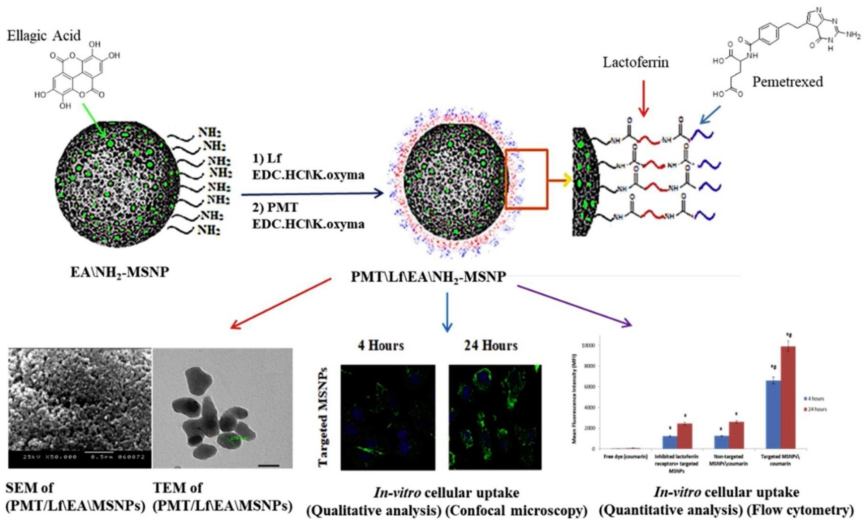
2.1.1. Silica Nanoparticles
2.1.2. Magnetic Nanoparticles
2.1.3. Other Inorganic Nanoparticles
2.2. Lipid-Based Nanoparticles Modified with Lf
2.2.1. Liposomes
2.2.2. Solid Lipid Nanoparticles and Nanostructured Lipid Carriers
2.3. Polymer-Based Nanoparticles Modified or Loaded with Lf
2.3.1. Natural Polymer-Based Nanoparticles
2.3.2. Synthetic Polymer-Based NPs

2.4. Drug–Lf Conjugates

| Material Base | Carriers | Anti-Cancer Dg | Results | Ref. |
|---|---|---|---|---|
| 1. Inorganic NPs | ||||
| 1.1. Silica NPs | Ultra-small size with large pore silica NPs (USLPs) | Doxorubicin (DOX) | Enhanced BBB permeation, enhanced internalization and apoptosis in U87 cells | [18] |
| Temozolomide (TMZ) | Improved BBB permeation and apoptosis | [19] | ||
| Mesoporous silica nanoparticles (MSNs) | Pemetrexed (PMT) and ellagic acid (EA) | Enhanced cellular uptake and cytotoxicity in MCF-7 breast cancer cells | [23] | |
| 1.2. Magnetic NPs | Maghemite and magnetite NPs | – | Enhanced cytotoxicity in 4T1 breast cancer cells | [27] |
| Mesoporous maghemite nanoparticles | DOX | Improved anti-cancer efficacy in vitro and in vivo | [28] | |
| Superparamagnetic iron oxide nanoparticles; graphene oxide (GO) sheets | DOX | Improved intracellular uptake and cytotoxicity to C6 glioma cells | [29] | |
| Superparamagnetic iron oxide (SPIO) nanoparticles; 10,12-pentacosadiynoic acid (PCDA) | Curcumin (CUR) | Enhanced CUR accumulation in brain and inhibited tumors in vivo | [30] | |
| Iron oxide nanoparticles, polyacrylic acid (PAA) and polyvinyl alcohol (PVA) | CUR and DOX | Enhanced cytotoxicity in RG2 glioma cells and reduced tumor growth | [31] | |
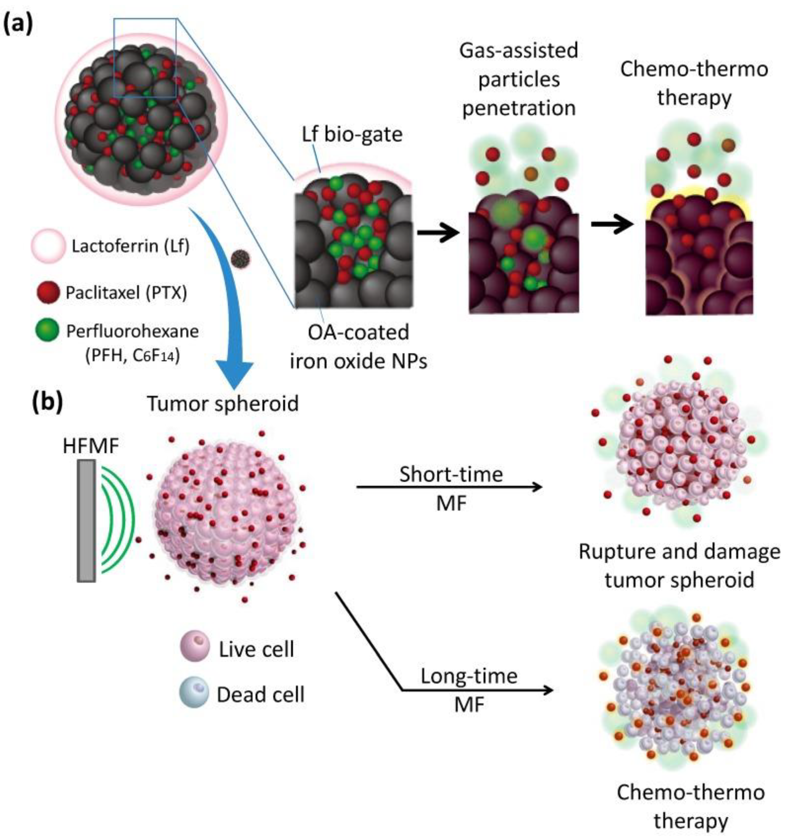
| Mesoporous iron oxide nanoparticles (MIONs) | Perfluorohexane (PFH) and paclitaxel (PTX) | Enhanced drug accumulation in tumor and and anti-cancer efficacy in vitro and in vivo | [32] | |
| YOF: Nd3+-MnO2 core–shell nanoparticles | Indocyanine green and glucose oxidase | Exhibited synergistic effect of starvation/photodynamic therapy to enhance anticancer efficacy in vitro | [36] | |
| Hollow mesoporous copper sulfide nanoparticles | TMZ and glucose oxidase | Exhibited synergistic chemo/chemodynamic/photothermal/starvation therapy to enhance anticancer efficacy in vitro and in vivo | [37] | |
| 2. Lipid-based NPs | ||||
| 2.1. Liposomes | Liposomes | DOX | Enhanced BBB permeability and cellular uptake | [41,42] |
| Liposomes coated with RGD peptide | Docetaxel (DTX) | Enhanced accumulation in gliomas and improved mice survival | [43] | |
| Liposome coated with muscone | DTX | Enhanced cellular internalization and anticancer efficacy in vitro and in vivo | [46] | |
| PEGylated liposomes | DOX | Enhanced uptake and cytotoxicity in ASGP-R positive cells; improved tumor inhibition in vivo | [49,50] | |
| PEGylated liposomes | DOX | Increased cellular uptake and tumor accumulation; enhanced tumor radiochemotherapy in vivo | [51] | |
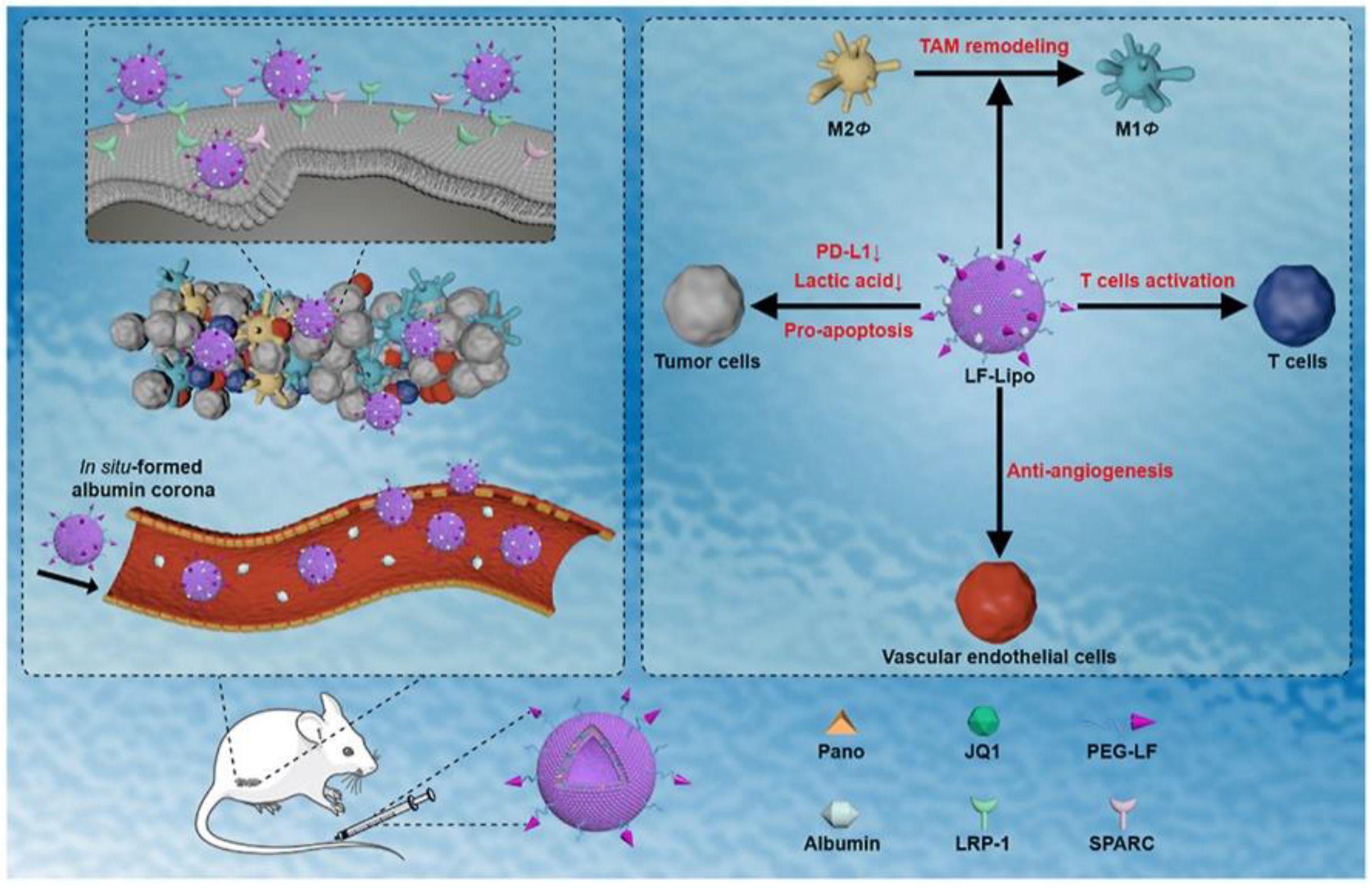
| Liposomes | Panobinostat and JQ1 | Activated anti-tumor immunity responses and inhibited tumor growth and metastasis | [54] | |
| 2.2. SLNs and NLCs | SLNs | Paclitacel (PTX) | Enhanced cytotoxicity in BEAS-2B human bronchial epithelial cells and drug accumulation in rats’ lungs | [59] |
| SLNs | DTX | Enhanced cellular uptake, cytotoxicity, and apoptosis in U87 MG cells | [60] | |
| SLNs | Tamoxifen and carmustine | Enhanced cellular uptake and cytotoxicity in U87 MG cells | [62] | |
| SLNs modified with wheat germ agglutinin (WGA) | Etoposide | Enhanced BBB permeation and antiproliferative activity in U87 MG cells | [63] | |
| NLCs modified with RGD peptide | TMZ | Enhanced cellular uptake and cytotoxicity in U87 MG cells; improved tumor inhibition in vivo | [64] | |
| 3. Polymer-based NPs | ||||
| 3.1. Natural polymer | Chitosan NPs | lactoperoxidase (LPO) | Improved cytotoxicity and apoptosis in HepG-2, Caco-2, PC-3, and MCF-7 cells | [68] |
| Sodium alginate | Pemetrexed, honokiol, and rosuvastatin | Enhanced cellular uptake into MCF-7 breast cancer cells and improved anticancer efficacy in vitro and in vivo | [69] | |
| Bovine serum albumin NPs modified with mPEG2000 | DOX | Enhanced cellular uptake and cytotoxicity in C6 glioma cells | [70] | |
| Mannopyranoside | Shikonin (SHK) and JQ1 | Enhanced accumulation in tumors and anticancer efficacy in vivo | [71] | |
| 3.2. Synthetic NPs | PEG–PLA NPs | PTX | Enhanced cellular uptake and cytotoxicity in C6 glioma cells | [76] |
| PLGA NPs | Etoposide | Enhanced BBB permeation and cytotoxicity in U87 MG cells | [77] | |
| PAEEP-PLLA NPs | Perfluoropentane | Enhanced cellular uptake in C6 glioma cells; exhibited strong, long-lasting, and tumor-enhanced ultrasonic contrast ability in vivo | [79] | |
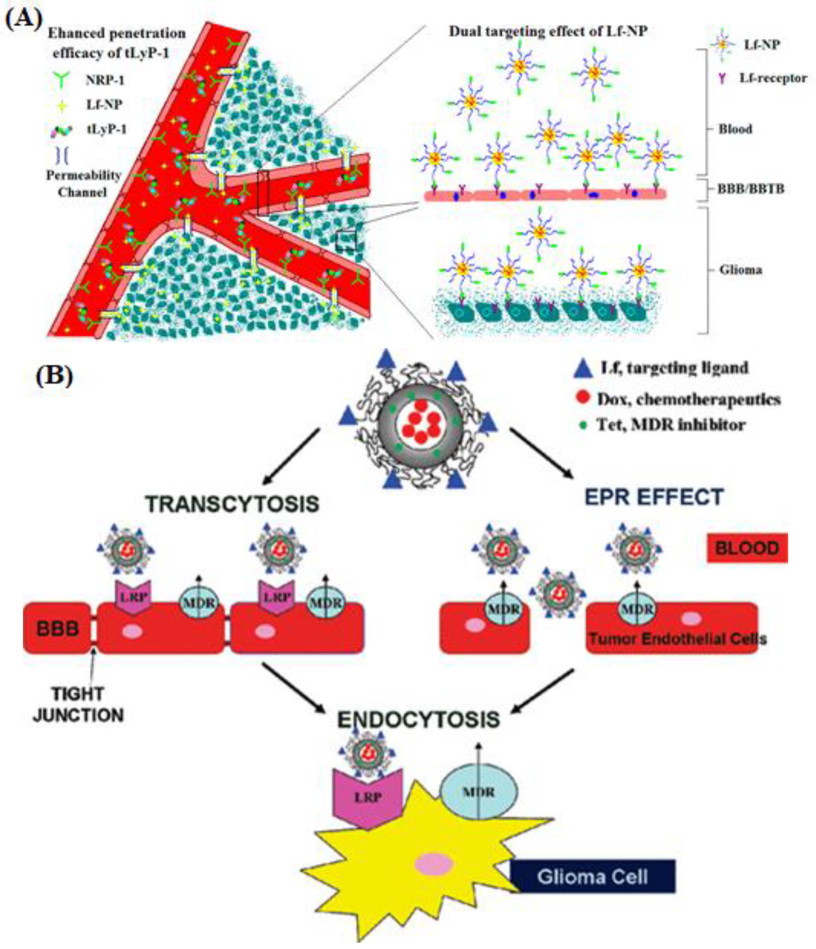
| Biodegradable polymersomes | DOX and tetrandrine (TET) | Enhanced cellular uptake and cytotoxicity in C6 glioma cells and improved anticancer efficacy in vivo | [81] | |
| Carboxymethyl cellulose | Pemetrexed and honokiol | Enhanced anticancer efficacy in vitro and in vivo | [82] | |
| 5.0G poly(propylene imine) (PPI) dendrimers | Methotrexate (MTX) | Enhanced drug accumulation in lungs | [85] | |
| Generation 3-diaminobutyric polypropylenimine dendrimers | Plasmid DNA encoding tumor necrosis factor alpha (TNFα) | Improved anticancer efficacy in vivo | [87] | |
| TNFα, tumor-necrosis factor related apoptosis-inducing ligand (TRAIL), and interleukine (IL)-12 genes | Improved anticancer efficacy in vivo with no side effects | [88] | ||
| 4. Drug-Lf Conjugates | – | CUR | Improved aqueous solubility, cellular uptake, and cytotoxicity in HCT116 human colon cancer cells | [90] |
| – | DOX | Enhanced uptake and cytotoxicity in DU145 prostate cancer cells; improved tumor inhibition with lower systemic toxicity in mice | [91] | |
| – | Celastrol and DTX | Suppressed tumor growth and prolonged survival in EAT-bearing mice | [92] | |
| – | MTX and exemestane | Enhanced cellular uptake and cytotoxicity in MCF-7 breast cancer cells | [93] | |
| – | Heparin | Improved circulation time and exhibited antiangiogenic activity in vitro and in vivo | [97] | |
3. Nanoparticles Carrying Lf as an Active Agent
| Material Base | Carriers | Results | Ref. |
|---|---|---|---|
| Inorganic NPs | Fe NPs | Enhanced cytotoxicity when combined with lactoperoxidase-loaded Cu NPs | [99] |
| Polymer-based NPs | Chitosan NPs coated with lactoperoxidase | Enhanced cytotoxicity in Caco-2, HepG-2, MCF-7, and PC-3 cells | [68] |
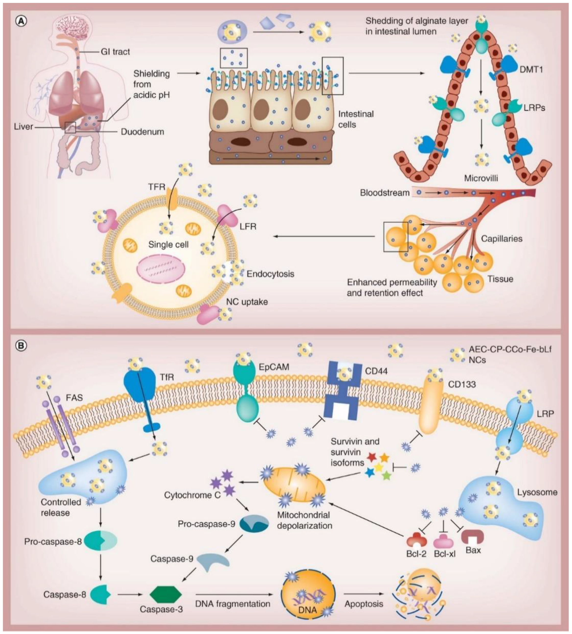
| Chitosan/alginate nanocapsules | Decreased viability of CaCo-2 cells and cancer stem cell markers in survivin, triple-positive CD133, and CD44 cancer stem-like cells; treated tumors developed in mice | [101] | |
| Chitosan NPs | The cytototoxicity depends on the locations targeted | [102] | |
| Complex | Bovine lactoferrin–oleic acid complex | Demonstrated anticancer effect in MCF-7, HepG2, and HT29 cells | [103] |
| Human lactoferrin–oleic acid complex | Enhanced tumor inhibition in vivo with higher safety | [104] |
4. Future Perspectives and Limitations
5. Conclusions
Author Contributions
Funding
Institutional Review Board Statement
Informed Consent Statement
Data Availability Statement
Conflicts of Interest
References
- Farnaud, S.; Evans, R.W. Lactoferrin—A multifunctional protein with antimicrobial properties. Mol. Immunol. 2003, 40, 395–405. [Google Scholar] [CrossRef]
- Kell, D.B.; Heyden, E.L.; Pretorius, E. The biology of lactoferrin, an iron-binding protein that can help defend against viruses and bacteria. Front. Immunol. 2020, 11, 1221. [Google Scholar] [CrossRef] [PubMed]
- Sienkiewicz, M.; Jaśkiewicz, A.; Tarasiuk, A.; Fichna, J. Lactoferrin: An overview of its main functions, immunomodulatory and antimicrobial role, and clinical significance. Crit. Rev. Food Sci. Nutr. 2022, 62, 6016–6033. [Google Scholar] [CrossRef] [PubMed]
- Adlerova, L.; Bartoskova, A.; Faldyna, M. Lactoferrin: A review. Vet. Med. 2008, 53, 457–468. [Google Scholar] [CrossRef]
- Zhang, Y.; Lima, C.F.; Rodrigues, L.R. Anticancer effects of lactoferrin: Underlying mechanisms and future trends in cancer therapy. Nutr. Rev. 2014, 72, 763–773. [Google Scholar] [CrossRef] [PubMed]
- Duarte, D.; Nicolau, A.; Teixeira, J.; Rodrigues, L. The effect of bovine milk lactoferrin on human breast cancer cell lines. J. Dairy Sci. 2011, 94, 66–76. [Google Scholar] [CrossRef]
- Eliassen, L.T.; Berge, G.; Leknessund, A.; Wikman, M.; Lindin, I.; Løkke, C.; Ponthan, F.; Johnsen, J.I.; Sveinbjørnsson, B.; Kogner, P. The antimicrobial peptide, lactoferricin B, is cytotoxic to neuroblastoma cells in vitro and inhibits xenograft growth in vivo. Int. J. Cancer 2006, 119, 493–500. [Google Scholar] [CrossRef]
- Lin, T.-Y.; Chiou, S.-H.; Chen, M.; Kuo, C.-D. Human lactoferrin exerts bi-directional actions on PC12 cell survival via ERK1/2 pathway. Biochem. Biophys. Res. Commun. 2005, 337, 330–336. [Google Scholar] [CrossRef]
- Sakai, T.; Banno, Y.; Kato, Y.; Nozawa, Y.; Kawaguchi, M. Pepsin-digested bovine lactoferrin induces apoptotic cell death with JNK/SAPK activation in oral cancer cells. J. Pharmacol. Sci. 2005, 98, 41–48. [Google Scholar] [CrossRef]
- Xu, X.; Jiang, H.; Li, H.; Zhang, T.; Zhou, Q.; Liu, N. Apoptosis of stomach cancer cell SGC-7901 and regulation of Akt signaling way induced by bovine lactoferrin. J. Dairy Sci. 2010, 93, 2344–2350. [Google Scholar] [CrossRef]
- Yamada, Y.; Sato, R.; Kobayashi, S.; Hankanga, C.; Inanami, O.; Kuwabara, M.; Momota, Y.; Tomizawa, N.; Yasuda, J. The antiproliferative effect of bovine lactoferrin on canine mammary gland tumor cells. J. Vet. Med. Sci. 2008, 70, 443–448. [Google Scholar] [CrossRef] [PubMed]
- Ando, K.; Hasegawa, K.; Shindo, K.I.; Furusawa, T.; Fujino, T.; Kikugawa, K.; Nakano, H.; Takeuchi, O.; Akira, S.; Akiyama, T. Human lactoferrin activates NF-κB through the Toll-like receptor 4 pathway while it interferes with the lipopolysaccharide-stimulated TLR4 signaling. FEBS J. 2010, 277, 2051–2066. [Google Scholar] [CrossRef] [PubMed]
- Kondapi, A.K. Targeting cancer with lactoferrin nanoparticles: Recent advances. Nanomedicine 2020, 15, 2071–2083. [Google Scholar] [CrossRef] [PubMed]
- Grodzinski, P.; Kircher, M.; Goldberg, M.; Gabizon, A. Integrating nanotechnology into cancer care. ACS Nano 2019, 13, 7370–7376. [Google Scholar] [CrossRef]
- Chatterjee, P.; Kumar, S. Current developments in nanotechnology for cancer treatment. Mater. Today Proc. 2022, 48, 1754–1758. [Google Scholar] [CrossRef]
- Schwartzbaum, J.A.; Fisher, J.L.; Aldape, K.D.; Wrensch, M. Epidemiology and molecular pathology of glioma. Nat. Clin. Pract. Neurol. 2006, 2, 494–503. [Google Scholar] [CrossRef]
- Zhang, F.; Xu, C.-L.; Liu, C.-M. Drug delivery strategies to enhance the permeability of the blood–brain barrier for treatment of glioma. Drug Des. Dev. Ther. 2015, 9, 2089. [Google Scholar] [CrossRef]
- Janjua, T.I.; Ahmed-Cox, A.; Meka, A.K.; Mansfeld, F.M.; Forgham, H.; Ignacio, R.M.C.; Cao, Y.; McCarroll, J.A.; Mazzieri, R.; Kavallaris, M. Facile synthesis of lactoferrin conjugated ultra small large pore silica nanoparticles for the treatment of glioblastoma. Nanoscale 2021, 13, 16909–16922. [Google Scholar] [CrossRef]
- Janjua, T.I.; Cao, Y.; Ahmed-Cox, A.; Raza, A.; Moniruzzaman, M.; Akhter, D.T.; Fletcher, N.L.; Kavallaris, M.; Thurecht, K.J.; Popat, A. Efficient delivery of Temozolomide using ultrasmall large-pore silica nanoparticles for glioblastoma. J. Control. Release 2023, 357, 161–174. [Google Scholar] [CrossRef]
- Wu, S.-H.; Hung, Y.; Mou, C.-Y. Mesoporous silica nanoparticles as nanocarriers. Chem. Commun. 2011, 47, 9972–9985. [Google Scholar] [CrossRef]
- Wang, Y.; Zhao, Q.; Han, N.; Bai, L.; Li, J.; Liu, J.; Che, E.; Hu, L.; Zhang, Q.; Jiang, T. Mesoporous silica nanoparticles in drug delivery and biomedical applications. Nanomed. Nanotechnol. Biol. Med. 2015, 11, 313–327. [Google Scholar] [CrossRef]
- Rosenholm, J.M.; Sahlgren, C.; Lindén, M. Towards multifunctional, targeted drug delivery systems using mesoporous silica nanoparticles–opportunities & challenges. Nanoscale 2010, 2, 1870–1883. [Google Scholar] [PubMed]
- Ali, O.M.; Bekhit, A.A.; Khattab, S.N.; Helmy, M.W.; Abdel-Ghany, Y.S.; Teleb, M.; Elzoghby, A.O. Synthesis of lactoferrin mesoporous silica nanoparticles for pemetrexed/ellagic acid synergistic breast cancer therapy. Colloids Surf. B Biointerfaces 2020, 188, 110824. [Google Scholar] [CrossRef]
- Shubayev, V.I.; Pisanic, T.R., II; Jin, S. Magnetic nanoparticles for theragnostics. Adv. Drug Deliv. Rev. 2009, 61, 467–477. [Google Scholar] [CrossRef] [PubMed]
- Mornet, S.; Vasseur, S.; Grasset, F.; Duguet, E. Magnetic nanoparticle design for medical diagnosis and therapy. J. Mater. Chem. 2004, 14, 2161–2175. [Google Scholar] [CrossRef]
- Pantic, I. Magnetic nanoparticles in cancer diagnosis and treatment: Novel approaches. Rev. Adv. Mater. Sci. 2010, 26, 67–73. [Google Scholar]
- Sharifi, M.; Rezayat, S.M.; Akhtari, K.; Hasan, A.; Falahati, M. Fabrication and evaluation of anti-cancer efficacy of lactoferrin-coated maghemite and magnetite nanoparticles. J. Biomol. Struct. Dyn. 2020, 38, 2945–2954. [Google Scholar] [CrossRef] [PubMed]
- Sharifi, M.; Jafari, S.; Hasan, A.; Paray, B.A.; Gong, G.; Zheng, Y.; Falahati, M. Antimetastatic activity of lactoferrin-coated mesoporous maghemite nanoparticles in breast cancer enabled by combination therapy. ACS Biomater. Sci. Eng. 2020, 6, 3574–3584. [Google Scholar] [CrossRef]
- Song, M.-M.; Xu, H.-L.; Liang, J.-X.; Xiang, H.-H.; Liu, R.; Shen, Y.-X. Lactoferrin modified graphene oxide iron oxide nanocomposite for glioma-targeted drug delivery. Mater. Sci. Eng. C 2017, 77, 904–911. [Google Scholar] [CrossRef]
- Fang, J.H.; Chiu, T.L.; Huang, W.C.; Lai, Y.H.; Hu, S.H.; Chen, Y.Y.; Chen, S.Y. Dual-Targeting Lactoferrin-Conjugated Polymerized Magnetic Polydiacetylene-Assembled Nanocarriers with Self-Responsive Fluorescence/Magnetic Resonance Imaging for In Vivo Brain Tumor Therapy. Adv. Healthc. Mater. 2016, 5, 688–695. [Google Scholar] [CrossRef]
- Fang, J.H.; Lai, Y.H.; Chiu, T.L.; Chen, Y.Y.; Hu, S.H.; Chen, S.Y. Magnetic Core–Shell Nanocapsules with Dual-Targeting Capabilities and Co-Delivery of Multiple Drugs to Treat Brain Gliomas. Adv. Healthc. Mater. 2014, 3, 1250–1260. [Google Scholar] [CrossRef] [PubMed]
- Su, Y.-L.; Fang, J.-H.; Liao, C.-Y.; Lin, C.-T.; Li, Y.-T.; Hu, S.-H. Targeted mesoporous iron oxide nanoparticles-encapsulated perfluorohexane and a hydrophobic drug for deep tumor penetration and therapy. Theranostics 2015, 5, 1233–1248. [Google Scholar] [CrossRef] [PubMed]
- Kan, L.K.; Drummond, K.; Hunn, M.; Williams, D.; O’Brien, T.J.; Monif, M. Potential biomarkers and challenges in glioma diagnosis, therapy and prognosis. BMJ Neurol. Open 2020, 2, e000069. [Google Scholar] [CrossRef] [PubMed]
- Pucelik, B.; Sułek, A.; Barzowska, A.; Dąbrowski, J.M. Recent advances in strategies for overcoming hypoxia in photodynamic therapy of cancer. Cancer Lett. 2020, 492, 116–135. [Google Scholar] [CrossRef] [PubMed]
- Jena, L.; McErlean, E.; McCarthy, H. Delivery across the blood-brain barrier: Nanomedicine for glioblastoma multiforme. Drug Deliv. Transl. Res. 2020, 10, 304–318. [Google Scholar] [CrossRef] [PubMed]
- Lv, Z.; Jin, L.; Gao, W.; Cao, Y.; Zhang, H.; Xue, D.; Yin, N.; Zhang, T.; Wang, Y.; Zhang, H. Novel YOF-based theranostic agents with a cascade effect for NIR-II fluorescence imaging and synergistic starvation/photodynamic therapy of orthotopic gliomas. ACS Appl. Mater. Interfaces 2022, 14, 30523–30532. [Google Scholar] [CrossRef] [PubMed]
- Cao, Y.; Jin, L.; Zhang, S.; Lv, Z.; Yin, N.; Zhang, H.; Zhang, T.; Wang, Y.; Chen, Y.; Liu, X. Blood-brain Barrier Permeable and Multi-stimuli Responsive Nanoplatform for Orthotopic Glioma Inhibition by Synergistic Enhanced Chemo-/Chemodynamic/Photothermal/Starvation Therapy. Eur. J. Pharm. Sci. 2023, 180, 106319. [Google Scholar] [CrossRef]
- Allen, T.M.; Cullis, P.R. Liposomal drug delivery systems: From concept to clinical applications. Adv. Drug Deliv. Rev. 2013, 65, 36–48. [Google Scholar] [CrossRef]
- Saraf, S.; Jain, A.; Tiwari, A.; Verma, A.; Panda, P.K.; Jain, S.K. Advances in liposomal drug delivery to cancer: An overview. J. Drug Deliv. Sci. Technol. 2020, 56, 101549. [Google Scholar] [CrossRef]
- Guimarães, D.; Cavaco-Paulo, A.; Nogueira, E. Design of liposomes as drug delivery system for therapeutic applications. Int. J. Pharm. 2021, 601, 120571. [Google Scholar] [CrossRef]
- Chen, H.; Qin, Y.; Zhang, Q.; Jiang, W.; Tang, L.; Liu, J.; He, Q. Lactoferrin modified doxorubicin-loaded procationic liposomes for the treatment of gliomas. Eur. J. Pharm. Sci. 2011, 44, 164–173. [Google Scholar] [CrossRef] [PubMed]
- Chen, H.; Tang, L.; Qin, Y.; Yin, Y.; Tang, J.; Tang, W.; Sun, X.; Zhang, Z.; Liu, J.; He, Q. Lactoferrin-modified procationic liposomes as a novel drug carrier for brain delivery. Eur. J. Pharm. Sci. 2010, 40, 94–102. [Google Scholar] [CrossRef]
- Qi, N.; Zhang, S.; Zhou, X.; Duan, W.; Gao, D.; Feng, J.; Li, A. Combined integrin αvβ3 and lactoferrin receptor targeted docetaxel liposomes enhance the brain targeting effect and anti-glioma effect. J. Nanobiotechnology 2021, 19, 446. [Google Scholar] [CrossRef] [PubMed]
- Wang, G.-Y.; Wang, N.; Liao, H.-N. Effects of muscone on the expression of P-gp, MMP-9 on blood–brain barrier model in vitro. Cell. Mol. Neurobiol. 2015, 35, 1105–1115. [Google Scholar] [CrossRef] [PubMed]
- Chen, Z.-Z.; Lu, Y.; Du, S.-Y.; Shang, K.-X.; Cai, C.-B. Influence of borneol and muscone on geniposide transport through MDCK and MDCK-MDR1 cells as blood–brain barrier in vitro model. Int. J. Pharm. 2013, 456, 73–79. [Google Scholar] [CrossRef] [PubMed]
- Qi, N.; Duan, W.; Gao, D.; Ma, N.; Zhang, J.; Feng, J.; Li, A. “Guide” of muscone modification enhanced brain-targeting efficacy and anti-glioma effect of lactoferrin modified DTX liposomes. Bioeng. Transl. Med. 2023, 8, e10393. [Google Scholar] [CrossRef] [PubMed]
- Poelstra, K.; Prakash, J.; Beljaars, L. Drug targeting to the diseased liver. J. Control. Release 2012, 161, 188–197. [Google Scholar] [CrossRef]
- D’souza, A.A.; Devarajan, P.V. Asialoglycoprotein receptor mediated hepatocyte targeting—Strategies and applications. J. Control. Release 2015, 203, 126–139. [Google Scholar] [CrossRef] [PubMed]
- Wei, M.; Xu, Y.; Zou, Q.; Tu, L.; Tang, C.; Xu, T.; Deng, L.; Wu, C. Hepatocellular carcinoma targeting effect of PEGylated liposomes modified with lactoferrin. Eur. J. Pharm. Sci. 2012, 46, 131–141. [Google Scholar] [CrossRef]
- Wei, M.; Guo, X.; Tu, L.; Zou, Q.; Li, Q.; Tang, C.; Chen, B.; Xu, Y.; Wu, C. Lactoferrin-modified PEGylated liposomes loaded with doxorubicin for targeting delivery to hepatocellular carcinoma. Int. J. Nanomed. 2015, 10, 5123. [Google Scholar]
- Zhang, Z.; Yang, J.; Min, Q.; Ling, C.; Maiti, D.; Xu, J.; Qin, L.; Yang, K. Holo-Lactoferrin Modified Liposome for Relieving Tumor Hypoxia and Enhancing Radiochemotherapy of Cancer. Small 2019, 15, 1803703. [Google Scholar] [CrossRef] [PubMed]
- Chisolm, D.A.; Weinmann, A.S. Connections between metabolism and epigenetics in programming cellular differentiation. Annu. Rev. Immunol. 2018, 36, 221–246. [Google Scholar] [CrossRef]
- Miranda-Gonçalves, V.; Lameirinhas, A.; Henrique, R.; Jerónimo, C. Metabolism and epigenetic interplay in cancer: Regulation and putative therapeutic targets. Front. Genet. 2018, 9, 427. [Google Scholar] [CrossRef] [PubMed]
- He, Y.; Fang, Y.; Zhang, M.; Zhao, Y.; Tu, B.; Shi, M.; Muhitdinov, B.; Asrorov, A.; Xu, Q.; Huang, Y. Remodeling “cold” tumor immune microenvironment via epigenetic-based therapy using targeted liposomes with in situ formed albumin corona. Acta Pharm. Sin. B 2022, 12, 2057–2073. [Google Scholar] [CrossRef] [PubMed]
- Lindemann, R.K.; Gabrielli, B.; Johnstone, R.W. Histone-deacetylase inhibitors for the treatment of cancer. Cell Cycle 2004, 3, 777–786. [Google Scholar] [CrossRef]
- Liu, K.; Zhou, Z.; Gao, H.; Yang, F.; Qian, Y.; Jin, H.; Guo, Y.; Liu, Y.; Li, H.; Zhang, C. JQ1, a BET-bromodomain inhibitor, inhibits human cancer growth and suppresses PD-L1 expression. Cell Biol. Int. 2019, 43, 642–650. [Google Scholar] [CrossRef]
- Akbari, J.; Saeedi, M.; Ahmadi, F.; Hashemi, S.M.H.; Babaei, A.; Yaddollahi, S.; Rostamkalaei, S.S.; Asare-Addo, K.; Nokhodchi, A. Solid lipid nanoparticles and nanostructured lipid carriers: A review of the methods of manufacture and routes of administration. Pharm. Dev. Technol. 2022, 27, 525–544. [Google Scholar] [CrossRef] [PubMed]
- Ghasemiyeh, P.; Mohammadi-Samani, S. Solid lipid nanoparticles and nanostructured lipid carriers as novel drug delivery systems: Applications, advantages and disadvantages. Res. Pharm. Sci. 2018, 13, 288. [Google Scholar]
- Pandey, V.; Gajbhiye, K.R.; Soni, V. Lactoferrin-appended solid lipid nanoparticles of paclitaxel for effective management of bronchogenic carcinoma. Drug Deliv. 2015, 22, 199–205. [Google Scholar] [CrossRef]
- Singh, I.; Swami, R.; Pooja, D.; Jeengar, M.K.; Khan, W.; Sistla, R. Lactoferrin bioconjugated solid lipid nanoparticles: A new drug delivery system for potential brain targeting. J. Drug Target. 2016, 24, 212–223. [Google Scholar] [CrossRef]
- Kayyali, R.; Marriott, C.; Wiseman, H. Tamoxifen decreases drug efflux from liposomes: Relevance to its ability to reverse multidrug resistance in cancer cells? FEBS Lett. 1994, 344, 221–224. [Google Scholar] [CrossRef]
- Kuo, Y.-C.; Cheng, S.-J. Brain targeted delivery of carmustine using solid lipid nanoparticles modified with tamoxifen and lactoferrin for antitumor proliferation. Int. J. Pharm. 2016, 499, 10–19. [Google Scholar] [CrossRef] [PubMed]
- Kuo, Y.-C.; Wang, I.-H. Using catanionic solid lipid nanoparticles with wheat germ agglutinin and lactoferrin for targeted delivery of etoposide to glioblastoma multiforme. J. Taiwan Inst. Chem. Eng. 2017, 77, 73–82. [Google Scholar] [CrossRef]
- Zhang, J.; Xiao, X.; Zhu, J.; Gao, Z.; Lai, X.; Zhu, X.; Mao, G. Lactoferrin-and RGD-comodified, temozolomide and vincristine-coloaded nanostructured lipid carriers for gliomatosis cerebri combination therapy. Int. J. Nanomed. 2018, 13, 3039. [Google Scholar] [CrossRef] [PubMed]
- Nagpal, K.; Singh, S.K.; Mishra, D.N. Chitosan nanoparticles: A promising system in novel drug delivery. Chem. Pharm. Bull. 2010, 58, 1423–1430. [Google Scholar] [CrossRef] [PubMed]
- Garg, U.; Chauhan, S.; Nagaich, U.; Jain, N. Current advances in chitosan nanoparticles based drug delivery and targeting. Adv. Pharm. Bull. 2019, 9, 195. [Google Scholar] [CrossRef]
- Wang, J.J.; Zeng, Z.W.; Xiao, R.Z.; Xie, T.; Zhou, G.L.; Zhan, X.R.; Wang, S.L. Recent advances of chitosan nanoparticles as drug carriers. Int. J. Nanomed. 2011, 6, 765–774. [Google Scholar]
- Abu-Serie, M.M.; El-Fakharany, E.M. Efficiency of novel nanocombinations of bovine milk proteins (lactoperoxidase and lactoferrin) for combating different human cancer cell lines. Sci. Rep. 2017, 7, 16769. [Google Scholar] [CrossRef]
- Salah, M.; Sallam, M.A.; Abdelmoneem, M.A.; Teleb, M.; Elkhodairy, K.A.; Bekhit, A.A.; Khafaga, A.F.; Noreldin, A.E.; Elzoghby, A.O.; Khattab, S.N. Sequential Delivery of Novel Triple Drug Combination via Crosslinked Alginate/Lactoferrin Nanohybrids for Enhanced Breast Cancer Treatment. Pharmaceutics 2022, 14, 2404. [Google Scholar] [CrossRef]
- Su, Z.; Xing, L.; Chen, Y.; Xu, Y.; Yang, F.; Zhang, C.; Ping, Q.; Xiao, Y. Lactoferrin-modified poly (ethylene glycol)-grafted BSA nanoparticles as a dual-targeting carrier for treating brain gliomas. Mol. Pharm. 2014, 11, 1823–1834. [Google Scholar] [CrossRef]
- Wang, H.; Tang, Y.; Fang, Y.; Zhang, M.; Wang, H.; He, Z.; Wang, B.; Xu, Q.; Huang, Y. Reprogramming tumor immune microenvironment (TIME) and metabolism via biomimetic targeting codelivery of shikonin/JQ1. Nano Lett. 2019, 19, 2935–2944. [Google Scholar] [CrossRef] [PubMed]
- Yin, S.-Y.; Efferth, T.; Jian, F.-Y.; Chen, Y.-H.; Liu, C.-I.; Wang, A.H.; Chen, Y.-R.; Hsiao, P.-W.; Yang, N.-S. Immunogenicity of mammary tumor cells can be induced by shikonin via direct binding-interference with hnRNPA1. Oncotarget 2016, 7, 43629. [Google Scholar] [CrossRef] [PubMed]
- Andújar, I.; Ríos, J.L.; Giner, R.M.; Recio, M.C. Pharmacological properties of shikonin–a review of literature since 2002. Planta Med. 2013, 79, 1685–1697. [Google Scholar] [CrossRef] [PubMed]
- Vališ, K.; Talacko, P.; Grobárová, V.; Černý, J.; Novák, P. Shikonin regulates C-MYC and GLUT1 expression through the MST1-YAP1-TEAD1 axis. Exp. Cell Res. 2016, 349, 273–281. [Google Scholar] [CrossRef] [PubMed]
- Otto, C.; Schmidt, S.; Kastner, C.; Denk, S.; Kettler, J.; Müller, N.; Germer, C.; Wolf, E.; Gallant, P.; Wiegering, A. Targeting bromodomain-containing protein 4 (BRD4) inhibits MYC expression in colorectal cancer cells. Neoplasia 2019, 21, 1110–1120. [Google Scholar] [CrossRef]
- Miao, D.; Jiang, M.; Liu, Z.; Gu, G.; Hu, Q.; Kang, T.; Song, Q.; Yao, L.; Li, W.; Gao, X. Co-administration of dual-targeting nanoparticles with penetration enhancement peptide for antiglioblastoma therapy. Mol. Pharm. 2014, 11, 90–101. [Google Scholar] [CrossRef]
- Kuo, Y.-C.; Chen, Y.-C. Targeting delivery of etoposide to inhibit the growth of human glioblastoma multiforme using lactoferrin-and folic acid-grafted poly (lactide-co-glycolide) nanoparticles. Int. J. Pharm. 2015, 479, 138–149. [Google Scholar] [CrossRef]
- Guo, J.; Schlich, M.; Cryan, J.F.; O’Driscoll, C.M. Targeted drug delivery via folate receptors for the treatment of brain cancer: Can the promise deliver? J. Pharm. Sci. 2017, 106, 3413–3420. [Google Scholar] [CrossRef]
- Luo, B.; Liang, H.; Zhang, S.; Qin, X.; Liu, X.; Liu, W.; Zeng, F.; Wu, Y.; Yang, X. Novel lactoferrin-conjugated amphiphilic poly (aminoethyl ethylene phosphate)/poly (L-lactide) copolymer nanobubbles for tumor-targeting ultrasonic imaging. Int. J. Nanomed. 2015, 10, 5805. [Google Scholar]
- Arvanitis, C.D.; Ferraro, G.B.; Jain, R.K. The blood–brain barrier and blood–tumour barrier in brain tumours and metastases. Nat. Rev. Cancer 2020, 20, 26–41. [Google Scholar] [CrossRef]
- Pang, Z.; Feng, L.; Hua, R.; Chen, J.; Gao, H.; Pan, S.; Jiang, X.; Zhang, P. Lactoferrin-conjugated biodegradable polymersome holding doxorubicin and tetrandrine for chemotherapy of glioma rats. Mol. Pharm. 2010, 7, 1995–2005. [Google Scholar] [CrossRef] [PubMed]
- Atallah, M.A.; Sallam, M.A.; Abdelmoneem, M.A.; Teleb, M.; Elkhodairy, K.A.; Bekhit, A.A.; Khafaga, A.F.; Noreldin, A.E.; Elzoghby, A.O.; Khattab, S.N. Green self-assembled lactoferrin carboxymethyl cellulose nanogels for synergistic chemo/herbal breast cancer therapy. Colloids Surf. B Biointerfaces 2022, 217, 112657. [Google Scholar] [CrossRef] [PubMed]
- Kesharwani, P.; Jain, K.; Jain, N.K. Dendrimer as nanocarrier for drug delivery. Prog. Polym. Sci. 2014, 39, 268–307. [Google Scholar] [CrossRef]
- Hsu, H.J.; Bugno, J.; Lee, S.r.; Hong, S. Dendrimer-based nanocarriers: A versatile platform for drug delivery. Wiley Interdiscip. Rev. Nanomed. Nanobiotechnology 2017, 9, e1409. [Google Scholar] [CrossRef] [PubMed]
- Kurmi, B.D.; Gajbhiye, V.; Kayat, J.; Jain, N.K. Lactoferrin-conjugated dendritic nanoconstructs for lung targeting of methotrexate. J. Pharm. Sci. 2011, 100, 2311–2320. [Google Scholar] [CrossRef]
- Luo, D.; Saltzman, W.M. Synthetic DNA delivery systems. Nat. Biotechnol. 2000, 18, 33–37. [Google Scholar] [CrossRef]
- Koppu, S.; Oh, Y.J.; Edrada-Ebel, R.; Blatchford, D.R.; Tetley, L.; Tate, R.J.; Dufès, C. Tumor regression after systemic administration of a novel tumor-targeted gene delivery system carrying a therapeutic plasmid DNA. J. Control. Release 2010, 143, 215–221. [Google Scholar] [CrossRef]
- Altwaijry, N.; Somani, S.; Parkinson, J.A.; Tate, R.J.; Keating, P.; Warzecha, M.; Mackenzie, G.R.; Leung, H.Y.; Dufès, C. Regression of prostate tumors after intravenous administration of lactoferrin-bearing polypropylenimine dendriplexes encoding TNF-α, TRAIL, and interleukin-12. Drug Deliv. 2018, 25, 679–689. [Google Scholar] [CrossRef]
- Kanyshkova, T.; Buneva, V.; Nevinsky, G. Lactoferrin and its biological functions. Biochemistry 2001, 66, 1–7. [Google Scholar]
- Chaharband, F.; Kamalinia, G.; Atyabi, F.; Mortazavi, S.A.; Mirzaie, Z.H.; Dinarvand, R. Formulation and in vitro evaluation of curcumin-lactoferrin conjugated nanostructures for cancerous cells. Artif. Cells Nanomed. Biotechnol. 2018, 46, 626–636. [Google Scholar] [CrossRef]
- Shankaranarayanan, J.S.; Kanwar, J.R.; Al-Juhaishi, A.J.A.; Kanwar, R.K. Doxorubicin conjugated to immunomodulatory anticancer lactoferrin displays improved cytotoxicity overcoming prostate cancer chemo resistance and inhibits tumour development in TRAMP mice. Sci. Rep. 2016, 6, 32062. [Google Scholar] [CrossRef] [PubMed]
- Abdelmoneem, M.A.; Abd Elwakil, M.M.; Khattab, S.N.; Helmy, M.W.; Bekhit, A.A.; Abdulkader, M.A.; Zaky, A.; Teleb, M.; Elkhodairy, K.A.; Albericio, F. Lactoferrin-dual drug nanoconjugate: Synergistic anti-tumor efficacy of docetaxel and the NF-κB inhibitor celastrol. Mater. Sci. Eng. C 2021, 118, 111422. [Google Scholar] [CrossRef] [PubMed]
- Mokhtar, S.; Khattab, S.N.; Elkhodairy, K.A.; Teleb, M.; Bekhit, A.A.; Elzoghby, A.O.; Sallam, M.A. Methotrexate-lactoferrin targeted exemestane cubosomes for synergistic breast cancer therapy. Front. Chem. 2022, 10, 847573. [Google Scholar] [CrossRef] [PubMed]
- Bick, R.L.; Frenkel, E.P. Clinical aspects of heparin-induced thrombocytopenia and thrombosis and other side effects of heparin therapy. Clin. Appl. Thromb. /Hemost. 1999, 5, S7–S15. [Google Scholar] [CrossRef] [PubMed]
- Walenga, J.M.; Bick, R.L. Heparin-induced thrombocytopenia, paradoxical thromboembolism, and other side effects of heparin therapy. Med. Clin. N. Am. 1998, 82, 635–658. [Google Scholar] [CrossRef]
- Alban, S. Adverse effects of heparin. In Heparin—A Century of Progress; Springer: Berlin/Heidelberg, Germany, 2012; pp. 211–263. [Google Scholar]
- Hwang, H.H.; Kim, H.S.; Lee, D.Y. Gastrointestinally absorbable lactoferrin-heparin conjugate with anti-angiogenic activity for treatment of brain tumor. J. Control. Release 2023, 355, 730–744. [Google Scholar] [CrossRef]
- Rodrigues, L.; Teixeira, J.; Schmitt, F.; Paulsson, M.; Månsson, H.L. Lactoferrin and cancer disease prevention. Crit. Rev. Food Sci. Nutr. 2008, 49, 203–217. [Google Scholar] [CrossRef]
- El-Fakharany, E.M.; Abu-Serie, M.M.; Habashy, N.H.; Eltarahony, M. Augmenting apoptosis-mediated anticancer activity of lactoperoxidase and lactoferrin by nanocombination with copper and iron hybrid nanometals. Sci. Rep. 2022, 12, 13153. [Google Scholar] [CrossRef]
- Sah, N.; Khan, Z.; Khan, G.; Bisen, P. Structural, functional and therapeutic biology of survivin. Cancer Lett. 2006, 244, 164–171. [Google Scholar] [CrossRef]
- Kanwar, J.R.; Mahidhara, G.; Roy, K.; Sasidharan, S.; Krishnakumar, S.; Prasad, N.; Sehgal, R.; Kanwar, R.K. Fe-bLf nanoformulation targets survivin to kill colon cancer stem cells and maintains absorption of iron, calcium and zinc. Nanomedicine 2015, 10, 35–55. [Google Scholar] [CrossRef]
- Tammam, S.N.; Azzazy, H.M.; Lamprecht, A. Nuclear and cytoplasmic delivery of lactoferrin in glioma using chitosan nanoparticles: Cellular location dependent-action of lactoferrin. Eur. J. Pharm. Biopharm. 2018, 129, 74–79. [Google Scholar] [CrossRef] [PubMed]
- Fang, B.; Zhang, M.; Tian, M.; Jiang, L.; Guo, H.Y.; Ren, F.Z. Bovine lactoferrin binds oleic acid to form an anti-tumor complex similar to HAMLET. Biochim. Et Biophys. Acta (BBA)-Mol. Cell Biol. Lipids 2014, 1841, 535–543. [Google Scholar] [CrossRef] [PubMed]
- Elizarova, A.; Sokolov, A.; Kostevich, V.; Kisseleva, E.; Zelenskiy, E.; Zakharova, E.; Panasenko, O.; Budevich, A.; Semak, I.; Egorov, V. Interaction of lactoferrin with unsaturated fatty acids: In vitro and in vivo study of human lactoferrin/oleic acid complex cytotoxicity. Materials 2021, 14, 1602. [Google Scholar] [CrossRef] [PubMed]
- Dao, H.M.; Sahakijpijarn, S.; Chrostowski, R.R.; Moon, C.; Mangolini, F.; Cui, Z.; Williams III, R.O. Aggregation of Lactoferrin Caused by Droplet Atomization Process via a Two-Fluid Nozzle: The Detrimental Effect of Air–Water Interfaces. Mol. Pharm. 2022, 19, 2662–2675. [Google Scholar] [CrossRef]
- Golshahi, L.; Lynch, K.; Dennis, J.; Finlay, W. In vitro lung delivery of bacteriophages KS4-M and ΦKZ using dry powder inhalers for treatment of Burkholderia cepacia complex and Pseudomonas aeruginosa infections in cystic fibrosis. J. Appl. Microbiol. 2011, 110, 106–117. [Google Scholar] [CrossRef]
- Chen, H.-L.; Yen, C.-C.; Wang, S.-M.; Tsai, T.-C.; Lai, Z.-L.; Sun, J.-Y.; Lin, W.; Hsu, W.-H.; Chen, C.-M. Aerosolized bovine lactoferrin reduces lung injury and fibrosis in mice exposed to hyperoxia. Biometals 2014, 27, 1057–1068. [Google Scholar] [CrossRef]
- Marshall, L.J.; Oguejiofor, W.; Price, R.; Shur, J. Investigation of the enhanced antimicrobial activity of combination dry powder inhaler formulations of lactoferrin. Int. J. Pharm. 2016, 514, 399–406. [Google Scholar] [CrossRef]


Disclaimer/Publisher’s Note: The statements, opinions and data contained in all publications are solely those of the individual author(s) and contributor(s) and not of MDPI and/or the editor(s). MDPI and/or the editor(s) disclaim responsibility for any injury to people or property resulting from any ideas, methods, instructions or products referred to in the content. |
© 2023 by the authors. Licensee MDPI, Basel, Switzerland. This article is an open access article distributed under the terms and conditions of the Creative Commons Attribution (CC BY) license (https://creativecommons.org/licenses/by/4.0/).
Share and Cite
Tran, T.H.; Tran, P.T.T.; Truong, D.H. Lactoferrin and Nanotechnology: The Potential for Cancer Treatment. Pharmaceutics 2023, 15, 1362. https://doi.org/10.3390/pharmaceutics15051362
Tran TH, Tran PTT, Truong DH. Lactoferrin and Nanotechnology: The Potential for Cancer Treatment. Pharmaceutics. 2023; 15(5):1362. https://doi.org/10.3390/pharmaceutics15051362
Chicago/Turabian StyleTran, Tuan Hiep, Phuong Thi Thu Tran, and Duy Hieu Truong. 2023. "Lactoferrin and Nanotechnology: The Potential for Cancer Treatment" Pharmaceutics 15, no. 5: 1362. https://doi.org/10.3390/pharmaceutics15051362