
Electrospun Nanofibers for Dura Mater Regeneration: A Mini Review on Current Progress


Abstract
:1. Electrospinning: History, Setup, and Principle
- (i)
- Electrospun nanofibers have a diameter ranging from nano to micro scale.
- (ii)
- Electrospun nanofibers possess high porosity.
- (iii)
- The fibers produced by the electrospinning technique have a large aspect ratio and a high surface-to-volume ratio.
- (iv)
- Electrospun nanofibers possess superior mechanical properties and flexibility.
- (v)
- The electrospinning process enables the production of nano/microfibers with an infinite number of chemical compositions.
- (vi)
- Various types of morphology can be prepared by modifying the spinneret.
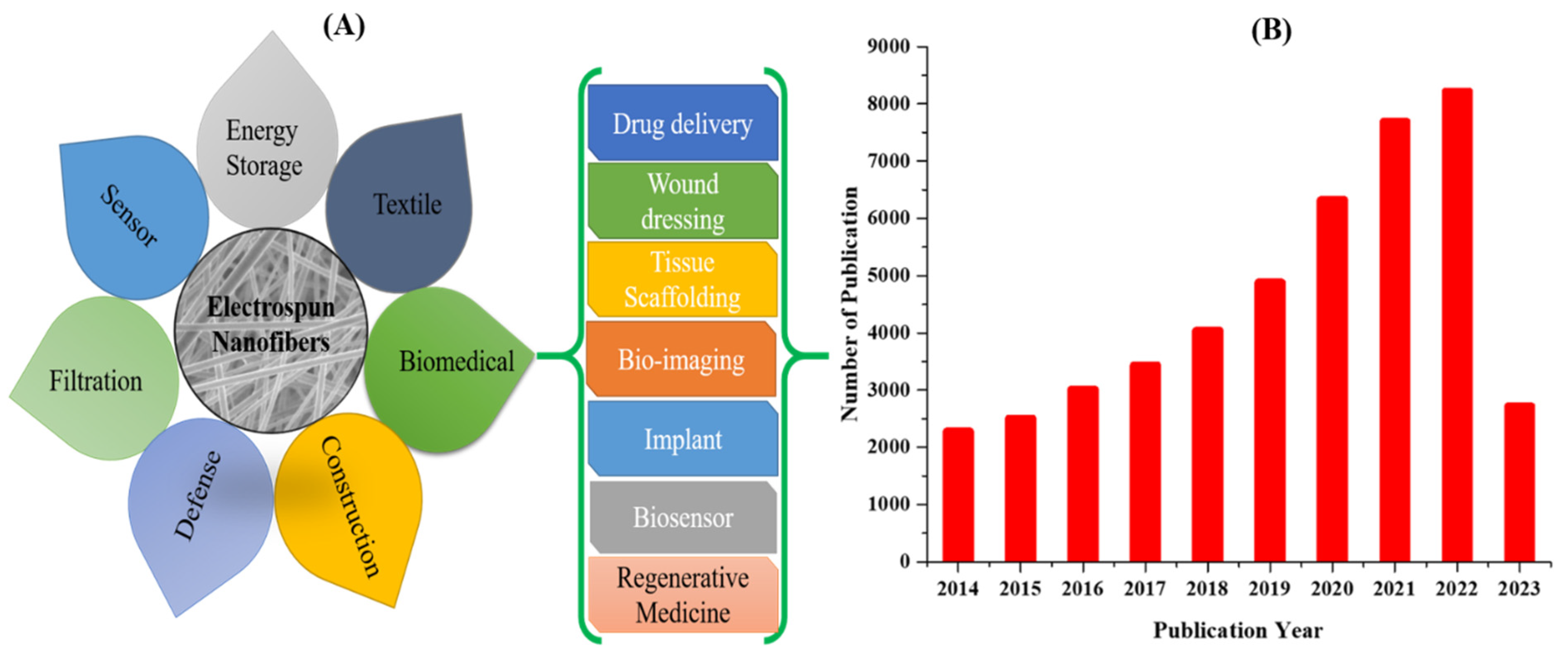
2. Biomedical Applications of Electrospun Nanofibers
Electrospun Nanofibers for Dura Mater Repair
- -
- It must be biocompatible.
- -
- It should mimic the physiochemical structure of the dura mater.
- -
- It should adequately restore the continuity of the dura mater and prevent the leakage of CSF.
- -
- It should lower the chances of infection.
- -
- The mechanical properties of the substrate should facilitate the suturing.
- -
- It should enhance tissue regeneration.
- -
- It should promote the adhesion and migration of dural fibroblasts.
- -
- It should minimize local tissue inflammation.
- -
- It should not induce adverse reactions.
- -
- It should be safe, inexpensive, and easy to handle.
- -
- Biocompatibility without inducing an immune or inflammatory response.
- -
- Controlled risks of infection.
- -
- Appropriate mechanical properties, which are resistant to tearing
- -
- Anti-leakage of CSF.
3. Conclusions, Challenges, and Future Perspectives
Author Contributions
Funding
Institutional Review Board Statement
Informed Consent Statement
Data Availability Statement
Conflicts of Interest
References
- Xue, J.; Wu, T.; Dai, Y.; Xia, Y. Electrospinning and Electrospun Nanofibers: Methods, Materials, and Applications. Chem. Rev. 2019, 119, 5298–5415. [Google Scholar] [CrossRef] [PubMed]
- Shepa, I.; Mudra, E.; Dusza, J. Electrospinning through the prism of time. Mater. Today Chem. 2021, 21, 100543. [Google Scholar] [CrossRef]
- Fan, P.; Ye, C.; Xu, L. One-dimensional nanostructured electrode materials based on electrospinning technology for supercapacitors. Diam. Relat. Mater. 2023, 134, 109803. [Google Scholar] [CrossRef]
- Wang, X.; Ding, B.; Sun, G.; Wang, M.; Yu, J. Electro-spinning/netting: A strategy for the fabrication of three-dimensional polymer nano-fiber/nets. Prog. Mater. Sci. 2013, 58, 1173–1243. [Google Scholar] [CrossRef]
- Pant, B.; Park, M. Electrospun Nanofibers for Drug Delivery Applications. In Innovative Approaches for Nanobiotechnology in Healthcare Systems; Amna, T., Hassan, M.S., Eds.; IGI Global: Hershey, PA, USA, 2022; Chapter 2; pp. 33–51. [Google Scholar] [CrossRef]
- Shi, S.; Si, Y.; Han, Y.; Wu, T.; Iqbal, M.I.; Fei, B.; Li, R.K.Y.; Hu, J.; Qu, J. Recent Progress in Protective Membranes Fabricated via Electrospinning: Advanced Materials, Biomimetic Structures, and Functional Applications. Adv. Mater. 2022, 34, 2107938. [Google Scholar] [CrossRef]
- Rayleigh, L. XX. On the equilibrium of liquid conducting masses charged with electricity. Lond. Edinb. Dublin Philos. Mag. J. Sci. 1882, 14, 184–186. [Google Scholar] [CrossRef]
- Cooley, J.F. Apparatus for Electrically Dispersing Fluids. U.S. Patent 692,631, 4 February 1902. [Google Scholar]
- Si, Y.; Shi, S.; Hu, J. Applications of electrospinning in human health: From detection, protection, regulation to reconstruction. Nano Today 2023, 48, 101723. [Google Scholar] [CrossRef]
- Taylor, G.I. Electrically driven jets. Proc. R. Soc. Lond. A Math. Phys. Sci. 1969, 313, 453–475. [Google Scholar]
- Odularu, A.T. Basic Principles of Electrospinning, Mechanisms, Nanofibre Production, and Anticancer Drug Delivery. J. Chem. 2022, 2022, 9283325. [Google Scholar] [CrossRef]
- Tucker, N.; Stanger, J.J.; Staiger, M.P.; Razzaq, H.; Hofman, K. The History of the Science and Technology of Electrospinning from 1600 to 1995. J. Eng. Fibers Fabr. 2012, 7, 155892501200702S155892501200710. [Google Scholar] [CrossRef]
- Pant, B.; Park, M.; Park, S.-J. Drug Delivery Applications of Core-Sheath Nanofibers Prepared by Coaxial Electrospinning: A Review. Pharmaceutics 2019, 11, 305. [Google Scholar] [CrossRef] [PubMed]
- Tijing, L.D.; Yao, M.; Ren, J.; Park, C.-H.; Kim, C.S.; Shon, H.K. Nanofibers for Water and Wastewater Treatment: Recent Advances and Developments. In Water and Wastewater Treatment Technologies; Bui, X.-T., Chiemchaisri, C., Fujioka, T., Varjani, S., Eds.; Springer: Singapore, 2019; pp. 431–468. [Google Scholar] [CrossRef]
- Lingayya, H.; Sruti, O.; Aishwarya, B.M.; Kala, N.G.; Keshamma, E. Electrospun Nanofibers: Characteristic Agents and Their Applications. In Nanofibers; Brajesh, K., Ed.; IntechOpen: Rijeka, Croatia, 2021; Chapter 5. [Google Scholar] [CrossRef]
- Pant, B.; Pant, H.R.; Pandeya, D.R.; Panthi, G.; Nam, K.T.; Hong, S.T.; Kim, C.S.; Kim, H.Y. Characterization and antibacterial properties of Ag NPs loaded nylon-6 nanocomposite prepared by one-step electrospinning process. Colloids Surf. A Physicochem. Eng. Asp. 2012, 395, 94–99. [Google Scholar] [CrossRef]
- Pant, B.; Park, M.; Kim, H.-Y.; Park, S.-J. Ag-ZnO photocatalyst anchored on carbon nanofibers: Synthesis, characterization, and photocatalytic activities. Synth. Met. 2016, 220, 533–537. [Google Scholar] [CrossRef]
- Pant, B.; Barakat, N.A.M.; Pant, H.R.; Park, M.; Saud, P.S.; Kim, J.-W.; Kim, H.-Y. Synthesis and photocatalytic activities of CdS/TiO2 nanoparticles supported on carbon nanofibers for high efficient adsorption and simultaneous decomposition of organic dyes. J. Colloid Interface Sci. 2014, 434, 159–166. [Google Scholar] [CrossRef] [PubMed]
- Pant, B.; Pant, H.R.; Barakat, N.A.M.; Park, M.; Jeon, K.; Choi, Y.; Kim, H.-Y. Carbon nanofibers decorated with binary semiconductor (TiO2/ZnO) nanocomposites for the effective removal of organic pollutants and the enhancement of antibacterial activities. Ceram. Int. 2013, 39, 7029–7035. [Google Scholar] [CrossRef]
- Pant, B.; Park, M.; Ojha, G.P.; Park, J.; Kuk, Y.-S.; Lee, E.-J.; Kim, H.-Y.; Park, S.-J. Carbon nanofibers wrapped with zinc oxide nano-flakes as promising electrode material for supercapacitors. J. Colloid Interface Sci. 2018, 522, 40–47. [Google Scholar] [CrossRef]
- Pant, B.; Park, M.; Park, S.-J. TiO2 NPs Assembled into a Carbon Nanofiber Composite Electrode by a One-Step Electrospinning Process for Supercapacitor Applications. Polymers 2019, 11, 899. [Google Scholar] [CrossRef]
- Pant, B.; Ojha, G.P.; Kim, H.-Y.; Park, M.; Park, S.-J. Fly-ash-incorporated electrospun zinc oxide nanofibers: Potential material for environmental remediation. Environ. Pollut. 2019, 245, 163–172. [Google Scholar] [CrossRef]
- Aliheidari, N.; Aliahmad, N.; Agarwal, M.; Dalir, H. Electrospun Nanofibers for Label-Free Sensor Applications. Sensors 2019, 19, 3587. [Google Scholar] [CrossRef]
- Baji, A.; Agarwal, K.; Oopath, S.V. Emerging Developments in the Use of Electrospun Fibers and Membranes for Protective Clothing Applications. Polymers 2020, 12, 492. [Google Scholar] [CrossRef]
- Li, Y.; Dong, T.; Li, Z.; Ni, S.; Zhou, F.; Alimi, O.A.; Chen, S.; Duan, B.; Kuss, M.; Wu, S. Review of advances in electrospinning-based strategies for spinal cord regeneration. Mater. Today Chem. 2022, 24, 100944. [Google Scholar] [CrossRef]
- Haider, A.; Haider, S.; Kang, I.-K. A comprehensive review summarizing the effect of electrospinning parameters and potential applications of nanofibers in biomedical and biotechnology. Arab. J. Chem. 2018, 11, 1165–1188. [Google Scholar] [CrossRef]
- Park, M.; Kuk, Y.-S.; Kwon, O.H.; Acharya, J.; Ojha, G.P.; Ko, J.-K.; Kong, H.-S.; Pant, B. Fly Ash-Incorporated Polystyrene Nanofiber Membrane as a Fire-Retardant Material: Valorization of Discarded Materials. Nanomaterials 2022, 12, 3811. [Google Scholar]
- Pant, B.; Pant, H.R.; Park, M.; Liu, Y.; Choi, J.-W.; Barakat, N.A.M.; Kim, H.-Y. Electrospun CdS–TiO2 doped carbon nanofibers for visible-light-induced photocatalytic hydrolysis of ammonia borane. Catal. Commun. 2014, 50, 63–68. [Google Scholar] [CrossRef]
- Pant, B.; Ojha, G.P.; Kuk, Y.-S.; Kwon, O.H.; Park, Y.W.; Park, M. Synthesis and Characterization of ZnO-TiO2/Carbon Fiber Composite with Enhanced Photocatalytic Properties. Nanomaterials 2020, 10, 1960. [Google Scholar] [CrossRef] [PubMed]
- Pant, B.; Park, M.; Ojha, G.P.; Kim, D.-U.; Kim, H.-Y.; Park, S.-J. Electrospun salicylic acid/polyurethane composite nanofibers for biomedical applications. Int. J. Polym. Mater. Polym. Biomater. 2018, 67, 739–744. [Google Scholar] [CrossRef]
- Keshvardoostchokami, M.; Majidi, S.S.; Huo, P.; Ramachandran, R.; Chen, M.; Liu, B. Electrospun Nanofibers of Natural and Synthetic Polymers as Artificial Extracellular Matrix for Tissue Engineering. Nanomaterials 2021, 11, 21. [Google Scholar] [CrossRef]
- Dehghani, F.; Annabi, N. Engineering porous scaffolds using gas-based techniques. Curr. Opin. Biotechnol. 2011, 22, 661–666. [Google Scholar] [CrossRef]
- Jia, G.; Huang, H.; Niu, J.; Chen, C.; Weng, J.; Yu, F.; Wang, D.; Kang, B.; Wang, T.; Yuan, G.; et al. Exploring the interconnectivity of biomimetic hierarchical porous Mg scaffolds for bone tissue engineering: Effects of pore size distribution on mechanical properties, degradation behavior and cell migration ability. J. Magnes. Alloys 2021, 9, 1954–1966. [Google Scholar] [CrossRef]
- Pant, H.R.; Neupane, M.P.; Pant, B.; Panthi, G.; Oh, H.-J.; Lee, M.H.; Kim, H.Y. Fabrication of highly porous poly (ɛ-caprolactone) fibers for novel tissue scaffold via water-bath electrospinning. Colloids Surf. B Biointerfaces 2011, 88, 587–592. [Google Scholar] [CrossRef]
- John, J.V.; McCarthy, A.; Karan, A.; Xie, J. Electrospun Nanofibers for Wound Management. ChemNanoMat 2022, 8, e202100349. [Google Scholar] [CrossRef] [PubMed]
- Nayl, A.A.; Abd-Elhamid, A.I.; Awwad, N.S.; Abdelgawad, M.A.; Wu, J.; Mo, X.; Gomha, S.M.; Aly, A.A.; Bräse, S. Recent Progress and Potential Biomedical Applications of Electrospun Nanofibers in Regeneration of Tissues and Organs. Polymers 2022, 14, 1508. [Google Scholar] [CrossRef] [PubMed]
- Fathi-Azarbayjani, A.; Qun, L.; Chan, Y.W.; Chan, S.Y. Novel vitamin and gold-loaded nanofiber facial mask for topical delivery. AAPS PharmSciTech 2010, 11, 1164–1170. [Google Scholar] [CrossRef] [PubMed]
- Jung, S.; Pant, B.; Climans, M.; Curtis Shaw, G.; Lee, E.-J.; Kim, N.; Park, M. Transformation of electrospun Keratin/PVA nanofiber membranes into multilayered 3D Scaffolds: Physiochemical studies and corneal implant applications. Int. J. Pharm. 2021, 610, 121228. [Google Scholar] [CrossRef]
- Mercante, L.A.; Pavinatto, A.; Pereira, T.S.; Migliorini, F.L.; dos Santos, D.M.; Correa, D.S. Nanofibers interfaces for biosensing: Design and applications. Sens. Actuators Rep. 2021, 3, 100048. [Google Scholar] [CrossRef]
- Sundera Murthe, S.; Mohamed Saheed, M.S.; Perumal, V.; Mohamed Saheed, M.S.; Mohamed, N.M. 11—Electrospun Nanofibers for Biosensing Applications. In Nanobiosensors for Biomolecular Targeting; Gopinath, S.C.B., Lakshmipriya, T., Eds.; Elsevier: Amsterdam, The Netherlands, 2019; pp. 253–267. [Google Scholar] [CrossRef]
- Pant, B.; Park, M.; Park, S.-J. One-Step Synthesis of Silver Nanoparticles Embedded Polyurethane Nano-Fiber/Net Structured Membrane as an Effective Antibacterial Medium. Polymers 2019, 11, 1185. [Google Scholar] [CrossRef]
- Townsend-Nicholson, A.; Jayasinghe, S.N. Cell electrospinning: A unique biotechnique for encapsulating living organisms for generating active biological microthreads/scaffolds. Biomacromolecules 2006, 7, 3364–3369. [Google Scholar] [CrossRef]
- Nosoudi, N.; Hart, C.; McKnight, I.; Esmaeilpour, M.; Ghomian, T.; Zadeh, A.; Raines, R.; Ramirez Vick, J.E. Differentiation of adipose-derived stem cells to chondrocytes using electrospraying. Sci. Rep. 2021, 11, 24301. [Google Scholar] [CrossRef]
- Fan, Y.; Tian, X.; Zheng, L.; Jin, X.; Zhang, Q.; Xu, S.; Liu, P.; Yang, N.; Bai, H.; Wang, H. Yeast encapsulation in nanofiber via electrospinning: Shape transformation, cell activity and immobilized efficiency. Mater. Sci. Eng. C 2021, 120, 111747. [Google Scholar] [CrossRef]
- Jayasinghe, S.N. Cell electrospinning: A novel tool for functionalising fibres, scaffolds and membranes with living cells and other advanced materials for regenerative biology and medicine. Analyst 2013, 138, 2215–2223. [Google Scholar] [CrossRef]
- Sahu, S.; Sharma, A.; Mukherjee, S.; Kumar, D.; Sen, F.; Nagraik, R.; Kumar, A.P. Role of Nanofibers in Encapsulation of the Whole Cell. Int. J. Polym. Sci. 2021, 2021, 4250122. [Google Scholar] [CrossRef]
- Stojanov, S.; Berlec, A. Electrospun Nanofibers as Carriers of Microorganisms, Stem Cells, Proteins, and Nucleic Acids in Therapeutic and Other Applications. Front. Bioeng. Biotechnol. 2020, 8, 130. [Google Scholar] [CrossRef] [PubMed]
- Hong, J.; Yeo, M.; Yang, G.H.; Kim, G. Cell-Electrospinning and Its Application for Tissue Engineering. Int. J. Mol. Sci. 2019, 20, 6208. [Google Scholar] [CrossRef] [PubMed]
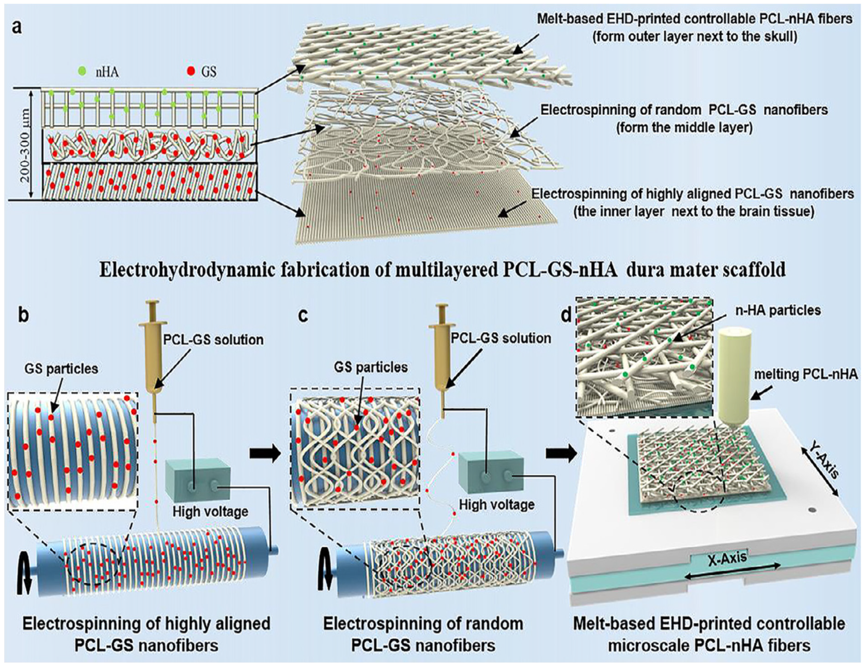
- Su, Y.; Li, Z.; Zhu, H.; He, J.; Wei, B.; Li, D. Electrohydrodynamic Fabrication of Triple-layered Polycaprolactone Dura Mater Substitute with Antibacterial and Enhanced Osteogenic Capability. Chin. J. Mech. Eng. Addit. Manuf. Front. 2022, 1, 100026. [Google Scholar] [CrossRef]
- Wang, W.; Ao, Q. Research and application progress on dural substitutes. J. Neurorestoratol. 2019, 7, 161–170. [Google Scholar] [CrossRef]
- Liu, W.; Wang, X.; Su, J.; Jiang, Q.; Wang, J.; Xu, Y.; Zheng, Y.; Zhong, Z.; Lin, H. In vivo Evaluation of Fibrous Collagen Dura Substitutes. Front. Bioeng. Biotechnol. 2021, 9, 628129. [Google Scholar] [CrossRef]
- Wang, Y.-f.; Guo, H.-f.; Ying, D.-j. Multilayer scaffold of electrospun PLA–PCL–collagen nanofibers as a dural substitute. J. Biomed. Mater. Res. Part B Appl. Biomater. 2013, 101, 1359–1366. [Google Scholar] [CrossRef]
- Li, J.; Tian, J.; Li, C.; Chen, L.; Zhao, Y. A hydrogel spinal dural patch with potential anti-inflammatory, pain relieving and antibacterial effects. Bioact. Mater. 2022, 14, 389–401. [Google Scholar] [CrossRef]
- Costa, B.S.; Cavalcanti-Mendes Gde, A.; Abreu, M.S.; Sousa, A.A. Clinical experience with a novel bovine collagen dura mater substitute. Arq. De Neuro-Psiquiatr. 2011, 69, 217–220. [Google Scholar] [CrossRef]
- Mukai, T.; Shirahama, N.; Tominaga, B.; Ohno, K.; Koyama, Y.; Takakuda, K. Development of watertight and bioabsorbable synthetic dural substitutes. Artif. Organs 2008, 32, 473–483. [Google Scholar] [CrossRef]
- Shi, Z.; Xu, T.; Yuan, Y.; Deng, K.; Liu, M.; Ke, Y.; Luo, C.; Yuan, T.; Ayyad, A. A New Absorbable Synthetic Substitute with Biomimetic Design for Dural Tissue Repair. Artif. Organs 2016, 40, 403–413. [Google Scholar] [CrossRef] [PubMed]
- Khandpur, U.; Ray, W.; MacEwan, M. Clinical performance of a novel fully synthetic dura substitute. Neurosurg.—Cases Rev. 2019, 2, 18. [Google Scholar] [CrossRef]
- Berjano, R.; Vinas, F.C.; Dujovny, M. A review of dural substitutes used in neurosurgery. Crit. Rev. Neurosurg. CR 1999, 9, 217–222. [Google Scholar] [CrossRef] [PubMed]
- McCall, T.D.; Fults, D.W.; Schmidt, R.H. Use of resorbable collagen dural substitutes in the presence of cranial and spinal infections-report of 3 cases. Surg. Neurol. 2008, 70, 92–96. [Google Scholar] [CrossRef] [PubMed]
- Yamada, M.; Noguchi-Shinohara, M.; Hamaguchi, T.; Nozaki, I.; Kitamoto, T.; Sato, T.; Nakamura, Y.; Mizusawa, H. Dura mater graft-associated Creutzfeldt-Jakob disease in Japan: Clinicopathological and molecular characterization of the two distinct subtypes. Neuropathol. Off. J. Jpn. Soc. Neuropathol. 2009, 29, 609–618. [Google Scholar] [CrossRef]
- Zerris, V.A.; James, K.S.; Roberts, J.B.; Bell, E.; Heilman, C.B. Repair of the dura mater with processed collagen devices. J. Biomed. Mater. Res. Part B Appl. Biomater. 2007, 83, 580–588. [Google Scholar] [CrossRef]
- Martínez-Lage, J.F.; Poza, M.; Sola, J.; Tortosa, J.G.; Brown, P.; Cervenáková, L.; Esteban, J.A.; Mendoza, A. Accidental transmission of Creutzfeldt-Jakob disease by dural cadaveric grafts. J. Neurol. Neurosurg. Psychiatry 1994, 57, 1091–1094. [Google Scholar] [CrossRef]
- Thadani, V.; Penar, P.L.; Partington, J.; Kalb, R.; Janssen, R.; Schonberger, L.B.; Rabkin, C.S.; Prichard, J.W. Creutzfeldt-Jakob disease probably acquired from a cadaveric dura mater graft. Case report. J. Neurosurg. 1988, 69, 766–769. [Google Scholar] [CrossRef]
- Stendel, R.; Danne, M.; Fiss, I.; Klein, I.; Schilling, A.; Hammersen, S.; Pietilae, T.; Jänisch, W.; Hopfenmüller, W. Efficacy and safety of a collagen matrix for cranial and spinal dural reconstruction using different fixation techniques. J. Neurosurg. 2008, 109, 215–221. [Google Scholar] [CrossRef]
- Narotam, P.K.; Qiao, F.; Nathoo, N. Collagen matrix duraplasty for posterior fossa surgery: Evaluation of surgical technique in 52 adult patients. Clinical article. J. Neurosurg. 2009, 111, 380–386. [Google Scholar] [CrossRef]
- Schmalz, P.; Griessenauer, C.; Ogilvy, C.S.; Thomas, A.J. Use of an Absorbable Synthetic Polymer Dural Substitute for Repair of Dural Defects: A Technical Note. Cureus 2018, 10, e2127. [Google Scholar] [CrossRef] [PubMed]
- MacEwan, M.R.; Kovacs, T.; Osbun, J.; Ray, W.Z. Comparative analysis of a fully-synthetic nanofabricated dura substitute and bovine collagen dura substitute in a large animal model of dural repair. Interdiscip. Neurosurg. 2018, 13, 145–150. [Google Scholar] [CrossRef]
- Zhang, X.; Meng, Y.; Gong, B.; Wang, T.; Lu, Y.; Zhang, L.; Xue, J. Electrospun nanofibers for manipulating soft tissue regeneration. J. Mater. Chem. B 2022, 10, 7281–7308. [Google Scholar] [CrossRef] [PubMed]
- Ohbayashi, N.; Inagawa, T.; Katoh, Y.; Kumano, K.; Nagasako, R.; Hada, H. Complication of silastic dural substitute 20 years after dural plasty. Surg. Neurol. 1994, 41, 338–341. [Google Scholar] [CrossRef]
- Degen, J.W.; Gagnon, G.J.; Voyadzis, J.M.; McRae, D.A.; Lunsden, M.; Dieterich, S.; Molzahn, I.; Henderson, F.C. CyberKnife stereotactic radiosurgical treatment of spinal tumors for pain control and quality of life. J. Neurosurg. Spine 2005, 2, 540–549. [Google Scholar] [CrossRef]
- Troy, E.; Tilbury, M.A.; Power, A.M.; Wall, J.G. Nature-Based Biomaterials and Their Application in Biomedicine. Polymers 2021, 13, 3321. [Google Scholar] [CrossRef]
- Teo, A.J.T.; Mishra, A.; Park, I.; Kim, Y.-J.; Park, W.-T.; Yoon, Y.-J. Polymeric Biomaterials for Medical Implants and Devices. ACS Biomater. Sci. Eng. 2016, 2, 454–472. [Google Scholar] [CrossRef]
- Xu, C.; Ma, X.; Chen, S.; Tao, M.; Yuan, L.; Jing, Y. Bacterial Cellulose Membranes Used as Artificial Substitutes for Dural Defection in Rabbits. Int. J. Mol. Sci. 2014, 15, 10855–10867. [Google Scholar] [CrossRef]
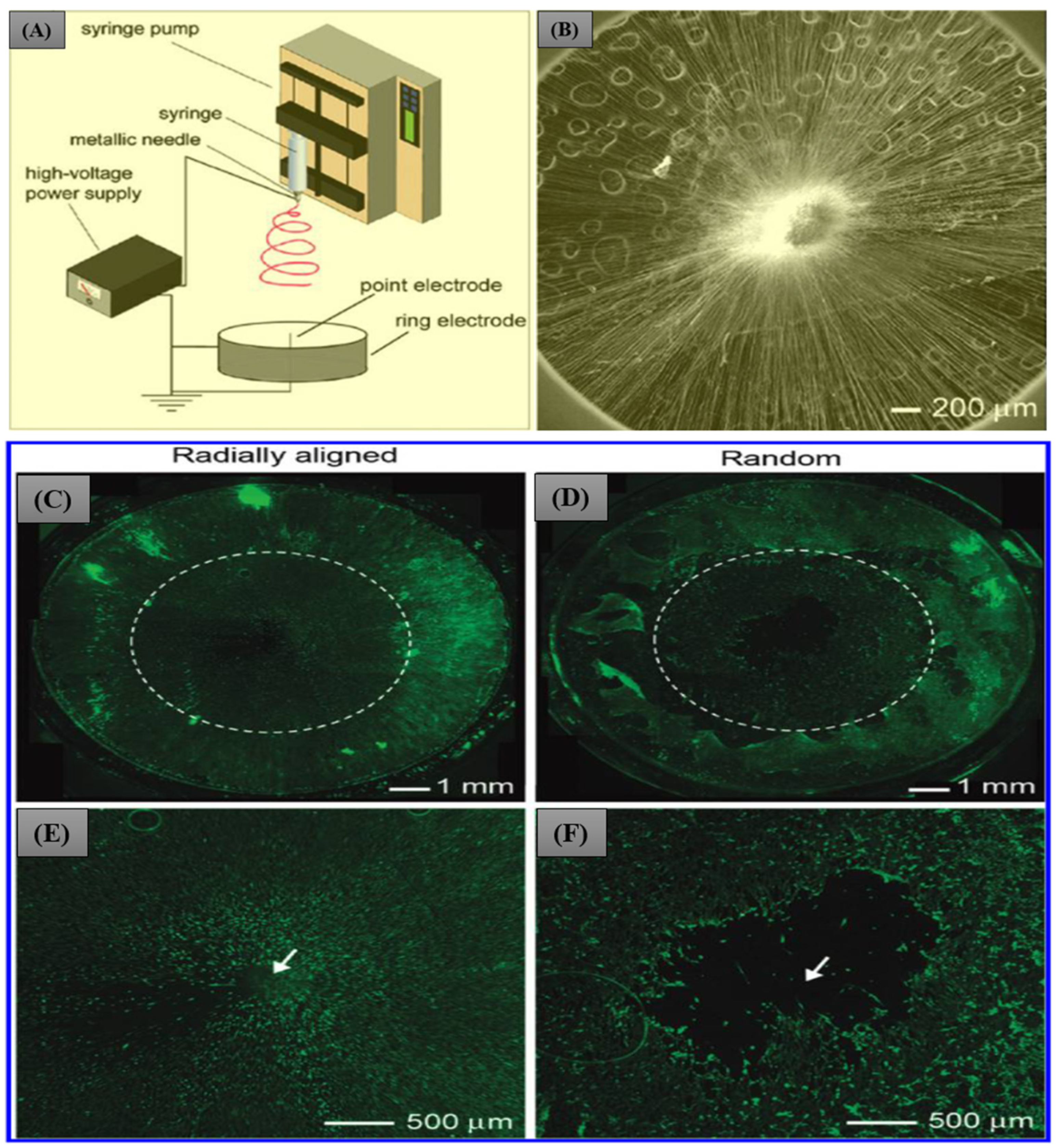
- Xie, J.; MacEwan, M.R.; Ray, W.Z.; Liu, W.; Siewe, D.Y.; Xia, Y. Radially Aligned, Electrospun Nanofibers as Dural Substitutes for Wound Closure and Tissue Regeneration Applications. ACS Nano 2010, 4, 5027–5036. [Google Scholar] [CrossRef]
- Shimada, Y.; Hongo, M.; Miyakoshi, N.; Sugawara, T.; Kasukawa, Y.; Ando, S.; Ishikawa, Y.; Itoi, E. Dural substitute with polyglycolic acid mesh and fibrin glue for dural repair: Technical note and preliminary results. J. Orthop. Sci. Off. J. Jpn. Orthop. Assoc. 2006, 11, 454–458. [Google Scholar] [CrossRef]
- Bernd, H.E.; Kunze, C.; Freier, T.; Sternberg, K.; Kramer, S.; Behrend, D.; Prall, F.; Donat, M.; Kramp, B. Poly(3-hydroxybutyrate) (PHB) patches for covering anterior skull base defects—An animal study with minipigs. Acta Oto-Laryngol. 2009, 129, 1010–1017. [Google Scholar] [CrossRef]
- Yu, F.; Li, Q.; Yin, S.; Liao, X.; Huang, F.; Chen, D.; Cao, Y.; Cen, L. Reconstructing spinal dura-like tissue using electrospun poly(lactide-co-glycolide) membranes and dermal fibroblasts to seamlessly repair spinal dural defects in goats. J. Biomater. Appl. 2015, 30, 311–326. [Google Scholar] [CrossRef] [PubMed]
- Zhu, Y.; Wang, A.; Shen, W.; Patel, S.; Zhang, R.; Young, W.; Li, S. Nanofibrous Patches for Spinal Cord Regeneration. Adv. Funct. Mater. 2010, 20, 1433–1440. [Google Scholar] [CrossRef] [PubMed]
- Suwanprateeb, J.; Luangwattanawilai, T.; Theeranattapong, T.; Suvannapruk, W.; Chumnanvej, S.; Hemstapat, W. Bilayer oxidized regenerated cellulose/poly ε-caprolactone knitted fabric-reinforced composite for use as an artificial dural substitute. J. Mater. Sci. Mater. Med. 2016, 27, 122. [Google Scholar] [CrossRef]
- Yamada, K.; Miyamoto, S.; Nagata, I.; Kikuchi, H.; Ikada, Y.; Iwata, H.; Yamamoto, K. Development of a dural substitute from synthetic bioabsorbable polymers. J. Neurosurg. 1997, 86, 1012–1017. [Google Scholar] [CrossRef] [PubMed]
- Yamada, K.; Miyamoto, S.; Takayama, M.; Nagata, I.; Hashimoto, N.; Ikada, Y.; Kikuchi, H. Clinical application of a new bioabsorbable artificial dura mater. J. Neurosurg. 2002, 96, 731–735. [Google Scholar] [CrossRef]
- Bhatia, S.; Bergethon, P.R.; Blease, S.; Kemper, T.; Rosiello, A.; Zimbardi, G.P.; Franzblau, C.; Spatz, E.L. A synthetic dural prosthesis constructed from hydroxyethylmethacrylate hydrogels. J. Neurosurg. 1995, 83, 897–902. [Google Scholar] [CrossRef]
- Yu, H.C.; Zhang, H.; Ren, K.; Ying, Z.; Zhu, F.; Qian, J.; Ji, J.; Wu, Z.L.; Zheng, Q. Ultrathin κ-Carrageenan/Chitosan Hydrogel Films with High Toughness and Antiadhesion Property. ACS Appl. Mater. Interfaces 2018, 10, 9002–9009. [Google Scholar] [CrossRef]
- Spencer, K.C.; Sy, J.C.; Ramadi, K.B.; Graybiel, A.M.; Langer, R.; Cima, M.J. Characterization of Mechanically Matched Hydrogel Coatings to Improve the Biocompatibility of Neural Implants. Sci. Rep. 2017, 7, 1952. [Google Scholar] [CrossRef]
- Boogaarts, J.D.; Grotenhuis, J.A.; Bartels, R.H.; Beems, T. Use of a novel absorbable hydrogel for augmentation of dural repair: Results of a preliminary clinical study. Neurosurgery 2005, 57, 146–151. [Google Scholar] [CrossRef]
- Strong, M.J.; Carnahan, M.A.; D’Alessio, K.; Butlin, J.D.G.; Butt, M.T.; Asher, A.L. Preclinical characterization and safety of a novel hydrogel for augmenting dural repair. Mater. Res. Express 2015, 2, 095401. [Google Scholar] [CrossRef]
- Huang, Y.-C.; Liu, Z.-H.; Kuo, C.-Y.; Chen, J.-P. Photo-Crosslinked Hyaluronic Acid/Carboxymethyl Cellulose Composite Hydrogel as a Dural Substitute to Prevent Post-Surgical Adhesion. Int. J. Mol. Sci. 2022, 23, 6177. [Google Scholar] [CrossRef] [PubMed]
- Lugoloobi, I.; Yuanhao, W.; Marriam, I.; Hu, J.; Tebyetekerwa, M.; Ramakrishna, S. Electrospun biomedical nanofibers and their future as intelligent biomaterials. Curr. Opin. Biomed. Eng. 2022, 24, 100418. [Google Scholar] [CrossRef]
- Chuan, D.; Wang, Y.; Fan, R.; Zhou, L.; Chen, H.; Xu, J.; Guo, G. Fabrication and Properties of a Biomimetic Dura Matter Substitute Based on Stereocomplex Poly(Lactic Acid) Nanofibers. Int. J. Nanomed. 2020, 15, 3729–3740. [Google Scholar] [CrossRef]
- Protasoni, M.; Sangiorgi, S.; Cividini, A.; Culuvaris, G.T.; Tomei, G.; Dell’Orbo, C.; Raspanti, M.; Balbi, S.; Reguzzoni, M. The collagenic architecture of human dura mater. J. Neurosurg. 2011, 114, 1723–1730. [Google Scholar] [CrossRef] [PubMed]
- Xu, Y.; Shi, G.; Tang, J.; Cheng, R.; Shen, X.; Gu, Y.; Wu, L.; Xi, K.; Zhao, Y.; Cui, W.; et al. ECM-inspired micro/nanofibers for modulating cell function and tissue generation. Sci. Adv. 2020, 6, eabc2036. [Google Scholar] [CrossRef]
- Kurpinski, K.; Patel, S. Dura mater regeneration with a novel synthetic, bilayered nanofibrous dural substitute: An experimental study. Nanomedicine 2011, 6, 325–337. [Google Scholar] [CrossRef] [PubMed]
- Lv, F.Y.; Dong, R.H.; Li, Z.J.; Qin, C.C.; Yan, X.; He, X.X.; Zhou, Y.; Yan, S.Y.; Long, Y.Z. In situ precise electrospinning of medical glue fibers as nonsuture dural repair with high sealing capability and flexibility. Int. J. Nanomed. 2016, 11, 4213–4220. [Google Scholar] [CrossRef]
- Ma, H.; Sun, Y.; Tang, Y.; Shen, Y.; Kan, Z.; Li, Q.; Fang, S.; Lu, Y.; Zhou, X.; Li, Z. Robust Electrospun Nanofibers from Chemosynthetic Poly(4-hydroxybutyrate) as Artificial Dural Substitute. Macromol. Biosci. 2021, 21, e2100134. [Google Scholar] [CrossRef]
- Campbell, B.; Anderson, Z.; Han, D.; Nebor, I.; Forbes, J.; Steckl, A.J. Electrospinning of cyanoacrylate tissue adhesives for human dural repair in endonasal surgery. J. Biomed. Mater. Res. Part B Appl. Biomater. 2022, 110, 660–667. [Google Scholar] [CrossRef]
- Zhao, Z.; Wu, T.; Cui, Y.; Zhao, R.; Wan, Q.; Xu, R. Design and Fabrication of Nanofibrous Dura Mater with Antifibrosis and Neuroprotection Effects on SH-SY5Y Cells. Polymers 2022, 14, 1882. [Google Scholar] [CrossRef] [PubMed]
- Jing, Y.; Ma, X.; Xu, C.; Tian, H.-l.; Chen, S.-w. Repair of dural defects with electrospun bacterial cellulose membranes in a rabbit experimental model. Mater. Sci. Eng. C 2020, 117, 111246. [Google Scholar] [CrossRef] [PubMed]
- Shi, R.; Xue, J.; Wang, H.; Wang, R.; Gong, M.; Chen, D.; Zhang, L.; Tian, W. Fabrication and evaluation of a homogeneous electrospun PCL–gelatin hybrid membrane as an anti-adhesion barrier for craniectomy. J. Mater. Chem. B 2015, 3, 4063–4073. [Google Scholar] [CrossRef] [PubMed]
- Zwirner, J.; Scholze, M.; Waddell, J.N.; Ondruschka, B.; Hammer, N. Mechanical Properties of Human Dura Mater in Tension—An Analysis at an Age Range of 2 to 94 Years. Sci. Rep. 2019, 9, 16655. [Google Scholar] [CrossRef]
- Xue, W.; Shi, W.; Kong, Y.; Kuss, M.; Duan, B. Anisotropic scaffolds for peripheral nerve and spinal cord regeneration. Bioact. Mater. 2021, 6, 4141–4160. [Google Scholar] [CrossRef]
- Rezvani Ghomi, E.; Khosravi, F.; Neisiany, R.E.; Shakiba, M.; Zare, M.; Lakshminarayanan, R.; Chellappan, V.; Abdouss, M.; Ramakrishna, S. Advances in electrospinning of aligned nanofiber scaffolds used for wound dressings. Curr. Opin. Biomed. Eng. 2022, 22, 100393. [Google Scholar] [CrossRef]
- Alvarado, A.G.; Chauhan, G. Nanofiber alignment for biomedical applications. Mater. Today Proc. 2022, 48, 79–83. [Google Scholar] [CrossRef]
- Li, X.; Wang, X.; Yao, D.; Jiang, J.; Guo, X.; Gao, Y.; Li, Q.; Shen, C. Effects of aligned and random fibers with different diameter on cell behaviors. Colloids Surf. B Biointerfaces 2018, 171, 461–467. [Google Scholar] [CrossRef]
- Robinson, A.J.; Pérez-Nava, A.; Ali, S.C.; González-Campos, J.B.; Holloway, J.L.; Cosgriff-Hernandez, E.M. Comparative analysis of fiber alignment methods in electrospinning. Matter 2021, 4, 821–844. [Google Scholar] [CrossRef]
- Shih, T.-Y.; Yang, J.-D.; Chen, J.-H. Synthesis, Characterization and Evaluation of Segmented Polycaprolactone for Development of Dura Substitute. Procedia Eng. 2012, 36, 144–149. [Google Scholar] [CrossRef]
- Chumnanvej, S.; Luangwattanawilai, T.; Rawiwet, V.; Suwanprateeb, J.; Rattanapinyopituk, K.; Huaijantug, S.; Yinharnmingmongkol, C.; Hemstapat, R. In vivo evaluation of bilayer ORC/PCL composites in a rabbit model for using as a dural substitute. Neurol. Res. 2020, 42, 879–889. [Google Scholar] [CrossRef] [PubMed]
- Wang, Y.; Guo, Q.; Wang, W.; Wang, Y.; Fang, K.; Wan, Q.; Li, H.; Wu, T. Potential use of bioactive nanofibrous dural substitutes with controlled release of IGF-1 for neuroprotection after traumatic brain injury. Nanoscale 2022, 14, 18217–18230. [Google Scholar] [CrossRef] [PubMed]
- Venugopal, J.; Ramakrishna, S. Biocompatible nanofiber matrices for the engineering of a dermal substitute for skin regeneration. Tissue Eng. 2005, 11, 847–854. [Google Scholar] [CrossRef]
- Li, W.J.; Danielson, K.G.; Alexander, P.G.; Tuan, R.S. Biological response of chondrocytes cultured in three-dimensional nanofibrous poly(epsilon-caprolactone) scaffolds. J. Biomed. Mater. Res. Part A 2003, 67, 1105–1114. [Google Scholar] [CrossRef] [PubMed]
- Matsumoto, K.; Nakamura, T.; Fukuda, S.; Sekine, T.; Ueda, H.; Shimizu, Y. A gelatin coated collagen-polyglycolic acid composite membrane as a dural substitute. ASAIO J. 2001, 47, 641–645. [Google Scholar] [CrossRef] [PubMed]
- Jin, S.; Pu, Y.; Guo, Z.; Zhu, W.; Li, S.; Zhou, X.; Gao, W.; He, B. A double-layer dura mater based on poly(caprolactone-co-lactide) film and polyurethane sponge: Preparation, characterization, and biodegradation study. J. Mater. Chem. B 2021, 9, 3863–3873. [Google Scholar] [CrossRef] [PubMed]
- Bhattarai, N.; Li, Z.; Gunn, J.; Leung, M.; Cooper, A.; Edmondson, D.; Veiseh, O.; Chen, M.-H.; Zhang, Y.; Ellenbogen, R.G.; et al. Natural-Synthetic Polyblend Nanofibers for Biomedical Applications. Adv. Mater. 2009, 21, 2792–2797. [Google Scholar] [CrossRef]
- Deng, K.; Yang, Y.; Ke, Y.; Luo, C.; Liu, M.; Deng, Y.; Tian, Q.; Yuan, Y.; Yuan, T.; Xu, T. A novel biomimetic composite substitute of PLLA/gelatin nanofiber membrane for dura repairing. Neurol. Res. 2017, 39, 819–829. [Google Scholar] [CrossRef]
- Lee, C.H.; Shin, H.J.; Cho, I.H.; Kang, Y.-M.; Kim, I.A.; Park, K.-D.; Shin, J.-W. Nanofiber alignment and direction of mechanical strain affect the ECM production of human ACL fibroblast. Biomaterials 2005, 26, 1261–1270. [Google Scholar] [CrossRef]
- Liu, Y.; Franco, A.; Huang, L.; Gersappe, D.; Clark, R.A.F.; Rafailovich, M.H. Control of cell migration in two and three dimensions using substrate morphology. Exp. Cell Res. 2009, 315, 2544–2557. [Google Scholar] [CrossRef]
- Kim, J.I.; Hwang, T.I.; Aguilar, L.E.; Park, C.H.; Kim, C.S. A Controlled Design of Aligned and Random Nanofibers for 3D Bi-functionalized Nerve Conduits Fabricated via a Novel Electrospinning Set-up. Sci. Rep. 2016, 6, 23761. [Google Scholar] [CrossRef] [PubMed]
- Badami, A.S.; Kreke, M.R.; Thompson, M.S.; Riffle, J.S.; Goldstein, A.S. Effect of fiber diameter on spreading, proliferation, and differentiation of osteoblastic cells on electrospun poly(lactic acid) substrates. Biomaterials 2006, 27, 596–606. [Google Scholar] [CrossRef]
- Fischer, R.S.; Sun, X.; Baird, M.A.; Hourwitz, M.J.; Seo, B.R.; Pasapera, A.M.; Mehta, S.B.; Losert, W.; Fischbach, C.; Fourkas, J.T.; et al. Contractility, focal adhesion orientation, and stress fiber orientation drive cancer cell polarity and migration along wavy ECM substrates. Proc. Natl. Acad. Sci. USA 2021, 118, e2021135118. [Google Scholar] [CrossRef] [PubMed]
- Bal-Ozturk, A.; Cecen, B.; Avci-Adali, M.; Topkaya, S.N.; Alarcin, E.; Yasayan, G.; Ethan, Y.C.; Bulkurcuoglu, B.; Akpek, A.; Avci, H.; et al. Tissue Adhesives: From Research to Clinical Translation. Nano Today 2021, 36, 101049. [Google Scholar] [CrossRef] [PubMed]
- Bhagat, V.; Becker, M.L. Degradable Adhesives for Surgery and Tissue Engineering. Biomacromolecules 2017, 18, 3009–3039. [Google Scholar] [CrossRef] [PubMed]
- Xie, J.; Li, X.; Xia, Y. Putting Electrospun Nanofibers to Work for Biomedical Research. Macromol. Rapid Commun. 2008, 29, 1775–1792. [Google Scholar] [CrossRef] [PubMed]
- Shahriar, S.M.S.; Mondal, J.; Hasan, M.N.; Revuri, V.; Lee, D.Y.; Lee, Y.-K. Electrospinning Nanofibers for Therapeutics Delivery. Nanomaterials 2019, 9, 532. [Google Scholar] [CrossRef]
- Albiñana-Cunningham, J.N.; Ripalda-Cemboráin, P.; Labiano, T.; Echeveste, J.I.; Granero-Moltó, F.; Alfonso-Olmos, M. Mechanical barriers and transforming growth factor beta inhibitor on epidural fibrosis in a rabbit laminectomy model. J. Orthop. Surg. Res. 2018, 13, 72. [Google Scholar] [CrossRef]
- Crowley, S.T.; Fukushima, Y.; Uchida, S.; Kataoka, K.; Itaka, K. Enhancement of Motor Function Recovery after Spinal Cord Injury in Mice by Delivery of Brain-Derived Neurotrophic Factor mRNA. Mol. Ther.—Nucleic Acids 2019, 17, 465–476. [Google Scholar] [CrossRef]
- Nurata, H.; Cemil, B.; Kurt, G.; Uçankuş, N.L.; Dogulu, F.; Ömeroğlu, S. The role of fibroblast growth factor-2 in healing the dura mater after inducing cerebrospinal fluid leakage in rats. J. Clin. Neurosci. 2009, 16, 542–544. [Google Scholar] [CrossRef]
- Mohtaram, N.K.; Ko, J.; Agbay, A.; Rattray, D.; Neill, P.O.; Rajwani, A.; Vasandani, R.; Thu, H.L.; Jun, M.B.G.; Willerth, S.M. Development of a glial cell-derived neurotrophic factor-releasing artificial dura for neural tissue engineering applications. J. Mater. Chem. B 2015, 3, 7974–7985. [Google Scholar] [CrossRef]
- Shi, R.; Huang, Y.; Zhang, J.; Wu, C.; Gong, M.; Tian, W.; Zhang, L. Effective delivery of mitomycin-C and meloxicam by double-layer electrospun membranes for the prevention of epidural adhesions. J. Biomed. Mater. Res. Part B Appl. Biomater. 2020, 108, 353–366. [Google Scholar] [CrossRef] [PubMed]
- Liao, J.; Li, X.; He, W.; Guo, Q.; Fan, Y. A biomimetic triple-layered biocomposite with effective multifunction for dura repair. Acta Biomater. 2021, 130, 248–267. [Google Scholar] [CrossRef] [PubMed]
- Sanpakitwattana, A.; Suvannapruk, W.; Chumnanvej, S.; Hemstapat, R.; Suwanprateeb, J. Cefazolin Loaded Oxidized Regenerated Cellulose/Polycaprolactone Bilayered Composite for Use as Potential Antibacterial Dural Substitute. Polymers 2022, 14, 4449. [Google Scholar] [CrossRef]
- Huang, Y.; Shi, R.; Gong, M.; Zhang, J.; Li, W.; Song, Q.; Wu, C.; Tian, W. Icariin-loaded electrospun PCL/gelatin sub-microfiber mat for preventing epidural adhesions after laminectomy. Int. J. Nanomed. 2018, 13, 4831–4844. [Google Scholar] [CrossRef] [PubMed]
- Fan, Q.; Wu, H.; Kong, Q. Superhydrophilic PLGA-Graft-PVP/PC Nanofiber Membranes for the Prevention of Epidural Adhesion. Int. J. Nanomed. 2022, 17, 1423–1435. [Google Scholar] [CrossRef] [PubMed]
- Tseng, Y.Y.; Liao, J.Y.; Chen, W.A.; Kao, Y.C.; Liu, S.J. Biodegradable poly([D,L]-lactide-co-glycolide) nanofibers for the sustainable delivery of lidocaine into the epidural space after laminectomy. Nanomedicine 2014, 9, 77–87. [Google Scholar] [CrossRef]


| SN | Materials | Results | Ref. |
|---|---|---|---|
| 1 | PLCL, PPG and sodium acetate | -Bilayer dural substitute having aligned nanofibers on one side and random nanofibers on the other. -Significantly high strength and durability compared to commercially available collagen matrix. -In vitro fibroblast and in vivo dural healing were enhanced by the aligned nanofibers. | [92] |
| 2 | PCL | -Proposed a new setup for producing aligned nanofibers by electrospinning. -Migration of cells from periphery to the center. -Potentially allowing for fast regeneration and formation of neodura. | [74] |
| 3 | poly(lactide-co-glycolide) | -A package with two layers of electrospun membranes, dermal fibroblast and mussel adhesive protein and used in a goat. -Electrospun PLGA and chitosan coated PLGA membranes were used as inner and outer membranes, respectively. -Guided tissue growth and regeneration in the defects were observed. | [77] |
| 4 | PLA-PCL-Collagen | -Three-layered scaffold (PLA-PCL-collagen serve as inner to outer layer). -Sufficient mechanical strength and biocompatible. | [52] |
| 5 | n-octyl-2-cyanoacrylate/poly(methyl methacrylate) | -High compactness and flexibility -Experiments on egg membranes and goat meninges showed rapid and effective recovery in dural defect. | [93] |
| 6 | PCL/GS/nHA | -Biomimetic triple-layered membrane. -Comparable mechanical properties to natural dura mater. -Good biocompatibility with anti-infection properties. | [49] |
| 7 | PLLA, poly(D-lactic acid)-grafted tetracalcium phosphate | -Tensile strength close to human dura mater. -Non-toxic and neuron compatible. | [89] |
| 8 | poly(4-hydroxybutyrate) (P4HB) | -Good mechanical properties that match the natural dura mater. -Induces fast cellular migration, adhesion, and proliferation of fibroblasts in vitro. -Implantation in rats demonstrates excellent biocompatibility of the P4HB membrane with proper biodegradation behaviors. | [94] |
| 9 | n-octyl- 2-cyanoacrylate (NOCA) | -Studied for in situ dural closures after neurosurgery. -The fiber membrane showed significantly higher sealing capabilities of defects in human dura. | [95] |
| 10 | PLGA/CS | -Can inhibit the excessive proliferation of fibroblasts, as well as provide a sustained -Protective effect on the human neuroblastoma (SH-SY5Y) cells treated with oxygen–glucose deprivation/reperfusion | [96] |
| 11 | Bacterial cellulose | -First study on using bacterial cellulose for rabbit dural defect. -Good biocompatibility in vitro and in vivo. -Implantation study showed no relevant complications. -Mild local inflammatory reaction detected. | [97] |
| 12 | PCL/gelatin | -The mechanical strength was increase with the PCL content whereas biocompatibility was increased with gelatin content. -Subcutaneous implantation in rabbit for 6 months exhibited adjustable biodegradable behavior. | [98] |
| S.N. | Materials | Results | Ref. |
|---|---|---|---|
| 1 | mitomycin-C and meloxicam loaded PCL/CS fibers | -Drug-loaded double-layered membrane. -Bottom layer is loaded with meloxicam to prevent dural inflammation. -Top layer is loaded with mitomycin-C to inhibit the DNA synthesis. -Both drugs were released for 12 days. -It prevented the epidural adhesion formation. | [127] |
| 2 | tetramethylpyrazine/PLGA/CS NFs | -Excellent biocompatibility, adequate mechanical properties and good antifibrotic effects. -Inhibits excessive proliferation of fibroblasts. -Brought anti-adhesive effects and inhibited the formation of scar tissue. | [96] |
| 3 | icariin-loaded PCL/gelatin NFs | -Prevent fibroblast adhesion and proliferation. -In vivo studies with rabbit laminectomy models showed the release of ICA in a controlled and sustained manner. | [130] |
| 4 | PCL/hyaluronic acid methacryloyl (HAMA)/IGF-1 NFs | -Long-term release of growth factor. -hydrophobic membrane with good mechanical properties. -Improve the microenvironment of neurite growth and promote the survival of neural cells. | [107] |
| 5 | PLGA-Graft-PVP/PC NFs | -No cytotoxic effect -Safe and effective physical barrier for preventing epidural adhesion. | [131] |
| 6 | PCL-BSA-GDNF | -Random and aligned fibers -GDNF release -Support the culture and differentiation of hiPSC-derived neural progenitors | [126] |
| 7 | rolipram/PLLA/PLGA NFs | -Aligned nanofibers in inner and random fiber in outer layer. -Effective in guiding axon growth and angiogenesis and releasing drug. | [78] |
| 8 | SIS loaded PLLA/CS/gelatin NFs | -Combination of hydrogel and electrospun nanofibers with triple-layered structure. -SIS helped to improve bioactivity. -Good dura reconstruction potential with interesting features such as leakage blockade, adhesion prevention, and antibacterial properties. | [128] |
| 9 | cefazolin loaded ORC/PCL NFs | -Initial burst release one the first day followed by constant and slow release of cefazolin. -Antibacterial activity for 4 days. | [129] |
| 10 | lidocaine embedded PLGA NFs | -Biodegradable nanofiber membrane for epidural analgesia. -Sustainable release of lidocaine for more than two weeks. | [132] |
Disclaimer/Publisher’s Note: The statements, opinions and data contained in all publications are solely those of the individual author(s) and contributor(s) and not of MDPI and/or the editor(s). MDPI and/or the editor(s) disclaim responsibility for any injury to people or property resulting from any ideas, methods, instructions or products referred to in the content. |
© 2023 by the authors. Licensee MDPI, Basel, Switzerland. This article is an open access article distributed under the terms and conditions of the Creative Commons Attribution (CC BY) license (https://creativecommons.org/licenses/by/4.0/).
Share and Cite
Pant, B.; Park, M.; Kim, A.A. Electrospun Nanofibers for Dura Mater Regeneration: A Mini Review on Current Progress. Pharmaceutics 2023, 15, 1347. https://doi.org/10.3390/pharmaceutics15051347
Pant B, Park M, Kim AA. Electrospun Nanofibers for Dura Mater Regeneration: A Mini Review on Current Progress. Pharmaceutics. 2023; 15(5):1347. https://doi.org/10.3390/pharmaceutics15051347
Chicago/Turabian StylePant, Bishweshwar, Mira Park, and Allison A. Kim. 2023. "Electrospun Nanofibers for Dura Mater Regeneration: A Mini Review on Current Progress" Pharmaceutics 15, no. 5: 1347. https://doi.org/10.3390/pharmaceutics15051347