Review

An Overview of the Stability and Delivery Challenges of Commercial Nucleic Acid Therapeutics

1
Institute of Drug Metabolism and Pharmaceutical Analysis, College of Pharmaceutical Sciences, Zhejiang University, Hangzhou 310027, China
2
Dr. Rajendra Gode College of Pharmacy, Amravati 444602, India
*
Author to whom correspondence should be addressed.
Pharmaceutics 2023, 15(4), 1158; https://doi.org/10.3390/pharmaceutics15041158
Submission received: 20 January 2023 / Revised: 21 March 2023 / Accepted: 24 March 2023 / Published: 6 April 2023

Abstract

:
Nucleic acid (NA)-based biopharmaceuticals have emerged as promising therapeutic modalities. NA therapeutics are a diverse class of RNA and DNA and include antisense oligonucleotides, siRNA, miRNA, mRNA, small activating RNA, and gene therapies. Meanwhile, NA therapeutics have posed significant stability and delivery challenges and are expensive. This article discusses the challenges and opportunities for achieving stable formulations of NAs with novel drug delivery systems (DDSs). Here we review the current progress in the stability issues and the significance of novel DDSs associated with NA-based biopharmaceuticals, as well as mRNA vaccines. We also highlight the European Medicines Agency (EMA) and US Food and Drug Administration (FDA)-approved NA-based therapeutics with their formulation profiles. NA therapeutics could impact future markets if the remaining challenges and requirements are addressed. Regardless of the limited information available for NA therapeutics, reviewing and collating the relevant facts and figures generates a precious resource for formulation experts familiar with the NA therapeutics’ stability profile, their delivery challenges, and regulatory acceptance.

Graphical Abstract

1. Introduction

Biopharmaceuticals are at the supreme level of the pharmaceutical market due to their high efficacy, high specificity, and low toxicity profiles [1]. Recently, nucleic acid (NA) therapeutics have emerged as promising candidates for several severe diseases and disorders. NAs are present in all living organisms, including humans, animals, and plants [2]. NAs are naturally occurring chemical compounds; certain small NAs are also synthesized in the laboratory. NAs can be broken down into sugars, phosphoric acid, and a mixture of organic bases (e.g., purines and pyrimidines). NAs have been developed as therapeutic agents and carefully characterized to provide the intended quality, efficacy, and safety profile. NAs are complex and delicate molecules that require sophisticated processes with clever handling during manufacturing, which makes these drugs more expensive. The stability of NAs during manufacturing, handling, shipping, and long-term storage is a major subject of discussion. Excipients play a key role in designing NA therapeutics by improving the manufacturability, stability, quality, and safe delivery of the products [3].
Due to their complex nature, NAs require special attention as active pharmaceutical ingredients (APIs). The alteration in NA quality as a result of physicochemical degradation makes their formulation development challenging. Therefore, several aspects must be considered, including active drug concentration, excipients, delivery routes, and novel drug delivery systems (DDSs). The use of excipients at optimized concentrations aims to maintain the stability of NA therapeutics [4]. However, a key obstacle for the formulation expert is to formulate stable NA therapeutics with the narrow range of excipients usually employed in parenteral settings. Therefore, the launch of novel and ideal excipients to maintain the integrity of significant scientific contribution. Specifically, a critical manufacturing hurdle is precision in the reproducibility of chemical synthesis, the assurance of reproducibility, and the integrity of subsequent batches of the NA therapeutics. For example, the synthesis of thiophosphate derivatives of oligonucleotides results in a mixture of 2n diastereomers, in which each diastereomer might interact in a slightly different manner. Here, chirality impacts the physical and biological properties of NAs, such as the binding affinity, nuclease stability, etc. [5]. In addition, NA therapeutics are still related with the dilemma of complex drug delivery. This is a fundamental setback preventing the widespread implementation of NA therapeutics. Naked NAs are quickly degraded into physiological fluids and do not accumulate in target tissues [6,7,8]. Despite these issues, the current NA dosage forms and novel DDSs have enabled the successful launch of NA therapeutics globally. The application of novel DDSs not only improves the long-term storage stability of NAs but also preserves their in vivo efficacy. Therefore, it could be assumed that it is important to conduct an up-to-date survey of the excipients in approved NA therapeutics with novel DDSs. It could serve the biopharmaceutical industry by minimizing the time spent on pre-formulation studies and speeding up the development of stable NA formulations.
Between 2004 and 2021, there have been 23 NA therapeutics approved via the United States Food and Drug Administration (FDA) and/or the European Medicines Agency (EMA) approaches. Among these, fomivirsen (Vitravene) was removed from the European and US markets in 2002 and 2006, respectively, owing to the demand having been undermined. In addition, Macugen (pegaptanib sodium injection), Glybera (alipogene tiparvovec), and Kynamro (mipomersen sodium) were withdrawn from the market in 2019, 2017, and 2022, respectively [9]. Therapeutic NAs formulated in liquid, suspension, and freeze-dried forms, as well as vector-like liposomes/lipids and nanoparticles (NPs), are divided into their functional classes. Antisense oligonucleotides are a major division of approved NA therapeutics. On the other hand, gene therapy has revealed exciting treatment opportunities for numerous severe and rare diseases that have not been cured thus far, although the safety of gene therapy is a major concern. Continuous monitoring is needed to overcome the challenges posed by these new drugs and to increase their contribution as novel therapeutic modalities in the biopharmaceutical industry [10].
To date, one of the major unresolved issues for approved NAs is the high cost of these drugs. For example, nusinersen costs USD $750,000 for the first year and USD $375,000 in subsequent years. Likewise, eteplirsen costs USD $300,000 annually. The expense for high-efficacy, life-saving drugs, such as nusinersen, is likely to be acceptable. In contrast, eteplirsen has a narrow efficacy; therefore, justifying the cost of eteplirsen would be difficult [11,12].

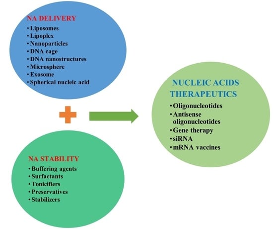
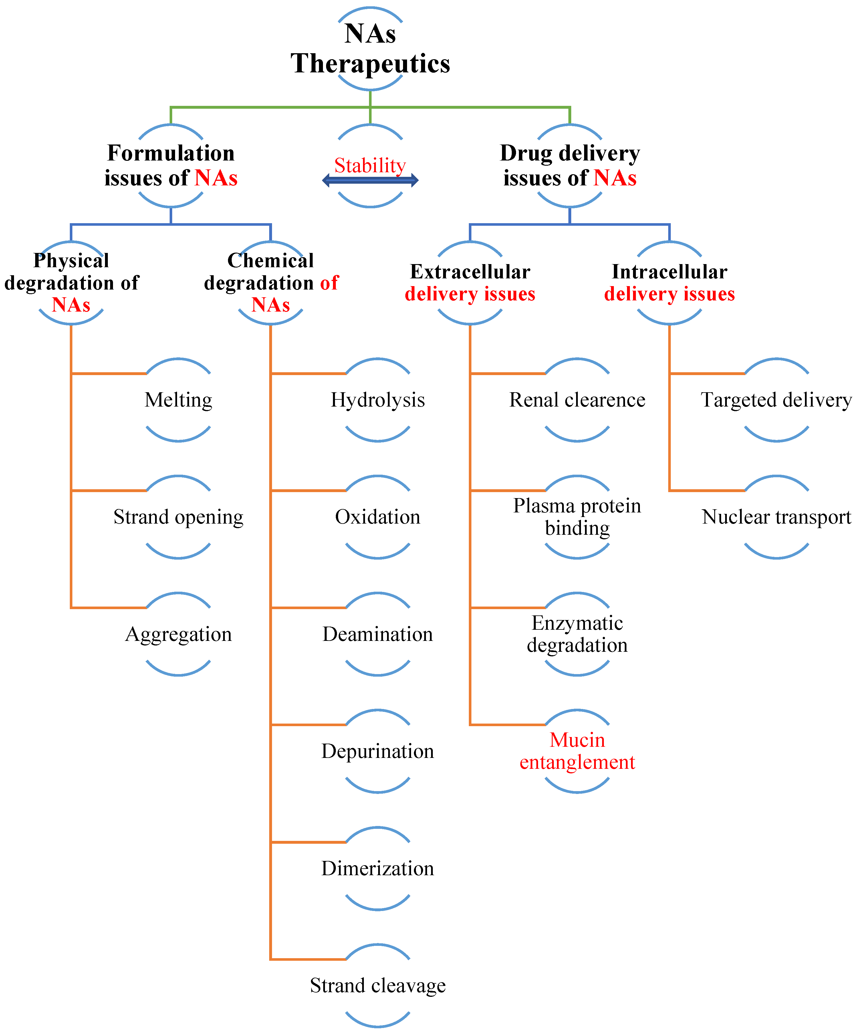
2. Lasting Challenges and Considerations of NA Therapeutics

2.1. NA Therapeutics Stability

NAs could have unique stability issues, similar to protein drugs, due to their complex and fragile nature (Figure 1). Naked or unmodified NAs have extremely short half-lives in circulation due to enzymatic (e.g., nucleases, such as deoxyribonucleases, and RNAse, such as ribonucleases) and chemical (e.g., oxidation, hydrolysis, deamidation, depurination, and strand cleavage) degradation. Therefore, chemical modifications are usually necessary to improve NA stability. Currently, messenger RNA (mRNA) vaccines have become the frontrunners in fighting coronavirus (COVID-19). However, mRNAs alone are prone to nuclease degradation and phosphate backbone hydrolysis through the intramolecular attack of the 2′-hydroxyl group in physiological fluids [13], which is responsible for the short half-life and low efficiency of the mRNA therapeutics due to incompatibility with nuclease, high molecular weight, high negativity, and their hydrophilic and acid-labile nature [14]. Therefore, lipid nanoparticles (LNPs) are employed in COVID-19 mRNA vaccines. A key disadvantage of the approved COVID-19 mRNA-LNP vaccines is the need to be kept under (ultra) cold storage conditions [15], and the stability of mRNA vaccines during storage, handling, and shipping at ambient temperatures is a primary concern [16,17]. As discussed earlier, mRNA could also undergo hydrolytic degradation during storage, handling, and shipping at ambient temperatures. Thus, DDSs, such as LNP, act as a shield for mRNA to prevent degradation and assure its potency.
In the case of antisense oligonucleotides (ASOs), modifications are usually made to the 2′-carbon of the sugar ring or phosphodiester bond. Phosphorothioate modification provides key protection from nucleases and extends the half-life with better stability. A similar phosphonoacetate modification is possible at the ASO backbone, which is totally resistant to nuclease degradation. Another approach to enhance ASO stability is the ribose sugar modification at the 2′-position of the ring, e.g., locked nucleic acids (LNAs) [18,19]. However, in phosphorodiamidate morpholino oligomers (PMOs), ribose sugars are exchanged with phosphodiester bonds and morpholino groups (e.g., casimersen), sometimes referred to as splice switching oligonucleotides (SSOs). For example, nusinersen and eteplirsen are inclusion- and skipping-type SSOs, respectively. In the case of peptide nucleic acids (PNAs), the ribose-phosphate backbone is exchanged with a polyamide backbone [20,21]. In addition, short interfering RNA (siRNA) chemical modifications at various sites, such as phosphate, nucleobases, ribose, poly-2-O-(2,4-dini-trophenyl)-RNA, and oxygen ring replacement with a sulfur group, exhibit enhanced resistance to RNase with better stability. Fluorine, sugar, methoxy, or deoxy modifications at 2′ positions of the ring are other approaches to improve siRNA serum stability [22,23,24]. On the other hand, targeting the liver is both a major advantage and disadvantage (e.g., immunotoxicity, immunogenesis, degradation of LNP against the harsh GIT conditions) of NA therapeutics [25]. Due to their different particle size and lipid composition, nanocarriers have organ-specific targeting. LNP is likely delivering the drug to the liver. Rizvi et al. [26] measured the organ distribution of protein activity produced by firefly luciferase encoding mRNA-LNP (Luc mRNA-LNP) and found that robust luciferase expression was detected in the liver. Yang et al. [27] used the lipopolyplex (LPP) nanoparticles, formulated by SW-01(a positively charged cationic compound), ionized and non-ionized lipids, carrying with mRNA encoding luciferase which was evaluated for biodistribution pattern. After intramuscular injection (i.m.) into the hind legs of mice, a strong luciferase expression was noticed at the injection site (muscle) but not in the liver.
In addition, the ASOs are modified with PS-linkages (e.g., Kynamro, Tegsedi), and siRNA (e.g., Oxlumo) are well targeted to hepatic delivery. Synthetic therapeutic oligonucleotides (STOs) could control numerous intracellular and extracellular obstacles to interact with their biological RNA targets inside cells. STOs depend on passive exchanges of phosphorothioate (PS) oligonucleotides with cell-surface and plasma proteins to endorse delivery to the kidney and liver. PS-mediated drug delivery was also useful in delivering NA therapeutics (e.g., Spinraza) to CNS tissues [28].
Additionally, gene-based therapies present exclusive challenges due to several factors, including membrane fluidity and permeability and multiple and complex antigen epitopes. Aggregation, oxidation, deamidation, hydrolysis, and adsorption of gene therapies are frequent pathways responsible for degradation. During deamidation, a succinimidyl intermediate is formed due to nucleophilic attack by the adjacent amide over the amide group of asparagine, and the succinimidyl intermediate is hydrolyzed to isoaspartic and aspartic acids [29]. Thus, this causes instabilities in cell-based therapeutics [30], which may lead to the loss of therapeutic efficacy and produce severe immunogenic responses in patients [31]. Therefore, an astute consideration of the degradation processes and triggering factors and the selection of suitable excipients are crucial to protect NA therapeutics against destabilization.
The challenges involved in the successful formulation of NA therapeutics include a variety of physicochemical degradation pathways. The mechanisms of physicochemical degradation with various environmental factors influencing NA stability have been broadly studied. Physical instability is due to exposure to different temperatures, pH, buffer concentrations, presence of oxygen, ultraviolet light, transition metal cations (e.g., copper, ferrous, zinc, magnesium, and nickel), and various stress conditions. For example, uracil is the deamidate product of cytosine, and the process is 100-fold quicker in single-stranded NAs. At higher temperatures and above pH 7, depurination is observed to be sequence-independent. Metal contamination is a significant factor in the degradation of NAs. During the manufacture of NAs, trace amounts of metals present in various raw materials can be established. Transition metal cations have been found to form metal-base pairs by chelation and initiate the breakage of NA strands. Chelation occurs between the NA strands by connecting the cytosines and two adenines, or mixtures thereof [32,33]. Among the transition metals present, copper and ferrous metal ions seem to be strong degradation catalysts. For example, DNA degradation is observed via the intermediate formation of the DNA-copper-hydroperoxide complex by copper ions and ferrous ions, which have been found to be responsible for the degradation of calf thymus DNA due to molecular oxygen [34]. Chemical instability is prevalently caused by hydrolysis, oxidation, depyrimidination, and deamination [35]. In hydrolysis, phosphoester and N-glycosidic bonds are more prone to hydrolytic cleavage. Nucleophilic phosphodiester cleavage is caused by either inter- or intramolecular reactions. Oxidative degradation primarily involves reactive oxygen species (ROS) reactions in which Fenton-type processes are the most frequent origin of ROS. In addition, the formation of covalent intrastrand purine dimers [36] and oxidation through free radicals are considered chief degradative processes for NA therapeutics [37]. Irradiation with NAs was found to generate hydroxyl free radicals that also caused damage [38].
As specified, NA therapeutics are usually formulated as freeze-dried powders, similar to many protein drugs [39,40]. NAs are generally more stable in a freeze-dried (lyophilized) form than in a liquid state. In the freeze-dried form, many of these degradation pathways may be avoided or retarded. Therefore, freeze-drying is advised to be a practical method to sort out stability problems during long-term storage [41]. siRNA has been administered as a dry powder inhalation (DPI) to facilitate the delivery of drugs in lung therapy [42,43]. In addition, Gennova biopharmaceuticals-based mRNA HGCO19 vaccine will be in a lyophilized form. Recently, the approved tozinameran (BNT162b2, Pfizer/BioNTech) and elasomeran (mRNA-1273, Moderna) mRNA vaccines have been considering sucrose as a lyoprotectant [44] due to the surprising loss of mRNA stability and delivery efficiency following the lyophilization of LNP [45,46]. Therefore, it is necessary to optimize freeze-drying parameters and choose a suitable lyoprotectant to achieve a stable product.
Figure 1. Challenges in the formulation and delivery of NA therapeutics [34].
Pharmaceutics 15 01158 g001

2.2. NA Therapeutics Delivery

The use of NA therapeutics has climbed significantly in the last decade, but there are still clinical challenges, such as poor pharmacokinetics and pharmacodynamics [47,48,49]. Incompetent delivery to target organs is a key hurdle preventing the prevalent usage of NA therapeutics (Figure 1). The design of delivery vehicles with particular features renders them stable, efficient, and safe in transfection.
The delivery of NA therapeutics can be influenced by their attributes, such as a negative charge, hydrophilicity, and susceptibility to enzyme degradation [50]. In addition, off-target side effects must be cautiously monitored. Therefore, these issues need to be addressed for the timely development of smart NA formulation. The most commonly engaged strategies to boost NA delivery include chemical, ribose sugar, nucleobases, backbone, and terminal modifications with cell-penetrating moieties. The approaches which have been developed most recently include liposomes, lipoplexes, NPs, DNA cages, DNA nanostructures (framework nucleic acids) [51], microspheres, exosomes [11], gene therapy, spherical NA, red blood cells, biological solids, stimuli-responsive nanotechnology, polyplexes, extracellular vesicles (natural and engineered), micelleplexes, heteroduplex oligonucleotides, niosomes (unilamellar and multilamellar), carbon nanotubes, carbon nanodots, and aptamers [52,53,54,55,56,57,58,59,60,61,62,63]. Furthermore, Maurer et al. developed magnetic hybrid niosomes (iron-oxide NPs) for siRNA delivery to treat breast cancer [64,65]. All these NA delivery systems have contributed to overcoming several challenges related to NA therapeutics, which include protecting them from degradation and avoiding renal excretion, thereby improving the safety profile.
NP-based drug delivery represents a highly adaptable platform for a variety of therapeutics [66,67]. This includes lipid-based NPs (e.g., liposomes, cubosomes, ionizable, and solid LNPs), polymeric NPs (e.g., natural and synthetic polymers), metal NPs (e.g., gold, silver, and iron), gold NPs (e.g., spherical and nonspherical (such as nanostars, nanorods, and nanocubes)), porous NPs (e.g., porous silicon and mesoporous silica NPs), and metal-organic frameworks (e.g., NU-100) [68,69,70]. The most frequently employed biodegradable natural polymers are alginate, hyaluronic acids, and chitosan. The current use of synthetic polymers over natural polymers has received noticeably more attention due to their better mechanical and reproducible properties, i.e., dendrimers (e.g., poly-(β-amino ester) (PβAE), poly-(L-lysine) (PLL), and polyamidoamine (PAMAM)), PLGA (polylactic-co-glycolic acid), and PEIs (polyethylene imines) [71,72].
Novel DDSs could enhance the solubility, bioavailability, safety, and PK profiles of systemically administered drugs, leading to enhanced therapeutic efficacy [73]. Therefore, formulation scientists are paying attention to easy and smart DDSs. The current scenario shows rapid commercialization and increasing popularity of nanomedicine dominance over the other DDSs [74,75,76,77]. However, liposomes face parallel challenges, including restrictions from the risk of causing immune responses and biodistribution [78]. Due to the suitability of liposomes to both hydrophilic and lipophilic drug candidates, they are used as carriers of choice for various therapeutics [79]. The leading pathway for siRNA therapeutics delivery to the liver was found to be N-acetyl galactosamine (GalNAc), which has proven to be long-acting, therefore, improving the safety profile of the NA therapeutics [80]. In addition, lipid-based carriers (e.g., cationic lipids) [81], polyplexes (many mRNA-based drugs under clinical trials), multivalent cationic non-viral vectors, and polymeric vehicles [82,83,84] represent the most commonly preferred alternative to viral vectors in gene therapy [85,86,87]. Viral vectors with antigen-encoding RNA in place of viral genes are prepared by genetic modification of viruses and are effective delivery vehicles. Viral vectors are categorized as replicating and non-replicating vectors [88]. Various RNA viruses, such as adenovirus, picornavirus, flavivirus, and alphavirus, have been used as viral vectors for mRNA delivery. Viral vectors have several limitations, including host genome integration (genotoxicity) and allergic reactions. Therefore, viral vectors have been replaced by non-viral vectors. Non-viral vectors can be classified as hybrid, polymer-based, and lipid-based [36]. They have several benefits over viral vectors, such as ease of production, multi-dosing capabilities, lesser toxicity, and the lack of immunogenicity [89,90]. Therefore, non-viral vectors could be designed with multiple components to overcome physiological barriers [91].
In 1995, the liposome Doxil emerged as the first NP therapeutic. In 2005, human albumin was employed in NP formulations (e.g., Abraxane). Recently, in 2017 [92], the FDA approved the first gene therapies (e.g., Kymriah), which were prepared from the white blood cells of the patients. The great commercial achievement of these DDSs has fascinated many professionals in the field [93]. DDSs play a crucial role in biopharmaceutical formulations, including proteins, monoclonal antibodies, and, recently, NAs. The advanced DDSs promote the high quality and efficacy of the drugs and extend the shelf life of these new molecular entities. Among the approved NA therapeutics, many are delivered as novel DDSs. Patisiran (Onpattro®) is the first commercially available LNP formulation delivered in liposome vesicles. In addition, some gene therapies are associated with adenovirus vectors (e.g., voretigene neparvovecrzyl and onasemnogene abeparvovec-xioi), herpes simplex virus vectors (e.g., talimogene laherparepvec), and gamma retroviral vectors (e.g., strimvelis) [94,95,96]. Concerning other gene therapies, adeno-associated virus (AAV) has emerged as the principal vector due to the sustainability of the viral genome and its lack of pathogenicity. AAV vectors have special features, such as requiring a helper virus for replication and avoiding pathogenicity. They also have a low tendency for gene integration and, therefore, avoid genotoxicity [97]. The FDA approved Luxturna® as AAV2-based and Zolgensma® as AAV9-based gene therapy, and many more AAV-based gene therapies are under clinical trials [98].
Recently, numerous RNA and DNA vaccines have entered the clinical stages. Among them, mRNA vaccines have become therapeutic targets of interest in many severe diseases and disorders. The safe and efficient delivery of therapeutic mRNAs is one of the key challenges for their wide implementation in humans. mRNAs have concerns such as sensitivity to catalytic hydrolysis, intracellular delivery, and high instability under certain physiological conditions [99]. Recently, developments in mRNA vaccines have been made through their formulation with LNP, which not only provides enhanced delivery and protection but also performs the role of an adjuvant in vaccine reactogenicity [100,101], i.e., the LNP-based delivery of mRNA vaccines against influenza and Zika [102]. In addition, nanoscale delivery platforms (e.g., lipid-derived NPs, polymeric NPs, polymer-lipid hybrid NPs, metal NPs, and peptide complexes) have prolonged the viability of mRNA-based therapeutics, which permit their promising application in protein replacement therapy, genome editing, and cancer immunotherapy [103]. Currently, a major vaccine development platform is advancing with self-assembling drug delivery vehicles, such as cationic monovalent lipids (e.g., N-[1-(2,3-dioleyloxy)propyl]-N,N,N-trimethylammonium chloride (DOTMA), 1,2-dioleoyl-3-tri-methylammonium propane (DOTAP), cationic multivalent lipids (e.g., 2,3-dioleyloxy-N-(2(sperminecarboxaminino)ethyl)-N,N-dimethyl-1-propanaminium trifluroacetate (DOSPA), dioctadecylamidoglycylspermine (DOGS), neutral lipids (e.g., 1,2-dioleoyl-snglycero-3-phosphoethanolamine (DOPE), dioleoyl phosphatidylcholine (DOPC), and polymeric (e.g., poly(beta-amino esters)), poly(poly-polyesters), poly(colactic glycolic acid), and dioleoyl(DOPC). This involves the use of mRNA-lipid-derived NPs as principal components. In addition to cationic and neutral lipids, anionic lipids have been useful in gene delivery (e.g., dioleoylphosphatidic acid (DOPA), dihexadecyl phosphate (DHPG), dioleoylphosphatidylglycerol (DOPG), and dioleoylphosphatidylserine (DOPS) [104,105,106,107].
In particular, LNP-based therapeutics have been proven to be highly biocompatible over polymeric and metal-based delivery systems. It is well-reported that the practice of polymer and metal-based NPs may exert adverse effects [108]. Many vaccine candidates are under clinical trials (e.g., COVAC1, CvnCoV, and LUNAR®-COV-19) [109,110,111,112]. Recently, the FDA approved the outstanding vaccine candidate’s tozinameran and elasomeran initially under emergency use authorization (EUA) in late 2020 and subsequently granted full approval in mid-2021 for the control of the worldwide COVID-19 pandemic. This has proven to be a breakthrough for the global health emergency of COVID-19. In collaboration with the National Institute of Allergy and Infectious Diseases, Moderna developed the lipid NP-encapsulated mRNA-based vaccine named elasomeran [113]. Simultaneously, Pfizer’s mRNA vaccine candidate tozinameran satisfied the entire preliminary efficacy endpoints and was granted the Medicines and Healthcare Products Regulatory Agency (MHRA) approval. The tozinameran vaccine is supplied as a frozen lipid NP suspension. The development of the vaccines was complex, and the Pfizer vaccine (tozinameran) must be stored at −80 to −60 °C, while the Moderna vaccine (elasomeran) must be stored at −25 to −15 °C, making them difficult to handle worldwide, especially in tropical regions.
Therefore, merging NA therapeutics with suitable novel DDSs has become imperative for improving the efficacy with targeted drug delivery and potentially lowering the dose regimens, which may equally reduce the cost.

3. Commercially Approved NA Therapeutics

NA therapeutics can be generally categorized based on the origin and size of the drug. This includes oligonucleotides, ASOs, siRNA, gene therapy, and mRNA vaccines (Table 1) [114]. Oligonucleotides are short DNA or RNA molecules. ASOs are a short single-stranded DNA. The siRNAs are small, double-stranded RNAs with each strand being 20–30 nucleotides. mRNA molecules are composed of thousands of nucleotides with high molecular weights. The modes of action of NAs are different from that of other drugs. NAs are directly delivered to target cells and tissues. Due to their high molecular weight and highly hydrophilic nature, they do not penetrate cells easily and are prone to degradation. Therefore, NAs need high-quality formulations with suitable DDSs to protect them from degradation and to ensure delivery to targeted cells or tissues [115,116]. NA therapeutic formulation development is especially challenging in terms of physical, chemical, and conformational instability. Therefore, an appropriate choice of excipients and drug delivery could lead to high-quality and stable NA therapeutics.
Usually, excipients are the key components of a formulation, of which the active drugs comprise only a tiny proportion of the total composition [117]. The key functions of excipients are to improve the safety, stability, and efficacy of therapeutics. A few excipients may be added to formulations to provide tonicity to minimize pain upon injection or to target the easy delivery in the body upon administration (i.e., buffers for pH control, protectants for higher stability, bulking agents for freeze-drying, a surfactant for adsorption control, and salts for osmolality adjustment) [118]. Excipients are comprised of integral components of any formulation. As noted by a model excipient, it is chemically compatible, safe, stable, economical, and multifunctional [119]. In the case of NA formulations, they not only function to regulate shifts in pH but can also stabilize NAs by a variety of mechanisms [92].
The ability of excipients to stabilize therapeutic NAs is notable. The names of excipients used in approved NA therapeutics are shown in Table 2 [120], which summarizes the common excipients included in NA therapeutics for each functional category. The information is briefly tabulated as the commercial name, APIs, dosage form, therapeutic class, excipient compositions, strength, dosage, pH range, administration route, storage condition, and date of approval by the governing authorities (e.g., FDA and EMA).
NA therapeutics are manufactured in different dosage forms (e.g., solution, suspension, freeze-dried, or vector-like liposome/lipids, and NPs). Excipients can play key roles when employed in optimized concentrations [121]. The compositions of approved NA therapeutics are reported, and the relevant information has been gathered from accredited sources, such as DrugBank.com (10 December 2022) and FDA labels (www.fda.gov, 12 December 2022).
The selection of excipients for any formulation requires the verification of some basic factors, such as the API concentration, dosage form (liquid or freeze-dried), administration route, and compatibility. In the case of parenteral formulations, the choice of excipients is fairly limited. Therefore, the proper selection and optimization of excipients are crucial factors for protecting against physicochemical instabilities.
The pH of the formulation has a strong impact on the NA stability. The degradation pathway usually involves a two-step process, such as β-elimination and depurination, which are acid- and base-catalyzed, respectively. A cytosine-deaminated product by sodium bisulfite was detected in acidic buffers (i.e., pH < 6.0) [122]. At an alkaline pH (i.e., pH > 13), the bisulfite adduct is transformed into uracil through bisulfite elimination, whereas an N-glycoside bond cleavage is responsible for the conversion of the pyrimidine-sulfite adduct into a basic site [123]. Therefore, buffers are necessary to maintain the pH at which the specific NA has maximal stability. A pH in a physiological range is more suitable for the easy administration of a drug. It was recommended that a low buffer concentration would minimize the risk of a pH shift [124]. The approved NA therapeutics have specific pH values, i.e., patisiran, viltolarsen, vutrisiran, and lumasiran sodium at pH 7.0; voretigene neparvovecrzyl at pH 7.3; nusinersen at pH 7.2; mipomersen sodium, eteplirsen, inotersen, casimersen, and golodirsen at pH 7.5; volanesorsen sodium at pH 8.0.
The selection of any suitable buffer depends on compatibility with the NA and its formulation excipients. Occasionally, the absence of buffers in NA formulations might negatively impact the quality profile of formulations. In 2015, Garidel et al. tried a successful buffer-free strategy in freeze-dried protein formulations [125]. This might be an important consideration for NA drugs.
The majority of NA therapeutics, including pegaptanib, nusinersen, eteplirsen, vutrisiran, and golodirsen, use monobasic and dibasic sodium phosphate as the buffer. On the other hand, eteplirsen and golodirsen have used monobasic potassium phosphate as the buffering agent to prevent a pH shift. In addition, they also contain sodium chloride, sodium citrate, and/or potassium chloride in optimal concentration ranges as tonicifiers and may protect them from pH oscillations during storage [126].
Primarily, for pH adjustment, mipomersen sodium, inotersen, volanesorsen sodium, defibrotide sodium, inclisiran sodium, casimersen, vutrisiran, and viltolarsen contain sodium hydroxide and hydrochloric acid as excipients. Defibrotide sodium and viltolarsen additionally contain sodium citrate and sodium chloride as tonicifiers, respectively.
In contrast, few NA therapeutics, such as siRNA-based patisiran and both mRNA-based vaccines, are versatile in terms of excipients. All three products are composed of four lipids showing only differences in the storage conditions. For example, patisiran has diverse excipients, such as cholesterol, (6Z, 9Z, 28Z, 31Z)-heptatriaconta-6,9,28,31 tetraen-19-yl-4-(dimethylamino)-butanoate-(DLin-MC3-DMA), functioning as a conjugation for delivery [127], and 1,2-distearoyl-sn-glycero-3-phosphocholine (DSPC) as a helper lipid to protect NPs from aggregation, with diffusible α-(3′-{[1,2-di(myristyloxy)propanoxy]-carbonylamino}propyl)-ω-methoxy, polyoxyethylene (PEG 2000 C-DMG). In addition, all these vaccines play key roles in efficient delivery and enhancing active pharmaceutical ingredients. The carriers may also regulate the safety profile of these drugs to a great extent, which helps to improve the ultimate drug performance and add a valuable contribution to safety, as well as targeted delivery.
In addition, the latest COVID-19 mRNA vaccines contain cholesterol, the helper lipid DSPC, and the diffusible PEG-lipid ((2-[(polyethylene glycol)-2000]-N,N-ditetradecylacetamide, PEG 2000-DMA in tozinameran, or PEG 2000-DMG, 1,2-dimyristoyl-rac-glycero3-methoxypolyethylene glycol-2000 in elasomeran)) [15]. Cholesterol is responsible for lipid membrane fluidity. PEG chain-linked phospholipids function as excipients that furnish a hydrophilic layer and prolong the half-life of mRNA. An optimized concentration of sucrose and sodium phosphate is used to stabilize liposomal NPs during shipping [126]. The ionizable lipids SM-102 and ALC-0315 are employed in the LNP formulations of elasomeran and tozinameran, respectively [15]. Tozinameran and elasomeran contain formulations of ionizable:cholesterol:neutral lipid:PEGylated lipid at molar ratios of 46.3:42.7:9.4:1.6 mol% and 50:38.5:10:1.5 mol%, respectively, along with a lipid:mRNA ratio of 0.05 (w/w). In the mRNA vaccine formulation, sucrose functions as a cryoprotectant during freeze–thaw cycles [128,129]. Similarly, CVnCoV (CureVac/BAYER), currently in phase 2b/3 clinical trials, has molar ratios similar to those of elasomeran [14,130,131,132,133]. Currently, several LNP-based mRNA vaccines are in the clinical pipeline to prevent COVID-19, such as ARCT-021 by Arcturus Therapeutics [134], ARCoV by Walvax Biotechnology/Suzhou Abogen Biosciences [135], DS-5670 by Daiichi Sankyo/the University of Tokyo [136], and LNP-nCoV RNA by Imperial College London [137]. In addition, LNP-based mRNA vaccines, such as mRNA-1325 and mRNA-1893 for Zika virus, mRNA-1944 for chikungunya, CV7202 mRNA for rabies, VAL-506440/H10N8 antigen mRNA, and VAL-339851/H7N9 antigen mRNA for influenza are under Phase 1 clinical trial [138]. LNPs composed of biodegradable ionizable lipids could be a promising next-generation delivery system.
A surfactant can stabilize NA by increasing its solubility while minimizing interfacial and nonspecific interactions. The voretigene neparvovecrzyl gene therapy has used poloxamer 188 as a surfactant at optimal concentrations. In addition, patisiran, tozinameran, and elasomeran have PEG 2000 C-DMG, ALC-0159, and PEG-DMG, respectively.
Talimogene laherparepvec contains sorbitol as a stabilizer with sodium dihydrogen and disodium hydrogen phosphate dihydrate as buffering agents. In addition to buffer, nusinersen contains calcium chloride dihydrate with magnesium chloride hexahydrate as complexing agents, which may chelate trace amounts of transition metals present in the buffer to prevent degradation [139]. However, the alipogene tiparvovec injection has used potassium dihydrogen phosphate, disodium phosphate, sucrose, potassium, and sodium chloride as the excipients for formulation. Unfortunately, the sponsor allowed the approval to expire due to the poor commercial absorption of the drug in the market in 2017 [140,141].
In addition to the drug delivery approach, the selection of suitable administration routes is also a significant factor in ensuring efficient and safe drug delivery. Primarily, NA therapeutics are designed for parenteral administration because they are often hard to administer via non-parenteral routes [142]. Therefore, NAs must be formulated as a stable liquid or freeze-dried powder to deliver these drugs safely and efficiently to their target site [143]. To target drug delivery sites, NAs need to be carefully tailored. The novel trends are set to subcutaneous (SC) administration, and the percentage of NA therapeutics approved for SC administration has risen gradually. Among the approved NA therapeutics, 35% of each have been intended for SC and IV administration (Figure 2). Recently, the approved mRNA vaccines for COVID-19 have been administered intramuscularly (IM). NA formulations with a high strength (i.e., >80 mg/mL) are more frequently delivered subcutaneously. Therefore, the NA concentrations in the formulations may depend on the therapeutic effect and the selected administration route for drug delivery. For example, nusinersen (2.4 mg/mL) was administered via the intrathecal route (IT), pegaptanib (0.4 mg/mL) via the intravitreal route (IVI), and mipomersen sodium (200 mg/mL) via the SC route.
It is important to include tonicifiers in NA therapeutics to maintain isotonicity for superior parenteral administration. Most NA therapeutics use potassium chloride and/or sodium chloride as tonicifying agents; however, additional excipients could be added to advance the quality of the therapeutics.
Another crucial aspect is the storage condition. Long-term storage can affect the physicochemical stabilities of NA therapeutics. Sulfur substitutions on the phosphate backbone could easily be exchanged back to dissolved oxygen at elevated temperatures, making the products more prone to nuclease attack. The majority of NA therapeutics are stored at 2–8 °C. The exceptions include voretigene neparvovecrzyl, stored at ≤−65 °C, alipogene tiparvovec stored at −25 °C to −15 °C, defibrotide sodium stored at 20–25 °C, and givosiran, vutrisiran, and lumasiran stored at 2–25 °C. The new group mRNA vaccines tozinameran (Pfizer-BioNTech) and elasomeran (Moderna) are recommended to be stored at −80 °C to −60 °C and −25 °C to −15 °C, respectively. A recent study concluded that mRNA vaccines are stored at a low temperature to avoid or slow down mRNA degradation by hydrolysis during storage [128].

4. Conclusions and Future Perspectives

Over the past decade, the impact of NA therapeutics has been facilitated by breakthroughs associated with high-end manufacturing and drug delivery in the field of pharmaceuticals. Although excellent growth has been made in NA delivery, including intracellular delivery, several challenges and requirements remain.
This article also discusses the prime considerations pertaining to novel excipients and analyzes their function and rationale in NA therapeutics. To date, 23 NA therapeutics have been approved; however, the publicly accessible awareness of NA therapeutics is extremely limited. Therefore, we recommend that scientists carefully monitor the stability during all stages of NA therapeutics development. Simultaneously, the trend of novel excipients and SC administration is also garnering attention, predominantly in terms of highly concentrated NA therapeutics. In addition, it is important to monitor deficiencies caused by the administration of various NA therapeutics. For example, vutrisiran has caused vitamin-A deficiency in patients. Therefore, the provision of vitamin-A supplementation is a primary concern during the therapeutic application of vutrisiran. In future publications, we aim to discuss the adverse events and the precautionary actions taken during NA therapeutics therapy.
Looking at the current scenario, the development of NAs as therapeutics focuses on long-term stability and the efficiency of DDSs, such as liposomes, NPs, microspheres, or gene therapy. However, there is a need to address several challenges posed by DDSs, such as manufacturing complexity, cost-effectiveness, and safety. In addition, self-amplifying RNA would be a next-generation vaccine platform but requires smart drug delivery vehicles to maintain the long-term stability and efficiency of the drug. In the near future, developing long-acting DDSs to improve the PK and enhance the targeting efficiency at cellular and tissue sites is critical. Formulation scientists could merge advanced techniques, like artificial intelligence and machine learning, with the DDSs to make them more intelligent and potentially more affordable, and easier to use for patients. This continuous advancement presents the hope that remedies for rare or currently untreatable diseases will soon be possible and affordable.

Author Contributions

R.G.I.: Conceptualization, Data curation, Formal analysis, Investigation, Visualization, Software, Methodology, Writing-original draft; W.-J.F.: Conceptualization, Validation, Funding acquisition, Project administration, Supervision, Writing-Review and editing. All authors have read and agreed to the published version of the manuscript.

Funding

This research was funded by the Ministry of Science and Technology of China (Grant No. 2018ZX09J18107-002) and the National Natural Science Foundation of China (Grant No. 81741144).

Acknowledgments

The authors are thankful to the Ministry of Science and Technology of China (Grant No. 2018ZX09J18107-002) and the National Natural Science Foundation of China (Grant No. 81741144) for their financial support.

Conflicts of Interest

The authors declare no conflict of interest.

Abbreviations

AAV: adeno-associated virus; API, active pharmaceutical ingredient; ASOs, antisense oligonucleotides; COVID-19, coronavirus disease-2019; DDSs, drug delivery systems; DSPC, 1,2-distearoyl-sn-glycero-3-phosphocholine; EMA, European Medicines Agency; EUA, emergency use authorization; FDA, United States Food and Drug Administration; IL, intralesional; IM, intramuscular; IOI, intraocular injection; IV, intravenous; IVI, intravitreal; IT, intrathecal; LNP, lipid nanoparticle; mRNA, messenger RNA; NA, nucleic acid; NPs, nanoparticles; PEG-2000­C-DMG, α-(3′-{[1,2-di(myristyloxy)propanoxy]-carbonylamino}propyl)-ω-methoxy, polyoxyethylene; PEG2000-DMG, 1,2-dimyristoyl-rac-glycero3-methoxypolyethylene glycol-2000; PS, phosphorothioate; ROS, reactive oxygen species; siRNA, short interfering RNA; SC, subcutaneous; SSO, splice switching oligonucleotide; Synthetic therapeutic oligonucleotides, STOs.

References

  1. Bjeloevi, M.; Pobirk, A.; Planinšek, O.; Grabnar, P. Excipients in freeze-dried biopharmaceuticals: Contributions toward formulation stability and lyophilization cycle optimization. Int. J. Pharm. 2020, 576, 119029. [Google Scholar] [CrossRef] [PubMed]
  2. Rao, V.A.; Kim, J.J.; Patel, D.S.; Rains, K.; Estoll, C.R. A comprehensive scientific Survey of excipients used in currently marketed, therapeutic biological drug products. Pharm. Res. 2020, 37, 200. [Google Scholar] [CrossRef]
  3. Gervasi, V.; Agnol, R.D.; Cullen, S.; McCoy, T.; Vucen, S.; Crean, A. Parenteral protein formulations: An overview of approved products within the European Union. Eur. J. Pharm. Biopharm. 2018, 131, 8–24. [Google Scholar] [CrossRef]
  4. Cui, Y.; Cui, P.; Chen, B.; Li, S.; Guan, H. Monoclonal antibodies: Formulations of marketed products and recent advances in novel delivery system. Drug Dev. Ind. Pharm. 2017, 43, 519–530. [Google Scholar] [CrossRef]
  5. Wan, W.B.; Migawa, M.T.; Vasquez, G.; Murray, H.M.; Nichols, J.G.; Gaus, H.; Berdeja, A.; Lee, S.; Hart, C.E.; Lima, W.F.; et al. Synthesis, biophysical properties and biological activity of second-generation antisense oligonucleotides containing chiral phosphorothioate linkages. Nucleic Acids Res. 2014, 42, 13456–13468. [Google Scholar] [CrossRef] [PubMed] [Green Version]
  6. Whitehead, K.A.; Langer, R.; Anderson, D.G. Knocking down barriers: Advances in siRNA delivery. Nat. Rev. Drug Discov. 2009, 8, 129–138. [Google Scholar] [CrossRef] [PubMed]
  7. Cullis, P.R.; Hope, M.J. Lipid nanoparticle systems for enabling gene therapies. Mol. Ther. 2017, 25, 1467–1475. [Google Scholar] [CrossRef] [Green Version]
  8. Mitragotri, S.; Burke, P.A.; Langer, R. Overcoming the challenges in administering biopharmaceuticals: Formulation and delivery strategies. Nat. Rev. Drug Discov. 2014, 13, 655–672. [Google Scholar] [CrossRef] [Green Version]
  9. Walsh, G.; Walsh, E. Biopharmaceuticals benchmarks 2022. Nat. Biotechnol. 2022, 40, 1722–1760. [Google Scholar] [CrossRef]
  10. Cring, M.R.; Sheffield, V.C. Gene therapy and gene correction: Targets, progress, and challenges for treating human diseases. Gene Ther. 2020, 29, 3–12. [Google Scholar] [CrossRef]
  11. Ke, W.; Afonin, K.A. Exosomes as natural delivery carriers for programmable therapeutic nucleic acid nanoparticles (NANPs). Adv. Drug Deliv. Rev. 2021, 176, 113835. [Google Scholar] [CrossRef] [PubMed]
  12. Prasad, V. Nusinersen for spinal muscular atrophy: Are we paying too much for too little? JAMA Pediatr. 2018, 172, 123–125. [Google Scholar] [CrossRef] [PubMed]
  13. Han, X.; Mitchell, M.J.; Nie, G. Nanomaterials for therapeutic RNA delivery. Matter 2020, 3, 1948–1975. [Google Scholar] [CrossRef]
  14. Weng, Y.; Li, C.; Yang, T.; Hu, B.; Zhang, M.; Guo, S.; Xiao, H.; Liang, X.-J.; Huang, Y. The challenge and prospect of mRNA therapeutics landscape. Biotechnol. Adv. 2020, 40, 107534. [Google Scholar] [CrossRef] [PubMed]
  15. Schoenmaker, L.; Witzigmann, D.; Kulkarni, J.; Verbeke, R.; Kersten, G.; Jiskoot, W.; Crommelin, D. mRNA-lipid nanoparticle COVID-19 vaccines: Structure and stability. Int. J. Pharm. 2021, 601, 120586. [Google Scholar] [CrossRef]
  16. Crommelin, D.J.; Anchordoquy, T.J.; Volkin, D.B.; Jiskoot, W.; Mastrobattista, E. Addressing the cold reality of mRNA vaccine stability. J. Pharm. Sci. 2021, 110, 997–1001. [Google Scholar] [CrossRef] [PubMed]
  17. Verbeke, R.; Lentacker, I.; De Smedt, S.C.; Dewitte, H. The dawn of mRNA vaccines: The COVID-19 case. J. Control. Release 2021, 333, 511–520. [Google Scholar] [CrossRef]
  18. Morris, G.; O’Brien, D.; Henshall, D.C. Opportunities and challenges for microRNA-targeting therapeutics for epilepsy. Trends Pharmacol. Sci. 2021, 42, 605–616. [Google Scholar] [CrossRef]
  19. Crooke, S.T.; Liang, X.-H.; Baker, B.F.; Crooke, R.M. Antisense technology: A review. J. Biol. Chem. 2021, 296, 100416. [Google Scholar] [CrossRef]
  20. Nielsen, P.; Egholm, M.; Berg, R.; Buchardt, O. Sequence-selective recognition of DNA by strand displacement with a thymine-substituted polyamide. Science 1991, 254, 1497–1500. [Google Scholar] [CrossRef]
  21. Egholm, M.; Buchardt, O.; Christensen, L.; Behrens, C.; Freier, S.M.; Driver, D.A.; Berg, R.H.; Kim, S.K.; Norden, B.; Nielsen, P.E. PNA hybridizes to complementary oligonucleotides obeying the Watson–Crick hydrogen-bonding rules. Nature 1993, 365, 566–568. [Google Scholar] [CrossRef] [PubMed]
  22. Alshaer, W.; Zureigat, H.; Al Karaki, A.; Al-Kadash, A.; Gharaibeh, L.; Ma’mon, M.H.; Aljabali, A.A.; Awidi, A. siRNA: Mechanism of action, challenges, and therapeutic approaches. Eur. J. Pharmacol. 2021, 905, 174178. [Google Scholar] [CrossRef]
  23. Parashar, D.; Rajendran, V.; Shukla, R.; Sistla, R. Lipid-based nanocarriers for delivery of small interfering RNA for T therapeutic use. Eur. J. Pharm. Sci. 2020, 142, 105159. [Google Scholar] [CrossRef]
  24. Bruno, K. Using drug-excipient interactions for siRNA delivery. Adv. Drug Deliv. Rev. 2011, 63, 1210–1226. [Google Scholar] [CrossRef]
  25. Samaridou, E.; Heyes, J.; Lutwyche, P. Lipid nanoparticles for nucleic acid delivery: Current perspectives. Adv. Drug Deliv. Rev. 2020, 154–155, 37–63. [Google Scholar] [CrossRef] [PubMed]
  26. Rizvi, F.; Everton, E.; Smith, A.R.; Liu, H.; Osota, E.; Beattie, M.; Tam, Y.; Pardi, N.; Weissman, D.; Gouon-Evans, V. Murine liver repair via transient activation of regenerative pathways in hepatocytes using lipid nanoparticle-complexed nucleoside-modified mRNA. Nat. Commun. 2021, 12, 613. [Google Scholar] [CrossRef]
  27. Yang, R.; Deng, Y.; Huang, B.; Huang, L.; Lin, A.; Li, Y.; Wang, W.; Liu, J.; Lu, S.; Zhan, Z.; et al. A core-shell structured COVID-19 mRNA vaccine with favorable biodistribution pattern and promising immunity. Signal Transduct. Target. Ther. 2021, 6, 213. [Google Scholar] [CrossRef] [PubMed]
  28. Gökirmak, T.; Nikan, M.; Wiechmann, S.; Prakash, T.P.; Tanowitz, M.; Seth, P.P. Overcoming the challenges of tissue delivery for oligonucleotide therapeutics. Trends Pharmacol. Sci. 2021, 42, 588–604. [Google Scholar] [CrossRef] [PubMed]
  29. Mohammad, R. Key considerations in formulation development for gene therapy products. Drug Discov. Today 2021, 27, 292–303. [Google Scholar] [CrossRef]
  30. Amer, M.H.; White, L.J.; Shakesheff, K.M. The effect of injection using narrow-bore needles on mammalian cells: Administration and formulation considerations for cell therapies. J. Pharm. Pharmacol. 2015, 67, 640–650. [Google Scholar] [CrossRef] [Green Version]
  31. Falconer, R.J. Advances in liquid formulations of parenteral therapeutic proteins. Biotechnol. Adv. 2019, 37, 107412. [Google Scholar] [CrossRef]
  32. Medley, C.D.; Muralidhara, B.K.; Chico, S.; Durban, S.; Mehelic, P.; Demarest, C. Quantitation of plasmid DNA deposited on gold particles for particle mediated epidermal delivery using ICP-MS. Anal. Bioanal. Chem. 2010, 398, 527–535. [Google Scholar] [CrossRef] [PubMed]
  33. Boerner, L.J.; Zaleski, J.M. Metal complex-DNA interactions: From transcription inhibition to photoactivated cleavage. Curr. Opin. Chem. Biol. 2005, 9, 135–144. [Google Scholar] [CrossRef] [PubMed]
  34. Muralidhara, B.K.; Baid, R.; Bishop, S.M.; Huang, M.; Wang, W.; Nema, S. Critical considerations for developing nucleic acid macromolecule based drug products. Drug Discov. Today 2016, 21, 430–444. [Google Scholar] [CrossRef]
  35. Daugherty, A.L.; Mrsny, R.J. Formulation and delivery issues for monoclonal B antibody therapeutics. Adv. Drug Del. Rev. 2006, 58, 686–706. [Google Scholar] [CrossRef] [PubMed]
  36. Pogocki, D.; Schoneich, C. Chemical stability of nucleic acid–derived drugs. J. Pharm. Sci. 2000, 89, 443–456. [Google Scholar] [CrossRef]
  37. Evans, R.K.; Xu, Z.; Bohannon, K.E.; Wang, B.; Bruner, M.W.; Volkin, D.B. Evaluation of degradation pathways for plasmid DNA in pharmaceutical formulations via accelerated stability studies. J. Pharm. Sci. 2000, 89, 76–87. [Google Scholar] [CrossRef]
  38. Frelon, S.; Douki, T.; Favier, A.; Cadet, J. Hydroxyl radical is not the main reactive species involved in the degradation of DNA bases by copper in the presence of hydrogen peroxide. Chem. Res. Toxicol. 2003, 16, 191–197. [Google Scholar] [CrossRef]
  39. Nugraheni, R.; Mulyadi, N.; Yusuf, H. Freeze-dried liposome formulation for small molecules, nucleic acid, and protein delivery. Sys. Rev. Pharm. 2020, 11, 143–151. [Google Scholar]
  40. Liang, W.; Chan, A.Y.; Chow, M.Y.; Lo, F.F.; Qiu, Y.; Kwok, P.C.L.; Lam, J.K. Spray freeze drying of small nucleic acids as inhaled powder for pulmonary delivery. Asian J. Pharm. Sci. 2018, 13, 163–172. [Google Scholar] [CrossRef]
  41. Preston, K.B.; Randolph, T.W. Stability of lyophilized and spray dried vaccine formulations. Adv. Drug Deliv. Rev. 2021, 171, 50–61. [Google Scholar] [CrossRef] [PubMed]
  42. Kubczak, M.; Michlewska, S.; Bryszewska, M.; Aigner, A.; Ionov, M. Nanoparticles for local delivery of siRNA in lung therapy. Adv. Drug Deliv. Rev. 2021, 179, 114038. [Google Scholar] [CrossRef] [PubMed]
  43. Zoulikha, M.; Xiao, Q.; Boafo, G.F.; Sallam, M.A.; Chen, Z.; He, W. Pulmonary delivery of siRNA against acute lung injury/acute respiratory distress syndrome. Acta Pharm. Sin. B 2021, 12, 600–620. [Google Scholar] [CrossRef]
  44. Uddin, M.; Roni, M. Challenges of storage and stability of mRNA-based COVID-19 vaccines. Vaccines 2021, 9, 1033. [Google Scholar] [CrossRef]
  45. Packer, M.; Gyawali, D.; Yerabolu, R.; Schariter, J. White. A novel mechanism for the loss of mRNA activity in lipid nanoparticle delivery systems. Nat. Commun. 2021, 12, 6777. [Google Scholar] [CrossRef] [PubMed]
  46. Zhao, P.; Hou, X.; Yan, J.; Du, S.; Xue, Y.; Li, W.; Xiang, G.; Dong, Y. Long-term storage of lipid-like nanoparticles for mRNA delivery. Bioact. Mater. 2020, 5, 358–363. [Google Scholar] [CrossRef]
  47. Yadav, K.; Singh, M.R.; Rai, V.K.; Srivastava, N.; Yadav, N.P. Commercial aspects and market potential of novel delivery systems for bioactives and biological agents. In Advances and Avenues in the Development of Novel Carriers for Bioactives and Biological Agents; Academic Press: Cambridge, MA, USA, 2020; pp. 595–620. [Google Scholar]
  48. Megahed, M.; El-Sawy, H.; El-Say, K. The promising expedition of the delivery systems for monoclonal antibodies. In Advances in the Development of Novel Carriers for Bioactives and Biological Agents; Academic Press: Cambridge, MA, USA, 2020; pp. 69–103. [Google Scholar]
  49. Herkt, M.; Thum, T. Pharmacokinetics and proceedings in clinical application of nucleic acid therapeutics. Mol. Ther. 2021, 29, 521–539. [Google Scholar] [CrossRef]
  50. Zhou, S.; Chen, W.; Cole, J.; Zhu, G. Delivery of nucleic acid therapeutics for cancer immunotherapy. Med. Drug Discov. 2020, 6, 100023. [Google Scholar] [CrossRef]
  51. Chen, L.; Zhang, J.; Lin, Z.; Zhang, Z.; Mao, M.; Wu, J.; Li, Q.; Zhang, Y.; Fan, C. Pharmaceutical applications of framework nucleic acids. Acta Pharm. Sin. B 2021, 12, 76–91. [Google Scholar] [CrossRef]
  52. Roberts, T.C.; Langer, R.; Wood, M.J.A. Advances in oligonucleotide drug delivery. Nat. Rev. Drug Discov. 2020, 19, 673–694. [Google Scholar] [CrossRef]
  53. Hu, Q.; Li, H.; Wang, L.; Gu, H.; Fan, C. DNA nanotechnology-enabled drug delivery systems. Chem. Rev. 2019, 119, 6459–6506. [Google Scholar] [CrossRef] [PubMed]
  54. Scheideler, M.; Vidakovic, I.; Prassl, R. Lipid nanocarriers for microRNA delivery. Chem. Phys. Lipids 2020, 226, 104837. [Google Scholar] [CrossRef] [PubMed]
  55. Zhang, Y.; Davis, D.A.; AboulFotouh, K.; Wang, J.; Williams, D.; Bhambhani, A.; Zakrewsky, M.; Maniruzzaman, M.; Cui, Z.; Williams, R.O. Williams. Novel formulations and drug delivery systems to administer biological solids. Adv. Drug Deliv. Rev. 2021, 172, 183–210. [Google Scholar] [CrossRef] [PubMed]
  56. Schulz-Siegmund, M.; Aigner, A. Nucleic acid delivery with extracellular vesicles. Adv. Drug Deliv. Rev. 2021, 173, 89–111. [Google Scholar] [CrossRef] [PubMed]
  57. Pereira-Silva, M.; Jarak, I.; Alvarez-Lorenzo, C.; Concheiro, A.; Santos, A.C.; Veiga, F.; Figueiras, A. Micelleplexes as nucleic acid delivery systems for cancer-targeted therapies. J. Control. Release 2020, 323, 442–462. [Google Scholar] [CrossRef]
  58. Asami, Y.; Yoshioka, K.; Nishina, K.; Nagata, T.; Yokota, T. Drug delivery system of therapeutic oligonucleotides. Drug Discov. Ther. 2016, 10, 256–262. [Google Scholar] [CrossRef] [Green Version]
  59. Gupta, A.; Andresen, J.L.; Manan, R.S.; Langer, R. Nucleic acid delivery for therapeutic applications. Adv. Drug Deliv. Rev. 2021, 178, 113834. [Google Scholar] [CrossRef]
  60. Fattal, E.; Fay, F. Nanomedicine-based delivery strategies for nucleic acid gene inhibitors in inflammatory diseases. Adv. Drug Deliv. Rev. 2021, 175, 113809. [Google Scholar] [CrossRef]
  61. Patel, P.; Agrawal, Y. Targeting nanocarriers containing antisense oligonucleotides to cancer cell. J. Drug Deliv. Sci. Technol. 2017, 37, 97–114. [Google Scholar] [CrossRef]
  62. Zafar, A.; Alruwaili, N.; Imam, S.; Alharbi, K.; Afzal, M.; Alotaibi, N.; Yasir, M.; Elmowafy, M.; Alshehri, S. Novel nanotechnology approaches for diagnosis and therapy of breast, ovarian and cervical cancer in female: A review. J. Drug Del. Sci. Tech. 2021, 61, 102198. [Google Scholar] [CrossRef]
  63. Subhan, M.; Torchilin, V. siRNA based drug design, quality, delivery and clinical translation. Nanomed. Nanotech. Bio. Med. 2020, 29, 102239. [Google Scholar] [CrossRef] [PubMed]
  64. Maurer, V.; Altin, S.; Seleci, D.A.; Zarinwall, A.; Temel, B.; Vogt, P.; Strauß, S.; Stahl, F.; Scheper, T.; Bucan, V. In-vitro application of magnetic hybrid niosomes: Targeted siRNA-delivery for enhanced breast cancer therapy. Pharmaceutics 2021, 13, 394. [Google Scholar] [CrossRef]
  65. Aparajay, P.; Dev, A. Functionalized niosomes as a smart delivery device in cancer and fungal infection. Eur. J. Pharm. Sci. 2022, 168, 106052. [Google Scholar] [CrossRef] [PubMed]
  66. Johnson, M.B.; Chandler, M.; Afonin, K.A. Nucleic acid nanoparticles (NANPs) as molecular tools to direct desirable and avoid undesirable immunological effects. Adv. Drug Deliv. Rev. 2021, 173, 427–438. [Google Scholar] [CrossRef] [PubMed]
  67. Zhang, J.; Salaita, K. Smart nucleic acids as future therapeutics. Trends Biotechnol. 2021, 39, 1289–1307. [Google Scholar] [CrossRef]
  68. He, S.; Wu, L.; Li, X.; Sun, H.; Xiong, T.; Liu, J.; Huang, C.; Xu, H.; Sun, H.; Chen, W.; et al. Metal–organic frameworks for advanced drug delivery. Acta Pharm. Sin. B 2021, 11, 2362–2395. [Google Scholar] [CrossRef]
  69. Fan, Y.; Marioli, M.; Zhang, K. Analytical characterization of liposomes and other lipid nanoparticles for drug delivery. J. Pharm. Biomed. Anal. 2021, 192, 113642. [Google Scholar] [CrossRef]
  70. Magar, K.T.; Boafo, G.F.; Li, X.; Chen, Z.; He, W. Liposome-based delivery of biological drugs. Chin. Chem. Lett. 2021, 33, 587–596. [Google Scholar] [CrossRef]
  71. Wu, L.; Zhou, W.; Lin, L.; Chen, A.; Feng, J.; Qu, X.; Zhang, H.; Yue, J. Delivery of therapeutic oligonucleotides in nanoscale. Bioact. Mater. 2022, 7, 292–323. [Google Scholar] [CrossRef]
  72. Anwar, M.; Muhammad, F.; Akhtar, B. Biodegradable nanoparticles as drug delivery devices. Drug Deliv. Sci. Technol. 2021, 64, 1026388. [Google Scholar] [CrossRef]
  73. Subhan, M.; Torchilin, V. Efficient nanocarriers of siRNA therapeutics for cancer treatment. Transl. Res. 2019, 214, 62–91. [Google Scholar] [CrossRef] [PubMed]
  74. Aminu, N.; Bello, I.; Umar, N.M.; Tanko, N.; Aminu, A.; Audu, M.M. The influence of nanoparticulate drug delivery systems in drug therapy. J. Drug Deliv. Sci. Technol. 2020, 60, 101961. [Google Scholar] [CrossRef]
  75. Jin, J.-O.; Kim, G.; Hwang, J.; Han, K.H.; Kwak, M.; Lee, P.C. Nucleic acid nanotechnology for cancer treatment. BBA-Rev. Cancer 2020, 1874, 188377. [Google Scholar] [CrossRef] [PubMed]
  76. Patnaik, S.; Gorain, B.; Padhi, S.; Choudhury, H.; Gabr, G.A.; Shadab; Mishra, D.K.; Kesharwani, P. Recent update of toxicity aspects of nanoparticulate systems for drug delivery. Eur. J. Pharm. Biopharm. 2021, 161, 100–119. [Google Scholar] [CrossRef]
  77. Barani, M.; Bilal, M.; Sabir, F.; Rahdar, A.; Kyzas, G.Z. Nanotechnology in ovarian cancer: Diagnosis and treatment. Life Sci. 2021, 266, 118914. [Google Scholar] [CrossRef]
  78. Ickenstein, L.M.; Garidel, P. Lipid-based nanoparticle formulations for small molecules and RNA drugs. Expert Opin. Drug Deliv. 2019, 16, 1205–1226. [Google Scholar] [CrossRef]
  79. Saraf, S.; Jain, A.; Tiwari, A.; Verma, A.; Panda, P.K.; Jain, S.K. Advances in liposomal drug delivery to cancer: An overview. J. Drug Deliv. Sci. Technol. 2020, 56, 101549. [Google Scholar] [CrossRef]
  80. Springer, A.; Dowdy, S. GalNAc-siRNA conjugates: Leading the way for delivery of RNAi therapeutics. Nucleic Acid Ther. 2018, 28, 109–118. [Google Scholar] [CrossRef] [Green Version]
  81. Ponti, F.; Campolungo, M.; Melchiori, C.; Bono, N.; Candiani, G. Cationic lipids for gene delivery: Many players, one goal. Chem. Phys. Lipids 2021, 235, 105032. [Google Scholar] [CrossRef]
  82. Piotrowski-Daspit, A.S.; Kauffman, A.C.; Bracaglia, L.G.; Saltzman, W.M. Polymeric vehicles for nucleic acid delivery. Adv. Drug Deliv. Rev. 2020, 156, 119–132. [Google Scholar] [CrossRef]
  83. Blakney, A.K.; McKay, P.F.; Hu, K.; Samnuan, K.; Jain, N.; Brown, A.; Thomas, A.; Rogers, P.; Polra, K.; Sallah, H.; et al. Polymeric and lipid nanoparticles for delivery of self-amplifying RNA vaccines. J. Control. Release 2021, 338, 201–210. [Google Scholar] [CrossRef] [PubMed]
  84. Shah, A.; Aftab, S.; Nisar, J.; Naeem, M.; Faiza, A.; Iftikhar, J. Nanocarriers for targeted drug delivery. J. Drug Del. Sci. Tech. 2021, 62, 102426. [Google Scholar] [CrossRef]
  85. Junquera, E.; Aicart, E. Recent progress in gene therapy to deliver nucleic acids with multivalent cationic vectors. Adv. Colloid Interface Sci. 2016, 233, 161–175. [Google Scholar] [CrossRef]
  86. Hayat, S.M.G.; Farahani, N.; Safdarian, E.; Roointan, A.; Sahebkar, A. Gene delivery using lipoplexes and polyplexes: Principles, limitations and solutions. Crit. Rev. Eukaryot. Gene Expr. 2019, 29, 29–36. [Google Scholar] [CrossRef] [PubMed]
  87. Tan, X.; Jia, F.; Wang, P.; Zhang, K. Nucleic acid-based drug delivery strategies. J. Control. Release 2020, 323, 240–252. [Google Scholar] [CrossRef]
  88. Ndwandwe, D.; Wiysonge, C. COVID-19 vaccines. Curr. Opin. Immunol. 2021, 71, 111–116. [Google Scholar] [CrossRef]
  89. Begum, A.A.; Toth, I.; Hussein, W.M.; Moyle, P.M. Advances in targeted gene delivery. Curr. Drug Deliv. 2019, 16, 588–608. [Google Scholar] [CrossRef]
  90. Swingle, K.L.; Hamilton, A.G.; Mitchell, M.J. Lipid nanoparticle-mediated delivery of mRNA therapeutics and vaccines. Trends Mol. Med. 2021, 27, 616–617. [Google Scholar] [CrossRef]
  91. Gupta, V.; Lourenço, S.P.; Hidalgo, I.J. Development of gene therapy vectors: Remaining challenges. J. Pharm. Sci. 2021, 110, 1915–1920. [Google Scholar] [CrossRef]
  92. Zbacnik, T.J.; Holcomb, R.E.; Katayama, D.S.; Murphy, B.M.; Payne, R.W.; Coccaro, R.C.; Evans, G.J.; Matsuura, J.E.; Henry, C.S.; Manning, M.C. Role of buffers in protein formulations. J. Pharm. Sci. 2017, 106, 713–733. [Google Scholar] [CrossRef]
  93. Zhong, H.; Chan, G.; Hu, Y.; Hu, H.; Ouyang, D. A comprehensive map of FDA-approved pharmaceutical products. Pharmaceutics 2018, 10, 263. [Google Scholar] [CrossRef] [PubMed] [Green Version]
  94. Le, T.K.; Paris, C.; Khan, K.S.; Robson, F.; Ng, W.-L.; Rocchi, P. Nucleic acid-based technologies targeting coronaviruses. Trends Biochem. Sci. 2021, 46, 351–365. [Google Scholar] [CrossRef] [PubMed]
  95. Roncarolo, M.G. Gene Therapy. N. Eng. J. Med. 2019. Available online: https://www.xianjichina.com/news/details_141521.html (accessed on 14 November 2022).
  96. van den Berg, A.I.S.; Yun, C.O.; Schiffelers, R.M.; Hennink, W.E. Polymeric delivery systems for nucleic acid therapeutics: Approaching the clinic. J. Control. Release 2021, 331, 121–141. [Google Scholar] [CrossRef] [PubMed]
  97. Kulkarni, J.A.; Witzigmann, D.; Thomson, S.B.; Chen, S.; Leavitt, B.R.; Cullis, P.R.; van der Meel, R. The current landscape of nucleic acid therapeutics. Nat. Nanotechnol. 2021, 16, 630–643. [Google Scholar] [CrossRef]
  98. Srivastava, A.; Mallela, K.M.; Deorkar, N.; Brophy, G. Manufacturing challenges and rational formulation development for AAV viral vectors. J. Pharm. Sci. 2021, 110, 2609–2624. [Google Scholar] [CrossRef]
  99. Reichmuth, A.M.; Oberli, M.A.; Jaklenec, A.; Langer, R.; Blankschtein, D. mRNA vaccine delivery using lipid nanoparticles. Ther. Deliv. 2016, 7, 319–334. [Google Scholar] [CrossRef] [Green Version]
  100. Pilkington, E.H.; Suys, E.J.; Trevaskis, N.L.; Wheatley, A.K.; Zukancic, D.; Algarni, A.; Al-Wassiti, H.; Davis, T.P.; Pouton, C.W.; Kent, S.J.; et al. From influenza to COVID-19: Lipid nanoparticle mRNA vaccines at the frontiers of infectious diseases. Acta Biomater. 2021, 131, 16–40. [Google Scholar] [CrossRef]
  101. Buschmann, M.; Carrasco, M.; Alishetty, S.; Paige, M.; Alameh, M.; Weissman, D. Nanomaterial delivery systems for mRNA vaccines. Vaccines 2021, 9, 65. [Google Scholar] [CrossRef] [PubMed]
  102. Rosa, S.S.; Prazeres, D.M.F.; Azevedo, A.M.; Marques, M.P.C. mRNA vaccines manufacturing: Challenges and bottlenecks. Vaccine 2021, 39, 2190–2200. [Google Scholar] [CrossRef]
  103. Li, B.; Zhang, X.; Dong, Y. Nanoscale platforms for messenger RNA delivery. WIREs Nanomed. Nanobiotechnol. 2019, 18, e1530. [Google Scholar] [CrossRef]
  104. Quemener, A.; Bachelot, L.; Forestier, A.; Donnou-Fournet, E.; Gilot, D.; Galibert, M. The powerful world of antisense oligonucleotides: From bench to bedside. Wiley Interdiscip. Rev. RNA 2020, 11, e1594. [Google Scholar] [CrossRef] [PubMed]
  105. Beg, S.; Almalki, W.H.; Khatoon, F.; Alharbi, K.S.; Alghamdi, S.; Akhter, H.; Khalilullah, H.; Baothman, A.A.; Hafeez, A.; Rahman, M.; et al. Lipid and polymer-based nanocomplexes in nucleic acid delivery as cancer vaccines. Drug Discov. Today 2021, 26, 1891–1903. [Google Scholar] [CrossRef] [PubMed]
  106. Mukalel, A.; Riley, R.; Zhang, R.; Mitchell, M. Nanoparticles for nucleic acid delivery: Applications in cancer immunotherapy. Cancer Lett. 2019, 458, 102–112. [Google Scholar] [CrossRef] [PubMed]
  107. Rafael, D.; Andrade, F.; Arranja, A.; Luís, S.; Videira, M. Lipoplexes and polyplexes: Gene therapy. In Encyclopedia of Biomedical Polymers and Polymeric Biomaterials; CRC Press: Boca Raton, FL, USA, 2015; pp. 4335–4347. [Google Scholar]
  108. Yang, Y.; Qin, Z.; Zeng, W.; Yang, T.; Cao, Y.; Mei, C.; Kuang, Y. Toxicity assessment of nanoparticles in various systems and organs. Nanotechnol. Rev. 2017, 6, 279–289. [Google Scholar] [CrossRef]
  109. Kim, J.; Eygeris, Y.; Gupta, M.; Sahay, G. Self-assembled mRNA vaccines. Adv. Drug Deliv. Rev. 2021, 170, 83–112. [Google Scholar] [CrossRef] [PubMed]
  110. Machhi, J.; Shahjin, F.; Das, S.; Patel, M.; Abdelmoaty, M.; Cohen, J. Nanocarrier vaccines for SARS-CoV-2. Adv. Drug Deliv. Rev. 2021, 171, 215–239. [Google Scholar] [CrossRef] [PubMed]
  111. Han, H.; Nwagwu, C.; Anyim, O.; Ekweremadu, C.; Kim, S. COVID-19 and cancer: From basic mechanisms to vaccine development using nanotechnology. Int. Immunopharmacol. 2021, 90, 107247. [Google Scholar] [CrossRef]
  112. Hassett, K.J.; Higgins, J.; Woods, A.; Levy, B.; Xia, Y.; Hsiao, C.J.; Acosta, E.; Almarsson, Ö.; Moore, M.J.; Brito, L.A. Impact of lipid nanoparticle size on mRNA vaccine immunogenicity. J. Control. Release 2021, 335, 237–246. [Google Scholar] [CrossRef] [PubMed]
  113. Piyush, R.; Rajarshi, K.; Chatterjee, A.; Khan, R.; Ray, S. Nucleic acid-based therapy for coronavirus disease. Heliyon 2020, 6, e05007. [Google Scholar] [CrossRef]
  114. Available online: https://go.drugbank.com (accessed on 10 December 2022).
  115. Yamada, Y. Nucleic acid drugs—Current status, issues, and expectations for exosomes. Cancers 2021, 13, 5002. [Google Scholar] [CrossRef]
  116. Geary, R.S. Antisense oligonucleotide pharmacokinetics and metabolism. Expert Opin. Drug Metab. Toxicol. 2009, 5, 381–391. [Google Scholar] [CrossRef] [PubMed]
  117. Nema, S.; Brendel, R.J.; Chan, E.; Maa, Y.-F.; Overcashier, D.; Hsu, C.C. Excipients and their role in approved injectable products: Current usage and future directions. PDA J. Pharm. Sci. Technol. 2011, 65, 287–332. [Google Scholar] [CrossRef]
  118. Kamerzell, T.; Esfandiary, R.; Joshi, S.; Middaugh, C.; Volkin, D. Protein–excipient interactions: Mechanisms and biophysical characterization applied to protein formulation development. Adv. Drug Deliv. Rev. 2011, 63, 1118–1159. [Google Scholar] [CrossRef]
  119. Rayaprolu, B.M.; Strawser, J.J.; Anyarambhatla, G. Excipients in parenteral formulations: Selection considerations and effective utilization with small molecules and biologics. Drug Dev. Ind. Pharm. 2018, 44, 1565–1571. [Google Scholar] [CrossRef] [PubMed]
  120. Available online: https://USFDA.org (accessed on 12 December 2022).
  121. Poecheim, J.; Graeser, K.A.; Hoernschemeyer, J.; Becker, G.; Storch, K.; Printz, M. Development of stable liquid formulations for oligonucleotides. Eur. J. Pharm. Biopharm. 2018, 129, 80–87. [Google Scholar] [CrossRef] [PubMed]
  122. Shapiro, R.; Klein, R.S. The deamination of cytidine and cytosine by acidic buffer solutions-mutagenic implications. Biochemistry 1966, 5, 2358–2362. [Google Scholar] [CrossRef] [PubMed]
  123. Tanaka, K.; Okamoto, A. Degradation of DNA by bisulfite treatment. Bioorganic Med. Chem. Lett. 2007, 17, 1912–1915. [Google Scholar] [CrossRef] [PubMed]
  124. Thorat, A.; Munjal, B.; Geders, T.; Suryanarayanan, R. Freezing-induced protein aggregation—Role of pH shift and potential T mitigation strategies. J. Control. Release 2020, 323, 591–599. [Google Scholar] [CrossRef]
  125. Garidel, P.; Pevestorf, B.; Bahrenburg, S. Stability of buffer-free freeze-dried formulations: A feasibility study of a monoclonal antibody at high protein concentrations. Eur. J. Pharm. Biopharm. 2015, 97, 125–139. [Google Scholar] [CrossRef]
  126. Rosales-Mendoza, S.; Wong-Arce, A.; de Lourdes Betancourt-Mendiola, M. RNA-based vaccines against SARS-CoV-2. In Biomedical Innovations to Combat COVID-19; Academic Press: Cambridge, MA, USA, 2022; pp. 129–152. [Google Scholar]
  127. Hovorka, S.; Schoneich, C. Oxidative degradation of pharmaceuticals: Theory, mechanisms and inhibition. J. Pharm. Sci. 2001, 90, 253–269. [Google Scholar] [CrossRef]
  128. Ball, R.; Bajaj, P.; Whitehead, K. Achieving long-term stability of lipid nano particles: Examining the effect of pH, temperature, and lyophilization. Int. J. Nanomed. 2017, 12, 305–315. [Google Scholar] [CrossRef] [Green Version]
  129. Suzuki, Y.; Ishihara, H. Difference in the lipid nanoparticle technology employed in three approved siRNA (patisiran) and mRNA (COVID-19 vaccine) drugs. Drug Metab. Pharmacokinet. 2021, 41, 100424. [Google Scholar] [CrossRef]
  130. Kon, E.; Elia, U.; Peer, D. Principles for designing an optimal mRNA lipid nanoparticle vaccine. Curr. Opin. Biotechnol. 2022, 73, 329–336. [Google Scholar] [CrossRef] [PubMed]
  131. Jain, S.; Venkataraman, A.; Wechsler, M.E.; Peppas, N.A. Peppas. Messenger RNA-based vaccines: Past, present, and future directions in the context of the COVID-19 pandemic. Adv. Drug Deliv. Rev. 2021, 179, 114000. [Google Scholar] [CrossRef]
  132. Rauch, S.; Roth, N.; Schwendt, K.; Fotin-Mleczek, M.; Mueller, S.; Petsch, B. mRNA- based SARS-CoV-2 vaccine candidate CVnCoV induces high levels of virus-neutralizing antibodies and mediates protection in rodents. NPJ Vaccine 2021, 6, 57. [Google Scholar] [CrossRef] [PubMed]
  133. Dolgin, E. CureVac COVID vaccine let-down spotlights mRNA design challenges. Nature 2021, 594, 483. [Google Scholar] [CrossRef]
  134. de Alwis, R.; Gan, E.; Chen, S.; Leong, Y.; Tan, H.; Zhang, S.; Yau, C.; Low, J.; Kalimuddin, S.; Matsuda, D. A single dose of self-transcribing and replicating RNA-based SARS-CoV-2 vaccine produces protective adaptive immunity in mice. Mol. Ther. 2021, 29, 1970–1983. [Google Scholar] [CrossRef]
  135. Zhang, N.-N.; Li, X.-F.; Deng, Y.-Q.; Zhao, H.; Huang, Y.-J.; Yang, G.; Huang, W.-J.; Gao, P.; Zhou, C.; Zhang, R.-R.; et al. A thermostable mRNA vaccine against COVID-19. Cell 2020, 182, 1271–1283.e16. [Google Scholar] [CrossRef]
  136. Kobiyama, K.; Imai, M.; Jounai, N.; Nakayama, M.; Hioki, K.; Iwatsuki-Horimoto, K.; Yamayoshi, S.; Tsuchida, J.; Niwa, T.; Suzuki, T.; et al. Optimization of an LNP-mRNA vaccine candidate targeting SARS-CoV-2 receptor-binding domain. bioRxiv 2021, 2021. [Google Scholar] [CrossRef]
  137. McKay, P.F.; Hu, K.; Blakney, A.K.; Samnuan, K.; Brown, J.C.; Penn, R.; Zhou, J.; Bouton, C.R.; Rogers, P.; Polra, K.; et al. Self amplifying RNA SARS-CoV-2 lipid nanoparticle vaccine candidate induces high neutralizing antibody titers in mice. Nat. Commun. 2020, 11, 3523. [Google Scholar] [CrossRef] [PubMed]
  138. Shuai, Q.; Zhu, F.; Zhao, M.; Yan, Y. mRNA delivery via non-viral carriers for biomedical applications. Int. J. Pharm. 2021, 607, 121020. [Google Scholar] [CrossRef] [PubMed]
  139. Guzman-Villanueva, D.; El-Sherbiny, I.M.; Herrera-Ruiz, D.; Vlassov, A.V.; Smyth, H.D. Formulation approaches to short interfering RNA and MicroRNA: Challenges and implications. J. Pharm. Sci. 2012, 101, 4046–4066. [Google Scholar] [CrossRef] [PubMed]
  140. Available online: http://www.xenon-pharma.com/glybera/ (accessed on 15 October 2022).
  141. Available online: http://www.drugs.com/uk/glybera.html (accessed on 10 December 2022).
  142. Muralidhara, B.K.; Wong, M. Critical considerations in the formulation development of parenteral biologic drugs. Drug Discov. Today 2020, 25, 574–581. [Google Scholar] [CrossRef] [PubMed]
  143. Mastrobattista, E. Formulation and delivery solutions for the next generation biotherapeutics. J. Control. Release 2021, 336, 583–597. [Google Scholar] [CrossRef]
Figure 2. Route of administration allocation for approved NA therapeutics.
Pharmaceutics 15 01158 g002
Table 1. Classification of approved nucleic acid (NA) therapeutics.
Biologic
Classification
Drug Name (Brand Name)SubunitsMol FormulaTargetIndicationDrug
Delivery
Approving AgencyApproval
Year
OligonucleotidesPegaptanib
(Macugen)
27C294H342F13N107Na28O188P28[C2H4O]2nSelectively binds to VEGF165Neovascular (wet) age-related macular degeneration.NakedFDA2004
Mipomersen
(Kynamro)
20C230H324N67O122P19S19mRNA of apoB-100Familial hypercholesterolemiaNakedFDA2013
Defibrotide
(Defitelio)
-C20H21N4O6PAdenosine receptors A1, A2a, A2bSevere hepatic veno-occlusive diseaseNakedFDA2016
Antisense oligonucleotidesFomivirsen
(Vitravene)
21C204H243N63O114P20S20Na2030 kDa and 54 kDa immediate-early protein 2Cytomegalovirus (CMV) retinitis in patients with AIDSNakedFDA1998
Eteplirsen
(Exondys 51)
30C364H569N177O122P30Forcing the exclusion of exon 51 from the mature DMD mRNADuchenne muscular dystrophyNakedFDA2016
Nusinersen
(Spinraza)
18C234H323N61O128P17S17Na17Survival motor neuron-2 proteinSpinal muscular atrophyNakedFDA2016
Inotersen
(Tegsedi)
20C230H318N69O121P19S19Transthyretin mRNAPolyneuropathyNakedFDA2018
Volanesorsen
(Waylivra)
-C230H320N63O125P19S19Binds to apo C-III mRNAFamilial chylomicronemia syndromeNakedEMA2019
Golodirsen
(Vyondys 53)
25C305H481N138O112P25DystrophinDuchenne muscular dystrophyNakedFDA2019
Viltolarsen
(VILTEPSO)
21C244H381N113O88P20DMD gene (exon 53 viltolarsen target site)Duchenne muscular dystrophyNakedFDA2020
Casimersen
(Amondys 45)
20C268H424N124O95P22DMD gene (exon 45)Duchenne muscular dystrophyNakedFDA2021
Short interfering RNA (siRNA)Patisiran
(Onpattro)
21C412H480N148Na40O290P40Transthyretin mRNAPolyneuropathyLNPFDA2018
Givosiran
(Givlaari)
21C524H694F16N173O316P43S6ALAS1 mRNAAcute hepatic porphyriaN-acetylgalactosamineFDA2019
Lumasiran (Oxlumo)-C530H669F10N173O320P43S6Na43hydroxyacid oxidase-1 (HAO1) mRNA in hepatocytesPrimary hyperoxaluria type 1N-acetylgalactosamineFDA2020
Inclisiran
(Leqvio)
-C520H679 F21N175O309P43S6Inhibit hepatic translation proprotein convertase subtilisin-Kexin type 9 (PCSK9)Primary hypercholesterolemiaN-acetylgalactosamineEMA/FDA2020/2021
AMVUTTRA (Vutrisiran) C530H715F9N171O323P43S6Transthyretin mRNAAmyloidogenic Transthyretin AmyloidosisNakedFDA2022
Gene therapyVoretigene neparvovecrzyl
(Luxturna)
--Human retinal pigment epithelial 65 kDa protein (RPE65) encoded geneRetinal dystrophyAdeno-associated virus vectorFDA/EMA2017/2018
Onasemnogene abeparvovec-xioi (Zolgensma)--Gene encoding copy delivery to the human SMN proteinSpinal muscular atrophyAdeno-associated virusFDA2019
Alipogene tiparvovec (Glybera)---Severe pancreatitisNakedEMA2012
Talimogene laherparepvec (Imlygic)--For the production of immune response stimulatory protein, human GM-CSFIn recurrent melanomaHerpes simplex virus 1FDA/EMA2015
Strimvelis--Activate ADA enzymeAdenosine deaminase-severe combined immunodeficiency (ADA-SCID)Gamma retroviral vectorEMA2016
mRNA vaccinesTozinameran (Comirnaty)
(BNT162b2)
4284-SARS-CoV-2S glycoproteinCOVID-19LNPFDA/EMA2021
Elasomeran (Spikevax) (mRNA-1273)4284-SARS-CoV-2S antigenCOVID-19LNPFDA/EMA2021
Table 2. In brief therapeutic information of approved nucleic acid (NA) therapeutics.
APIDosage FormExcipientsStrengthDosagepH RangeRoute of
Administration
Pegaptanib
(0.3 mg)
Injection/solutionDibasic sodium phosphate heptahydrate, monobasic sodium phosphate monohydrate, sodium chloride
sodium hydroxide, and hydrochloric acid
0.4 mg/mL, 3.47 mg/mL0.3 mg/90 µL6.0 to 7.0IVI
Mipomersen sodium
(200 mg)
Injection/solutionHydrochloric acid and sodium hydroxide 200 mg/mL200 mg/mL solution7.5 to 8.5SC
Eteplirsen
(50 mg)
Injection/solution0.2 mg potassium phosphate monobasic, 0.2 mg potassium chloride, 8 mg sodium chloride, and 1.14 mg sodium phosphate dibasic, anhydrous
hydrochloric acid, and sodium hydroxide
50 mg/mL100 mg/2 mL, 500 mg/10 mL7.5IV
Nusinersen
(2.4 mg)
Injection/solution0.16 mg magnesium chloride hexahydrate USP, 0.22 mg potassium chloride USP, 0.21 mg calcium chloride dihydrate USP, 8.77 mg sodium chloride USP, 0.10 mg sodium phosphate dibasic anhydrous USP, 0.05 mg sodium phosphate monobasic dihydrate USP, hydrochloric acid and sodium hydroxide2.4 mg/mL12 mg/5 mL~7.2IT
Defibrotide sodium
(80 mg)
Injection/solution10 mg sodium citrate USP, hydrochloric acid, and sodium hydroxide80 mg/mL200 mg/2.5 mL6.8–7.8IV
Inotersen
(284 mg)
Injection/solutionPhosphate buffer,
hydrochloric acid, and sodium hydroxide
284 mg/1.5 mL284 mg/1.5 mL7.5 to 8.5SC
Patisiran
(2.0 mg)
Injection/liposome13.0 mg (6Z,9Z,28Z,31Z)-heptatriaconta-6,9,28,31 tetraen-19-yl-4-(dimethylamino) butanoate (DLin-MC3-DMA), 3.3 mg 1,2-distearoyl-sn-glycero-3-phosphocholine (DSPC), 6.2 mg cholesterol USP, 1.6 mg α-(3′-{[1,2-di(myristyloxy)propanoxy] carbonylamino}propyl)-ω-methoxy, polyoxyethylene (PEG 2000 C-DMG), 0.2 mg potassium phosphate monobasic anhydrous NF, 2.3 mg sodium phosphate dibasic heptahydrate USP, and 8.8 mg sodium chloride USP2 mg/mL10 mg/5 mL~7.0IV
Givosiran
(189 mg)
Injection/solutionWater for injection189 mg/mL189 mg/mL-SC
Volanesorsen sodium
(200 mg)
SolutionSodium hydroxide and hydrochloric acid 200 mg of Volanesorsen sodium/mL285 mg of Volanesorsen/1.5 ml8.0SC
Golodirsen
(50 mg)
Injection0.2 mg potassium phosphate monobasic, 0.2 mg potassium chloride, 8 mg sodium chloride, and 1.14 mg sodium phosphate dibasic anhydrous, sodium hydroxide,
and hydrochloric acid
50 mg/mL100 mg/2 mL7.5IV
Viltolarsen
(50 mg)
Injection/solution9 mg (0.9%) sodium chloride, sodium hydroxide, and hydrochloric acid50 mg/mL250 mg/5 mL7.0 to 7.5IV
Casimersen
(50 mg)
Injection/solution0.2 mg potassium chloride, 0.2 mg potassium phosphate monobasic, 8 mg sodium chloride, and 1.14 mg sodium phosphate dibasic50 mg/mL100 mg/2 mL7.5IV
Voretigene neparvovecrzylSolution/suspension10 mM sodium phosphate, 180 mM sodium chloride, and 0.001% poloxamer 1885 × 1012 vg/mL0.3 mL,
0.5 mL in 2 mL
7.3IOI
Onasemnogene abeparvovec-xioiSuspensionOnasemnogene abeparvovec (20000000000000 1/1 mL) + Onasemnogene abeparvovec (20000000000000 1/1 mL) + isopropyl alcohol (0.7 mL/1 mL)2.0 × 10¹³ vg/mL--IV
Talimogene laherparepvecInjection/suspension2.44 mg sodium dihydrogen phosphate dihydrate, 15.4 mg disodium hydrogen phosphate dihydrate, 8.5 mg sodium chloride, 40 mg myoinositol, and 20 mg sorbitol106(PFU)/1 mL106(PFU)/1 mL (For initial dose),
108(PFU)/1 mL
(For subsequent dose)
-SC/IL
Alipogene tiparvovecInjection/solutionPotassium dihydrogen phosphate, potassium chloride, sodium chloride, disodium phosphate, and sucrose3 × 10 12-genome copies/mL3 × 10 12-genome copies/mL-IM
Lumasiran sodiumSolutionSodium hydroxide and phosphoric acid94.5 mg/0.5 mL94.5 mg/0.5 mL7.0SC
Inclisiran sodium
(284 mg)
SolutionSodium hydroxide and con. phosphoric acid189 mg/mL284 mg/1.5 mL-SC
AMVUTTRA (Vutrisiran sodium)Injection0.7 mg sodium phosphate dibasic dihydrate, 0.2 mg sodium phosphate
monobasic dihydrate, 3.2 mg sodium chloride
26.5 mg/0.5 mL26.5 mg/0.5 mL7.0SC
TozinameranSuspension0.09 mg 1,2-distearoyl-sn-glycero-3-phosphocholine, 0.05 mg 2[(polyethylene glycol)-2000]- N,N-ditetradecylacetamide, 0.43 mg (4-hydroxybutyl)azanediyl)-bis(hexane-6,1-diyl)bis(2-hexyldecanoate), 0.2 mg cholesterol), 0.01 mg potassium chloride, 0.01 mg monobasic potassium phosphate, 0.07 mg dibasic sodium phosphate dihydrate, 6 mg sucrose, and 0.36 mg sodium chloride30 mcg of mRNA0.5 mg/1 mL6.9 to 7.9IM
ElasomeranSuspensionLipids, cholesterol, 1.93 mg (SM-102, polyethylene glycol-2000, 1,2-distearoyl-sn-glycero-3-phosphocholine), 0.31 mg tromethamine, 1.18 mg tromethamine HCl, 0.043 mg acetic acid, 0.12 mg sodium acetate, dimyristoyl glycerol, and 43.5 mg sucrose100 mcg of mRNA0.2 mg/1 mL7.0 to 8.0IM
(IVI, intravitreal; SC, subcutaneous; IV, intravenous; IM, intramuscular; IL, intralesional; IOI, intraocular injection; IT, intrathecal.).
Disclaimer/Publisher’s Note: The statements, opinions and data contained in all publications are solely those of the individual author(s) and contributor(s) and not of MDPI and/or the editor(s). MDPI and/or the editor(s) disclaim responsibility for any injury to people or property resulting from any ideas, methods, instructions or products referred to in the content.

