Review

Utilization of Functionalized Metal–Organic Framework Nanoparticle as Targeted Drug Delivery System for Cancer Therapy

1
Institute of Applied Technology and Sustainable Development, Nguyen Tat Thanh University, Ho Chi Minh City 700000, Vietnam
2
Faculty of Environmental and Food Engineering, Nguyen Tat Thanh University, Ho Chi Minh City 700000, Vietnam
3
Center for Advanced Chemistry, Institute of Research and Development, Duy Tan University, 03 Quang Trung, Da Nang 550000, Vietnam
4
Faculty of Natural Sciences, Duy Tan University, 03 Quang Trung, Da Nang 550000, Vietnam
5
The Faculty of Chemical Engineering, Industrial University of Ho Chi Minh City, Ho Chi Minh City 700000, Vietnam
6
Faculty of Pharmacy, University of Medicine and Pharmacy at Ho Chi Minh City, Ho Chi Minh City 700000, Vietnam
*
Authors to whom correspondence should be addressed.
Pharmaceutics 2023, 15(3), 931; https://doi.org/10.3390/pharmaceutics15030931
Submission received: 27 January 2023 / Revised: 9 March 2023 / Accepted: 12 March 2023 / Published: 13 March 2023
(This article belongs to the Special Issue Targeted Drug Delivery to Improve Cancer Therapy)

Abstract

:
Cancer is a multifaceted disease that results from the complex interaction between genetic and environmental factors. Cancer is a mortal disease with the biggest clinical, societal, and economic burden. Research on better methods of the detection, diagnosis, and treatment of cancer is crucial. Recent advancements in material science have led to the development of metal–organic frameworks, also known as MOFs. MOFs have recently been established as promising and adaptable delivery platforms and target vehicles for cancer therapy. These MOFs have been constructed in a fashion that offers them the capability of drug release that is stimuli-responsive. This feature has the potential to be exploited for cancer therapy that is externally led. This review presents an in-depth summary of the research that has been conducted to date in the field of MOF-based nanoplatforms for cancer therapeutics.

1. Introduction

To maximize therapeutic efficacy and minimize side effects, a substantial amount of work has been devoted to the development of novel micro- or nano-platforms for regulated and smart drug release systems, thanks in large part to the explosive expansion of materials chemistry [1]. High interest has been drawn to metal–organic frameworks (MOFs) ever since they were first reported in 1989 by Hoskins and Robson [2]. These materials are made by merging metal clusters or metal ions with organic ligands via coordinative bonds, resulting in a two- or three-dimensional topology that offers architectural control at the molecular scale. There are already over 20,000 unique MOF frameworks documented in the Cambridge database [3]. A wide range of metal–organic frameworks (MOFs) with tailored physicochemical properties (e.g., hydrophobicity or hydrophilicity, morphology, pore diameter, surface area) can be fabricated for critical applications, including separations [4], gas storage [5], analytical chemistry [6], catalysis [7,8,9], sensing [10], energy [11], imaging, and biomedicine [12].
A multifunctional nanomaterial is not merely an improved form of the initial capability [13]. In fact, multifunctional nanostructures incorporate multiple functions into a single particle to increase the carrier’s utility [14]. This layout has the potential to improve cancer treatment by allowing for a more precise detection, scanning, and management of the tumor’s microenvironment [15]. Multifunctional particles’ architectural backbones can be either organic (polymeric, liposomal, or proteinaceous) or inorganic (metal, nonmetallic, or biomimetic) [16,17,18]. The building blocks of peptide nanoparticles are amino acids, either organic or synthetic, that have been scaled down to the nanometer range. Due to their biodegradability, peptide nanoparticles have found widespread usage in cancer therapy, gene transfer, and target medication delivery [17]. However, while peptide-based delivery methods have shown promise in in vitro and in vivo research, this progress has been slower in transferring to clinical studies. Peptides are typically used for their therapeutic effects rather than as a delivery mechanism in peptide-based therapeutics [19]. In another advancement, biomaterials called metal–organic frameworks (MOFs) are created when metal cations or clusters are coordinated with organic ligands. MOFs have a large specific surface area and a high porosity, both of which promote greater contact with the cell membranes, leading to an increase in the amount of MOFs that are taken up by cells. Furthermore, positively charged MOF materials are able to connect to cell membranes via electrostatic interactions, and, as a result, are able to enter cells via the process of endocytosis [18]. MOFs delivery systems are on the verge of becoming one of the greatest promising approaches for use in biomedical applications.
The last decade has seen a massive increase in the utilization of MOFs in biological applications in terms of their fine-tunability, vast surface areas, and high loading capacities. In particular, a variety of applications for the delivery of drugs using MOFs are being investigated [12,20]. Initially, MOFs were employed to deliver medications in the form of tiny molecules, but more recent research has concentrated on the delivery of macromolecular cargo, including nucleic acids and proteins. Here, we explore the recent utilization of drug-delivery MOFs, with an emphasis on the alternatives that can be employed to build toward particular drug-delivery MOF applications. Among these choices are the MOF structure, the synthesis process, and the drug loading. Tuning, alterations, cellular targeting, biocompatibility, and uptake are other factors to take into account [12,21].
To battle diseases such as cancer, scientists are constantly looking for novel treatments, early diagnoses, and early detection methods. The principal application of MOFs beyond this viewpoint is their potential as cutting-edge materials and systems for cancer therapy [22]. Also highlighted are several difficult and promising facets of MOF-based cancer diagnosis and therapy. There are also a few successful case studies that can provide a phase change to clinics, but this is a fascinating field of science with progressive breakthroughs that require intense emphasis to fully transfer from bench to bedside. This is a crucial step in identifying the restrictions and barriers to the use of cutting-edge materials, such as MOFs, for the treatment of cancer that has reached the clinical stage [21,23].
Cancer is a leading cause of human mortality and poses a risk to nearly every family on Earth. In 2020, the number of people diagnosed with cancer was 19.3 million, and approximately 10 million lost their lives to the disease [24]. Cancer treatment typically consists of early diagnosis with X-ray computed tomography imaging, optical imaging, magnetic resonance imaging, and other imaging methods applicable to biological subjects, and late treatment with radiotherapy, chemotherapy, surgery, gene therapy, immunotherapy, and combination therapy. Chemotherapy, along with cytoreductive surgery and radiotherapy, is the most prevalent treatment for cancer. In particular, an abundance of immunotherapeutic and chemotherapeutic medications has been established and approved to be utilized by the Food and Drug Administration of the United States (FDA) because of the rising cancer rate. Nanotechnology devices are on par in size with macromolecules (such as enzymes and receptors) found in living organisms. Due to their microscopic size, nanoscale devices can easily interact with biomolecules and can also reach places previously inaccessible in the body, expanding the possibilities for illness detection and therapy [25]. Highly effective modulators of biomolecules have been produced by chemical biologists; however, many of these may not demonstrate perfect functionality in vivo because of an inadequate stability, solubility, poor pharmacokinetics, biocompatibility, and/or off-target activity. Scientists working in the field of nanotechnology have created devices to address the problems associated with the transport and pharmacokinetics of poorly behaving molecules. These devices work by enclosing active cargos and directing them into certain tissues, cells, or organelles [26].
Recently, MOFs have been developed as nanocarriers of medicines for cancer treatment [27,28]. The advantages of MOFs are their huge specific surface area, high porosity, and size controllability. They may be utilized as photothermal agents, photosensitizers, and Fenton reaction catalysts in chemodynamic therapy (CDT), photodynamic therapy (PDT), and photothermal therapy (PTT) (Figure 1). The poly(acrylic acid-mannose acrylamide) that further functionalized MOF-808 provided highly effective selective drug delivery with high cytotoxicity in HepG2 human hepatocellular carcinoma cells [14]. To increase the biocompatibility, extend blood circulation time, and target the encapsulated medication to the folate-expressing MCF-7 breast cancer, UiO-66 nanoparticles and folate-conjugated pluronic F127 were integrated [29]. Encapsulated in ZIF8 and loaded onto gelatin nanofibrous, phenamil, an activator of bone morphogenetic protein pathways, can kill MG-63 cells in vitro and suppress the formation of subcutaneous tumors in vivo [30]. In addition, MOFs can be loaded with therapeutic agents (drugs, photothermal agents, photosensitizers, etc.) for use in CT, PTT, PDT, CDT, and other tumor-specific treatments. In this article, we will discuss the most up-to-date findings about therapy-targeted MOFs as a foundation for cancer therapy.
This review presents an overview of various innovative nanomaterials generated for research and clinical application, highlights existing limitations and barriers that prevent the transfer from research to clinical usage, and addresses solutions for a more efficient utilization of nanoparticles in cancer therapy (Figure 2).

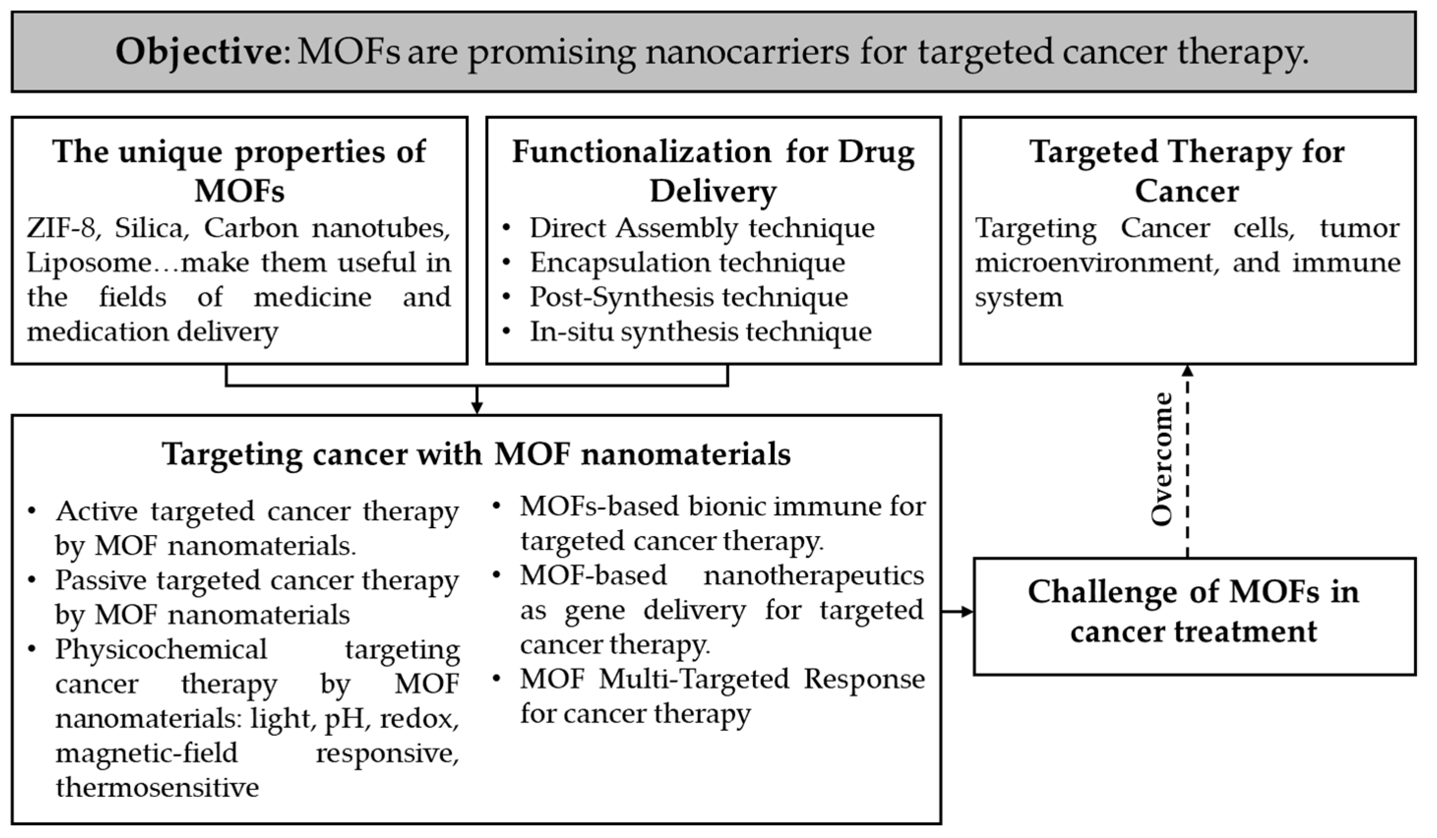
2. Basic Nanomaterial and Cancer and Target Therapy

2.1. Basics of Nanomaterials for Drug Delivery

Nanotechnology holds great promise for the management of chronic human diseases by delivering precise drugs to designated areas and targets. Recent years have seen many significant applications for the use of nanomedicine (biological, chemotherapeutic, and cancer immunotherapy agents) in the medical care of many diseases (Table 1). The review article offers a comprehensive summary of recent developments in the field of nanocarriers and drug delivery via nanocarrier technologies through a full assessment of the exploration and usage of nanostructured materials in enhancing both the effectiveness of old and new drugs and specific diagnosis through disease marker molecules [31].
Treatment delivery using MOFs has also been researched, starting with loading cancer drugs and controlling release. Small molecule medications, including the anticancer agents’ doxorubicin and curcumin, are still the main targets of applications of drug delivery MOFs. The development of macromolecular drugs with various MOFs, including plasmids, gelonin, siRNAs, and sgRNA-loaded Cas9 for CRISPR, has been accomplished via a method known as biomimetic mineralization. A burgeoning field of research focuses on using MOFs to deliver therapies of all sorts, such as cells, proteins, small chemicals, nucleic acids, gasotransmitters, and viruses [41].

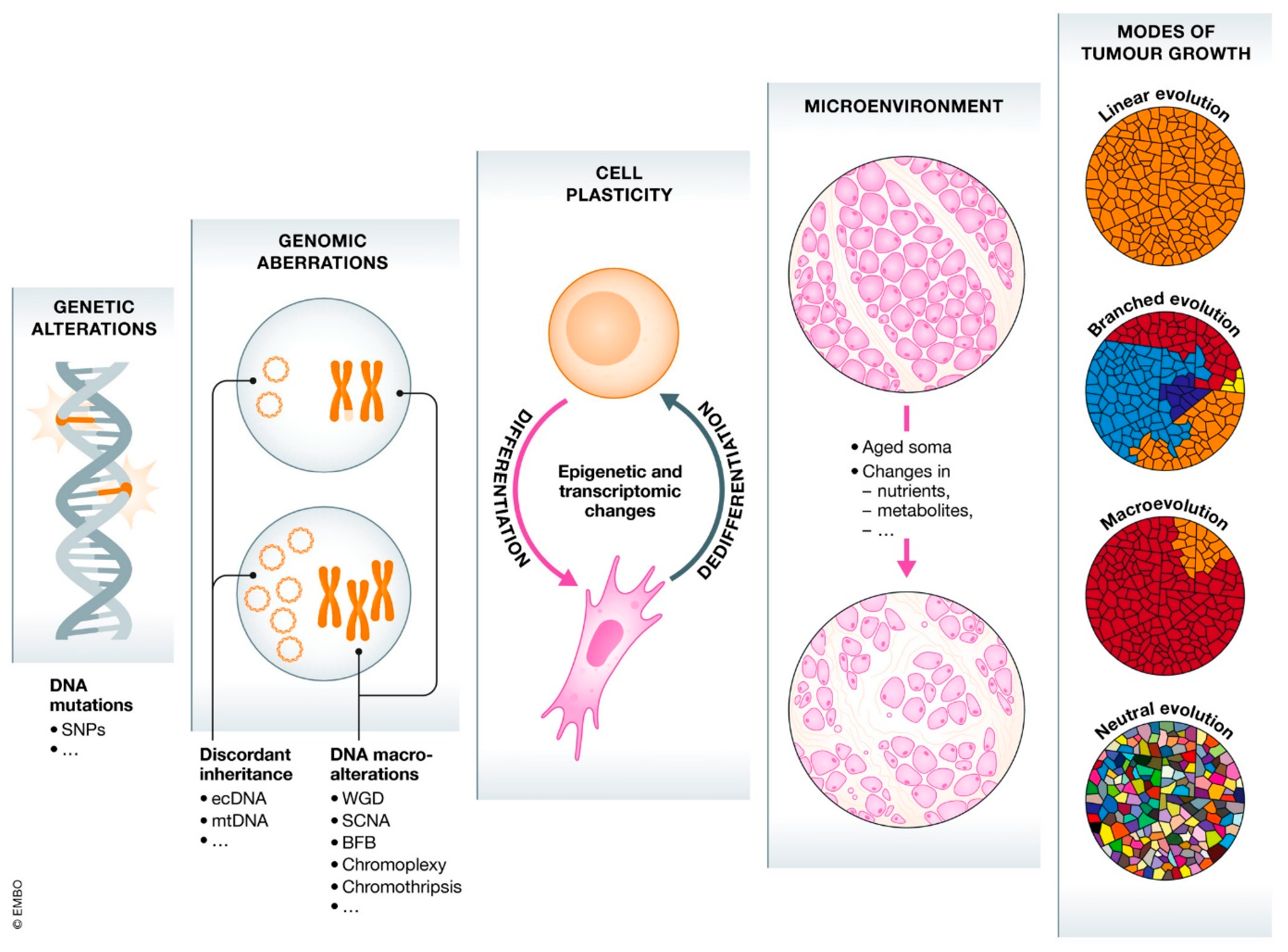
2.2. Basics of Cancer and Target Therapy

The evolutionary lens is reshaping our knowledge of cancer. Peter Nowell was an early pioneer of the theory of tumor evolution. According to Nowell’s concept, most cancers begin with a single neoplastic cell and then progress through a process of selection for somatic modifications, with the most aggressive clones eventually proliferating and surviving [42]. Incredible advances in genetics and cell biology in recent years are shedding new light on cancer that contradicts prior conceptions. The evolutionary viewpoint provides five fundamentals of evolution necessary for understanding cancer: (1) the formation of malignancies takes place through a process called somatic selection; (2) ecological principles can be used to describe the relationship between tumors and microenvironments; (3) principles of behavioral ecology give insight on the dynamics in which cancer clones interact with one another in terms of cooperation and competition; (4) natural selection may be credited for the rarity of cancer; and (5) the common occurrence of cancer is explained by evolutionary medicine [43,44,45,46,47]. Cancer is therefore a multifaceted disease due to its complex combination of genetic and environmental factors. It is now known that DNA damage is a fundamental cause of the abnormalities that eventually lead to cancer. As a result of these alterations, uncontrolled cell division occurs, which ultimately leads to tissue damage (Figure 3).
To combat the development and spread of cancer, scientists have developed targeted cancer medicines that work by interfering with certain molecules (“molecular targets”). Various terms, including “molecule-targeted pharmaceuticals”, “molecule targeted therapies,” “precision medicines,” and others, are used to refer to targeted cancer treatments [48,49]. The high degree of specificity achieved by targeted therapy is remarkable. This specificity permits the following comparisons between targeted therapies and conventional chemotherapy [49]:
  • In contrast to standard chemotherapies, which affect both rapidly dividing normal and malignant cells, targeted therapies focus on a narrow set of molecular targets that are suspected to play a role in cancer development and progression.
  • Targeted therapies are selected or engineered to interact with their target, while many mainstream chemotherapies were discovered because they kill cells.
Targeted therapies are frequently cytostatic (that is, they prevent the proliferation of cancerous cells), whereas conventional chemotherapy medicines are cytotoxic (that is, they kill tumor cells). Combinations of metal clusters and organic linkers fabric the structure of MOFs, which gives them many desirable properties (including a large surface area and pore volume, surface chemistry, and a tunable pore environment) and allows for their application in a wide range of imaging and drug delivery systems. Biocompatibility, large drug payloads, and the ability to hybridize with a wide range of functions make MOFs an attractive option for targeted drug delivery [8,50]. For biomedical applications, nanoscale MOFs that are Zr-linked, such as MOF-808, offer some significant benefits in particular [14]. To improve chemotherapy’s therapeutic efficiency and achieve a highly selective target in cancer cells, nanoparticles loaded with floxuridine and carboplatin and further functionalized with a poly (acrylic acid-mannose acrylamide) glycopolymer coating was developed. Specifically, in HepG2 human hepatocellular carcinoma cells, the modification boosted the absorption of the nanoparticles and provided a significant selective drug delivery with great cytotoxicity. These findings demonstrate that MOF-808 is a promising choice for future drug delivery investigations [14].

3. Synthesis, Functionalization, and Modification of MOF Nanomaterials for Targeted Cancer Drug Delivery

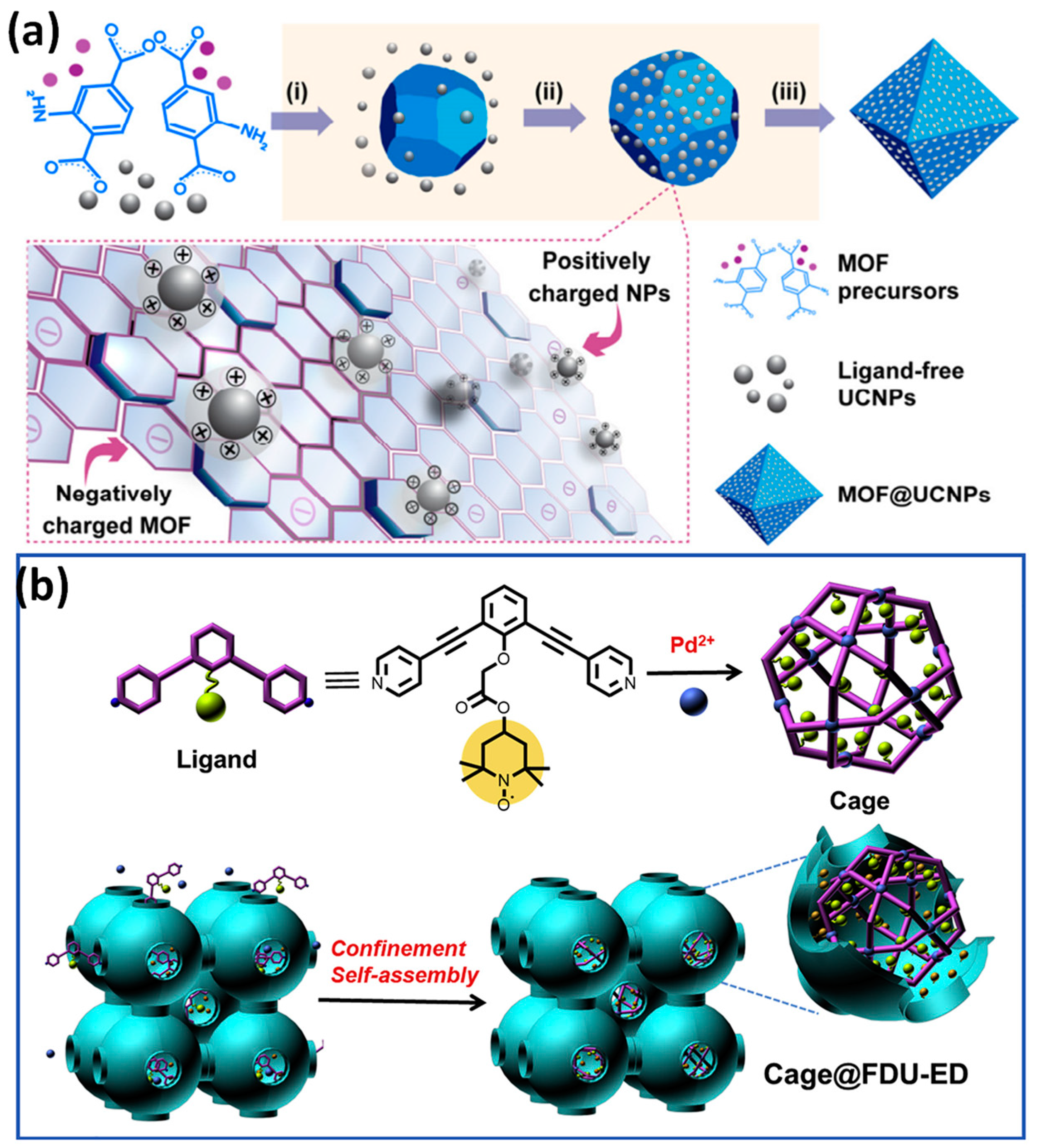
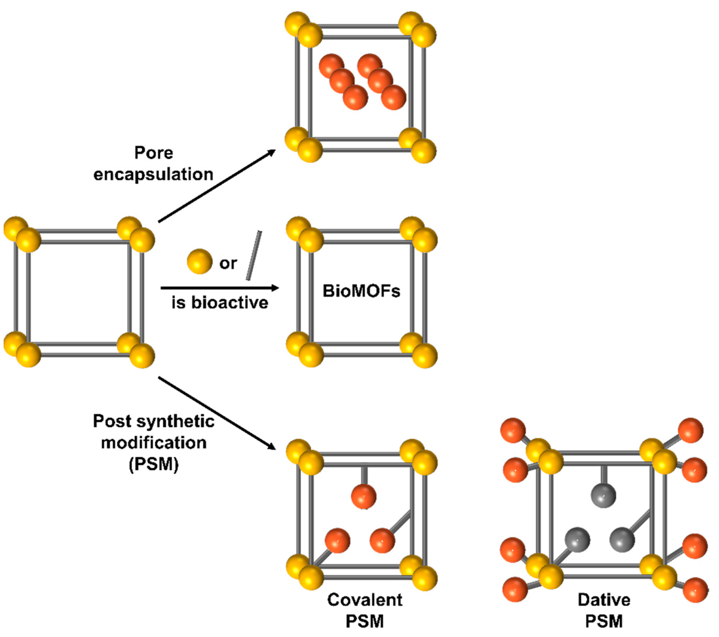
3.1. Direct Assembly Technique

In addition to being directly encapsulated, some cargo molecules or their prodrug form can be utilized as ligands to actively contribute to the development of framework structures through coordination bonds between the cargo’s accessible coordinated functions and particular metal nodes [51]. A few chemotherapy agents, including pamidronate, zoledronate, methotrexate, and several platinum-based anticancer agents, as well as photoactive cargos for phototherapy, have been effectively introduced into MOFs. Through the gradual breakdown of these cargo or prodrug ligands into active components in a physiological milieu, this type of MOF nanoparticle can achieve its entire therapeutic function. The most significant aspects of this technique are its uniform cargo distribution and increased loading within the NMOF matrix, but it is important to completely evaluate how to preserve the therapeutic action of those cargo ligands during the synthesis process [52,53].
Coordination modulation is currently widely used in MOF chemistry due to the exquisite control it provides for the synthetic chemist. The technique evolved from early attempts to regulate the particle size of MOFs into a set of diverse synthetic protocols to manufacture single crystals, simplify synthesis, induce defects, and regulate a variety of physical properties, and, additionally, it has recently provided conditions in complicated delivery systems.
Related methods are currently being developed, such as multivariate modulation to increase the storage of numerous cargo molecules and pore complexity from the MOFs porosity, which allows for the simultaneous control of surface chemistry and particle size. Coordination modulation is currently motivating other techniques to exercise kinetic control while applying to substitute materials because the capacity to influence self-assembly kinetics has the potential to be highly strong. Even in previously well-researched chemical regions, the kinetic control provided by the various modulation strategies should aid in the identification of novel materials [54].
The path is taken by self-assembly to create nanocomposites containing upconversion nanoparticles that are homogeneously paved over MOFs. This approach, which is primarily driven by electrostatic interactions, can be utilized to combine various upconversion NPs with various MOFs. The as-synthesized composites are helpful for applications such as luminescence-monitored medication delivery. They can also be utilized to create composites with distinctive architectures, such as MOF@upconversion NPs@MOF sandwiched nanocomposites (Figure 4) [55].
To create novel functionalized heterogeneous catalysts of Cage@FDU-ED during a reaction of (2,2,6,6-tetramethylpiperidin-1-yl)oxyl, metal-organic cages were placed inside mesoporous carbon with amino functions. The discovered bifunctional catalyst has an improved catalytic activity, selectivity, and recyclability due to the orthogonal properties of the segregated actively catalyzing locations, resulting in an overall transformation yield of up to a 96% conversion. It is possible to achieve a constant and highly effective chemical transformation by carefully designing the catalytic sites in both the mesoporous matrix and MOF cages separately (Figure 4) [56].

3.2. Encapsulation Technique

Active compounds, primarily anticancer medicines, have been effectively integrated into nanoMOFs utilizing the three primary methodologies depicted in Figure 5. This section will focus on the specifics of how biomolecules are enclosed within the pores of MOFs. Utilizing the high and modifiable porous structure of MOFs and encapsulating bioactive molecules within the pores of MOFs can be a simple and effective way to overcome the limitations of the surface attachment method. However, as the majority of biomacromolecules have diameters >2 nm, the greatest difficulty in capturing biomolecules comes in the fabrication of MOFs with large pore spaces. The International Union of Pure and Applied Chemistry (IUPAC) divides nanocomposites into three main categories determined by the size of their pores: macroporous (>50 nm), mesoporous (2–50 nm), and microporous (2 nm). Mesoporous MOFs are widely used as host matrices for biomolecules due to their ability to shield them from environmental perturbations such as pH and temperature shifts and organic solvents. In addition, the microenvironment around the encapsulated biomolecules can be changed by precisely controlling the structure or property (e.g., functionality, charge, or lipophilicity) of the pore walls of MOFs, creating optimal conditions for biomolecules’ activities or applications [57]. Particularly attractive for drug entrapment are Materials Institute Lavoisier (MIL) MOFs constructed from centers of trivalent metal and bridging ligands of polycarboxylic acid, which create extraordinary surface areas ranging from 1500 to 5900 m2/g and huge pore diameters ranging from 25 to 34 Å [58]. MIL MOFs have been successfully loaded with a wide variety of different sorts of active compounds, including anti-inflammatory, anticancer drugs, metallodrugs, antiviral, nitric oxide, and peptides (Table 2). Several additional MOFs, including those based on zinc, copper, and zirconium, were utilized as drug carriers. Due to the possible advantages that metal ions such as Gd, In, and Ni could impart, several MOFs based on them were also examined for drug encapsulation, and their imaging characteristics were also investigated. On the other hand, it has been noted that the toxicity of metals might be an obstacle to the use of certain MOFs in medical applications.

3.3. Post-Synthesis Technique

In the absence of functional groups, the structurally unmodified form of an MOF may restrict its usefulness. Toward this aim, post-synthetic modification (PSM) is performed to increase the functional groups attached to MOFs (Figure 5) [109,110], thereby extending their potential spectrum of applications. Briefly, PSM is a systematic approach to surface functionalization that is used to introduce functional groups into MOFs [111,112]. If suitable functional groups are installed, PSM has the potential to enhance the chemical and physical characteristics of materials. This alteration also serves to govern the overall utilization (colloidal stability or self-assembly under varying conditions) and its interactions with its surroundings, such as a target-specific accumulation [110]. Surface functionalization has many additional benefits, including (i) preventing nanoparticle aggregation; (ii) phase transfer, transferring nanoparticles from one solvent to another solvent (e.g., organic solvent to water); (iii) allowing nanomaterials to associate with particular biomolecules of attention, such as nucleic acid, in delivery, therapeutic use, and biological networks for imaging; and (iv) modification using fluorescent dyes for functionality [113].

3.4. In Situ Synthesis Technique

Given their host–guest characteristics and ease of chemical synthesis modification, one of the most important and potentially useful applications of MOFs is the delivery of drugs. The inorganic segment of MOFs regulates medication release, while the organic segment of MOFs can be customized to encapsulate a variety of pharmaceuticals. Even though these substances have demonstrated an adequate drug loading capacity and controllable drug distribution behavior, few studies on drug delivery in MOFs have been established up until this point. A nanoscale MOF was recently used for effective medication loading and delivery [114].

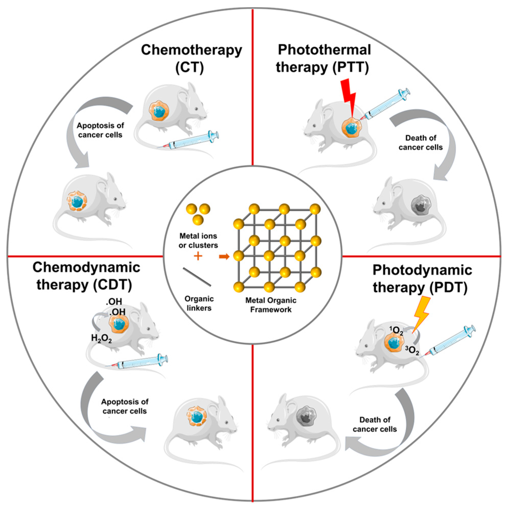
4. Applications of MOF Nanomaterials in Targeting Cancer Therapy

4.1. Active Targeted Cancer Therapy by MOF Nanomaterials

Using an active targeting method, nanoparticles (NPs) can increase the intracellular concentration of medications in malignant cells while minimizing damage in healthy cells. Bioscience-enhanced NPs are actively being developed for targeted drug administration, biomarkers for cancer using bimolecular profiling, and in vivo tumor imaging. The patient will need to take fewer doses more frequently, the drug will have a more consistent impact, there will be fewer side effects, and the levels of the drug in the blood will fluctuate less. These are all benefits of the active targeted release system. Active targeting entails adding various ligands to the medication or DDS, including vitamins, peptides, antibodies, sugars, and biological proteins. These ligands interact with cell receptors to form complexes that induce the drug to assemble within the target cells [115,116,117].
A methodical approach to creating an MOF that is two-photon active is through a click reaction of PCN-58-Ps. Hyaluronic acid is additionally added to PCN-58-Ps by coordination to give it cancer-cell-specific targeting characteristics. The improved composite of PCN-58-Ps-HA consequently demonstrates the strong activity of two-photon (up activation by a laser with a 910 nm wavelength) and light-activated ROS of 1O2 and O2•− production capabilities (Figure 6a,b). Future clinical applications of deep-tissue cancer imaging using two-photon PDT that is activated by NIR light and treatment have a large amount of potential due to the interaction of these two crucial variables inside the framework of PCN-58-Ps-HA [118].
Glycyrrhetinic acid (GA), lactobionic acid (LA), dual ligands of GA and LA, and folic acid (FA) were designed and built as effective multifunctional DDSs for combating hepatocellular carcinoma (HCC). Doxorubicin was loaded into the Zr (IV)-based NMOF (NH2-UiO-66) nanoscale. It was established that pure NH2-UiO-66 was safe when used with HSF cells based on biocompatibility experiments; however, DOX-loaded NMOF was discovered to be more harmful to HepG2 cells by flow cytometry (Figure 6c). The created dual-ligated NMOF demonstrated a pH change in response to the DOX release [119].

4.2. Passive Targeted Cancer Therapy by MOF Nanomaterials

To achieve screening and therapeutic uses in cancer nanobiotechnology in vivo, nanoparticles must be transported to cancer locations. To achieve this, two broad strategies have been employed: passive targeting and active targeting (Figure 7) [120,121,122]. Passive targeting exploits the biological characteristics of tumors to enable nanocarrier accumulation in tumors via an improved permeability and retention (EPR) [123]. For the accumulation of NPs in tumors, passive targeting relies on aberrant gap junctions (100–600 nm) in the endothelium of tumor blood arteries. MOFs can typically be adequately manipulated at the nanoscale for passive targeting. Park and colleagues explored the HeLa human cervical tumor cell absorption of a porphyrinic MOF (PCN-224) by altering the particle size to boost the cytotoxicity and internalization via passive targeting [124]. They found evidence that MOFs improve the photodynamic performance. The cytotoxicity of photosensitizers is minimal when MOFs are not included. The use of TCPP@PCN-24 in photodynamic therapy was found to be most successful when the particle size was 90 nm, whereas the use of this combination was found to be least effective when the particle size was 190 nm. Due to its superior retention impact in the tumor area, Duan also showed that particles of size 60 nm AZIF-8 have an anti-tumor effect superior to those of other sizes [125].

4.3. Physicochemical Targeting Cancer Therapy by MOF Nanomaterials

4.3.1. Light-Responsive Targeted Cancer Therapy by MOF Nanomaterials

Recent advances in light-mediated nanomedicines, particularly their minimally invasive abilities and great spatiotemporal accuracy, have made them attractive methods for precisely controlling the therapeutic activation and imaging probes both in vivo and in vitro (Figure 8) [126,127], particularly the light-activated MOF-based therapeutic system, which not only offers imaging-guided or combination therapies but also improves the laser penetration depth and targeting [128,129].
A biodegradable and biocompatible MOF was created for effective drug loading and controlled release by developing Au (gold nanorods) @ZIF-8 (crystalline zeolitic imidazolate framework-8). A significant drug loading efficiency of roughly 37% was achieved by the Au@ZIF-8 while loading doxorubicin. The ZIF-8 layer was swiftly destroyed by NIR light or a mildly acidic environment, which led to an on-demand medication release at the tumor location. More significantly, because of the synergistic effects of photothermal treatments and chemotherapy, highly efficient cancer treatment was accomplished in both in vitro cell experiments and in vivo tumor-generating naked mice experiments under the irradiation of a near-infrared laser. The in vivo study also demonstrated Au@ZIF-8’s good biocompatibility (Figure 8) [130].
The nanoparticle is made up of a cancer cell membrane shell and a metal–organic framework core coated in MnO2 nanosheets (CM-MMNPs). The H2O2 and H+ responsiveness of the MnO2 layer allows it to generate O2, enhancing the generation of O2-mediated singlet oxygen (1O2) for photodynamic therapy (PDT). Additionally, the resultant Mn2+ is a superior MRI contrast material. The CM-MMNPs are given cellular endocytosis that occurs with strong stability and integrity and a dependable homologous cell-targeting capability by the addition of membrane proteins and cell membranes. This multifunctional nanoparticle offers a new paradigm for targeted therapy, diagnosis, and treatment, and can treat cancer cells’ hypoxia with PDT [127].

4.3.2. pH-Responsive Targeted Cancer Therapy by MOF Nanomaterials

Chemodynamic therapy (CDT), a new therapeutic, is described as the generation of cytotoxic OH at tumor locations via a Fenton or Fenton-like reaction [131,132]. Because CDT relies on the Fenton-type process, which primarily catalyzes endogenous hydrogen peroxide (H2O2) to produce OH, the low H2O2 level in solid tumors will limit its usefulness. Therefore, combining GOx-induced fasting therapy with CDT is a brilliant move that has the potential to greatly boost synergistic therapeutic effects. A pH-responsive nanoplatform was developed by encapsulating GOx and natural hemoglobin (HB) together in ZIF-8 via co-precipitation to combine CDT and starvation therapy effectively [133,134]. Another study revealed the creation of a biodegradable mesoporous Fe (III) polycarboxylate MOF exhibiting the pH-sensitive and reversible aggregating ability to specifically target the pulmonary for drug administration in order to efficiently suppress lung cancers (Figure 9) [135]. Using a lung metastasis model, the nanoparticles were randomly aggregated in the capillaries and subsequently disaggregated after 24 h, allowing the encapsulated medication to be released. The pH-responsive property might not just facilitate the launching of medications in the appropriate location within tumor cells but may also be used to synthesize MOFs that disintegrate at a specific pH, resulting in rapid drug release [136].

4.3.3. Magnetic-Field-Responsive Targeted Cancer Therapy by MOF Nanomaterials

A hybrid magnetic nanocomposite with a well-controlled size distribution, typically 100 nm, is produced by combining MIL-88B-NH2 MOF-structured Fe3O4 magnetic NPs using tailored synthetic media, including F127 copolymer as a stabilizing agent and acetic acid as a modifying agent. The nano MOF has also been used as a nanocarrier for controlled medication release on demand and effective drug delivery. Carmustine and Mertansine, two glioblastoma medications, were perfectly inserted into the MOF structure’s pores to reduce their potent internal harmful effects, which restrict their clinical use. Moreover, localized heating was caused as a result of the magnetic local minimum included in the MOF NPs when a modified magnetic field was practiced on the magnetic nanocomposites loaded with DM1 to accomplish controlled drug release. To verify the therapeutic effectiveness of the DDS on U251 glioblastoma cells, in vitro cytotoxicity experiments were performed (Figure 10) [137].
An Fe2Mn(3-O) cluster was used to create an FeMn-based ferromagnetic MOF. FeMn-MIL-88B acquired its ferromagnetic properties with the addition of Mn. As a model drug, 5-Fluoruracil (5-FU) was entrapped in MOFs, and its regulated release that was responsive to both pH and H2S stimuli was achieved. In tumor microenvironment (TME) simulation media, FeMn-MIL-88B revealed an impressive capacity for loading 5-FU (43.8 wt%) and the quick release of the drug. Additionally, the carrier’s cytotoxicity profile against embryonic kidney cells of humans indicates no negative effects (100 g/mL). The low toxicity values (LD50; Mn = 1.5 g/kg, Fe = 30 g/kg, and terephthalic acid = 5 g/kg) of the MOF’s basic components can be attributed to the less hazardous effect on the cell viability (Figure 10) [138].

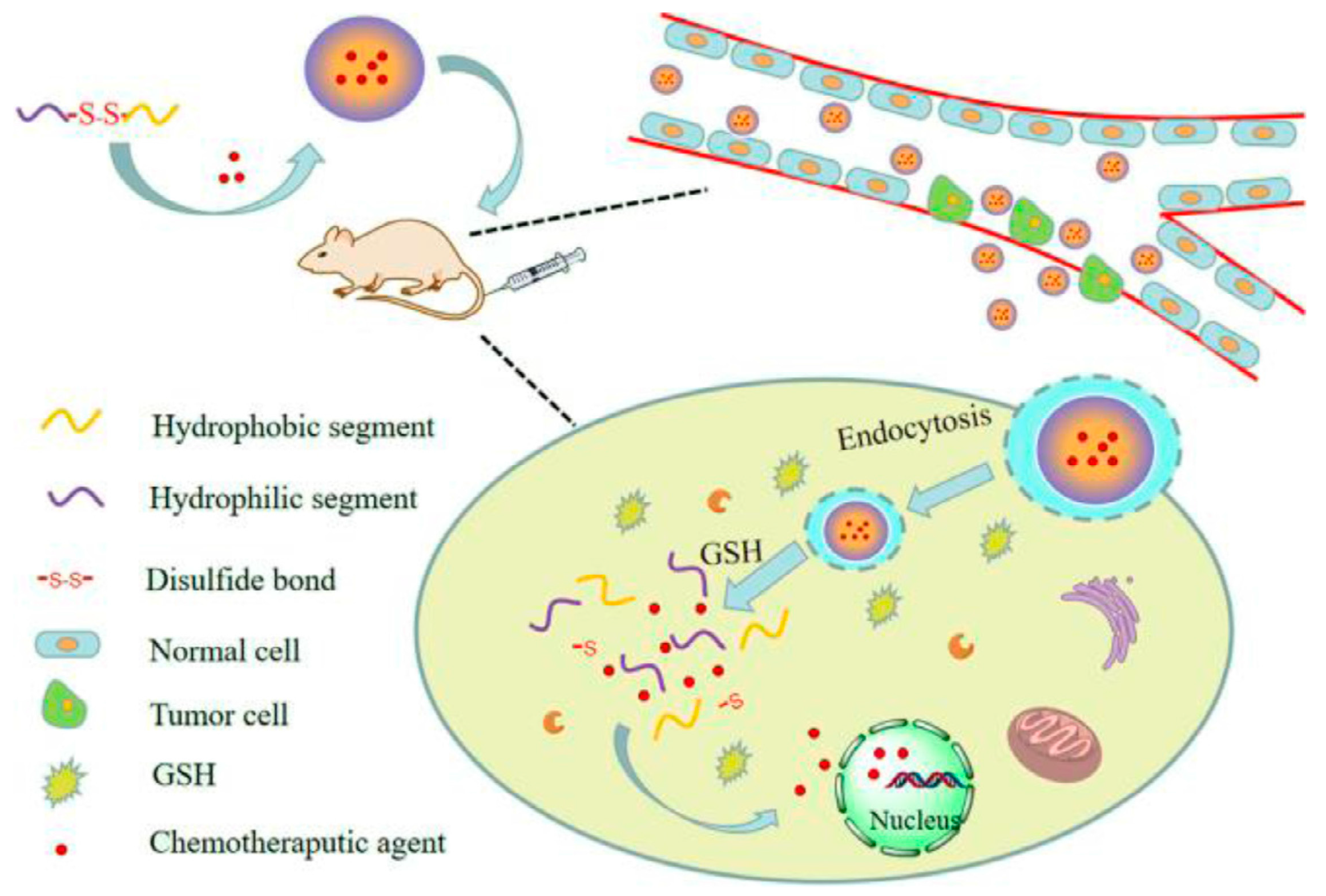
4.3.4. Redox-Responsive and Targeted Cancer Therapy by MOF Nanomaterials

The abnormal physiological properties of cancerous tissue are reflected in the presence of distinct cellular microenvironments, such as reducing, acidic, and enzyme environments [139]. With significant reducing capacities, the reduction states and oxidation of nicotinamide-adenine dinucleotide phosphate (NADPH) and glutathione (GSH) allowed them to primarily manage the reducing environment of cancer cells [140]. In a reducing environment, GSH that has a concentration greater than that of NADPH plays a crucial function in microenvironment regulation. GSH fragments and forms disulfide (S-S) links to regulate the cellular reducing environment. The intracellular tumor GSH concentration was higher than the extracellular tumor GSH concentration. Tumor tissues had four times greater GSH concentration than normal tissues. To take advantage of these features, numerous nanoscale drug delivery devices for monitoring the reducing environments of cancerous tissue and triggering drug release by disulfide bond breakdown in GSH-responsive nanomaterials have been constructed (Figure 11) [139,141]. Disulfide bonds have several important applications in MOF fashion, including the architecture of ligands and the alteration of surfaces [142,143,144]. Zhao et al. described the fabrication of Mn-S-S, a glutathione-responsive MOF system, using Mn2+ and dithiodiglycolic acid as ligands, thus inserting the S-S link into the MOF ligand [143]. The disulfide bond was cleaved in the existence of glutathione, which allowed for the medication (DOX) to be successfully released from its encapsulation. In addition, the Mn2+ in Mn-SS@MOF showed an improved T1 contrast in the medical diagnostic of magnetic resonance imaging (MRI). In addition, Liu et al. designed an MOF that is both redox-sensitive and tumor targeting by attaching folic acid and functional S-S anhydride to the organic linkages of UiO-66-NH2 MOFs [145]. Drugs in MOFs are released in response to redox stimuli, and this is achieved by the overexpression of GSH in tumor cells, which causes an assault on the thiolate moiety and cleaves the S-S bonds.

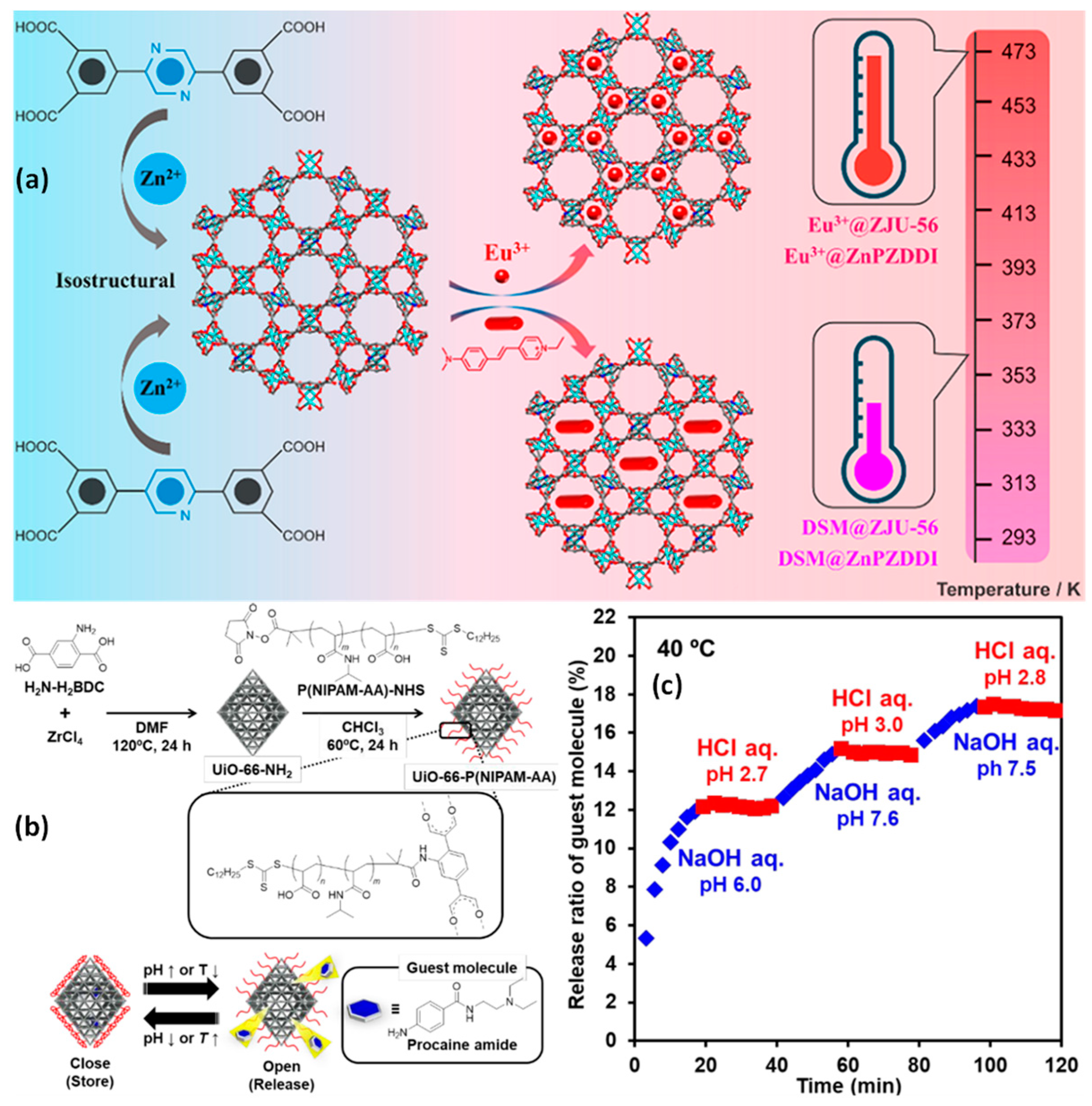
4.3.5. Thermosensitive MOFs for Targeted Cancer Therapy

An MOF contains organic linkers that are synthesized utilizing NbO-type Zn2+, which contains two structurally identical tetracarboxylate ligands with pyrazine or pyridine moieties. The trivalent europium ion (Eu3+) and the pyridinium hemicyanine dye 4-p-(dimethylamino)styryl]-1-methylpyridinium (DSM), both of which have cationic red-emitting units, were embedded in various composites, and their potential as ratiometric temperature probes was assessed. These dual-emitting composites’ temperature-responsive luminescence was examined, along with their characteristic features of temperature resolution, relative sensitivity, spectral repeatability, and luminous color change. The Eu3+@ZnPZDDI and Eu3+@ZJU-56 exhibit good sensor temperature ranges and high relative sensitivities, indicating that the composites can be extensively designed by integrating the guest and host units (Figure 12) [146].
By post-synthetic modifying, a copolymer comprising N-isopropyl acrylamide and acrylic acid was utilized to cover the MOF. The additional molecules can be released in an “on-off” fashion thanks to the polymer’s quickness as well as the transition from a reversible coil to a globule, which is pH- and temperature-responsive. When the polymer assumes a coil shape at low temperatures (25 °C) or a high pH of 6.86, the additional molecules are quickly freed from the MOF. The release of the attached molecules is blocked when the polymer takes on the shape of a globule with a pH of 4.01 and/or warm temperatures of over 40 °C. Even once the release has begun, it can be stopped by adding external stimulation (Figure 12) [147].

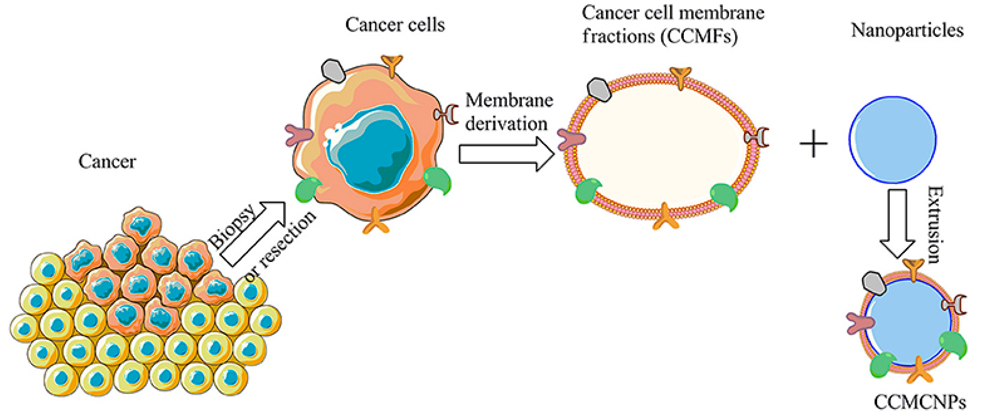
4.4. MOF-Based Bionic Immune for Targeted Cancer Therapy

The immune system is a sophisticated biological architecture crucial for recognizing and removing invading invaders, destroying abnormal cells, and preventing the development of tumors [148,149,150]. Therefore, immune cell therapy presents significant opportunities for treating diseases such as cancer, autoimmune diseases, inflammation, and infections. There are many different kinds of immune cells, each of which performs a specific function and has the potential to be used as a live treatment for a variety of disorders [151]. To optimize pharmacokinetics and degradation, a bionic nanoplatform can deliver drugs to the immune system (Figure 13). These techniques have strengthened drug bioavailability by providing extra protection and targeting, encouraging the enhancement and evolution of bionic solutions [152,153,154]. In expanding the variety of medications that can be loaded, Gong et al. came up with the innovative idea of developing a hybrid coating made up of macrophage and tumor-cell membranes. This coating combined the characteristics of both kinds of cells [155]. These cells targeted particular homogeneousness and metastasis, and also aggregated in inflammatory areas. Thus, this combination offers promise for nanobionic architecture. However, the bionic system of cancer cell membranes needs to be improved and made more open to innovation.

4.5. MOF-Based Nanotherapeutics as Gene Delivery for Targeted Cancer Therapy

The genetic advancements cleared the door for the introduction of genetic engineering, and, since 1980, gene therapy has become an increasingly prominent topic in cancer research. The three primary categories of gene therapy approaches are immunomodulatory, corrective, and cytoreductive [156]. Immunomodulation is the process of boosting the body’s immune barrier to effectively identify and destroy cancer cells. An important goal of corrective gene therapy is to restore the normal function of a gene whose mutation contributes to the development of cancer. Suicide gene therapy, in which a gene is inserted into cells that codes for an enzyme that converts a harmless prodrug into its toxic metabolite, is also being studied extensively as a means of treating cancer. It has been hypothesized that nucleic acids incorporated into MOF nanocarriers might be stabilized against degradation and taken up by cells more quickly. In addition, the steric and electrostatic impediment to aggregation could be enhanced by the surface configuration of an MOF with nucleic acids, resulting in an improved colloidal consistency. MOFs have found widespread application in the administration of drugs, the transport of siRNA, and the encapsulation of DNA/RNA, proteins, and polysaccharides, as well as in prokaryotic and eukaryotic organisms [157,158]. As an example, ICG@ZIF-8 was used by Liu et al. as a means of electrostatically adsorbing siRNA (ICG@ZIF-8@siRNA) to promote siRNA diffusion under laser control [159].

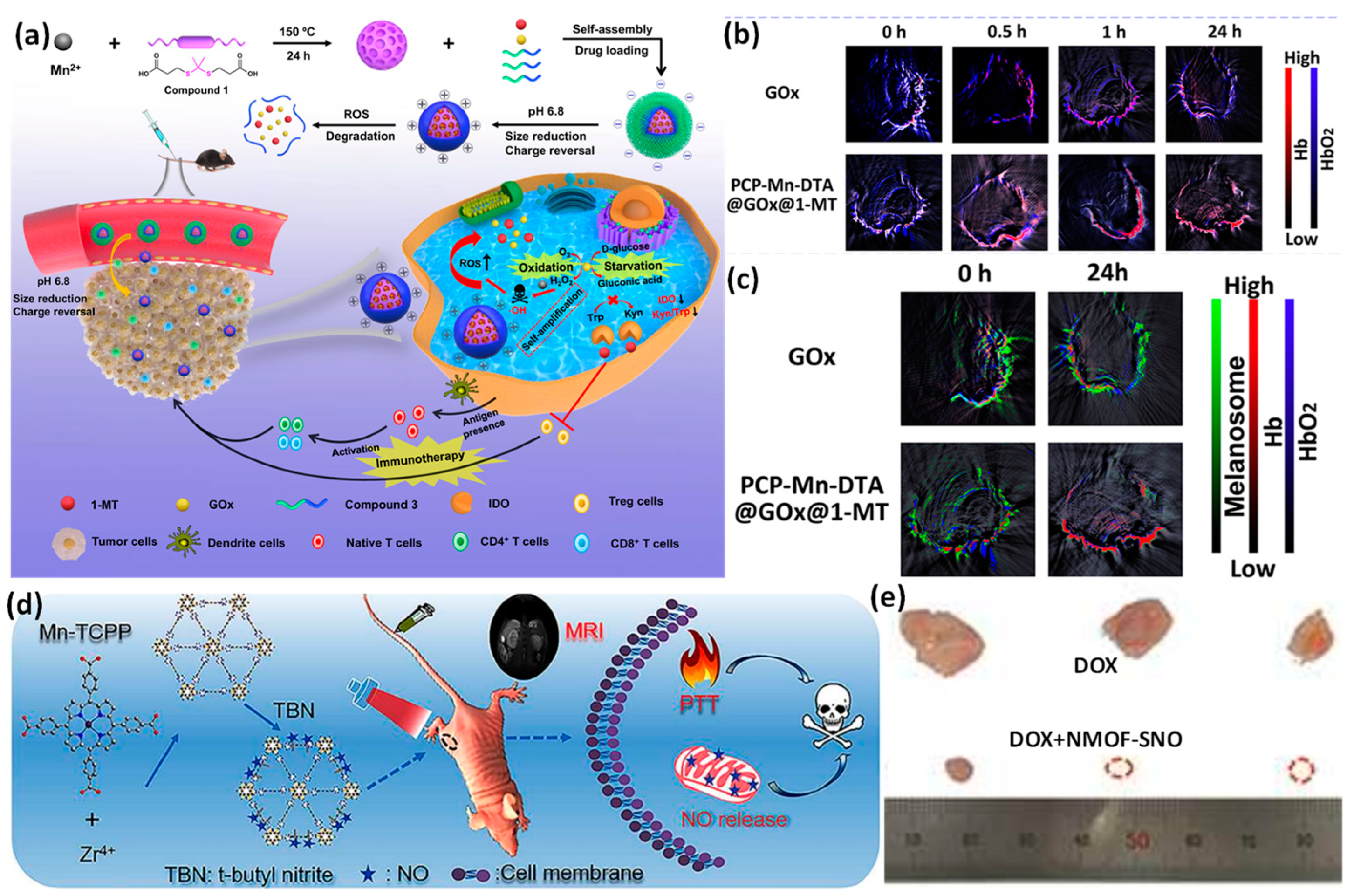
4.6. MOF Multi-Targeted Response for Cancer Therapy

With self-amplified releasing and improved penetrating, a pH and ROS dual-sensitive biodegradable MOF nanoreactor-based nanomaterial was created to provide GOx and 1-MT together for combination oxidation/starvation treatment and IDO-blockade malignancies immunotherapy (Figure 14a–c). The comprehensive in vivo and in vitro results validated PCP-Mn-DTA@GOx@1-MT nanomaterial to not only proficiently strengthen immune system activation with a decreased sensitivity by GOx-activated starvation/oxidation intervention and IDO-blockade immunotherapeutic, but to also strategically overwhelm biobarriers and increase the distribution efficiency through the mildly acidic tumor cells [160,161,162].
As a novel method for cancer therapeutic applications, a nanoscale MOF platform will merge magnetic resonance imaging, photothermal therapy, and spatiotemporally programmable NO delivery. The MOFs are generated as a proof of concept using biodegradable Mn-porphyrin and Zr4+ ions as linking ligands. Mn-porphyrin gives the NMOF a robust T1-weighted MR contrast performance and a significant photothermal transformation for effective PTT through the incorporation of paramagnetic Mn ions into porphyrin rings. For heat-sensitive NO production, S-Nitrosothiol (SNO) is attached to the NMOF surfaces. Additionally, a single NIR (near-infrared) light triggers both the PTT and regulated NO release concurrently for their effective synergistic therapy in a single step. The tumor-bearing mice’s MR images reveal that NMOF-SNO exhibits effective tumor accumulation after intravenous injection. Tumors in mice given NMOF-SNO injections are fully suppressed when applied by NIR laser, demonstrating the effectiveness of the drug [163].

5. Challenge of MOF Nanomaterials in Cancer Treatment

5.1. Toxicity and Biocompatibility

For application in biomedical and pharmaceutical applications, MOFs must present toxicologically compatible characteristics. The cytotoxicity of several representative MOFs was evaluated on zebrafish embryos. At 120 h after fertilization, it was discovered that the viability of embryos that had been subjected to Co and Mg-MOF-74, UiO-66 and 67, and MIL-100 and 101 was not significantly different from those without MOFs (the control group), even at a concentration as high as 200 μM. On the contrary, the embryo viability rate (EV) was significantly reduced by ZIF-7 and 8 and HKUST-1. At a concentration of 200 μM, ZIF-7 was marginally harmful, with EV= 79.2%, whereas ZIF-8 was more toxic, with EV = 33.3%. NanoHKUST-1 was extremely hazardous even at a concentration of 20 μM (EV = 0%). It was highlighted that the absorption of solubilized metal ions had a crucial role in determining the toxicity potential of MOFs [164]. Rats were used in the experiment to evaluate both the toxicity of trimesic acid and iron trimesic MIL-100 (Fe). MIL-100 and trimesic acid were administered intravenously at doses of approximately 220 mg/kg and 78 mg/kg, respectively, and the study revealed that weight growth and the animal behavior of treated rats were completely normal when matched with the group that served as the control. This led to the conclusion that the concerned ingredient is suitable for drug delivery [165].
The biocompatibility testing of MOF building blocks is necessary before MOFs can be used in biomedical applications because some forms may have a harmful influence due to the degradation of MOFs within cells [166]. Additionally, it is also essential to examine the biocompatibility of MOFs with different types of cells since the outcomes may differ. In vitro analyses were conducted to study the biocompatibility of three distinct metal systems of MIL-100 MOFs (Fe, Al, and Cr). Even at high doses, they did not produce in vitro cell toxicity in the liver (HepG2) and lung (Calu-3 and A549) cell lines. Only the toxicity of MIL-100(Fe) was seen in the Hep3B cell line [167]. However, the other research, observing the biocompatibility of ZIF-8 concerning six distinct cell cultures, each one representing a different part of the human body (skin, breast, blood, kidneys, bones, and connective tissues), showed that ZIF-8 at concentrations higher than the threshold level of 30 μg/mL exhibited cytotoxicity [168]. This might be related to the influence of liberated Zn2+ on the generation of mitochondrial reactive oxygen species. Despite the tremendous amount of research conducted in the field of nanomedicine and enhanced nanocarrier therapeutics directly attacking cancer, the process of bringing medication from the research lab to the industry is frequently intermittent and highly slow. Most of the issues can be traced back to the lack of rigorous monitoring of the safety and effectiveness-by-design strategy for nanomedicines.

5.2. Drug Release before Reaching the Target Cancer

The performance and safety of MOFs are significantly impacted by their chemical stability. The stability of MOFs is affected by several variables, including pH, temperature, humidity, solvents, metal ions, and biological molecules. Different stability levels may be needed depending on the application. For instance, MOFs must be able to survive the stomach’s acidity and the intestine’s alkalinity when given orally. MOFs must be resistant to metal ions and tear fluid for ocular administration [169]. The stability of MOFs for drug delivery has been improved through the development of several methods: the addition of protective layers such as polymers; the selection of more stable metal nodes or organic ligands; and the addition of functional groups or linkers to MOF structures or compositions [170]. The porosity, loading capacity, release kinetics, biocompatibility, and toxicity of MOFs may also be impacted by these tactics. Hence, for each application, a careful balance between stability and functionality needs to be struck [171].
Because of their modifiable surface, tunable size, high active ingredient loading, good biocompatibility, and, most importantly, their capacity to be selectively distributed in tumor cells via an increased retention and permeability, MOFs made up of bridging ligands and metal attachment sites were investigated as a new innovative system for the enhanced treatment and diagnosis of cancer. However, the following points need to be studied more deeply. However, controlling drug release during cancer cell targeting is quite important.
The body typically experiences severe adverse responses as a result of chemotherapy medications. Therefore, it is critical to increase the targeted drug delivery system’s stability to prevent both medication leakage outside of the tumor and significant detrimental effects on the body. Cell-membrane-coated biomimetic approaches are already common; however, they have more expensive material needs. The development of an MOF-based DDS suited for clinical applications, from synthesis to in vivo process monitoring, and quality control still needs to be carried out [116].

5.3. In Vivo Studies and Applications

Despite the unparalleled benefits, more focus needs to be placed on in vivo investigations of MOFs, such as the toxicity and biocompatibility. More information is required to fully comprehend the metabolic and mechanical operations of MOFs in the body since they degrade. Numerous research studies on the cytotoxicity of MOFs in vitro have been recently published. However, cell models do not reveal the same biocompatibility of MOFs in the body, despite the good biosafety at a given dose. The majority of research on the toxicity and metabolism of MOFs-based DDS have been limited to their anti-tumor activities in experimental animals.
The Fe3O4@C@PMOF metabolism in nude mice was studied in breast cancer nude mice. Fluorescent in vivo imaging with the nanoparticles is possible. The presence of fluorescent dots in the lymph and liver nodes proved that NPs could take part in both lymphatic and blood circulation. The tumor region then expanded to form the tissue with the maximum fluorescence intensity. The NPs were expelled from the body through stool after 8 days. The mice who received the injections acted normally, and their weight did not significantly drop. The primary organs of mice did not exhibit any pathological changes eight days after injection, and there was good biocompatibility [172].
The toxicity of a porphyrinic MOF nanosystem to the major organs, blood, and tissues of mice was assessed as part of a study on in vivo biosafety. All indications were in the usual range, proving the nanosystem’s high level of safety [173,174]. Iron (III) MOFs’ in vivo toxicity was examined using markers such as serum, enzymes, and histology, all of which were consistent with low toxic effects. The liver and spleen isolate the nanomembrane, which is further biodegraded into carboxylic acids and iron and subsequently eliminated directly in the feces or urine while still preserving iron homeostasis. This demonstrates that iron (III) carboxylate MOF NPs are non-toxic and biodegradable [175].

5.4. Quality Control from Laboratory Scale to Industrial Scale

Research on MOF’s biomedical efficiency is still being conducted in small-scale production and laboratory testing at this time. When MOFs are produced in large quantities, it can be challenging to control their quality, which can cause fluctuations in the pore size and material size. Additionally impacted are the drug loading capacity and release rate. Therefore, one of the most difficult issues is the construction of stable and manageable MOFs (Table 3).

6. Future Perspectives of MOF Nanomaterials in Cancer Treatment

Although the biofunctionalization of MOFs has been the subject of substantial investigation in the area of nanomedicine basic research, its use in clinical therapy still has a long way to go. MOFs with good biocompatibility and non-toxicity or low toxicity require optimizing the preparation and synthesis of the nanomaterials such that they can circulate for an extended period and are efficiently eliminated via metabolism. An additional experiment is necessary to fully understand the biodegradability and stability of MOFs in the body. There is a requirement for reinforcement in light of the effects of MOFs nanomedicines on the physiological activities of organisms. Going forward, building dynamics on MOF-responsive biomaterials and responding to specific physiological stimuli by the targeted and timed release of bioactive molecules have significant research significance and application potential.

7. Conclusions

Biomolecule–metal–organic framework composites have advanced substantially over the past decade to become novel platforms for an extensive variety of solutions in the identification and treatment of cancer. Here, we reviewed recent improvements in the fabrication of biomolecule–MOF composites and their application in cancer treatment. Integrating biomolecules into MOFs has been accomplished using a wide range of different approaches, including surface adhesion, covalent bonding, pore encapsulation, in situ synthesis, and bioengineering MOFs, and has led to significant progress in the development of targeted therapy for cancer. Due to the advent of MOFs, it is now possible to overcome the limitations of conventional cancer therapeutic approaches, such as vulnerability, inadequate absorption, low aqueous solubility, low selectivity, high lethality, and multidrug resistance. Although MOFs are gaining importance for cancer prevention and treatment, each inclusion approach has advantages and disadvantages. Their promise as medication carriers in cancer treatment is still in its infancy. In fact, a significant number of MOFs are now going through clinical or preclinical testing in preparation for being approved and made accessible for purchase. Global research on using nanostructured frameworks for the goal of generating patient-specific drug delivery systems will assist the synthesis and development of MOF–drug composites for real-world applications. With the assistance of future development strategies of MOF-based materials for cancer therapy, it will be possible to reduce the cost of cancer treatment while simultaneously increasing the patient survival and quality of life.

Author Contributions

Conceptualization, V.A.T. and G.N.L.V.; methodology, V.A.T. and G.N.L.V.; software, G.N.L.V. and V.A.T.; validation, V.A.T. and G.N.L.V.; formal analysis, V.A.T., G.N.L.V. and V.T.L.; investigation, V.A.T., G.N.L.V. and V.D.D.; resources, V.A.T. and V.D.D.; data curation, G.N.L.V. and V.T.L.; writing—original draft preparation, V.A.T. and G.N.L.V.; writing—review and editing, V.A.T. and G.N.L.V.; visualization, V.A.T. and G.N.L.V.; supervision, V.A.T. and G.N.L.V.; project administration, V.A.T. and G.N.L.V.; funding acquisition, V.A.T. All authors have read and agreed to the published version of the manuscript.

Funding

“Vy Anh Tran was funded by the Postdoctoral Scholarship Programme of Vingroup Innovation Foundation (VINIF), code VINIF.2022.STS.45” and “The APC was funded by voucher discount”.

Institutional Review Board Statement

Not applicable.

Informed Consent Statement

Not applicable.

Data Availability Statement

Not applicable.

Conflicts of Interest

The authors declare no conflict of interest.

References

  1. He, S.; Wu, L.; Li, X.; Sun, H.; Xiong, T.; Liu, J.; Huang, C.; Xu, H.; Sun, H.; Chen, W. Metal-organic frameworks for advanced drug delivery. Acta Pharm. Sin. B 2021, 11, 2362–2395. [Google Scholar] [CrossRef] [PubMed]
  2. Hoskins, B.F.; Robson, R. Infinite polymeric frameworks consisting of three dimensionally linked rod-like segments. J. Am. Chem. Soc. 1989, 111, 5962–5964. [Google Scholar] [CrossRef]
  3. Furukawa, H.; Cordova, K.E.; O’Keeffe, M.; Yaghi, O.M. The chemistry and applications of metal-organic frameworks. Science 2013, 341, 1230444. [Google Scholar] [CrossRef] [Green Version]
  4. Ding, N.; Li, H.; Feng, X.; Wang, Q.; Wang, S.; Ma, L.; Zhou, J.; Wang, B. Partitioning MOF-5 into confined and hydrophobic compartments for carbon capture under humid conditions. J. Am. Chem. Soc. 2016, 138, 10100–10103. [Google Scholar] [CrossRef] [PubMed]
  5. Connolly, B.M.; Madden, D.G.; Wheatley, A.E.; Fairen-Jimenez, D. Shaping the future of fuel: Monolithic metal–organic frameworks for high-density gas storage. J. Am. Chem. Soc. 2020, 142, 8541–8549. [Google Scholar] [CrossRef]
  6. Wang, X.; Ye, N. Recent advances in metal-organic frameworks and covalent organic frameworks for sample preparation and chromatographic analysis. Electrophoresis 2017, 38, 3059–3078. [Google Scholar] [CrossRef]
  7. Altass, H.M.; Ahmed, S.A.; Salama, R.S.; Moussa, Z.; Jassas, R.S.; Alsantali, R.I.; Al-Rooqi, M.M.; Ibrahim, A.A.; Khder, M.A.; Morad, M. Low temperature CO oxidation over highly active gold nanoparticles supported on reduced graphene oxide@ Mg-BTC nanocomposite. Catal. Lett. 2022, 153, 876–886. [Google Scholar] [CrossRef]
  8. Al-Thabaiti, S.A.; Mostafa, M.M.M.; Ahmed, A.I.; Salama, R.S. Synthesis of copper/chromium metal organic frameworks-Derivatives as an advanced electrode material for high-performance supercapacitors. Ceram. Int. 2023, 49, 5119–5129. [Google Scholar] [CrossRef]
  9. Zhu, J.; You, Y.; Zhang, W.; Pu, F.; Ren, J.; Qu, X. Boosting Endogenous Copper (I) for Biologically Safe and Efficient Bioorthogonal Catalysis via Self-Adaptive Metal–Organic Frameworks. J. Am. Chem. Soc. 2023, 3, 1955–1963. [Google Scholar] [CrossRef]
  10. Yue, D.; Zhu, J.; Chen, D.; Li, W.; Qin, B.; Zhang, B.; Liu, D.; Yang, X.; Zhang, Y.; Wang, Z. A water-stable zinc metal-organic framework as fluorescent probe for simultaneously sensing of glutathione and cysteine. Dye. Pigment. 2022, 206, 110655. [Google Scholar] [CrossRef]
  11. Yang, H.; Xie, Y.; Zhong, X.; Li, L. Fluorescence Properties of Stable Porous Zr(IV)-Metal-Organic Framework based on Fluorescent Imidazolate-Ligand. Inorg. Chem. Commun. 2023, 150, 110522. [Google Scholar] [CrossRef]
  12. Pandey, A.; Dhas, N.; Deshmukh, P.; Caro, C.; Patil, P.; García-Martín, M.L.; Padya, B.; Nikam, A.; Mehta, T.; Mutalik, S. Heterogeneous surface architectured metal-organic frameworks for cancer therapy, imaging, and biosensing: A state-of-the-art review. Coord. Chem. Rev. 2020, 409, 213212. [Google Scholar] [CrossRef]
  13. Gao, Y.; Wang, K.; Zhang, J.; Duan, X.; Sun, Q.; Men, K. Multifunctional nanoparticle for cancer therapy. MedComm 2023, 4, 187. [Google Scholar] [CrossRef] [PubMed]
  14. Demir Duman, F.; Monaco, A.; Foulkes, R.; Becer, C.R.; Forgan, R.S. Glycopolymer-functionalized MOF-808 nanoparticles as a cancer-targeted dual drug delivery system for carboplatin and floxuridine. ACS Appl. Nano Mater. 2022, 5, 13862–13873. [Google Scholar] [CrossRef] [PubMed]
  15. Falsafi, M.; Saljooghi, A.S.; Abnous, K.; Taghdisi, S.M.; Ramezani, M.; Alibolandi, M. Smart metal organic frameworks: Focus on cancer treatment. Biomater. Sci. 2021, 9, 1503–1529. [Google Scholar] [CrossRef] [PubMed]
  16. Kumar, V.B.; Tiwari, O.S.; Finkelstein-Zuta, G.; Rencus-Lazar, S.; Gazit, E. Design of Functional RGD Peptide-Based Biomaterials for Tissue Engineering. Pharmaceutics 2023, 15, 345. [Google Scholar] [CrossRef]
  17. Kumar, V.B.; Ozguney, B.; Vlachou, A.; Chen, Y.; Gazit, E.; Tamamis, P. Peptide Self-Assembled Nanocarriers for Cancer Drug Delivery. J. Phys. Chem. B 2023, 9, 1857–1871. [Google Scholar] [CrossRef]
  18. Tong, P.-H.; Zhu, L.; Zang, Y.; Li, J.; He, X.-P.; James, T.D. Metal–organic frameworks (MOFs) as host materials for the enhanced delivery of biomacromolecular therapeutics. Chem. Commun. 2021, 57, 12098–12110. [Google Scholar] [CrossRef]
  19. Samec, T.; Boulos, J.; Gilmore, S.; Hazelton, A.; Alexander-Bryant, A. Peptide-based delivery of therapeutics in cancer treatment. Mater. Today Bio 2022, 14, 100248. [Google Scholar] [CrossRef]
  20. Yan, C.; Jin, Y.; Zhao, C. Environment responsive metal–organic frameworks as drug delivery system for tumor therapy. Nanoscale Res. Lett. 2021, 16, 140. [Google Scholar] [CrossRef]
  21. Saeb, M.R.; Rabiee, N.; Mozafari, M.; Verpoort, F.; Voskressensky, L.G.; Luque, R. Metal–organic frameworks (MOFs) for cancer therapy. Materials 2021, 14, 7277. [Google Scholar] [CrossRef] [PubMed]
  22. Oroojalian, F.; Karimzadeh, S.; Javanbakht, S.; Hejazi, M.; Baradaran, B.; Webster, T.J.; Mokhtarzadeh, A.; Varma, R.S.; Kesharwani, P.; Sahebkar, A. Current trends in stimuli-responsive nanotheranostics based on metal–organic frameworks for cancer therapy. Mater. Today 2022, 57, 192–224. [Google Scholar] [CrossRef]
  23. Masoudifar, R.; Pouyanfar, N.; Liu, D.; Ahmadi, M.; Landi, B.; Akbari, M.; Moayeri-Jolandan, S.; Ghorbani-Bidkorpeh, F.; Asadian, E.; Shahbazi, M.-A. Surface engineered metal-organic frameworks as active targeting nanomedicines for mono- and multi-therapy. Appl. Mater. Today 2022, 29, 101646. [Google Scholar] [CrossRef]
  24. Sung, H.; Ferlay, J.; Siegel, R.L.; Laversanne, M.; Soerjomataram, I.; Jemal, A.; Bray, F. Global cancer statistics 2020: GLOBOCAN estimates of incidence and mortality worldwide for 36 cancers in 185 countries. CA Cancer J. Clin. 2021, 71, 209–249. [Google Scholar] [CrossRef]
  25. Jin, C.; Wang, K.; Oppong-Gyebi, A.; Hu, J. Application of nanotechnology in cancer diagnosis and therapy-a mini-review. Int. J. Med. Sci. 2020, 17, 2964. [Google Scholar] [CrossRef]
  26. Williams, R.M.; Chen, S.; Langenbacher, R.E.; Galassi, T.V.; Harvey, J.D.; Jena, P.V.; Budhathoki-Uprety, J.; Luo, M.; Heller, D.A. Harnessing nanotechnology to expand the toolbox of chemical biology. Nat. Chem. Biol. 2021, 17, 129–137. [Google Scholar] [CrossRef]
  27. Wu, M.X.; Yang, Y.W. Metal–organic framework (MOF)-based drug/cargo delivery and cancer therapy. Adv. Mater. 2017, 29, 1606134. [Google Scholar] [CrossRef]
  28. Wang, J.; Zhang, B.; Sun, J.; Hu, W.; Wang, H. Recent advances in porous nanostructures for cancer theranostics. Nano Today 2021, 38, 101146. [Google Scholar] [CrossRef]
  29. Trushina, D.B.; Sapach, A.Y.; Burachevskaia, O.A.; Medvedev, P.V.; Khmelenin, D.N.; Borodina, T.N.; Soldatov, M.A.; Butova, V.V. Doxorubicin-loaded core–shell UiO-66@ SiO2 metal–organic frameworks for targeted cellular uptake and cancer treatment. Pharmaceutics 2022, 14, 1325. [Google Scholar] [CrossRef]
  30. Pan, Y.; Huang, K.; Li, Y.; Liu, Y.; Yu, H.; Zou, R.; Yao, Q. Mesoporous porphyrinic metal-organic framework nanoparticles/3D nanofibrous scaffold as a versatile platform for bone tumor therapy. Mater. Today Chem. 2022, 24, 100829. [Google Scholar] [CrossRef]
  31. Patra, J.K.; Das, G.; Fraceto, L.F.; Campos, E.V.R.; Rodriguez-Torres, M.d.P.; Acosta-Torres, L.S.; Diaz-Torres, L.A.; Grillo, R.; Swamy, M.K.; Sharma, S. Nano based drug delivery systems: Recent developments and future prospects. J. Nanobiotechnol. 2018, 16, 71. [Google Scholar] [CrossRef] [PubMed] [Green Version]
  32. Tran, A.V.; Shim, K.; Vo Thi, T.T.; Kook, J.K.; An, S.S.A.; Lee, S.W. Targeted and controlled drug delivery by multifunctional mesoporous silica nanoparticles with internal fluorescent conjugates and external polydopamine and graphene oxide layers. Acta Biomater. 2018, 74, 397–413. [Google Scholar] [CrossRef] [PubMed]
  33. Yuan, Y.; Guo, B.; Hao, L.; Liu, N.; Lin, Y.; Guo, W.; Li, X.; Gu, B. Doxorubicin-loaded environmentally friendly carbon dots as a novel drug delivery system for nucleus targeted cancer therapy. Colloids Surf. B Biointerfaces 2017, 159, 349–359. [Google Scholar] [CrossRef] [PubMed]
  34. Tran, V.A.; Vo, G.V.; Tan, M.A.; Park, J.-S.; An, S.S.A.; Lee, S.-W. Dual Stimuli-Responsive Multifunctional Silicon Nanocarriers for Specifically Targeting Mitochondria in Human Cancer Cells. Pharmaceutics 2022, 14, 858. [Google Scholar] [CrossRef] [PubMed]
  35. Deng, W.; Chen, W.; Clement, S.; Guller, A.; Zhao, Z.; Engel, A.; Goldys, E.M. Controlled gene and drug release from a liposomal delivery platform triggered by X-ray radiation. Nat. Commun. 2018, 9, 2713. [Google Scholar] [CrossRef] [PubMed] [Green Version]
  36. Elbialy, N.S.; Fathy, M.M.; Al-Wafi, R.; Darwesh, R.; Abdel-dayem, U.A.; Aldhahri, M.; Noorwali, A.; Al-ghamdi, A.A. Multifunctional magnetic-gold nanoparticles for efficient combined targeted drug delivery and interstitial photothermal therapy. Int. J. Pharm. 2019, 554, 256–263. [Google Scholar] [CrossRef]
  37. Chadar, R.; Afzal, O.; Alqahtani, S.M.; Kesharwani, P. Carbon nanotubes as an emerging nanocarrier for the delivery of doxorubicin for improved chemotherapy. Colloids Surf. B Biointerfaces 2021, 208, 112044. [Google Scholar] [CrossRef]
  38. Shi, K.; Xue, B.; Jia, Y.; Yuan, L.; Han, R.; Yang, F.; Peng, J.; Qian, Z. Sustained co-delivery of gemcitabine and cis-platinum via biodegradable thermo-sensitive hydrogel for synergistic combination therapy of pancreatic cancer. Nano Res. 2019, 12, 1389–1399. [Google Scholar] [CrossRef]
  39. Barwal, I.; Kumar, R.; Kateriya, S.; Dinda, A.K.; Yadav, S.C. Targeted delivery system for cancer cells consist of multiple ligands conjugated genetically modified CCMV capsid on doxorubicin GNPs complex. Sci. Rep. 2016, 6, 37096. [Google Scholar] [CrossRef] [Green Version]
  40. Tran, V.A.; Lee, S.-W. pH-triggered degradation and release of doxorubicin from zeolitic imidazolate framework-8 (ZIF8) decorated with polyacrylic acid. RSC Adv. 2021, 11, 9222–9234. [Google Scholar] [CrossRef]
  41. Lawson, H.D.; Walton, S.P.; Chan, C. Metal–organic frameworks for drug delivery: A design perspective. ACS Appl. Mater. Interfaces 2021, 13, 7004–7020. [Google Scholar] [CrossRef] [PubMed]
  42. Nowell, P.C. The Clonal Evolution of Tumor Cell Populations: Acquired genetic lability permits stepwise selection of variant sublines and underlies tumor progression. Science 1976, 194, 23–28. [Google Scholar] [CrossRef]
  43. Aktipis, C.A.; Nesse, R.M. Evolutionary foundations for cancer biology. Evol. Appl. 2013, 6, 144–159. [Google Scholar] [CrossRef] [PubMed]
  44. Greaves, M.; Maley, C.C. Clonal evolution in cancer. Nature 2012, 481, 306–313. [Google Scholar] [CrossRef] [PubMed] [Green Version]
  45. Alexandrov, L.B.; Nik-Zainal, S.; Wedge, D.C.; Aparicio, S.A.; Behjati, S.; Biankin, A.V.; Bignell, G.R.; Bolli, N.; Borg, A.; Børresen-Dale, A.-L. Signatures of mutational processes in human cancer. Nature 2013, 500, 415–421. [Google Scholar] [CrossRef] [Green Version]
  46. Ujvari, B.; Roche, B.; Thomas, F. Ecology and Evolution of Cancer; Academic Press: Cambridge, MA, USA, 2017. [Google Scholar]
  47. Vendramin, R.; Litchfield, K.; Swanton, C. Cancer evolution: Darwin and beyond. EMBO J. 2021, 40, 108389. [Google Scholar] [CrossRef] [PubMed]
  48. Padma, V.V. An overview of targeted cancer therapy. BioMedicine 2015, 5, 19. [Google Scholar] [CrossRef]
  49. Olle, D.A. Treating Cancer with Immunotherapy and Targeted Therapy; Stylus Publishing, LLC: Sterling, VA, USA, 2022. [Google Scholar]
  50. Haddad, S.; Abánades Lázaro, I.; Fantham, M.; Mishra, A.; Silvestre-Albero, J.; Osterrieth, J.W.; Kaminski Schierle, G.S.; Kaminski, C.F.; Forgan, R.S.; Fairen-Jimenez, D. Design of a functionalized metal–organic framework system for enhanced targeted delivery to mitochondria. J. Am. Chem. Soc. 2020, 142, 6661–6674. [Google Scholar] [CrossRef] [Green Version]
  51. Peng, H.; Zhang, X.; Yang, P.; Zhao, J.; Zhang, W.; Feng, N.; Yang, W.; Tang, J. Defect self-assembly of metal-organic framework triggers ferroptosis to overcome resistance. Bioact. Mater. 2023, 19, 1–11. [Google Scholar] [CrossRef]
  52. Wang, L.; Zheng, M.; Xie, Z. Nanoscale metal–organic frameworks for drug delivery: A conventional platform with new promise. J. Mater. Chem. B 2018, 6, 707–717. [Google Scholar] [CrossRef]
  53. Katayama, Y.; Kalaj, M.; Barcus, K.S.; Cohen, S.M. Self-Assembly of Metal–Organic Framework (MOF) Nanoparticle Monolayers and Free-Standing Multilayers. J. Am. Chem. Soc. 2019, 141, 20000–20003. [Google Scholar] [CrossRef] [PubMed]
  54. Forgan, R.S. Modulated self-assembly of metal–organic frameworks. Chem. Sci. 2020, 11, 4546–4562. [Google Scholar] [CrossRef] [PubMed] [Green Version]
  55. Yuan, Z.; Zhang, L.; Li, S.; Zhang, W.; Lu, M.; Pan, Y.; Xie, X.; Huang, L.; Huang, W. Paving Metal–Organic Frameworks with Upconversion Nanoparticles via Self-Assembly. J. Am. Chem. Soc. 2018, 140, 15507–15515. [Google Scholar] [CrossRef]
  56. Zhu, F.-F.; Chen, L.-J.; Chen, S.; Wu, G.-Y.; Jiang, W.-L.; Shen, J.-C.; Qin, Y.; Xu, L.; Yang, H.-B. Confinement Self-Assembly of Metal-Organic Cages within Mesoporous Carbon for One-Pot Sequential Reactions. Chem 2020, 6, 2395–2406. [Google Scholar] [CrossRef]
  57. Liu, D.; Zou, D.; Zhu, H.; Zhang, J. Mesoporous metal–organic frameworks: Synthetic strategies and emerging applications. Small 2018, 14, 1801454. [Google Scholar] [CrossRef]
  58. Yang, Q.; Zhong, C. Molecular simulation of carbon dioxide/methane/hydrogen mixture adsorption in metal−organic frameworks. J. Phys. Chem. B 2006, 110, 17776–17783. [Google Scholar] [CrossRef] [PubMed]
  59. Mittal, A.; Roy, I.; Gandhi, S. Drug Delivery Applications of Metal-Organic Frameworks (MOFs). In Drug Carriers; InTech Open: Rijeka, Croatia, 2022. [Google Scholar] [CrossRef]
  60. Horcajada, P.; Serre, C.; Vallet-Regí, M.; Sebban, M.; Taulelle, F.; Férey, G. Metal–organic frameworks as efficient materials for drug delivery. Angew. Chem. Int. Ed. 2006, 118, 6120–6124. [Google Scholar] [CrossRef]
  61. Horcajada, P.; Serre, C.; Maurin, G.; Ramsahye, N.A.; Balas, F.; Vallet-Regí, M.; Sebban, M.; Taulelle, F.; Férey, G. Flexible porous metal-organic frameworks for a controlled drug delivery. J. Am. Chem. Soc. 2008, 130, 6774–6780. [Google Scholar] [CrossRef]
  62. Suresh, K.; Matzger, A.J. Enhanced drug delivery by dissolution of amorphous drug encapsulated in a water unstable metal–organic framework (MOF). Angew. Chem. Int. Ed. 2019, 58, 16790–16794. [Google Scholar] [CrossRef]
  63. Jiang, K.; Zhang, L.; Hu, Q.; Zhao, D.; Xia, T.; Lin, W.; Yang, Y.; Cui, Y.; Yang, Y.; Qian, G. Pressure controlled drug release in a Zr-cluster-based MOF. J. Mater. Chem. B 2016, 4, 6398–6401. [Google Scholar] [CrossRef]
  64. Horcajada, P.; Chalati, T.; Serre, C.; Gillet, B.; Sebrie, C.; Baati, T.; Eubank, J.F.; Heurtaux, D.; Clayette, P.; Kreuz, C. Porous metal–organic-framework nanoscale carriers as a potential platform for drug delivery and imaging. Nat. Mater. 2010, 9, 172–178. [Google Scholar] [CrossRef] [PubMed]
  65. André, V.n.; da Silva, A.R.F.; Fernandes, A.; Frade, R.; Garcia, C.; Rijo, P.; Antunes, A.M.; Rocha, J.o.; Duarte, M.T. Mg-and Mn-MOFs boost the antibiotic activity of nalidixic acid. ACS Appl. Bio Mater. 2019, 2, 2347–2354. [Google Scholar] [CrossRef] [PubMed]
  66. Lin, S.; Liu, X.; Tan, L.; Cui, Z.; Yang, X.; Yeung, K.W.; Pan, H.; Wu, S. Porous iron-carboxylate metal–organic framework: A novel bioplatform with sustained antibacterial efficacy and nontoxicity. ACS Appl. Mater. Interfaces 2017, 9, 19248–19257. [Google Scholar] [CrossRef] [PubMed]
  67. Nasrabadi, M.; Ghasemzadeh, M.A.; Monfared, M.R.Z. The preparation and characterization of UiO-66 metal–organic frameworks for the delivery of the drug ciprofloxacin and an evaluation of their antibacterial activities. New J. Chem. 2019, 43, 16033–16040. [Google Scholar] [CrossRef]
  68. Soltani, B.; Nabipour, H.; Nasab, N.A. Efficient storage of gentamicin in nanoscale zeolitic imidazolate framework-8 nanocarrier for pH-responsive drug release. J. Inorg. Organomet. Polym. Mater. 2018, 28, 1090–1097. [Google Scholar] [CrossRef]
  69. Nabipour, H.; Sadr, M.H.; Bardajee, G.R. Synthesis and characterization of nanoscale zeolitic imidazolate frameworks with ciprofloxacin and their applications as antimicrobial agents. New J. Chem. 2017, 41, 7364–7370. [Google Scholar] [CrossRef]
  70. Sava Gallis, D.F.; Butler, K.S.; Agola, J.O.; Pearce, C.J.; McBride, A.A. Antibacterial countermeasures via metal–organic framework-supported sustained therapeutic release. ACS Appl. Mater. Interfaces 2019, 11, 7782–7791. [Google Scholar] [CrossRef]
  71. Zhang, X.; Liu, L.; Huang, L.; Zhang, W.; Wang, R.; Yue, T.; Sun, J.; Li, G.; Wang, J. The highly efficient elimination of intracellular bacteria via a metal organic framework (MOF)-based three-in-one delivery system. Nanoscale 2019, 11, 9468–9477. [Google Scholar] [CrossRef]
  72. Wei, Y.; Chen, C.; Zhai, S.; Tan, M.; Zhao, J.; Zhu, X.; Wang, L.; Liu, Q.; Dai, T. Enrofloxacin/florfenicol loaded cyclodextrin metal-organic-framework for drug delivery and controlled release. Drug Deliv. 2021, 28, 372–379. [Google Scholar] [CrossRef]
  73. Lin, W.; Hu, Q.; Yu, J.; Jiang, K.; Yang, Y.; Xiang, S.; Cui, Y.; Yang, Y.; Wang, Z.; Qian, G. Low Cytotoxic Metal–Organic Frameworks as Temperature-Responsive Drug Carriers. ChemPlusChem 2016, 81, 804–810. [Google Scholar] [CrossRef]
  74. Anand, R.; Borghi, F.; Manoli, F.; Manet, I.; Agostoni, V.; Reschiglian, P.; Gref, R.; Monti, S. Host–guest interactions in Fe (III)-trimesate MOF nanoparticles loaded with doxorubicin. J. Phys. Chem. B 2014, 118, 8532–8539. [Google Scholar] [CrossRef] [PubMed]
  75. Chen, G.; Luo, J.; Cai, M.; Qin, L.; Wang, Y.; Gao, L.; Huang, P.; Yu, Y.; Ding, Y.; Dong, X. Investigation of metal-organic framework-5 (MOF-5) as an antitumor drug oridonin sustained release carrier. Molecules 2019, 24, 3369. [Google Scholar] [CrossRef] [PubMed] [Green Version]
  76. Rieter, W.J.; Pott, K.M.; Taylor, K.M.; Lin, W. Nanoscale coordination polymers for platinum-based anticancer drug delivery. J. Am. Chem. Soc. 2008, 130, 11584–11585. [Google Scholar] [CrossRef] [PubMed]
  77. Lin, W.; Hu, Q.; Jiang, K.; Yang, Y.; Yang, Y.; Cui, Y.; Qian, G. A porphyrin-based metal–organic framework as a pH-responsive drug carrier. J. Solid State Chem. 2016, 237, 307–312. [Google Scholar] [CrossRef]
  78. Zhu, X.; Gu, J.; Wang, Y.; Li, B.; Li, Y.; Zhao, W.; Shi, J. Inherent anchorages in UiO-66 nanoparticles for efficient capture of alendronate and its mediated release. Chem. Commun. 2014, 50, 8779–8782. [Google Scholar] [CrossRef]
  79. Gao, S.; Jin, Y.; Ge, K.; Li, Z.; Liu, H.; Dai, X.; Zhang, Y.; Chen, S.; Liang, X.; Zhang, J. Self-supply of O2 and H2O2 by a Nanocatalytic medicine to enhance combined chemo/Chemodynamic therapy. Adv. Sci. 2019, 6, 1902137. [Google Scholar] [CrossRef] [Green Version]
  80. Zhang, F.-M.; Dong, H.; Zhang, X.; Sun, X.-J.; Liu, M.; Yang, D.-D.; Liu, X.; Wei, J.-Z. Postsynthetic modification of ZIF-90 for potential targeted codelivery of two anticancer drugs. ACS Appl. Mater. Interfaces 2017, 9, 27332–27337. [Google Scholar] [CrossRef]
  81. Sun, C.-Y.; Qin, C.; Wang, X.-L.; Yang, G.-S.; Shao, K.-Z.; Lan, Y.-Q.; Su, Z.-M.; Huang, P.; Wang, C.-G.; Wang, E.-B. Zeolitic imidazolate framework-8 as efficient pH-sensitive drug delivery vehicle. Dalton Trans. 2012, 41, 6906–6909. [Google Scholar] [CrossRef]
  82. Zhuang, J.; Kuo, C.-H.; Chou, L.-Y.; Liu, D.-Y.; Weerapana, E.; Tsung, C.-K. Optimized Metal–Organic-Framework Nanospheres for Drug Delivery: Evaluation of Small-Molecule Encapsulation. ACS Nano 2014, 8, 2812–2819. [Google Scholar] [CrossRef]
  83. Zheng, H.; Zhang, Y.; Liu, L.; Wan, W.; Guo, P.; Nyström, A.M.; Zou, X. One-pot synthesis of metal–organic frameworks with encapsulated target molecules and their applications for controlled drug delivery. J. Am. Chem. Soc. 2016, 138, 962–968. [Google Scholar] [CrossRef]
  84. Chen, X.; Tong, R.; Shi, Z.; Yang, B.; Liu, H.; Ding, S.; Wang, X.; Lei, Q.; Wu, J.; Fang, W. MOF nanoparticles with encapsulated autophagy inhibitor in controlled drug delivery system for antitumor. ACS Appl. Mater. Interfaces 2018, 10, 2328–2337. [Google Scholar] [CrossRef] [PubMed]
  85. Imaz, I.; Rubio-Martínez, M.; García-Fernández, L.; García, F.; Ruiz-Molina, D.; Hernando, J.; Puntes, V.; Maspoch, D. Coordination polymer particles as potential drug delivery systems. Chem. Commun. 2010, 46, 4737–4739. [Google Scholar] [CrossRef] [PubMed] [Green Version]
  86. Chen, Y.; Li, P.; Modica, J.A.; Drout, R.J.; Farha, O.K. Acid-resistant mesoporous metal–organic framework toward oral insulin delivery: Protein encapsulation, protection, and release. J. Am. Chem. Soc. 2018, 140, 5678–5681. [Google Scholar] [CrossRef] [PubMed]
  87. Liu, X.; Yan, Z.; Zhang, Y.; Liu, Z.; Sun, Y.; Ren, J.; Qu, X. Two-dimensional metal–organic framework/enzyme hybrid nanocatalyst as a benign and self-activated cascade reagent for in vivo wound healing. Acs Nano 2019, 13, 5222–5230. [Google Scholar] [CrossRef] [PubMed]
  88. Zhou, Y.; Liu, L.; Cao, Y.; Yu, S.; He, C.; Chen, X. A Nanocomposite vehicle based on metal–organic framework nanoparticle incorporated biodegradable microspheres for enhanced oral insulin delivery. ACS Appl. Mater. Interfaces 2020, 12, 22581–22592. [Google Scholar] [CrossRef]
  89. Deng, H.; Grunder, S.; Cordova, K.E.; Valente, C.; Furukawa, H.; Hmadeh, M.; Gándara, F.; Whalley, A.C.; Liu, Z.; Asahina, S. Large-pore apertures in a series of metal-organic frameworks. Science 2012, 336, 1018–1023. [Google Scholar] [CrossRef] [Green Version]
  90. Lian, X.; Huang, Y.; Zhu, Y.; Fang, Y.; Zhao, R.; Joseph, E.; Li, J.; Pellois, J.P.; Zhou, H.C. Enzyme-MOF nanoreactor activates nontoxic paracetamol for cancer therapy. Angew. Chem. 2018, 130, 5827–5832. [Google Scholar] [CrossRef]
  91. Chen, Y.; Lykourinou, V.; Vetromile, C.; Hoang, T.; Ming, L.-J.; Larsen, R.W.; Ma, S. How can proteins enter the interior of a MOF? Investigation of cytochrome c translocation into a MOF consisting of mesoporous cages with microporous windows. J. Am. Chem. Soc. 2012, 134, 13188–13191. [Google Scholar] [CrossRef]
  92. Lykourinou, V.; Chen, Y.; Wang, X.-S.; Meng, L.; Hoang, T.; Ming, L.-J.; Musselman, R.L.; Ma, S. Immobilization of MP-11 into a mesoporous metal–organic framework, MP-11@ mesoMOF: A new platform for enzymatic catalysis. J. Am. Chem. Soc. 2011, 133, 10382–10385. [Google Scholar] [CrossRef]
  93. Wu, X.; Ge, J.; Yang, C.; Hou, M.; Liu, Z. Facile synthesis of multiple enzyme-containing metal–organic frameworks in a biomolecule-friendly environment. Chem. Commun. 2015, 51, 13408–13411. [Google Scholar] [CrossRef]
  94. Peng, S.; Liu, J.; Qin, Y.; Wang, H.; Cao, B.; Lu, L.; Yu, X. Metal–organic framework encapsulating hemoglobin as a high-stable and long-circulating oxygen carriers to treat hemorrhagic shock. ACS Appl. Mater. Interfaces 2019, 11, 35604–35612. [Google Scholar] [CrossRef] [PubMed]
  95. Zhang, X.; Zeng, Y.; Zheng, A.; Cai, Z.; Huang, A.; Zeng, J.; Liu, X.; Liu, J. A fluorescence based immunoassay for galectin-4 using gold nanoclusters and a composite consisting of glucose oxidase and a metal-organic framework. Microchim. Acta 2017, 184, 1933–1940. [Google Scholar] [CrossRef]
  96. Li, Y.; Xu, N.; Zhu, W.; Wang, L.; Liu, B.; Zhang, J.; Xie, Z.; Liu, W. Nanoscale melittin@ zeolitic imidazolate frameworks for enhanced anticancer activity and mechanism analysis. ACS Appl. Mater. Interfaces 2018, 10, 22974–22984. [Google Scholar] [CrossRef] [PubMed]
  97. Shieh, F.-K.; Wang, S.-C.; Yen, C.-I.; Wu, C.-C.; Dutta, S.; Chou, L.-Y.; Morabito, J.V.; Hu, P.; Hsu, M.-H.; Wu, K.C.-W. Imparting functionality to biocatalysts via embedding enzymes into nanoporous materials by a de novo approach: Size-selective sheltering of catalase in metal–organic framework microcrystals. J. Am. Chem. Soc. 2015, 137, 4276–4279. [Google Scholar] [CrossRef]
  98. Ni, K.; Luo, T.; Culbert, A.; Kaufmann, M.; Jiang, X.; Lin, W. Nanoscale metal–organic framework co-delivers TLR-7 agonists and anti-CD47 antibodies to modulate macrophages and orchestrate cancer immunotherapy. J. Am. Chem. Soc. 2020, 142, 12579–12584. [Google Scholar] [CrossRef] [PubMed]
  99. Feng, Y.; Wang, H.; Zhang, S.; Zhao, Y.; Gao, J.; Zheng, Y.; Zhao, P.; Zhang, Z.; Zaworotko, M.J.; Cheng, P. Antibodies@ MOFs: An in vitro protective coating for preparation and storage of biopharmaceuticals. Adv. Mater. 2019, 31, 1805148. [Google Scholar] [CrossRef]
  100. Zhang, Y.; Wang, F.; Ju, E.; Liu, Z.; Chen, Z.; Ren, J.; Qu, X. Metal-organic-framework-based vaccine platforms for enhanced systemic immune and memory response. Adv. Funct. Mater. 2016, 26, 6454–6461. [Google Scholar] [CrossRef]
  101. Xie, W.; Yin, T.; Chen, Y.-L.; Zhu, D.-M.; Zan, M.-H.; Chen, B.; Ji, L.-W.; Chen, L.; Guo, S.-S.; Huang, H.-M. Capture and “self-release” of circulating tumor cells using metal–organic framework materials. Nanoscale 2019, 11, 8293–8303. [Google Scholar] [CrossRef]
  102. Qi, Y.; Wang, L.; Guo, H.; Pan, Y.; Xie, Z.; Jin, N.; Huang, Y. Antigen-enabled facile preparation of MOF nanovaccine to activate the complement system for enhanced antigen-mediated immune response. Biomater. Sci. 2019, 7, 4022–4026. [Google Scholar] [CrossRef]
  103. Miao, Y.B.; Pan, W.Y.; Chen, K.H.; Wei, H.J.; Mi, F.L.; Lu, M.Y.; Chang, Y.; Sung, H.W. Engineering a Nanoscale Al-MOF-Armored Antigen Carried by a “Trojan Horse”-Like Platform for Oral Vaccination to Induce Potent and Long-Lasting Immunity. Adv. Funct. Mater. 2019, 29, 1904828. [Google Scholar] [CrossRef]
  104. Chen, Q.; Xu, M.; Zheng, W.; Xu, T.; Deng, H.; Liu, J. Se/Ru-decorated porous metal–organic framework nanoparticles for the delivery of pooled siRNAs to reversing multidrug resistance in taxol-resistant breast cancer cells. ACS Appl. Mater. Interfaces 2017, 9, 6712–6724. [Google Scholar] [CrossRef] [PubMed]
  105. Wang, S.; McGuirk, C.M.; Ross, M.B.; Wang, S.; Chen, P.; Xing, H.; Liu, Y.; Mirkin, C.A. General and direct method for preparing oligonucleotide-functionalized metal–organic framework nanoparticles. J. Am. Chem. Soc. 2017, 139, 9827–9830. [Google Scholar] [CrossRef] [PubMed] [Green Version]
  106. Li, Y.; Zhang, K.; Liu, P.; Chen, M.; Zhong, Y.; Ye, Q.; Wei, M.Q.; Zhao, H.; Tang, Z. Encapsulation of plasmid DNA by nanoscale metal–organic frameworks for efficient gene transportation and expression. Adv. Mater. 2019, 31, 1901570. [Google Scholar] [CrossRef] [PubMed]
  107. Zheng, J.; Li, B.; Ji, Y.; Chen, Y.; Lv, X.; Zhang, X.; Linhardt, R.J. Prolonged release and shelf-life of anticoagulant sulfated polysaccharides encapsulated with ZIF-8. Int. J. Biol. Macromol. 2021, 183, 1174–1183. [Google Scholar] [CrossRef] [PubMed]
  108. Astria, E.; Thonhofer, M.; Ricco, R.; Liang, W.; Chemelli, A.; Tarzia, A.; Alt, K.; Hagemeyer, C.E.; Rattenberger, J.; Schroettner, H. Carbohydrates@ MOFs. Mater. Horiz. 2019, 6, 969–977. [Google Scholar] [CrossRef] [Green Version]
  109. Tanabe, K.K.; Cohen, S.M. Postsynthetic modification of metal–organic frameworks—A progress report. Chem. Soc. Rev. 2011, 40, 498–519. [Google Scholar] [CrossRef]
  110. Jambovane, S.R.; Nune, S.K.; Kelly, R.T.; McGrail, B.P.; Wang, Z.; Nandasiri, M.I.; Katipamula, S.; Trader, C.; Schaef, H.T. Continuous, one-pot synthesis and post-synthetic modification of nanoMOFs using droplet nanoreactors. Sci. Rep. 2016, 6, 36657. [Google Scholar] [CrossRef] [Green Version]
  111. Deria, P.; Mondloch, J.E.; Karagiaridi, O.; Bury, W.; Hupp, J.T.; Farha, O.K. Beyond post-synthesis modification: Evolution of metal–organic frameworks via building block replacement. Chem. Soc. Rev. 2014, 43, 5896–5912. [Google Scholar] [CrossRef] [Green Version]
  112. Cohen, S.M. Postsynthetic methods for the functionalization of metal–organic frameworks. Chem. Rev. 2012, 112, 970–1000. [Google Scholar] [CrossRef]
  113. Sperling, R.A.; Parak, W.J. Surface modification, functionalization and bioconjugation of colloidal inorganic nanoparticles. Philos. Trans. R. Soc. A Math. Phys. Eng. Sci. 2010, 368, 1333–1383. [Google Scholar] [CrossRef]
  114. Motakef-Kazemi, N.; Shojaosadati, S.A.; Morsali, A. In situ synthesis of a drug-loaded MOF at room temperature. Microporous Mesoporous Mater. 2014, 186, 73–79. [Google Scholar] [CrossRef]
  115. Katayoun, D.; Abbas Hemmati, A. Active-targeted Nanotherapy as Smart Cancer Treatment. In Smart Drug Delivery System; Ali Demir, S., Ed.; IntechOpen: Rijeka, Croatia, 2016; Chapter 4. [Google Scholar]
  116. Cai, M.; Chen, G.; Qin, L.; Qu, C.; Dong, X.; Ni, J.; Yin, X. Metal Organic Frameworks as Drug Targeting Delivery Vehicles in the Treatment of Cancer. Pharmaceutics 2020, 12, 232. [Google Scholar] [CrossRef] [PubMed] [Green Version]
  117. Alavi, M.; Hamidi, M. Passive and active targeting in cancer therapy by liposomes and lipid nanoparticles. Drug Metab. Pers. Ther. 2019, 34, 32. [Google Scholar] [CrossRef] [PubMed]
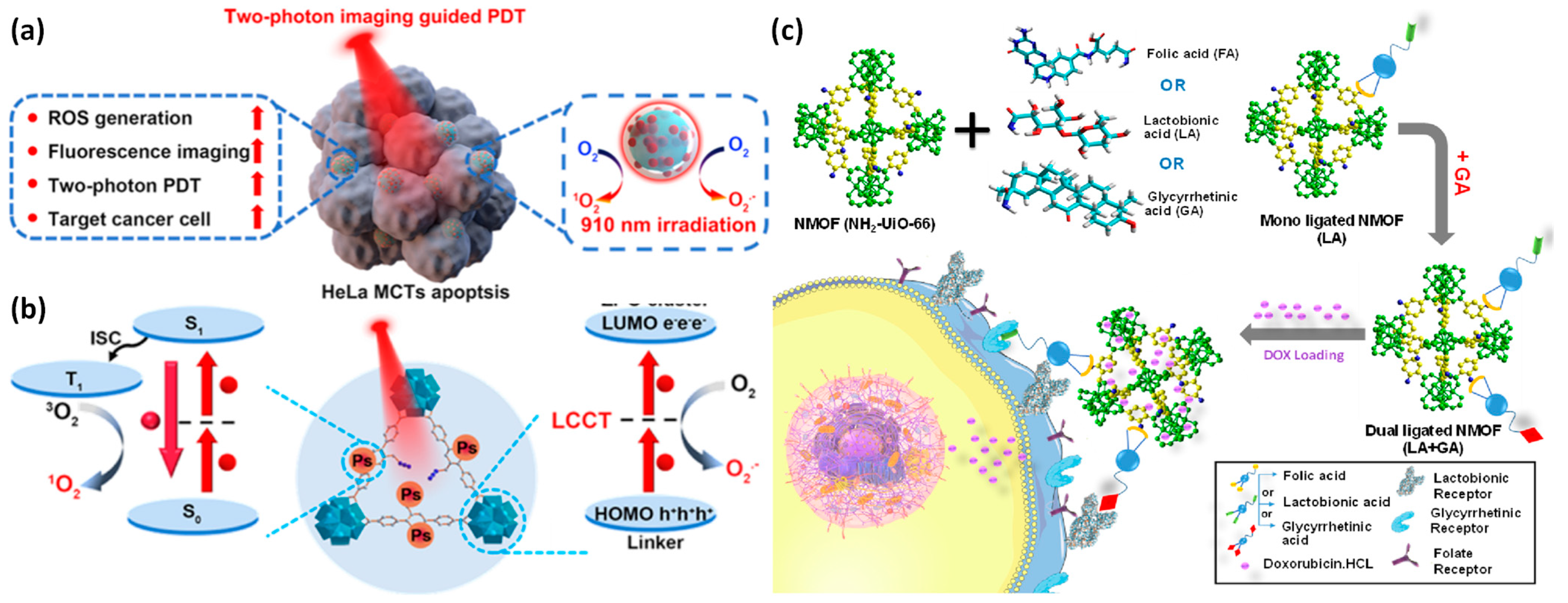
  118. Li, B.; Cao, H.; Zheng, J.; Ni, B.; Lu, X.; Tian, X.; Tian, Y.; Li, D. Click Modification of a Metal–Organic Framework for Two-Photon Photodynamic Therapy with Near-Infrared Excitation. ACS Appl. Mater. Interfaces 2021, 13, 9739–9747. [Google Scholar] [CrossRef]
  119. Fytory, M.; Arafa, K.K.; El Rouby, W.M.A.; Farghali, A.A.; Abdel-Hafiez, M.; El-Sherbiny, I.M. Dual-ligated metal organic framework as novel multifunctional nanovehicle for targeted drug delivery for hepatic cancer treatment. Sci. Rep. 2021, 11, 19808. [Google Scholar] [CrossRef] [PubMed]
  120. Cho, K.; Wang, X.; Nie, S.; Chen, Z.; Shin, D.M. Therapeutic nanoparticles for drug delivery in cancer. Clin. Cancer Res. 2008, 14, 1310–1316. [Google Scholar] [CrossRef] [Green Version]
  121. Rosenblum, D.; Joshi, N.; Tao, W.; Karp, J.M.; Peer, D. Progress and challenges towards targeted delivery of cancer therapeutics. Nat. Commun. 2018, 9, 1410. [Google Scholar] [CrossRef] [Green Version]
  122. Cordani, M.; Somoza, Á. Targeting autophagy using metallic nanoparticles: A promising strategy for cancer treatment. Cell. Mol. Life Sci. 2019, 76, 1215–1242. [Google Scholar] [CrossRef] [Green Version]
  123. Peer, D.; Karp, J.M.; Hong, S.; Farokhzad, O.C.; Margalit, R.; Langer, R. Nanocarriers as an emerging platform for cancer therapy. Nano-Enabled Med. Appl. 2020, 2, 751–760. [Google Scholar]
  124. Park, J.; Jiang, Q.; Feng, D.; Mao, L.; Zhou, H.-C. Size-controlled synthesis of porphyrinic metal–organic framework and functionalization for targeted photodynamic therapy. J. Am. Chem. Soc. 2016, 138, 3518–3525. [Google Scholar] [CrossRef]
  125. Duan, D.; Liu, H.; Xu, M.; Chen, M.; Han, Y.; Shi, Y.; Liu, Z. Size-controlled synthesis of drug-loaded zeolitic imidazolate framework in aqueous solution and size effect on their cancer theranostics in vivo. ACS Appl. Mater. Interfaces 2018, 10, 42165–42174. [Google Scholar] [CrossRef]
  126. Cornell, H.D.; Zhu, Y.; Ilic, S.; Lidman, N.E.; Yang, X.; Matson, J.B.; Morris, A.J. Green-light-responsive metal–organic frameworks for colorectal cancer treatment. Chem. Commun. 2022, 58, 5225–5228. [Google Scholar] [CrossRef] [PubMed]
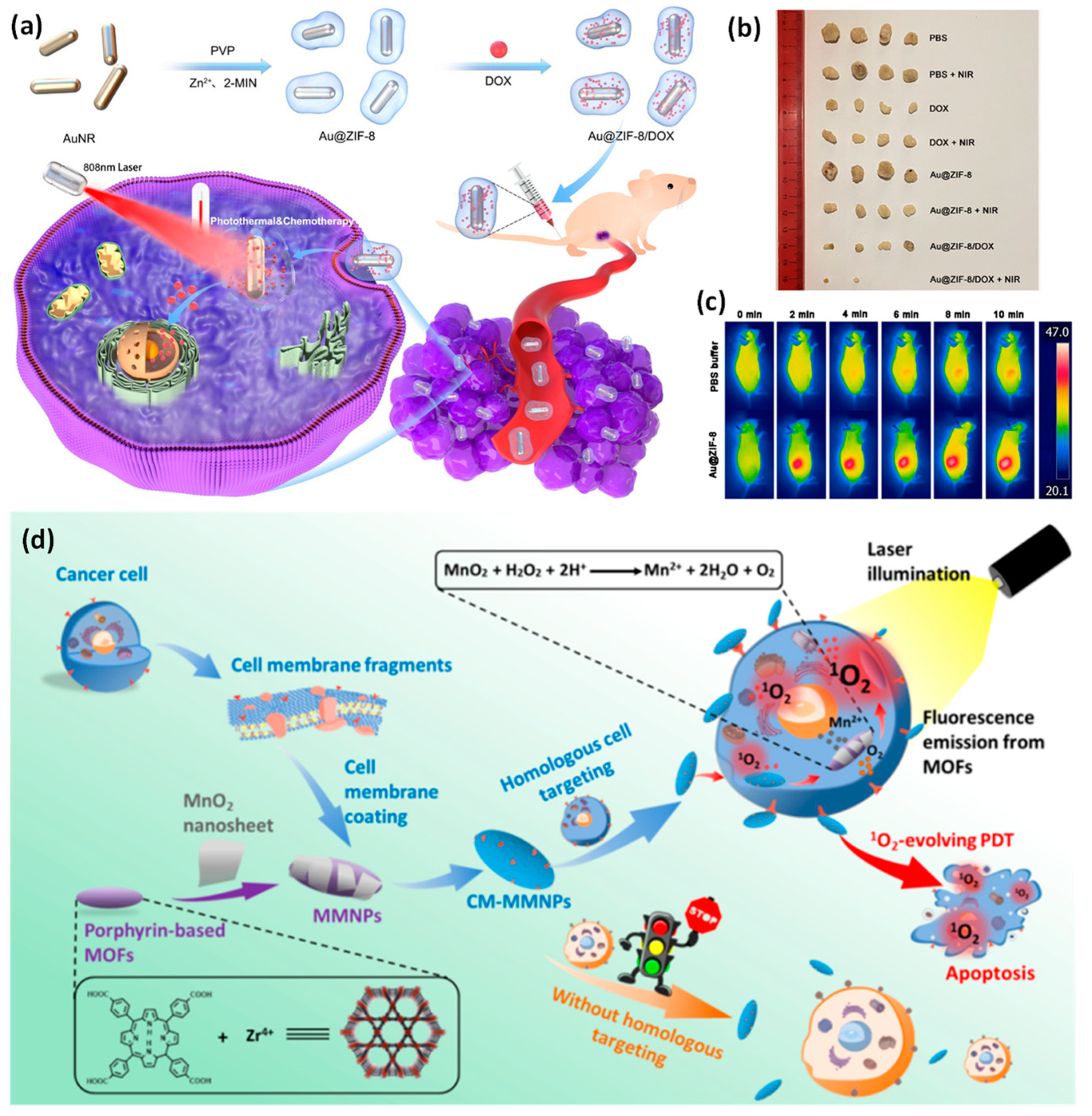
  127. Zhang, D.; Ye, Z.; Wei, L.; Luo, H.; Xiao, L. Cell Membrane-Coated Porphyrin Metal–Organic Frameworks for Cancer Cell Targeting and O2-Evolving Photodynamic Therapy. ACS Appl. Mater. Interfaces 2019, 11, 39594–39602. [Google Scholar] [CrossRef] [PubMed]
  128. Zhang, X.; Wang, S.; Cheng, G.; Yu, P.; Chang, J. Light-Responsive Nanomaterials for Cancer Therapy. Engineering 2022, 13, 18–30. [Google Scholar] [CrossRef]
  129. Zhao, D.; Zhang, W.; Yu, S.; Xia, S.-L.; Liu, Y.-N.; Yang, G.-J. Application of MOF-based nanotherapeutics in light-mediated cancer diagnosis and therapy. J. Nanobiotechnol. 2022, 20, 421. [Google Scholar] [CrossRef]
  130. Huang, J.; Xu, Z.; Jiang, Y.; Law, W.; Dong, B.; Zeng, X.; Ma, M.; Xu, G.; Zou, J.; Yang, C. Metal organic framework-coated gold nanorod as an on-demand drug delivery platform for chemo-photothermal cancer therapy. J. Nanobiotechnol. 2021, 19, 219. [Google Scholar] [CrossRef] [PubMed]
  131. Zhang, C.; Bu, W.; Ni, D.; Zhang, S.; Li, Q.; Yao, Z.; Zhang, J.; Yao, H.; Wang, Z.; Shi, J. Synthesis of iron nanometallic glasses and their application in cancer therapy by a localized Fenton reaction. Angew. Chem. 2016, 128, 2141–2146. [Google Scholar] [CrossRef]
  132. Ni, W.; Jiang, K.; Ke, Q.; Su, J.; Cao, X.; Zhang, L.; Li, C. Development of an intelligent heterojunction fenton catalyst for chemodynamic/starvation synergistic cancer therapy. J. Mater. Sci. Technol. 2023, 141, 11–20. [Google Scholar] [CrossRef]
  133. Ranji-Burachaloo, H.; Reyhani, A.; Gurr, P.A.; Dunstan, D.E.; Qiao, G.G. Combined Fenton and starvation therapies using hemoglobin and glucose oxidase. Nanoscale 2019, 11, 5705–5716. [Google Scholar] [CrossRef]
  134. Bian, Y.; Liu, B.; Liang, S.; Ding, B.; Zhao, Y.; Jiang, F.; Cheng, Z.; Al Kheraif, A.A.; Lin, J. Cu-based MOFs decorated dendritic mesoporous silica as tumor microenvironment responsive nanoreactor for enhanced tumor multimodal therapy. Chem. Eng. J. 2022, 435, 135046. [Google Scholar] [CrossRef]
  135. Nirosha Yalamandala, B.; Shen, W.T.; Min, S.H.; Chiang, W.H.; Chang, S.J.; Hu, S.H. Advances in Functional Metal-Organic Frameworks Based On-Demand Drug Delivery Systems for Tumor Therapeutics. Adv. NanoBiomed Res. 2021, 1, 2100014. [Google Scholar] [CrossRef]
  136. Zhou, Z.; Ke, Q.; Wu, M.; Zhang, L.; Jiang, K. Pore Space Partition Approach of ZIF-8 for pH Responsive Codelivery of Ursolic Acid and 5-Fluorouracil. ACS Mater. Lett. 2023, 5, 466–472. [Google Scholar] [CrossRef]
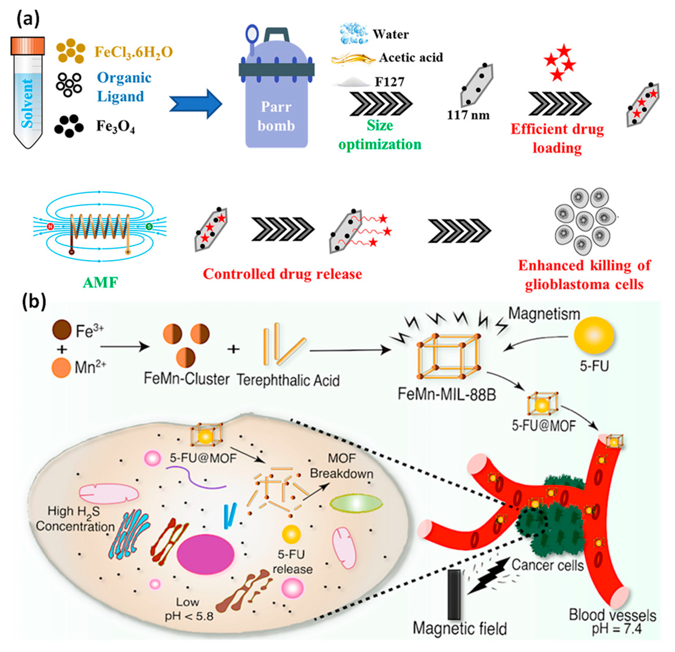
  137. Attia, M.; Glickman, R.D.; Romero, G.; Chen, B.; Brenner, A.J.; Ye, J.Y. Optimized metal-organic-framework based magnetic nanocomposites for efficient drug delivery and controlled release. J. Drug Deliv. Sci. Technol. 2022, 76, 103770. [Google Scholar] [CrossRef]
  138. Akbar, M.U.; Badar, M.; Zaheer, M. Programmable Drug Release from a Dual-Stimuli Responsive Magnetic Metal–Organic Framework. ACS Omega 2022, 7, 32588–32598. [Google Scholar] [CrossRef] [PubMed]
  139. Guo, X.; Cheng, Y.; Zhao, X.; Luo, Y.; Chen, J.; Yuan, W.-E. Advances in redox-responsive drug delivery systems of tumor microenvironment. J. Nanobiotechnol. 2018, 16, 74. [Google Scholar] [CrossRef] [PubMed] [Green Version]
  140. Wu, G.; Fang, Y.-Z.; Yang, S.; Lupton, J.R.; Turner, N.D. Glutathione metabolism and its implications for health. J. Nutr. 2004, 134, 489–492. [Google Scholar] [CrossRef] [Green Version]
  141. Li, R.; Peng, F.; Cai, J.; Yang, D.; Zhang, P. Redox dual-stimuli responsive drug delivery systems for improving tumor-targeting ability and reducing adverse side effects. Asian J. Pharm. Sci. 2020, 15, 311–325. [Google Scholar] [CrossRef]
  142. Arias-Duque, C.; Bladt, E.; Munoz, M.A.; Hernández-Garrido, J.C.; Cauqui, M.A.; Rodriguez-Izquierdo, J.M.; Blanco, G.; Bals, S.; Calvino, J.J.; Perez-Omil, J.A. Improving the redox response stability of ceria-zirconia nanocatalysts under harsh temperature conditions. Chem. Mater. 2017, 29, 9340–9350. [Google Scholar] [CrossRef]
  143. Zhao, J.; Yang, Y.; Han, X.; Liang, C.; Liu, J.; Song, X.; Ge, Z.; Liu, Z. Redox-sensitive nanoscale coordination polymers for drug delivery and cancer theranostics. ACS Appl. Mater. Interfaces 2017, 9, 23555–23563. [Google Scholar] [CrossRef]
  144. Lei, B.; Wang, M.; Jiang, Z.; Qi, W.; Su, R.; He, Z. Constructing redox-responsive metal–organic framework nanocarriers for anticancer drug delivery. ACS Appl. Mater. Interfaces 2018, 10, 16698–16706. [Google Scholar] [CrossRef]
  145. Liu, C.; Xu, X.; Zhou, J.; Yan, J.; Wang, D.; Zhang, H. Redox-responsive tumor targeted dual-drug loaded biocompatible metal–organic frameworks nanoparticles for enhancing anticancer effects. BMC Mater. 2020, 2, 7. [Google Scholar] [CrossRef]
  146. Wang, S.; Gong, M.; Han, X.; Zhao, D.; Liu, J.; Lu, Y.; Li, C.; Chen, B. Embedding Red Emitters in the NbO-Type Metal–Organic Frameworks for Highly Sensitive Luminescence Thermometry over Tunable Temperature Range. ACS Appl. Mater. Interfaces 2021, 13, 11078–11088. [Google Scholar] [CrossRef] [PubMed]
  147. Nagata, S.; Kokado, K.; Sada, K. Metal–organic framework tethering pH- and thermo-responsive polymer for ON–OFF controlled release of guest molecules. CrystEngComm 2020, 22, 1106–1111. [Google Scholar] [CrossRef]
  148. Wolf, A.J.; Underhill, D.M. Peptidoglycan recognition by the innate immune system. Nat. Rev. Immunol. 2018, 18, 243–254. [Google Scholar] [CrossRef]
  149. Iwasaki, A.; Foxman, E.F.; Molony, R.D. Early local immune defences in the respiratory tract. Nat. Rev. Immunol. 2017, 17, 7–20. [Google Scholar] [CrossRef] [PubMed] [Green Version]
  150. Feng, R.; Yu, F.; Xu, J.; Hu, X. Knowledge gaps in immune response and immunotherapy involving nanomaterials: Databases and artificial intelligence for material design. Biomaterials 2021, 266, 120469. [Google Scholar] [CrossRef]
  151. Li, C.; Qi, Y.; Zhang, Y.; Chen, Y.; Feng, J.; Zhang, X. Artificial Engineering of Immune Cells for Improved Immunotherapy. Adv. NanoBiomed Res. 2021, 1, 2000081. [Google Scholar] [CrossRef]
  152. Peng, T.; Yao, J. Development and application of bionic systems consisting of tumor-cell membranes. J. Zhejiang Univ. Sci. B 2022, 23, 770–777. [Google Scholar] [CrossRef]
  153. Jin, J.; Bhujwalla, Z.M. Biomimetic Nanoparticles Camouflaged in Cancer Cell Membranes and Their Applications in Cancer Theranostics. Front. Oncol. 2020, 9, 1560. [Google Scholar] [CrossRef] [Green Version]
  154. Ding, B.; Chen, H.; Tan, J.; Meng, Q.; Zheng, P.; Ma, P.a.; Lin, J. ZIF-8 Nanoparticles Evoke Pyroptosis for High-Efficiency Cancer Immunotherapy. Angew. Chem. Int. Ed. 2023, 62, 202215307. [Google Scholar] [CrossRef]
  155. Gong, C.; Yu, X.; You, B.; Wu, Y.; Wang, R.; Han, L.; Wang, Y.; Gao, S.; Yuan, Y. Macrophage-cancer hybrid membrane-coated nanoparticles for targeting lung metastasis in breast cancer therapy. J. Nanobiotechnol. 2020, 18, 92. [Google Scholar] [CrossRef]
  156. Harrington, K.; Spitzweg, C.; Bateman, A.; Morris, J.; Vile, R. Gene therapy for prostate cancer: Current status and future prospects. J. Urol. 2001, 166, 1220–1233. [Google Scholar] [CrossRef] [PubMed]
  157. Huxford, R.C.; Della Rocca, J.; Lin, W. Metal–organic frameworks as potential drug carriers. Curr. Opin. Chem. Biol. 2010, 14, 262–268. [Google Scholar] [CrossRef] [PubMed] [Green Version]
  158. Morris, W.; Briley, W.E.; Auyeung, E.; Cabezas, M.D.; Mirkin, C.A. Nucleic acid–metal organic framework (MOF) nanoparticle conjugates. J. Am. Chem. Soc. 2014, 136, 7261–7264. [Google Scholar] [CrossRef] [PubMed]
  159. Lin, G.; Zhang, Y.; Zhang, L.; Wang, J.; Tian, Y.; Cai, W.; Tang, S.; Chu, C.; Zhou, J.; Mi, P. Metal-organic frameworks nanoswitch: Toward photo-controllable endo/lysosomal rupture and release for enhanced cancer RNA interference. Nano Res. 2020, 13, 238–245. [Google Scholar] [CrossRef]
  160. Dai, L.; Yao, M.; Fu, Z.; Li, X.; Zheng, X.; Meng, S.; Yuan, Z.; Cai, K.; Yang, H.; Zhao, Y. Multifunctional metal-organic framework-based nanoreactor for starvation/oxidation improved indoleamine 2,3-dioxygenase-blockade tumor immunotherapy. Nat. Commun. 2022, 13, 2688. [Google Scholar] [CrossRef]
  161. Peng, M.; Ju, E.; Xu, Y.; Wang, Y.; Lv, S.; Shao, D.; Wang, H.; Tao, Y.; Zheng, Y.; Li, M. Dual-responsive disassembly of core-shell nanoparticles with self-supplied H2O2 and autocatalytic Fenton reaction for enhanced chemodynamic therapy. NPG Asia Mater. 2022, 14, 95. [Google Scholar] [CrossRef]
  162. Wang, Y.; Yan, J.; Wen, N.; Xiong, H.; Cai, S.; He, Q.; Hu, Y.; Peng, D.; Liu, Z.; Liu, Y. Metal-organic frameworks for stimuli-responsive drug delivery. Biomaterials 2020, 230, 119619. [Google Scholar] [CrossRef]
  163. Zhang, H.; Tian, X.-T.; Shang, Y.; Li, Y.-H.; Yin, X.-B. Theranostic Mn-Porphyrin Metal–Organic Frameworks for Magnetic Resonance Imaging-Guided Nitric Oxide and Photothermal Synergistic Therapy. ACS Appl. Mater. Interfaces 2018, 10, 28390–28398. [Google Scholar] [CrossRef]
  164. Ruyra, À.; Yazdi, A.; Espín, J.; Carné-Sánchez, A.; Roher, N.; Lorenzo, J.; Imaz, I.; Maspoch, D. Synthesis, culture medium stability, and in vitro and in vivo zebrafish embryo toxicity of metal–organic framework nanoparticles. Chem.—Eur. J. 2015, 21, 2508–2518. [Google Scholar] [CrossRef]
  165. Al-Ansari, D.E.; Al-Badr, M.; Zakaria, Z.Z.; Mohamed, N.A.; Nasrallah, G.K.; Yalcin, H.C.; Abou-Saleh, H. Evaluation of Metal–Organic Framework MIL-89 nanoparticles toxicity on embryonic zebrafish development. Toxicol. Rep. 2022, 9, 951–960. [Google Scholar] [CrossRef] [PubMed]
  166. Singh, N.; Qutub, S.; Khashab, N.M. Biocompatibility and biodegradability of metal organic frameworks for biomedical applications. J. Mater. Chem. B 2021, 9, 5925–5934. [Google Scholar] [CrossRef] [PubMed]
  167. Grall, R.; Hidalgo, T.; Delic, J.; Garcia-Marquez, A.; Chevillard, S.; Horcajada, P. In vitro biocompatibility of mesoporous metal (III.; Fe, Al, Cr) trimesate MOF nanocarriers. J. Mater. Chem. B 2015, 3, 8279–8292. [Google Scholar] [CrossRef] [PubMed]
  168. Hoop, M.; Walde, C.F.; Riccò, R.; Mushtaq, F.; Terzopoulou, A.; Chen, X.-Z.; deMello, A.J.; Doonan, C.J.; Falcaro, P.; Nelson, B.J. Biocompatibility characteristics of the metal organic framework ZIF-8 for therapeutical applications. Appl. Mater. Today 2018, 11, 13–21. [Google Scholar] [CrossRef] [Green Version]
  169. Awasthi, G.; Shivgotra, S.; Nikhar, S.; Sundarrajan, S.; Ramakrishna, S.; Kumar, P. Progressive Trends on the Biomedical Applications of Metal Organic Frameworks. Polymers 2022, 14, 4710. [Google Scholar] [CrossRef]
  170. Afrin, S.; Khan, M.W.; Haque, E.; Ren, B.; Ou, J.Z. Recent advances in the tuning of the organic framework materials-The selections of ligands, reaction conditions, and post-synthesis approaches. J. Colloid Interface Sci. 2022, 623, 378–404. [Google Scholar] [CrossRef]
  171. De, D.; Sahoo, P. The impact of MOFs in pH-dependent drug delivery systems: Progress in the last decade. Dalton Trans. 2022, 51, 9950–9965. [Google Scholar] [CrossRef]
  172. Zhang, H.; Li, Y.-H.; Chen, Y.; Wang, M.-M.; Wang, X.-S.; Yin, X.-B. Fluorescence and Magnetic Resonance Dual-Modality Imaging-Guided Photothermal and Photodynamic Dual-Therapy with Magnetic Porphyrin-Metal Organic Framework Nanocomposites. Sci. Rep. 2017, 7, 44153. [Google Scholar] [CrossRef] [Green Version]
  173. Chen, Z.-X.; Liu, M.-D.; Zhang, M.-K.; Wang, S.-B.; Xu, L.; Li, C.-X.; Gao, F.; Xie, B.-R.; Zhong, Z.-L.; Zhang, X.-Z. Interfering with Lactate-Fueled Respiration for Enhanced Photodynamic Tumor Therapy by a Porphyrinic MOF Nanoplatform. Adv. Funct. Mater. 2018, 28, 1803498. [Google Scholar] [CrossRef]
  174. Wang, H.; Yu, D.; Fang, J.; Cao, C.; Liu, Z.; Ren, J.; Qu, X. Renal-Clearable Porphyrinic Metal–Organic Framework Nanodots for Enhanced Photodynamic Therapy. ACS Nano 2019, 13, 9206–9217. [Google Scholar] [CrossRef]
  175. Baati, T.; Njim, L.; Neffati, F.; Kerkeni, A.; Bouttemi, M.; Gref, R.; Najjar, M.F.; Zakhama, A.; Couvreur, P.; Serre, C.; et al. In depth analysis of the in vivo toxicity of nanoparticles of porous iron(iii) metal–organic frameworks. Chem. Sci. 2013, 4, 1597–1607. [Google Scholar] [CrossRef]
  176. Zhang, S.; Rong, F.; Guo, C.; Duan, F.; He, L.; Wang, M.; Zhang, Z.; Kang, M.; Du, M. Metal–organic frameworks (MOFs) based electrochemical biosensors for early cancer diagnosis in vitro. Coord. Chem. Rev. 2021, 439, 213948. [Google Scholar] [CrossRef]
  177. Sharanyakanth, P.; Radhakrishnan, M. Synthesis of metal-organic frameworks (MOFs) and its application in food packaging: A critical review. Trends Food Sci. Technol. 2020, 104, 102–116. [Google Scholar] [CrossRef]
  178. Abuçafy, M.P.; da Silva, B.L.; Oshiro-Junior, J.A.; Manaia, E.B.; Chiari-Andréo, B.G.; Armando, R.A.; Frem, R.C.; Chiavacci, L.A. Advances in the use of MOFs for Cancer Diagnosis and Treatment: An Overview. Curr. Pharm. Des. 2020, 26, 4174–4184. [Google Scholar] [CrossRef] [PubMed]
  179. Rao, C.; Liao, D.; Pan, Y.; Zhong, Y.; Zhang, W.; Ouyang, Q.; Nezamzadeh-Ejhieh, A.; Liu, J. Novel formulations of metal-organic frameworks for controlled drug delivery. Expert Opin. Drug Deliv. 2022, 19, 1183–1202. [Google Scholar] [CrossRef]
  180. Wei, Q.; Wu, Y.; Liu, F.; Cao, J.; Liu, J. Advances in antitumor nanomedicine based on functional metal–organic frameworks beyond drug carriers. J. Mater. Chem. B 2022, 10, 676–699. [Google Scholar] [CrossRef]
  181. Mallakpour, S.; Nikkhoo, E.; Hussain, C.M. Application of MOF materials as drug delivery systems for cancer therapy and dermal treatment. Coord. Chem. Rev. 2022, 451, 214262. [Google Scholar] [CrossRef]
Figure 1. Some monotherapies are based on MOFs. Metal ions and an organic ligand are the two components that make up MOFs. The organic ligand joins the metal ions to form larger arrays. MOFs have several desirable structural properties that make them excellent candidates in the fields of drug administration and cancer therapy.
Pharmaceutics 15 00931 g001
Figure 2. The logical structure of the review.
Pharmaceutics 15 00931 g002
Figure 3. The gradient of cancer development. Microscopic to macroscopic (left to right) examples of the several factors of tumor evolution. Reproduced with permission from [47]. Copyright 2021 The EMBO Journal.
Pharmaceutics 15 00931 g003
Figure 4. (a) Upconversion nanoparticles (UCNPs) and MOF nanomaterial manufacturing is shown schematically. Direct mixing results in the formation of nanocomposites from the reaction intermediates of MOF and ligand-free UCNPs. The processes demonstrate the three hypothesized formation mechanisms: (i) nucleation of MOFs, (ii) electrostatic bonding of NPs to MOFs, and (iii) production of nanocomposite materials; (b) catalyst with two functions for single-pot sequential reactions using confinement self-assembly and M12L24 cage C in FDU-ED cavities mesoporous to produce cage@FDU-ED is shown schematically. Reproduced with permission from references [55,56].
Pharmaceutics 15 00931 g004
Figure 5. Combining drugs with hybrid metal–organic framework nanoparticles. The aforementioned three basic techniques have been successfully used to insert active compounds, particularly anticancer medicines, into the MOFs.
Pharmaceutics 15 00931 g005
Figure 6. (a) Schematic illustration displaying the PDT guided by two-photon fluorescence imaging, the target cancer cell, and the light-induced ROS creation; (b) mechanisms of ROS created by PCN-58-Ps-HA by light irradiation of two-photon; (c) drug delivery systems using functionalized Zr-based NMFOs (NH2-UiO-66) for the targeted therapy of hepatocellular cancer are represented systematically. Reproduced with permission from references [118,119].
Pharmaceutics 15 00931 g006
Figure 7. Cancer treatment involves passive and active targeting of nanoparticles. Extravasation of nanomaterials through enhanced permeability (EPR effect) of the tumor vasculature enables passive tumor targeting. By functionalizing nanomaterials aimed specifically at ligands that improve cell-specific identification and adherence, active tumor targeting (left inset) is possible. Reproduced with permission from [122]. Copyright 2018, Springer Nature.
Pharmaceutics 15 00931 g007
Figure 8. (a) A diagram of the core–shell Au@ZIF-8/DOX synthesis process for in vivo chemo-photothermal cancer treatment; (b) intravenous injections of Au@ZIF-8 or PBS solution followed by in vivo infrared thermal imaging of MCF-7 tumor-bearing mice following activation with a 1 W/cm2 808 nm laser for 10 min; (c) a characteristic image of tumors removed from mice treated in each group; (d) the nanostructure is made up of a cancerous cells membrane layer and a metal–organic framework core coated in MnO2 nanosheets for MRI and fluorescence dual-mode imaging, homologous targeting, and photodynamic therapy for the diagnosis and treatment of cancer cells. Reproduced with permission from references [127,130].
Pharmaceutics 15 00931 g008
Figure 9. To target the lung, an iron-based biodegradable MOF that exhibits pH-responsive as well as reversible aggregating activity was created. Within twenty-four hours, the nanoparticles were enabled to autonomously aggregate at the pulmonary capillaries and then disaggregate again. Reproduced with permission [135]. Copyright 2021, Wiley-VCH Verlag GmbH & Co. KGaA, Weinheim.
Pharmaceutics 15 00931 g009
Figure 10. (a) F127 copolymer was used as a stabilizing substance during the synthesis of MOF Fe3O4 NPs with MIL-88B-NH2 structures, and these nanoparticles were used to deliver drugs using an alternating magnetic field and localized heating effect; (b) a preassembled Fe2Mn(3-O) cluster was used to create an FeMn-based ferromagnetic MOF that controlled drug release by dual stimulation, ferromagnetic nature, and low toxicity. Reproduced with permission from references [137,138].
Pharmaceutics 15 00931 g010
Figure 11. The biological process that is accountable for the release of redox-sensitive drug delivery devices. Reproduced with permission from [141]. Copyright 2019 Elsevier B.V.
Pharmaceutics 15 00931 g011
Figure 12. (a) Illustration of the fabrication of nanocomposites made from DSM@MOFs and Eu3+@MOFs for dual-emitting sensor nanomaterial; (b) schematic illustration for making an MOF that is tethered by P(NIPAM-AA) (UiO-66-P(NIPAM-AA)) and released under regulated conditions utilizing UiO-66-P (NIPAM-AA); (c) procaine amide’s sequential release and halt exhibited by UiO-66-P/NIPAM-AA in water are time-dependent and affected by pH. Reproduced with permission from references [146,147].
Pharmaceutics 15 00931 g012
Figure 13. Diagram depicting the processes required for the synthesis of tumor cell membrane-coated nanomaterials. Reproduced with permission from [153]. Copyright 2020 Frontiers in Oncology.
Pharmaceutics 15 00931 g013
Figure 14. (a) Synthetic approach and graphical depiction of PCP-Mn-DTA@GOx@1-MT nanomaterial for combination starvation, immunotherapy, and oxidation; (b) hemoglobin (HB) and oxygenated hemoglobin (HBO2) photoacoustic recordings; (c) melanin signals in mouse tumor sites following intratumoral injecting with PCP-Mn-DTA@GOx@1-MT and GOx for multiple time frames; (d) a plan for creating MOF-SNO nanocomposite, releasing nitric oxide when exposed to NIR light, and using photothermal therapy; (e) pictures of the tumors in each group following treatment. Reproduced with permission from references [160,163].
Pharmaceutics 15 00931 g014
Table 1. Physicochemical properties and applications of nanomaterials for drug delivery.
Kind of MaterialSize
(nm)
ShapeSurface Area
(m2/g)
PropertiesApplicationRef.
Silica100–108Nanoparticles1156.4The photothermal heating effect, efficient endocytosis, (pH, NIR irradiation)-responsive, anchor effectChemo-photothermal therapy, active targeting[32]
Carbon
dots
4Nanodots-Electrostatic interactions,
pH-dependent release
Chemotherapy[33]
Silicon100Nanoparticles1407(pH and NIR)-responsiveness, mitochondrial targetingFluorescent image, chemo-photothermal therapy, active targeting[34]
Liposome165Nanoparticles-X-ray-triggered liposomesChemotherapy, radiotherapy, photodynamic therapy[35]
Magnetic-gold11–29Nanoparticles-Multifunctional magnetic gold, controlled-release mannerPassively magnetic targeting; chemmophotothermal therapy; magnetic resonance imaging (MRI)[36]
Carbon nanotubes0.4–2/2–100Cylindrical roll232.5π-π stacking, electrostatic interaction, pharmaco-toxicological propertiesChemotherapy[37]
Hydrogel35–60Sphere NPs-Thermo-sensitive micelles, reversible sol–gel transition,Chemotherapy[38]
Protein28Monodisperse nano-scaffold-Receptor-mediated internalization, fluorescent imageChemotherapy, active targeting[39]
ZIF-850–160Dodecahedral1925π–π stacking, hydrogen bonding, electrostatic interactions, fluorescent imaging, and pH-responsive drug releaseChemotherapy, passive targeting[40]
Table 2. A summary of documented MOFs for the delivery of medicinal substances [59].
Therapeutic DrugMOFsOrganic LinkerMetal IonDrug Encapsulation MethodRef.
1. Anti-inflammatory and analgesics drugs
IbuprofenMIL-1001,3,5-benzene tricarboxylic acid (BTC)Cr3+Post-synthetic (PS) encapsulation[60]
IbuprofenMIL-1011,4-benzene dicarboxylic acid (BDC)Cr3+PS encapsulation[60]
IbuprofenMIL-53BDCFe3+, Cr3+PS encapsulation[61]
Curcumin, SulindacMOF-5BDCZn2+PS encapsulation[62]
Diclofenac sodiumZJU-800F-H2PDAZr2+PS encapsulation[63]
2. Antiviral and antibacterial drugs
CidofovirMIL-101-NH22-amino-BDCFe3+PS encapsulation[64]
Nalidixic acidBio-MOFNalidixic acidMg2+, Mn2+Direct assembly[65]
VancomycinMIL-53BDCFe3+PS encapsulation[66]
CiprofloxacinUiO-66BDCZr4+PS encapsulation[67]
GentamicinZIF-82-methyl imidazolate Zn2+PS encapsulation[68]
CiprofloxacinZIF-82-methyl imidazolate Zn2+PS encapsulation[69]
CeftazidimeZIF-82-methyl imidazolate Zn2+One-pot synthesis (OPS)[70]
TetracyclineZIF-82-methyl imidazolate Zn2+OPS[71]
Enrofloxacin, Florfenicolγ-CD-MOFCyclodextrinK+PS encapsulation[72]
3. Anti-cancer drugs
NimesulideHKUST-1BTCCu2+PS encapsulation[73]
BusulfanMIL-100BTCFe3+PS encapsulation[64]
DoxorubicinMIL-100BTCFe3+PS encapsulation[74]
DoxorubicinMIL-89Muconic acidFe3+PS encapsulation[64]
OridoninMOF-5BDCZn2+PS encapsulation[75]
CisplatinNCP-1DisuccinatocisplatinTb3+Direct assembly[76]
MethotrexatePCN-221TCPPZr4+PS encapsulation[77]
AlendronateUiO-66BDCZr4+Covalent bonding[78]
DoxorubicinZIF-672-methyl imidazolateCo2+OPS[79]
5-Fluoro uracilZIF-67Imidazole-2-carboxaldehydeCo2+PS encapsulation[80]
DoxorubicinZIF-67Imidazole-2-carboxaldehydeCo2+Covalent bonding[80]
5-Fluoro uracilZIF-82-methyl imidazolateZn2+PS encapsulation[81]
CamptothecinZIF-82-methyl imidazolateZn2+OPS[82]
DoxorubicinZIF-82-methyl imidazolateZn2+OPS[83]
3-Methyl adenineZIF-82-methyl imidazolateZn2+OPS[84]
Doxorubicin, Camptothecin, DaunomycinZn(bix)bixZn2+OPS[85]
4. Peptides, Proteins, and enzymes
InsulinNU-10004,4′,4″,4‴-(pyrene-1,3,6,8-tetrayl)tetrabenzoic acidZr4+PS encapsulation[86]
Glucose oxidaseCu-TCCP(Fe)TCPP(Fe)Cu2+Surface attachment[87]
InsulinMIL-1001,3,5-benzene tricarboxylic acidFe3+PS encapsulation[88]
MyoglobinMOF-742,5-dioxido terephthalateZn2+, Mg2+PS entrapment[89]
TyrosinasePCN-333TATBAl3+PS entrapment[90]
Cytochrome cTb-meso MOFTriazine-1,3,5-tribenzoic acidTb3+PS entrapment[91]
Microperoxidase-11Tb-meso MOFTriazine-1,3,5-tribenzoic acidTb3+PS entrapment[92]
Glucose oxidaseZIF-82-methyl imidazolateZn2+OPS[93]
Horseradish peroxidaseZIF-82-methyl imidazolateZn2+OPS[94]
Hemoglobin, Glucose oxidaseZIF-82-methyl imidazolateZn2+Biomimetic mineralization[95]
MelittinZIF-82-methyl imidazolateZn2+OPS[96]
CatalaseZIF-90Imidazole-2-carboxaldehydeZn2+OPS[97]
5. Antibodies and antigens
αCD47Hf-DBP5,15-di(p-benzoato) porphyrinHf4+Surface attachment[98]
H-IgG,ZIF-90Imidazole-2-carboxaldehydeZn2+OPS[99]
G-IgGZIF-90Imidazole-2-carboxaldehydeZn2+OPS[99]
NivolumabZIF-82-methyl imidazolateZn2+Biomimetic mineralization[95]
OvalbuminZIF-82-methyl imidazolateZn2+One-pot synthesis[100]
anti-EpCAMMIL-100BTCFe3+Surface attachment[101]
OvalbuminUiO-AMBDC, 2-amino-BDCZr4+Surface attachment[102]
OvalbuminAl-MOFBDC, 2-amino-BDCAl3+OPS[103]
6. Nucleotides and Nucleic Acids
siRNAMIL-101BDCFe3+Covalent-linkage[104]
Terminal phosphate modified oligo-nucleotidesUiO-66BDCZr4+Covalent linkage[105]
Plasmid DNAZIF-82-methyl imidazolateZn2+OPS[106]
7. Carbohydrates
Heparin, Hyaluronic acidMAF-72-methyl imidazolateZn2+Biomimetic mineralization[107]
Meglumine, Carboxylate dextranZIF-82-methyl imidazolateZn2+OPS[108]
Table 3. Advantages and disadvantages of MOFs nanomaterial and example for cancer treatment.
Synthesis
Method
MOFAdvantagesShortcomingsRef.
ElectrochemicalZr-MOF
Zn-MOF
High loading capacity and controlled release of anticancer drugs.
Ability to accommodate imaging agents for theranostic applications.
Selective targeting and enhanced permeability to tumor sites.
Potential toxicity and biocompatibility issues.
Complex synthesis and functionalization procedures.
Limited clinical trials and regulatory approvals.
[21,176]
SolvothermalZn-MOF-74
MIL-100 (Fe)
UiO-66
High purity and crystallinity.
Control over size and shape.
Ability to incorporate functional groups.
Long reaction time
Discontinuity of the process.
Inhomogeneity of heating.
[21,177,178]
UltrasonicCu-MOF
Fe-MOF
High surface area and porosity.
Flexible and tunable chemical structure and architecture.
Ability to capture and degrade.
Ability to carry anticancer drugs and imaging agents.
Low mechanical and thermal stability.
Difficult to recycle and reuse.
Potential toxicity to living environments.
Possible immune reaction or poor biocompatibility in the human body.
[179,180]
DiffusionZr-MOF
MIL-100
Large surface area and porosity that can accommodate various drugs and imaging agents.
High chemical stability and biocompatibility.
Easily functionalized and modified with different ligands and nanoparticles.
Enable controlled drug release by external stimuli such as pH, temperature, and light.
Enhance the therapeutic efficacy and reduce the side effects of drugs by targeting specific cancer cells.
Low solubility and dispersibility in biological fluids.
Induce immune responses or toxicity in some cases.
Limited loading capacity or release rate for some drugs.
Suffer from aggregation or degradation in vivo.
[11,181]
Disclaimer/Publisher’s Note: The statements, opinions and data contained in all publications are solely those of the individual author(s) and contributor(s) and not of MDPI and/or the editor(s). MDPI and/or the editor(s) disclaim responsibility for any injury to people or property resulting from any ideas, methods, instructions or products referred to in the content.

