

Chitosan-Based Biomaterials for Tissue Regeneration
 , ,
, ,
Abstract
:
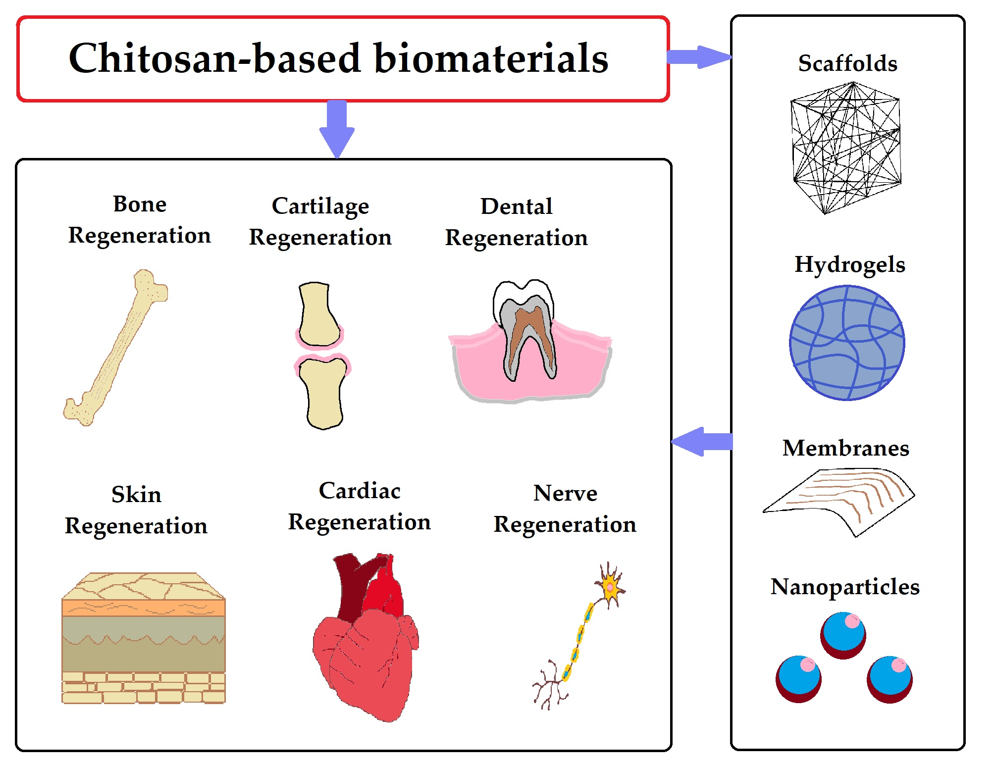
1. Introduction
2. Bone, Cartilage, and Dental Tissues
2.1. Bone Regeneration Using Chitosan-Based Biomaterials
2.2. Cartilage Regeneration Using Chitosan-Based Biomaterials
2.3. Dental Regeneration Using Chitosan-Based Biomaterials
3. Skin
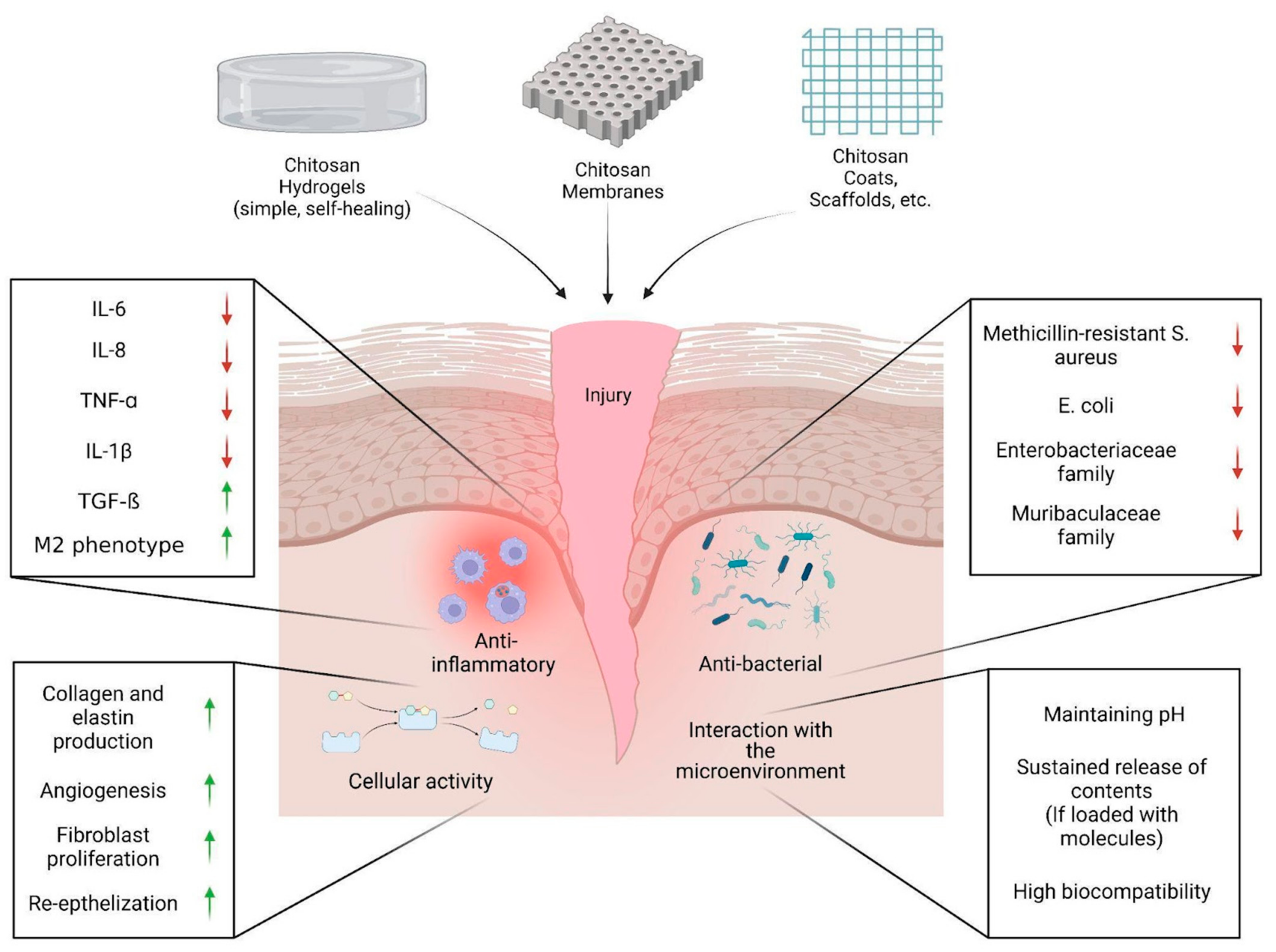
3.1. Skin Tissue Regeneration and Wound Healing Using Chitosan-Based Hydrogels
3.2. Skin Tissue Regeneration and Wound Healing Using Chitosan-Based Membranes
3.3. Skin Tissue Regeneration and Wound Healing Using Other Chitosan-Based Biomaterials
4. Cardiac and Nervous Tissues
4.1. Cardiac Tissue Regeneration Using Chitosan-Based Biomaterials
4.2. Nervous Tissue Regeneration Using Chitosan-Based Biomaterials
5. Conclusions and Future Perspectives
Author Contributions
Funding
Institutional Review Board Statement
Informed Consent Statement
Conflicts of Interest
References
- Islam, M.M.; Shahruzzaman, M.; Biswas, S.; Sakib, M.N.; Rashid, T.U. Chitosan based bioactive materials in tissue engineering applications—A review. Bioact. Mater. 2020, 5, 164–183. [Google Scholar] [CrossRef]
- Kou, S.G.; Peters, L.M.; Mucalo, M.R. Chitosan: A review of sources and preparation methods. Int. J. Biol. Macromol. 2021, 169, 85–94. [Google Scholar] [CrossRef]
- Baranwal, A.; Kumar, A.; Priyadharshini, A.; Oggu, G.S.; Bhatnagar, I.; Srivastava, A.; Chandra, P. Chitosan: An undisputed bio-fabrication material for tissue engineering and bio-sensing applications. Int. J. Biol. Macromol. 2018, 110, 110–123. [Google Scholar] [CrossRef]
- Sultankulov, B.; Berillo, D.; Sultankulova, K.; Tokay, T.; Saparov, A. Progress in the Development of Chitosan-Based Biomaterials for Tissue Engineering and Regenerative Medicine. Biomolecules 2019, 9, 470. [Google Scholar] [CrossRef] [PubMed] [Green Version]
- Abourehab, M.A.; Pramanik, S.; Abdelgawad, M.A.; Abualsoud, B.M.; Kadi, A.; Ansari, M.J.; Deepak, A. Recent advances of chitosan formulations in biomedical applications. Int. J. Mol. Sci. 2022, 23, 10975. [Google Scholar] [CrossRef] [PubMed]
- Atia, G.A.N.; Shalaby, H.K.; Zehravi, M.; Ghobashy, M.M.; Attia, H.A.N.; Ahmad, Z.; Khan, F.S.; Dey, A.; Mukerjee, N.; Alexiou, A. Drug-Loaded Chitosan Scaffolds for Periodontal Tissue Regeneration. Polymers 2022, 14, 3192. [Google Scholar] [CrossRef] [PubMed]
- Tang, G.; Tan, Z.; Zeng, W.; Wang, X.; Shi, C.; Liu, Y.; He, H.; Chen, R.; Ye, X. Recent advances of chitosan-based injectable hydrogels for bone and dental tissue regeneration. Front. Bioeng. Biotechnol. 2020, 8, 587658. [Google Scholar] [CrossRef]
- Wan, M.-C.; Qin, W.; Lei, C.; Li, Q.-H.; Meng, M.; Fang, M.; Song, W.; Chen, J.-H.; Tay, F.; Niu, L.-N. Biomaterials from the sea: Future building blocks for biomedical applications. Bioact. Mater. 2021, 6, 4255–4285. [Google Scholar] [CrossRef]
- Sami El-banna, F.; Mahfouz, M.E.; Leporatti, S.; El-Kemary, M.; AN Hanafy, N. Chitosan as a natural copolymer with unique properties for the development of hydrogels. Appl. Sci. 2019, 9, 2193. [Google Scholar] [CrossRef] [Green Version]
- Ahmed, S.; Ali, A.; Sheikh, J. A review on chitosan centred scaffolds and their applications in tissue engineering. Int. J. Biol. Macromol. 2018, 116, 849–862. [Google Scholar] [CrossRef]
- Choi, A.H.; Ben-Nissan, B. Marine-Derived Biomaterials for Tissue Engineering Applications; Springer: Berlin/Heidelberg, Germany, 2019; Volume 14. [Google Scholar]
- Lavanya, K.; Chandran, S.V.; Balagangadharan, K.; Selvamurugan, N. Temperature-and pH-responsive chitosan-based injectable hydrogels for bone tissue engineering. Mater. Sci. Eng. C 2020, 111, 110862. [Google Scholar] [CrossRef]
- Cao, Y.; Tan, Y.F.; Wong, Y.S.; Liew, M.W.J.; Venkatraman, S. Recent advances in chitosan-based carriers for gene delivery. Mar. Drugs 2019, 17, 381. [Google Scholar] [CrossRef] [Green Version]
- Jana, S.; Jana, S. Nanoengineering of Biomaterials: Drug Delivery & Biomedical Applications; John Wiley & Sons: Hoboken, NJ, USA, 2022. [Google Scholar]
- Berillo, D.; Zharkinbekov, Z.; Kim, Y.; Raziyeva, K.; Temirkhanova, K.; Saparov, A. Stimuli-Responsive Polymers for Transdermal, Transmucosal and Ocular Drug Delivery. Pharmaceutics 2021, 13, 2050. [Google Scholar] [CrossRef]
- Padhi, S.; Behera, A.; Hasnain, M.S.; Nayak, A.K. Uses of chitosan in drug delivery. In Chitosan in Biomedical Applications; Elsevier: Amsterdam, The Netherlands, 2022; pp. 139–162. [Google Scholar]
- Lang, X.; Wang, T.; Sun, M.; Chen, X.; Liu, Y. Advances and applications of chitosan-based nanomaterials as oral delivery carriers: A review. Int. J. Biol. Macromol. 2020, 154, 433–445. [Google Scholar] [CrossRef]
- Shanmuganathan, R.; Edison, T.N.J.I.; LewisOscar, F.; Kumar, P.; Shanmugam, S.; Pugazhendhi, A. Chitosan nanopolymers: An overview of drug delivery against cancer. Int. J. Biol. Macromol. 2019, 130, 727–736. [Google Scholar] [CrossRef]
- Kulkarni, N.; Jain, P.; Shindikar, A.; Suryawanshi, P.; Thorat, N. Advances in the colon-targeted chitosan based drug delivery systems for the treatment of inflammatory bowel disease. Carbohydr. Polym. 2022, 288, 119351. [Google Scholar] [CrossRef]
- Wilson, B.; Alobaid, B.N.M.; Geetha, K.M.; Jenita, J.L. Chitosan nanoparticles to enhance nasal absorption and brain targeting of sitagliptin to treat Alzheimer’s disease. J. Drug Deliv. Sci. Technol. 2021, 61, 102176. [Google Scholar] [CrossRef]
- Saylam, E.; Akkaya, Y.; Ilhan, E.; Cesur, S.; Guler, E.; Sahin, A.; Cam, M.E.; Ekren, N.; Oktar, F.N.; Gunduz, O. Levodopa-Loaded 3D-Printed Poly (Lactic) Acid/Chitosan Neural Tissue Scaffold as a Promising Drug Delivery System for the Treatment of Parkinson’s Disease. Appl. Sci. 2021, 11, 10727. [Google Scholar] [CrossRef]
- Sultankulov, B.; Berillo, D.; Kauanova, S.; Mikhalovsky, S.; Mikhalovska, L.; Saparov, A. Composite Cryogel with Polyelectrolyte Complexes for Growth Factor Delivery. Pharmaceutics 2019, 11, 650. [Google Scholar] [CrossRef] [Green Version]
- Sahranavard, M.; Zamanian, A.; Ghorbani, F.; Shahrezaee, M.H. A critical review on three dimensional-printed chitosan hydrogels for development of tissue engineering. Bioprinting 2020, 17, e00063. [Google Scholar] [CrossRef]
- Jiao, J.; Huang, J.; Zhang, Z. Hydrogels based on chitosan in tissue regeneration: How do they work? A mini review. J. Appl. Polym. Sci. 2019, 136, 47235. [Google Scholar] [CrossRef] [Green Version]
- Pahlevanzadeh, F.; Emadi, R.; Valiani, A.; Kharaziha, M.; Poursamar, S.A.; Bakhsheshi-Rad, H.R.; Ismail, A.F.; RamaKrishna, S.; Berto, F. Three-dimensional printing constructs based on the chitosan for tissue regeneration: State of the art, developing directions and prospect trends. Materials 2020, 13, 2663. [Google Scholar] [CrossRef] [PubMed]
- Aguilar, A.; Zein, N.; Harmouch, E.; Hafdi, B.; Bornert, F.; Offner, D.; Clauss, F.; Fioretti, F.; Huck, O.; Benkirane-Jessel, N. Application of chitosan in bone and dental engineering. Molecules 2019, 24, 3009. [Google Scholar] [CrossRef] [PubMed] [Green Version]
- Shen, Y.; Xu, Y.; Yi, B.; Wang, X.; Tang, H.; Chen, C.; Zhang, Y. Engineering a highly biomimetic chitosan-based cartilage scaffold by using short fibers and a cartilage-decellularized matrix. Biomacromolecules 2021, 22, 2284–2297. [Google Scholar] [CrossRef]
- Domengé, O.; Ragot, H.; Deloux, R.; Crépet, A.; Revet, G.; Boitard, S.E.; Simon, A.; Mougenot, N.; David, L.; Delair, T. Efficacy of epicardial implantation of acellular chitosan hydrogels in ischemic and nonischemic heart failure: Impact of the acetylation degree of chitosan. Acta Biomater. 2021, 119, 125–139. [Google Scholar] [CrossRef]
- Acosta, B.; Advincula, R.; Grande-Tovar, C. Chitosan-Based Scaffolds for the Treatment of Myocardial Infarction: A Systematic Review. Molecules 2023, 28, 1920. [Google Scholar] [CrossRef]
- Jiang, L.; Chen, D.; Wang, Z.; Zhang, Z.; Xia, Y.; Xue, H.; Liu, Y. Preparation of an electrically conductive graphene oxide/chitosan scaffold for cardiac tissue engineering. Appl. Biochem. Biotechnol. 2019, 188, 952–964. [Google Scholar] [CrossRef]
- Feng, P.; Luo, Y.; Ke, C.; Qiu, H.; Wang, W.; Zhu, Y.; Hou, R.; Xu, L.; Wu, S. Chitosan-based functional materials for skin wound repair: Mechanisms and applications. Front. Bioeng. Biotechnol. 2021, 9, 650598. [Google Scholar] [CrossRef]
- Tao, F.; Cheng, Y.; Shi, X.; Zheng, H.; Du, Y.; Xiang, W.; Deng, H. Applications of chitin and chitosan nanofibers in bone regenerative engineering. Carbohydr. Polym. 2020, 230, 115658. [Google Scholar] [CrossRef]
- Ogay, V.; Mun, E.A.; Kudaibergen, G.; Baidarbekov, M.; Kassymbek, K.; Zharkinbekov, Z.; Saparov, A. Progress and Prospects of Polymer-Based Drug Delivery Systems for Bone Tissue Regeneration. Polymers 2020, 12, 2881. [Google Scholar] [CrossRef]
- Zhao, Y.; Zhao, S.; Ma, Z.; Ding, C.; Chen, J.; Li, J. Chitosan-based scaffolds for facilitated endogenous bone re-generation. Pharmaceuticals 2022, 15, 1023. [Google Scholar] [CrossRef]
- Ye, H.; Zhu, J.; Deng, D.; Jin, S.; Li, J.; Man, Y. Enhanced osteogenesis and angiogenesis by PCL/chitosan/Sr-doped calcium phosphate electrospun nanocomposite membrane for guided bone regeneration. J. Biomater. Sci. Polym. Ed. 2019, 30, 1505–1522. [Google Scholar] [CrossRef]
- Jin, S.; Li, J.; Wang, J.; Jiang, J.; Zuo, Y.; Li, Y.; Yang, F. Electrospun silver ion-loaded calcium phosphate/chitosan antibacterial composite fibrous membranes for guided bone regeneration. Int. J. Nanomed. 2018, 13, 4591. [Google Scholar] [CrossRef] [Green Version]
- Ezati, M.; Safavipour, H.; Houshmand, B.; Faghihi, S. Development of a PCL/gelatin/chitosan/β-TCP electrospun composite for guided bone regeneration. Prog. Biomater. 2018, 7, 225–237. [Google Scholar] [CrossRef] [Green Version]
- Zhu, J.; Ye, H.; Deng, D.; Li, J.; Wu, Y. Electrospun metformin-loaded polycaprolactone/chitosan nanofibrous membranes as promoting guided bone regeneration membranes: Preparation and characterization of fibers, drug release, and osteogenic activity in vitro. J. Biomater. Appl. 2020, 34, 1282–1293. [Google Scholar] [CrossRef]
- Huang, Y.-z.; Ji, Y.-r.; Kang, Z.-w.; Li, F.; Ge, S.-f.; Yang, D.-P.; Ruan, J.; Fan, X.-q. Integrating eggshell-derived CaCO3/MgO nanocomposites and chitosan into a biomimetic scaffold for bone regeneration. Chem. Eng. J. 2020, 395, 125098. [Google Scholar] [CrossRef]
- Bakopoulou, A.; Georgopoulou, A.; Grivas, I.; Bekiari, C.; Prymak, O.; Loza, Κ.; Epple, M.; Papadopoulos, G.C.; Koidis, P.; Chatzinikolaidou, Μ. Dental pulp stem cells in chitosan/gelatin scaffolds for enhanced orofacial bone regeneration. Dent. Mater. 2019, 35, 310–327. [Google Scholar] [CrossRef]
- Georgopoulou, A.; Papadogiannis, F.; Batsali, A.; Marakis, J.; Alpantaki, K.; Eliopoulos, A.G.; Pontikoglou, C.; Chatzinikolaidou, M. Chitosan/gelatin scaffolds support bone regeneration. J. Mater. Sci. Mater. Med. 2018, 29, 59. [Google Scholar] [CrossRef]
- Lee, S.J.; Nah, H.; Heo, D.N.; Kim, K.-H.; Seok, J.M.; Heo, M.; Moon, H.-J.; Lee, D.; Lee, J.S.; An, S.Y. Induction of osteogenic differentiation in a rat calvarial bone defect model using an In situ forming graphene oxide incorporated glycol chitosan/oxidized hyaluronic acid injectable hydrogel. Carbon 2020, 168, 264–277. [Google Scholar] [CrossRef]
- Tao, J.; Zhang, Y.; Shen, A.; Yang, Y.; Diao, L.; Wang, L.; Cai, D.; Hu, Y. Injectable chitosan-based thermosensitive hydrogel/nanoparticle-loaded system for local delivery of vancomycin in the treatment of osteomyelitis. Int. J. Nanomed. 2020, 15, 5855. [Google Scholar] [CrossRef]
- Petit, C.; Batool, F.; Stutz, C.; Anton, N.; Klymchenko, A.; Vandamme, T.; Benkirane-Jessel, N.; Huck, O. Development of a thermosensitive statin loaded chitosan-based hydrogel promoting bone healing. Int. J. Pharm. 2020, 586, 119534. [Google Scholar] [CrossRef] [PubMed]
- Cui, Z.-K.; Kim, S.; Baljon, J.J.; Wu, B.M.; Aghaloo, T.; Lee, M. Microporous methacrylated glycol chitosan-montmorillonite nanocomposite hydrogel for bone tissue engineering. Nat. Commun. 2019, 10, 3523. [Google Scholar] [CrossRef] [PubMed] [Green Version]
- Tamburaci, S.; Tihminlioglu, F. Development of Si doped nano hydroxyapatite reinforced bilayer chitosan nanocomposite barrier membranes for guided bone regeneration. Mater. Sci. Eng. C 2021, 128, 112298. [Google Scholar] [CrossRef] [PubMed]
- Rad, M.M.; Khorasani, S.N.; Ghasemi-Mobarakeh, L.; Prabhakaran, M.P.; Foroughi, M.R.; Kharaziha, M.; Saadatkish, N.; Ramakrishna, S. Fabrication and characterization of two-layered nanofibrous membrane for guided bone and tissue regeneration application. Mater. Sci. Eng. C 2017, 80, 75–87. [Google Scholar]
- Govindasamy, K.; Dahlan, N.A.; Janarthanan, P.; Goh, K.L.; Chai, S.-P.; Pasbakhsh, P. Electrospun chitosan/polyethylene-oxide (PEO)/halloysites (HAL) membranes for bone regeneration applications. Appl. Clay Sci. 2020, 190, 105601. [Google Scholar] [CrossRef]
- Ahmadi, S.; Hivechi, A.; Bahrami, S.H.; Milan, P.B.; Ashraf, S.S. Cinnamon extract loaded electrospun chitosan/gelatin membrane with antibacterial activity. Int. J. Biol. Macromol. 2021, 173, 580–590. [Google Scholar] [CrossRef]
- Su, H.; Fujiwara, T.; Anderson, K.M.; Karydis, A.; Ghadri, M.N.; Bumgardner, J.D. A comparison of two types of electrospun chitosan membranes and a collagen membrane in vivo. Dent. Mater. 2021, 37, 60–70. [Google Scholar] [CrossRef]
- Guo, S.; He, L.; Yang, R.; Chen, B.; Xie, X.; Jiang, B.; Weidong, T.; Ding, Y. Enhanced effects of electrospun collagen-chitosan nanofiber membranes on guided bone regeneration. J. Biomater. Sci. Polym. Ed. 2020, 31, 155–168. [Google Scholar] [CrossRef]
- Macfarlane, E.; Seibel, M.J.; Zhou, H. Arthritis and the role of endogenous glucocorticoids. Bone Res. 2020, 8, 33. [Google Scholar] [CrossRef]
- Li, H.; Hu, C.; Yu, H.; Chen, C. Chitosan composite scaffolds for articular cartilage defect repair: A review. RSC Adv. 2018, 8, 3736–3749. [Google Scholar] [CrossRef]
- Huang, B.J.; Hu, J.C.; Athanasiou, K.A. Cell-based tissue engineering strategies used in the clinical repair of articular cartilage. Biomaterials 2016, 98, 1–22. [Google Scholar] [CrossRef] [Green Version]
- Shamekhi, M.A.; Mirzadeh, H.; Mahdavi, H.; Rabiee, A.; Mohebbi-Kalhori, D.; Eslaminejad, M.B. Graphene oxide containing chitosan scaffolds for cartilage tissue engineering. Int. J. Biol. Macromol. 2019, 127, 396–405. [Google Scholar] [CrossRef]
- Sadeghianmaryan, A.; Naghieh, S.; Sardroud, H.A.; Yazdanpanah, Z.; Soltani, Y.A.; Sernaglia, J.; Chen, X. Extrusion-based printing of chitosan scaffolds and their in vitro characterization for cartilage tissue engineering. Int. J. Biol. Macromol. 2020, 164, 3179–3192. [Google Scholar] [CrossRef]
- Yang, J.; Jing, X.; Wang, Z.; Liu, X.; Zhu, X.; Lei, T.; Li, X.; Guo, W.; Rao, H.; Chen, M. In vitro and in vivo study on an injectable glycol chitosan/dibenzaldehyde-terminated polyethylene glycol hydrogel in repairing articular cartilage defects. Front. Bioeng. Biotechnol. 2021, 9, 607709. [Google Scholar] [CrossRef]
- Boyer, C.; Réthoré, G.; Weiss, P.; d’Arros, C.; Lesoeur, J.; Vinatier, C.; Halgand, B.; Geffroy, O.; Fusellier, M.; Vaillant, G. A self-setting hydrogel of silylated chitosan and cellulose for the repair of osteochondral defects: From in vitro characterization to preclinical evaluation in dogs. Front. Bioeng. Biotechnol. 2020, 8, 23. [Google Scholar] [CrossRef] [Green Version]
- Guo, C.; Cao, Z.; Peng, Y.; Wu, R.; Xu, H.; Yuan, Z.; Xiong, H.; Wang, Y.; Wu, Y.; Li, W. Subchondral bone-inspired hydrogel scaffold for cartilage regeneration. Colloids Surf. B Biointerfaces 2022, 218, 112721. [Google Scholar] [CrossRef]
- Rajagopal, K.; Ramesh, S.; Walter, N.M.; Arora, A.; Katti, D.S.; Madhuri, V. In vivo cartilage regeneration in a multi-layered articular cartilage architecture mimicking scaffold. Bone Jt. Res. 2020, 9, 601–612. [Google Scholar] [CrossRef]
- Nazhvani, F.D.; Amirabad, L.M.; Azari, A.; Namazi, H.; Hosseinzadeh, S.; Samanipour, R.; Khojasteh, A.; Golchin, A.; Hashemi, S. Effects of in vitro low oxygen tension preconditioning of buccal fat pad stem cells on in Vivo articular cartilage tissue repair. Life Sci. 2021, 280, 119728. [Google Scholar] [CrossRef]
- Luo, M.; Chen, M.; Bai, J.; Chen, T.; He, S.; Peng, W.; Wang, J.; Zhi, W.; Weng, J. A bionic composite hydrogel with dual regulatory functions for the osteochondral repair. Colloids Surf. B: Biointerfaces 2022, 219, 112821. [Google Scholar] [CrossRef]
- Li, P.; Fu, L.; Liao, Z.; Peng, Y.; Ning, C.; Gao, C.; Zhang, D.; Sui, X.; Lin, Y.; Liu, S. Chitosan hydrogel/3D-printed poly (ε-caprolactone) hybrid scaffold containing synovial mesenchymal stem cells for cartilage regeneration based on tetrahedral framework nucleic acid recruitment. Biomaterials 2021, 278, 121131. [Google Scholar] [CrossRef]
- Houreh, A.B.; Masaeli, E.; Nasr-Esfahani, M.H. Chitosan/polycaprolactone multilayer hydrogel: A sustained Kartogenin delivery model for cartilage regeneration. Int. J. Biol. Macromol. 2021, 177, 589–600. [Google Scholar] [CrossRef] [PubMed]
- Yuan, Z.; Lyu, Z.; Zhang, W.; Zhang, J.; Wang, Y. Porous bioactive prosthesis with chitosan/mesoporous silica nanoparticles microspheres sequentially and sustainedly releasing platelet-derived growth factor-BB and kartogenin: A new treatment strategy for osteoarticular lesions. Front. Bioeng. Biotechnol. 2022, 10, 107. [Google Scholar] [CrossRef] [PubMed]
- Cui, P.; Pan, P.; Qin, L.; Wang, X.; Chen, X.; Deng, Y.; Zhang, X. Nanoengineered hydrogels as 3D biomimetic extracellular matrix with injectable and sustained delivery capability for cartilage regeneration. Bioact. Mater. 2023, 19, 487–498. [Google Scholar] [CrossRef] [PubMed]
- Zhang, Z.; Lin, S.; Yan, Y.; You, X.; Ye, H. Enhanced Efficacy of Transforming Growth Factor-β1 loaded an Injectable Cross-linked Thiolated chitosan and Carboxymethyl cellulose-based Hydrogels for Cartilage tissue engineering. J. Biomater. Sci. Polym. Ed. 2021, 32, 2402–2422. [Google Scholar] [CrossRef]
- Singh, B.N.; Nallakumarasamy, A.; Sinha, S.; Rastogi, A.; Mallick, S.P.; Divakar, S.; Srivastava, P. Generation of hybrid tissue engineered construct through embedding autologous chondrocyte loaded platelet rich plasma/alginate based hydrogel in porous scaffold for cartilage regeneration. Int. J. Biol. Macromol. 2022, 203, 389–405. [Google Scholar] [CrossRef]
- Stager, M.A.; Thomas, S.M.; Rotello-Kuri, N.; Payne, K.A.; Krebs, M.D. Polyelectrolyte Complex Hydrogels with Controlled Mechanics Affect Mesenchymal Stem Cell Differentiation Relevant to Growth Plate Injuries. Macromol. Biosci. 2022, 22, 2200126. [Google Scholar] [CrossRef]
- Erickson, C.B.; Newsom, J.P.; Fletcher, N.A.; Feuer, Z.M.; Yu, Y.; Rodriguez-Fontan, F.; Hadley Miller, N.; Krebs, M.D.; Payne, K.A. In vivo degradation rate of alginate-chitosan hydrogels influences tissue repair following physeal injury. J. Biomed. Mater. Res. Part B Appl. Biomater. 2020, 108, 2484–2494. [Google Scholar] [CrossRef]
- Erpaçal, B.; Adıgüzel, Ö.; Cangül, S.; Acartürk, M. A general overview of chitosan and its use in dentistry. Int. Biol. Biomed. J. 2019, 5, 1–11. [Google Scholar]
- Ghannam, M.G.; Alameddine, H.; Bordoni, B. Anatomy, head and neck, pulp (Tooth). In StatPearls [Internet]; StatPearls Publishing: Tampa, FL, USA, 2021. [Google Scholar]
- Moreira, M.S.; Sarra, G.; Carvalho, G.L.; Gonçalves, F.; Caballero-Flores, H.V.; Pedroni, A.C.F.; Lascala, C.A.; Catalani, L.H.; Marques, M.M. Physical and biological properties of a chitosan hydrogel scaffold associated to photobiomodulation therapy for dental pulp regeneration: An in vitro and in vivo study. BioMed Res. Int. 2021, 2021, 6684667. [Google Scholar] [CrossRef]
- Wells, C.; Dulong, C.; McCormack, S. Vital pulp therapy for endodontic treatment of mature teeth: A review of clinical effectiveness, cost-effectiveness, and guidelines. Can. Agency Drugs Technol. Health 2019, 31525010. [Google Scholar]
- Zhu, N.; Chatzistavrou, X.; Papagerakis, P.; Ge, L.; Qin, M.; Wang, Y. Silver-doped bioactive glass/chitosan hydrogel with potential application in dental pulp repair. ACS Biomater. Sci. Eng. 2019, 5, 4624–4633. [Google Scholar] [CrossRef]
- Ducret, M.; Montembault, A.; Josse, J.; Pasdeloup, M.; Celle, A.; Benchrih, R.; Mallein-Gerin, F.; Alliot-Licht, B.; David, L.; Farges, J.-C. Design and characterization of a chitosan-enriched fibrin hydrogel for human dental pulp regeneration. Dent. Mater. 2019, 35, 523–533. [Google Scholar] [CrossRef]
- Goldberg, M.; Kulkarni, A.B.; Young, M.; Boskey, A. Dentin: Structure, composition and mineralization. Front. Biosci. 2011, 3, 711–735. [Google Scholar] [CrossRef]
- Sheldahl, L.; Yapp, R.A. Histology and Embryology for Dental Hygiene; Open Oregon Educational Resources: Pendleton, OR, USA, 2020. [Google Scholar]
- Soares, D.G.; Anovazzi, G.; Bordini, E.A.F.; Zuta, U.O.; Leite, M.L.A.S.; Basso, F.G.; Hebling, J.; de Souza Costa, C.A. Biological analysis of simvastatin-releasing chitosan scaffold as a cell-free system for pulp-dentin regeneration. J. Endod. 2018, 44, 971–976. [Google Scholar] [CrossRef] [Green Version]
- Soares, D.G.; Bordini, E.A.F.; Bronze-Uhle, E.S.; Cassiano, F.B.; Silva, I.S.P.; Gallinari, M.d.O.; Matheus, H.R.; Almeida, J.M.d.; Cintra, L.; Hebling, J. Chitosan-calcium-simvastatin scaffold as an inductive cell-free platform. J. Dent. Res. 2021, 100, 1118–1126. [Google Scholar] [CrossRef]
- Bordini, E.A.F.; Cassiano, F.B.; Silva, I.S.P.; Usberti, F.R.; Anovazzi, G.; Pacheco, L.E.; Pansani, T.N.; Leite, M.L.; Hebling, J.; de Souza Costa, C.A. Synergistic potential of 1α, 25-dihydroxyvitamin D3 and calcium–aluminate–chitosan scaffolds with dental pulp cells. Clin. Oral Investig. 2020, 24, 663–674. [Google Scholar] [CrossRef]
- Ziotti, I.R.; Paschoini, V.L.; Corona, S.A.M.; Souza-Gabriel, A.E. Chitosan-induced biomodification on demineralized dentin to improve the adhesive interface. Restor. Dent. Endod. 2022, 47, e28. [Google Scholar] [CrossRef]
- Lacruz, R.S.; Habelitz, S.; Wright, J.T.; Paine, M.L. Dental enamel formation and implications for oral health and disease. Physiol. Rev. 2017, 97, 939–993. [Google Scholar] [CrossRef]
- Zhang, J.; Boyes, V.; Festy, F.; Lynch, R.J.; Watson, T.F.; Banerjee, A. In-vitro subsurface remineralisation of artificial enamel white spot lesions pre-treated with chitosan. Dent. Mater. 2018, 34, 1154–1167. [Google Scholar] [CrossRef] [Green Version]
- Muşat, V.; Anghel, E.M.; Zaharia, A.; Atkinson, I.; Mocioiu, O.C.; Buşilă, M.; Alexandru, P. A Chitosan-Agarose Polysaccharide-Based Hydrogel for Biomimetic Remineralization of Dental Enamel. Biomolecules 2021, 11, 1137. [Google Scholar] [CrossRef]
- Mohabatpour, F.; Yazdanpanah, Z.; Papagerakis, S.; Chen, X.; Papagerakis, P. Self-Crosslinkable Oxidized Alginate-Carboxymethyl Chitosan Hydrogels as an Injectable Cell Carrier for In Vitro Dental Enamel Regeneration. J. Funct. Biomater. 2022, 13, 71. [Google Scholar] [CrossRef] [PubMed]
- Yu, R.; Zhang, H.; Guo, B. Conductive Biomaterials as Bioactive Wound Dressing for Wound Healing and Skin Tissue Engineering. Nano-Micro Lett. 2021, 14, 1. [Google Scholar] [CrossRef] [PubMed]
- Raziyeva, K.; Kim, Y.; Zharkinbekov, Z.; Kassymbek, K.; Jimi, S.; Saparov, A. Immunology of acute and chronic wound healing. Biomolecules 2021, 11, 700. [Google Scholar] [CrossRef] [PubMed]
- Sarsenova, M.; Kim, Y.; Raziyeva, K.; Kazybay, B.; Ogay, V.; Saparov, A. Recent advances to enhance the immunomodulatory potential of mesenchymal stem cells. Front. Immunol. 2022, 13, 1010399. [Google Scholar] [CrossRef]
- Miguel, S.P.; Moreira, A.F.; Correia, I.J. Chitosan based-asymmetric membranes for wound healing: A review. Int. J. Biol. Macromol. 2019, 127, 460–475. [Google Scholar] [CrossRef]
- Vijayan, A.; Sabareeswaran, A.; Kumar, G.V. PEG grafted chitosan scaffold for dual growth factor delivery for enhanced wound healing. Sci. Rep. 2019, 9, 19165. [Google Scholar] [CrossRef] [Green Version]
- Ren, Y.; Huang, L.; Wang, Y.; Mei, L.; Fan, R.; He, M.; Wang, C.; Tong, A.; Chen, H.; Guo, G. Stereocomplexed electrospun nanofibers containing poly (lactic acid) modified quaternized chitosan for wound healing. Carbohydr. Polym. 2020, 247, 116754. [Google Scholar] [CrossRef]
- Jimi, S.; Jaguparov, A.; Nurkesh, A.; Sultankulov, B.; Saparov, A. Sequential Delivery of Cryogel Released Growth Factors and Cytokines Accelerates Wound Healing and Improves Tissue Regeneration. Front. Bioeng. Biotechnol. 2020, 8, 345. [Google Scholar] [CrossRef]
- Deng, P.; Yao, L.; Chen, J.; Tang, Z.; Zhou, J. Chitosan-based hydrogels with injectable, self-healing and antibacterial properties for wound healing. Carbohydr. Polym. 2022, 276, 118718. [Google Scholar] [CrossRef]
- Xie, M.; Zeng, Y.; Wu, H.; Wang, S.; Zhao, J. Multifunctional carboxymethyl chitosan/oxidized dextran/sodium alginate hydrogels as dressing for hemostasis and closure of infected wounds. Int. J. Biol. Macromol. 2022, 219, 1337–1350. [Google Scholar] [CrossRef]
- Li, H.; Zhou, X.; Luo, L.; Ding, Q.; Tang, S. Bio-orthogonally crosslinked catechol–chitosan hydrogel for effective hemostasis and wound healing. Carbohydr. Polym. 2022, 281, 119039. [Google Scholar] [CrossRef]
- Yang, Y.; Liang, Y.; Chen, J.; Duan, X.; Guo, B. Mussel-inspired adhesive antioxidant antibacterial hemostatic composite hydrogel wound dressing via photo-polymerization for infected skin wound healing. Bioact. Mater. 2022, 8, 341–354. [Google Scholar] [CrossRef]
- Kamimura, D.; Hirano, T.; Murakami, M. Interleukin-6. In Reference Module in Neuroscience and Biobehavioral Psychology; Elsevier: Amsterdam, The Netherlands, 2017. [Google Scholar]
- Liarte, S.; Bernabé-García, Á.; Nicolás, F.J. Role of TGF-β in skin chronic wounds: A keratinocyte perspective. Cells 2020, 9, 306. [Google Scholar] [CrossRef] [Green Version]
- Sahraneshin-Samani, F.; Kazemi-Ashtiani, M.; Karimi, H.; Shiravandi, A.; Baharvand, H.; Daemi, H. Regioselective sulfated chitosan produces a biocompatible and antibacterial wound dressing with low inflammatory response. Biomater. Adv. 2022, 139, 213020. [Google Scholar] [CrossRef]
- Del Olmo, J.A.; Alonso, J.M.; Sáez-Martínez, V.; Benito-Cid, S.; Moreno-Benítez, I.; Bengoa-Larrauri, M.; Pérez-González, R.; Vilas-Vilela, J.L.; Pérez-Álvarez, L. Self-healing, antibacterial and anti-inflammatory chitosan-PEG hydrogels for ulcerated skin wound healing and drug delivery. Biomater. Adv. 2022, 139, 212992. [Google Scholar] [CrossRef]
- Sheir, M.M.; Nasra, M.M.; Abdallah, O.Y. Chitosan alginate nanoparticles as a platform for the treatment of diabetic and non-diabetic pressure ulcers: Formulation and in vitro/in vivo evaluation. Int. J. Pharm. 2021, 607, 120963. [Google Scholar] [CrossRef]
- Guo, H.; Callaway, J.B.; Ting, J.P. Inflammasomes: Mechanism of action, role in disease, and therapeutics. Nat. Med. 2015, 21, 677–687. [Google Scholar] [CrossRef] [Green Version]
- Hao, M.; Ding, C.; Sun, S.; Peng, X.; Liu, W. Chitosan/Sodium Alginate/Velvet Antler Blood Peptides Hydrogel Promotes Diabetic Wound Healing via Regulating Angiogenesis, Inflammatory Response and Skin Flora. J. Inflamm. Res. 2022, 226, 4921–4938. [Google Scholar] [CrossRef]
- Guo, S.; Ren, Y.; Chang, R.; He, Y.; Zhang, D.; Guan, F.; Yao, M. Injectable self-healing adhesive chitosan hydrogel with antioxidative, antibacterial, and hemostatic activities for rapid hemostasis and skin wound healing. ACS Appl. Mater. Interfaces 2022, 14, 34455–34469. [Google Scholar] [CrossRef]
- Qin, D.; Zhang, A.; Wang, N.; Yao, Y.; Chen, X.; Liu, Y. Hydroxybutyl chitosan/oxidized glucomannan self-healing hydrogels as BMSCs-derived exosomes carriers for advanced stretchable wounds. Appl. Mater. Today 2022, 26, 101342. [Google Scholar] [CrossRef]
- Feng, W.; Wang, Z. Shear-thinning and self-healing chitosan-graphene oxide hydrogel for hemostasis and wound healing. Carbohydr. Polym. 2022, 294, 119824. [Google Scholar] [CrossRef] [PubMed]
- Weng, H.; Jia, W.; Li, M.; Chen, Z. New injectable chitosan-hyaluronic acid based hydrogels for hemostasis and wound healing. Carbohydr. Polym. 2022, 294, 119767. [Google Scholar] [CrossRef] [PubMed]
- Li, Z.; Chen, S.; Wu, B.; Liu, Z.; Cheng, L.; Bao, Y.; Ma, Y.; Chen, L.; Tong, X.; Dai, F. Multifunctional dual ionic-covalent membranes for wound healing. ACS Biomater. Sci. Eng. 2020, 6, 6949–6960. [Google Scholar] [CrossRef] [PubMed]
- Ionescu, O.M.; Iacob, A.-T.; Mignon, A.; Van Vlierberghe, S.; Baican, M.; Danu, M.; Ibănescu, C.; Simionescu, N.; Profire, L. Design, preparation and in vitro characterization of biomimetic and bioactive chitosan/polyethylene oxide based nanofibers as wound dressings. Int. J. Biol. Macromol. 2021, 193, 996–1008. [Google Scholar] [CrossRef]
- Doan, V.K.; Tran, C.M.; Ho, T.T.-P.; Nguyen, L.K.-K.; Nguyen, Y.N.; Tang, N.T.; Luong, T.D.; Dang, N.N.-T.; Tran, N.M.-P.; Vu, B.T. Optimization of Oligomer Chitosan/Polyvinylpyrrolidone Coating for Enhancing Antibacterial, Hemostatic Effects and Biocompatibility of Nanofibrous Wound Dressing. Polymers 2022, 14, 3541. [Google Scholar] [CrossRef]
- Teaima, M.H.; Elasaly, M.K.; Omar, S.A.; El-Nabarawi, M.A.; Shoueir, K.R. Wound healing activities of polyurethane modified chitosan nanofibers loaded with different concentrations of linezolid in an experimental model of diabetes. J. Drug Deliv. Sci. Technol. 2022, 67, 102982. [Google Scholar] [CrossRef]
- Bagheri, M.; Validi, M.; Gholipour, A.; Makvandi, P.; Sharifi, E. Chitosan nanofiber biocomposites for potential wound healing applications: Antioxidant activity with synergic antibacterial effect. Bioeng. Transl. Med. 2022, 7, e10254. [Google Scholar] [CrossRef]
- Cui, C.; Sun, S.; Li, X.; Chen, S.; Wu, S.; Zhou, F.; Ma, J. Optimizing the chitosan-PCL based membranes with random/aligned fiber structure for controlled ciprofloxacin delivery and wound healing. Int. J. Biol. Macromol. 2022, 205, 500–510. [Google Scholar] [CrossRef]
- Zhou, F.; Cui, C.; Sun, S.; Wu, S.; Chen, S.; Ma, J.; Li, C.M. Electrospun ZnO-loaded chitosan/PCL bilayer membranes with spatially designed structure for accelerated wound healing. Carbohydr. Polym. 2022, 282, 119131. [Google Scholar] [CrossRef]
- Zhou, L.; Cai, L.; Ruan, H.; Zhang, L.; Wang, J.; Jiang, H.; Wu, Y.; Feng, S.; Chen, J. Electrospun chitosan oligosaccharide/polycaprolactone nanofibers loaded with wound-healing compounds of Rutin and Quercetin as antibacterial dressings. Int. J. Biol. Macromol. 2021, 183, 1145–1154. [Google Scholar] [CrossRef]
- Motasadizadeh, H.; Azizi, S.; Shaabani, A.; Sarvestani, M.G.; Sedghi, R.; Dinarvand, R. Development of PVA/Chitosan-g-Poly (N-vinyl imidazole)/TiO2/curcumin nanofibers as high-performance wound dressing. Carbohydr. Polym. 2022, 296, 119956. [Google Scholar] [CrossRef]
- Wang, W.; Ding, D.; Zhou, K.; Zhang, M.; Zhang, W.; Yan, F.; Cheng, N. Prussian blue and collagen loaded chitosan nanofibers with NIR-controlled NO release and photothermal activities for wound healing. J. Mater. Sci. Technol. 2021, 93, 17–27. [Google Scholar] [CrossRef]
- Lv, H.; Zhao, M.; Li, Y.; Li, K.; Chen, S.; Zhao, W.; Wu, S.; Han, Y. Electrospun chitosan–polyvinyl alcohol nanofiber dressings loaded with bioactive ursolic acid promoting diabetic wound healing. Nanomaterials 2022, 12, 2933. [Google Scholar] [CrossRef]
- Wang, Y.; Ying, M.; Zhang, M.; Ren, X.; Kim, I.S. Development of Antibacterial and Hemostatic PCL/Zein/ZnO—Quaternary Ammonium Salts NPs Composite Mats as Wound Dressings. Macromol. Mater. Eng. 2021, 306, 2100587. [Google Scholar] [CrossRef]
- Chong, G.O.; Lee, Y.H.; Jeon, S.Y.; Yang, H.-Y.; An, S.-H. Efficacy of a chitosan tampon in the loop electrosurgical excision procedure: A prospective randomized controlled study. Sci. Rep. 2020, 10, 6017. [Google Scholar] [CrossRef] [Green Version]
- Sáez-Alcaide, L.M.; Molinero-Mourelle, P.; González-Serrano, J.; Rubio-Alonso, L.; Bornstein, M.M.; López-Quiles, J. Efficacy of a topical gel containing chitosan, chlorhexidine, allantoin and dexpanthenol for pain and inflammation control after third molar surgery: A randomized and placebo-controlled clinical trial. Med. Oral Patol. Oral Y Cir. Bucal 2020, 25, e644. [Google Scholar] [CrossRef]
- Sultana, T.; Hossain, M.; Rahaman, S.; Kim, Y.S.; Gwon, J.-G.; Lee, B.-T. Multi-functional nanocellulose-chitosan dressing loaded with antibacterial lawsone for rapid hemostasis and cutaneous wound healing. Carbohydr. Polym. 2021, 272, 118482. [Google Scholar] [CrossRef]
- Elgendy, I.Y.; Mahtta, D.; Pepine, C.J. Medical therapy for heart failure caused by ischemic heart disease. Circ. Res. 2019, 124, 1520–1535. [Google Scholar] [CrossRef]
- Raziyeva, K.; Kim, Y.; Zharkinbekov, Z.; Temirkhanova, K.; Saparov, A. Novel Therapies for the Treatment of Cardiac Fibrosis Following Myocardial Infarction. Biomedicines 2022, 10, 2178. [Google Scholar] [CrossRef]
- Kurakula, M.; Gorityala, S.; Patel, D.B.; Basim, P.; Patel, B.; Kumar Jha, S. Trends of chitosan based delivery systems in neuroregeneration and functional recovery in spinal cord injuries. Polysaccharides 2021, 2, 519–537. [Google Scholar] [CrossRef]
- Smagul, S.; Kim, Y.; Smagulova, A.; Raziyeva, K.; Nurkesh, A.; Saparov, A. Biomaterials Loaded with Growth Factors/Cytokines and Stem Cells for Cardiac Tissue Regeneration. Int. J. Mol. Sci. 2020, 21, 5952. [Google Scholar] [CrossRef] [PubMed]
- Zarei, M.; Samimi, A.; Khorram, M.; Abdi, M.M.; Golestaneh, S.I. Fabrication and characterization of conductive polypyrrole/chitosan/collagen electrospun nanofiber scaffold for tissue engineering application. Int. J. Biol. Macromol. 2021, 168, 175–186. [Google Scholar] [CrossRef] [PubMed]
- Sadeghianmaryan, A.; Naghieh, S.; Yazdanpanah, Z.; Sardroud, H.A.; Sharma, N.; Wilson, L.D.; Chen, X. Fabrication of chitosan/alginate/hydroxyapatite hybrid scaffolds using 3D printing and impregnating techniques for potential cartilage regeneration. Int. J. Biol. Macromol. 2022, 204, 62–75. [Google Scholar] [CrossRef] [PubMed]
- Lv, J.; Liu, W.; Shi, G.; Zhu, F.; He, X.; Zhu, Z.; Chen, H. Human cardiac extracellular matrix-chitosan-gelatin composite scaffold and its endothelialization. Exp. Ther. Med. 2020, 19, 1225–1234. [Google Scholar] [CrossRef] [PubMed] [Green Version]
- Savchenko, A.; Yin, R.T.; Kireev, D.; Efimov, I.R.; Molokanova, E. Graphene-Based Scaffolds: Fundamentals and Applications for Cardiovascular Tissue Engineering. Front. Bioeng. Biotechnol. 2021, 9, 797340. [Google Scholar] [CrossRef] [PubMed]
- Mombini, S.; Mohammadnejad, J.; Bakhshandeh, B.; Narmani, A.; Nourmohammadi, J.; Vahdat, S.; Zirak, S. Chitosan-PVA-CNT nanofibers as electrically conductive scaffolds for cardiovascular tissue engineering. Int. J. Biol. Macromol. 2019, 140, 278–287. [Google Scholar] [CrossRef]
- Tamimi, M.; Rajabi, S.; Pezeshki-Modaress, M. Cardiac ECM/chitosan/alginate ternary scaffolds for cardiac tissue engineering application. Int. J. Biol. Macromol. 2020, 164, 389–402. [Google Scholar] [CrossRef]
- Ahmadi, P.; Nazeri, N.; Derakhshan, M.A.; Ghanbari, H. Preparation and characterization of polyurethane/chitosan/CNT nanofibrous scaffold for cardiac tissue engineering. Int. J. Biol. Macromol. 2021, 180, 590–598. [Google Scholar] [CrossRef]
- Tohidi, H.; Maleki-Jirsaraei, N.; Simchi, A.; Mohandes, F.; Emami, Z.; Fassina, L.; Naro, F.; Conti, B.; Barbagallo, F. An Electroconductive, Thermosensitive, and Injectable Chitosan/Pluronic/Gold-Decorated Cellulose Nanofiber Hydrogel as an Efficient Carrier for Regeneration of Cardiac Tissue. Materials 2022, 15, 5122. [Google Scholar] [CrossRef]
- Patel, B.; Manne, R.; Patel, D.B.; Gorityala, S.; Palaniappan, A.; Kurakula, M. Chitosan as Functional Biomaterial for Designing Delivery Systems in Cardiac Therapies. Gels 2021, 7, 253. [Google Scholar] [CrossRef]
- Doescher, C.; Thai, A.; Cha, E.; Cheng, P.V.; Agrawal, D.K.; Thankam, F.G. Intelligent Hydrogels in Myocardial Regeneration and Engineering. Gels 2022, 8, 576. [Google Scholar] [CrossRef]
- Torabi, H.; Mehdikhani, M.; Varshosaz, J.; Shafiee, F. An innovative approach to fabricate a thermosensitive melatonin-loaded conductive pluronic/chitosan hydrogel for myocardial tissue engineering. J. Appl. Polym. Sci. 2021, 138, app50327. [Google Scholar] [CrossRef]
- Mohammadi Nasr, S.; Rabiee, N.; Hajebi, S.; Ahmadi, S.; Fatahi, Y.; Hosseini, M.; Bagherzadeh, M.; Ghadiri, A.M.; Rabiee, M.; Jajarmi, V.; et al. Biodegradable Nanopolymers in Cardiac Tissue Engineering: From Concept Towards Nanomedicine. Int. J. Nanomed. 2020, 15, 4205–4224. [Google Scholar] [CrossRef]
- Nezhad-Mokhtari, P.; Akrami-Hasan-Kohal, M.; Ghorbani, M. An injectable chitosan-based hydrogel scaffold containing gold nanoparticles for tissue engineering applications. Int. J. Biol. Macromol. 2020, 154, 198–205. [Google Scholar] [CrossRef]
- Saghebasl, S.; Akbarzadeh, A.; Gorabi, A.M.; Nikzamir, N.; SeyedSadjadi, M.; Mostafavi, E. Biodegradable functional macromolecules as promising scaffolds for cardiac tissue engineering. Polym. Adv. Technol. 2022, 33, 2044–2068. [Google Scholar] [CrossRef]
- He, S.; Wu, J.; Li, S.-H.; Wang, L.; Sun, Y.; Xie, J.; Ramnath, D.; Weisel, R.D.; Yau, T.M.; Sung, H.-W. The conductive function of biopolymer corrects myocardial scar conduction blockage and resynchronizes contraction to prevent heart failure. Biomaterials 2020, 258, 120285. [Google Scholar] [CrossRef] [PubMed]
- Yao, Y.; Yang, L.; Feng, L.-f.; Yue, Z.-w.; Zhao, N.-h.; Li, Z.; He, Z.X. IGF-1C domain–modified hydrogel enhanced the efficacy of stem cells in the treatment of AMI. Stem Cell Res. Ther. 2020, 11, 136. [Google Scholar] [CrossRef] [PubMed]
- Liu, Y.; Li, P.; Qiao, C.; Wu, T.; Sun, X.; Wen, M.; Zhang, W. Chitosan Hydrogel Enhances the Therapeutic Efficacy of Bone Marrow-Derived Mesenchymal Stem Cells for Myocardial Infarction by Alleviating Vascular Endothelial Cell Pyroptosis. J. Cardiovasc. Pharmacol. 2020, 75, 75–83. [Google Scholar] [CrossRef] [PubMed]
- Si, R.; Gao, C.; Guo, R.; Lin, C.; Li, J.; Guo, W. Human mesenchymal stem cells encapsulated-coacervated photoluminescent nanodots layered bioactive chitosan/collagen hydrogel matrices to indorse cardiac healing after acute myocardial infarction. J. Photochem. Photobiol. B Biol. 2020, 206, 111789. [Google Scholar] [CrossRef]
- Ojeda-Hernández, D.D.; Canales-Aguirre, A.A.; Matias-Guiu, J.; Gomez-Pinedo, U.; Mateos-Díaz, J.C. Potential of chitosan and its derivatives for biomedical applications in the central nervous system. Front. Bioeng. Biotechnol. 2020, 8, 389. [Google Scholar] [CrossRef]
- Gnavi, S.; Barwig, C.; Freier, T.; Haastert-Talini, K.; Grothe, C.; Geuna, S. The use of chitosan-based scaffolds to enhance regeneration in the nervous system. Int. Rev. Neurobiol. 2013, 109, 1–62. [Google Scholar]
- Cao, S.; Deng, Y.; Zhang, L.; Aleahmad, M. Chitosan nanoparticles, as biological macromolecule-based drug delivery systems to improve the healing potential of artificial neural guidance channels: A review. Int. J. Biol. Macromol. 2022, 201, 569–579. [Google Scholar] [CrossRef]
- Pooshidani, Y.; Zoghi, N.; Rajabi, M.; Haghbin Nazarpak, M.; Hassannejad, Z. Fabrication and evaluation of porous and conductive nanofibrous scaffolds for nerve tissue engineering. J. Mater. Sci. Mater. Med. 2021, 32, 46. [Google Scholar] [CrossRef]
- Ehterami, A.; Masoomikarimi, M.; Bastami, F.; Jafarisani, M.; Alizadeh, M.; Mehrabi, M.; Salehi, M. Fabrication and Characterization of Nanofibrous Poly (L-Lactic Acid)/Chitosan-Based Scaffold by Liquid–Liquid Phase Separation Technique for Nerve Tissue Engineering. Mol. Biotechnol. 2021, 63, 818–827. [Google Scholar] [CrossRef]
- Cheng, R.; Cao, Y.; Yan, Y.; Shen, Z.; Zhao, Y.; Zhang, Y.; Sang, S.; Han, Y. Fabrication and characterization of chitosan-based composite scaffolds for neural tissue engineering. Int. J. Polym. Mater. Polym. Biomater. 2022, 71, 831–841. [Google Scholar] [CrossRef]
- Si, J.; Yang, Y.; Xing, X.; Yang, F.; Shan, P. Controlled degradable chitosan/collagen composite scaffolds for application in nerve tissue regeneration. Polym. Degrad. Stab. 2019, 166, 73–85. [Google Scholar] [CrossRef]
- Yang, B.; Wang, P.B.; Mu, N.; Ma, K.; Wang, S.; Yang, C.Y.; Huang, Z.B.; Lai, Y.; Feng, H.; Yin, G.F.; et al. Graphene oxide-composited chitosan scaffold contributes to functional recovery of injured spinal cord in rats. Neural. Regen. Res. 2021, 16, 1829–1835. [Google Scholar] [CrossRef]
- Liu, F.-D.; Duan, H.-M.; Hao, F.; Zhao, W.; Gao, Y.-D.; Hao, P.; Yang, Z.-Y.; Li, X.-G. Biomimetic chitosan scaffolds with long-term controlled release of nerve growth factor repairs 20-mm-long sciatic nerve defects in rats. Neural Regen. Res. 2022, 17, 1146. [Google Scholar]
- Liu, Y.; Hsu, Y.-H.; Huang, A.P.-H.; Hsu, S.-h. Semi-interpenetrating polymer network of hyaluronan and chitosan self-healing hydrogels for central nervous system repair. ACS Appl. Mater. Interfaces 2020, 12, 40108–40120. [Google Scholar] [CrossRef]
- Revkova, V.A.; Grebenik, E.A.; Kalsin, V.A.; Demina, T.S.; Bardakova, K.N.; Shavkuta, B.S.; Melnikov, P.A.; Samoilova, E.M.; Konoplyannikov, M.A.; Efremov, Y.M. Chitosan-g-oligo (L, L-lactide) copolymer hydrogel potential for neural stem cell differentiation. Tissue Eng. Part A 2020, 26, 953–963. [Google Scholar] [CrossRef]
- Astaneh, M.E.; Goodarzi, A.; Khanmohammadi, M.; Shokati, A.; Mohandesnezhad, S.; Ataollahi, M.R.; Najafipour, S.; Farahani, M.S.; Ai, J. Chitosan/gelatin hydrogel and endometrial stem cells with subsequent atorvastatin injection impact in regenerating spinal cord tissue. J. Drug Deliv. Sci. Technol. 2020, 58, 101831. [Google Scholar] [CrossRef]
- Liu, H.; Zhao, Y.; Tong, J.; Shi, X.; Chen, Y.; Du, Y. Electrofabrication of flexible and mechanically strong tubular chitosan implants for peripheral nerve regeneration. J. Mater. Chem. B 2021, 9, 5537–5546. [Google Scholar] [CrossRef]
- Rao, F.; Wang, Y.; Zhang, D.; Lu, C.; Cao, Z.; Sui, J.; Wu, M.; Zhang, Y.; Pi, W.; Wang, B.; et al. Aligned chitosan nanofiber hydrogel grafted with peptides mimicking bioactive brain-derived neurotrophic factor and vascular endothelial growth factor repair long-distance sciatic nerve defects in rats. Theranostics 2020, 10, 1590–1603. [Google Scholar] [CrossRef]
- Xu, H.; Yu, Y.; Zhang, L.; Zheng, F.; Yin, Y.; Gao, Y.; Li, K.; Xu, J.; Wen, J.; Chen, H. Sustainable release of nerve growth factor for peripheral nerve regeneration using nerve conduits laden with Bioconjugated hyaluronic acid-chitosan hydrogel. Compos. Part B Eng. 2022, 230, 109509. [Google Scholar] [CrossRef]
- Salehi, M.; Bagher, Z.; Kamrava, S.K.; Ehterami, A.; Alizadeh, R.; Farhadi, M.; Falah, M.; Komeili, A. Alginate/chitosan hydrogel containing olfactory ectomesenchymal stem cells for sciatic nerve tissue engineering. J. Cell. Physiol. 2019, 234, 15357–15368. [Google Scholar] [CrossRef]
- Abbaszadeh-Goudarzi, G.; Haghi-Daredeh, S.; Ehterami, A.; Rahmati, M.; Nazarnezhad, S.; Hashemi, S.F.; Niyakan, M.; Vaez, A.; Salehi, M. Evaluating effect of alginate/chitosan hydrogel containing 4-Methylcatechol on peripheral nerve regeneration in rat model. Int. J. Polym. Mater. Polym. Biomater. 2021, 70, 1248–1257. [Google Scholar] [CrossRef]
- Huq, T.; Khan, A.; Brown, D.; Dhayagude, N.; He, Z.; Ni, Y. Sources, production and commercial applications of fungal chitosan: A review. J. Bioresour. Bioprod. 2022, 7, 85–98. [Google Scholar] [CrossRef]
- Morin-Crini, N.; Lichtfouse, E.; Torri, G.; Crini, G. Applications of chitosan in food, pharmaceuticals, medicine, cosmetics, agriculture, textiles, pulp and paper, biotechnology, and environmental chemistry. Environ. Chem. Lett. 2019, 17, 1667–1692. [Google Scholar] [CrossRef] [Green Version]
- Fatullayeva, S.; Tagiyev, D.; Zeynalov, N.; Mammadova, S.; Aliyeva, E. Recent advances of chitosan-based polymers in biomedical applications and environmental protection. J. Polym. Res. 2022, 29, 259. [Google Scholar] [CrossRef]
- Kantak, M.N.; Bharate, S.S. Analysis of clinical trials on biomaterial and therapeutic applications of chitosan: A review. Carbohydr. Polym. 2022, 278, 118999. [Google Scholar] [CrossRef]
- Fornasari, B.E.; Carta, G.; Gambarotta, G.; Raimondo, S. Natural-based biomaterials for peripheral nerve injury repair. Front. Bioeng. Biotechnol. 2020, 8, 554257. [Google Scholar] [CrossRef]
- Matica, M.A.; Aachmann, F.L.; Tøndervik, A.; Sletta, H.; Ostafe, V. Chitosan as a Wound Dressing Starting Material: Antimicrobial Properties and Mode of Action. Int. J. Mol. Sci. 2019, 20, 5889. [Google Scholar] [CrossRef] [Green Version]
- Shive, M.S.; Stanish, W.D.; McCormack, R.; Forriol, F.; Mohtadi, N.; Pelet, S.; Desnoyers, J.; Méthot, S.; Vehik, K.; Restrepo, A. BST-CarGel® treatment maintains cartilage repair superiority over microfracture at 5 years in a multicenter randomized controlled trial. Cartilage 2015, 6, 62–72. [Google Scholar] [CrossRef]
- Sridharan, B.; Sharma, B.; Detamore, M.S. A Road Map to Commercialization of Cartilage Therapy in the United States of America. Tissue Eng. Part B Rev. 2016, 22, 15–33. [Google Scholar] [CrossRef] [Green Version]


| Tissue | Formulation | Model | Effects | Reference |
|---|---|---|---|---|
| Bone | Composite scaffold of chitosan and magnesium oxide nanoparticle-coated eggshell particles loaded with BMP2 | Rat model of calvarial bone defects | Enhanced new osseous tissue formation, increased bone defect closure | [39] |
| Composite biomimetic scaffolds made of chitosan and gelatin and loaded with dental pulp cells | Mouse model of immunodeficiency | Increased mineralization, enhanced formation of the new bone | [40] | |
| Composite scaffolds made of chitosan and gelatin | Mouse model of femur orthotopic implantation | Enhanced formation of new extracellular matrix | [41] | |
| Injectable hydrogel made of glycol chitosan and oxidized hyaluronic acid and loaded with graphene oxide | Rat model of calvarial bone defects | Enhanced closure of bone defects | [42] | |
| Thermosensitive hydrogel/nanoparticle system made of chitosan and glycerol phosphate and loaded with vancomycin | Rabbit model of chronic osteomyelitis | Reduced bone inflammation, enhanced bone repair | [43] | |
| In situ forming hydrogel consisting of methacrylated glycol chitosan and montmorillonite | Mouse model of calvarial bone defects | Increased new osteoid bone formation | [45] | |
| Electrospun nanofiber membranes made of Triethylamine/tert-butyloxycarbonyl or butyryl-anhydride modified chitosan | Rat model of calvarial bone defects | Enhanced formation of new bone which appeared almost identical to a natural one | [50] | |
| Electrospun nanofiber membrane made of collagen and chitosan | Rat model of cranial bone injury | Enhanced healing of the osseous tissue | [51] | |
| Cartilage | 1.5% Ethylene glycol chitosan/4% Dibenzaldehyde-functionalized-polyethylene glycol hydrogel | Rat model of knee joint articular cartilage injury | Improved cell proliferation, thicker layer of regenerated tissue that fused well with adjacent cartilage, differentiation of stem cells into neonatal chondrocytes similar in morphology to hyaline chondrocytes | [57] |
| Multilayer scaffold of chitosan hydrogel and polycaprolactone mat conjugated with kartogenin | Human adipose-derived stem cells | Chondrogenic differentiation of SCs, increased expression of SOX9, COLL2, and ACAN | [64] | |
| Silanised hydroxypropymethyl cellulose and silanised chitosan hydrogel | Canine model of osteochondral defect | Improved osteochondral regeneration in load-bearing defects | [58] | |
| Chitosan-based hydrogel and mesoporous SiO2 nanoparticles loaded with anhydroicaritin | Rabbit model of cylindrical cartilage defect in trochlear groove | Increased extracellular matrix production, improved cartilage regeneration | [66] | |
| Multi-layered chitosan-gelatin scaffold | Rabbit model of bilateral osteochondral defects | Improved hyaline cartilage regeneration | [60] | |
| Chitosan hydrogel/3D-printed poly (ε-caprolactone) hybrid that recruited tetrahedral framework nucleic acid | Rabbit model of knee defects | Improved cartilage regeneration, impeded the development of osteoarthritis | [63] | |
| Alginate-chitosan hydrogels | Rat model of physeal injury | Decreased bony bar formation, increased chondrogenic differentiation in fast-degrading scaffold, increased bony bar formation in slow-degrading scaffold | [70] | |
| Chitosan/mesoporous silica nanoparticles microspheres loaded with kartogenin and platelet-derived growth factor BB | Rabbit model of focal cartilage defects | Improved chondrogenic differentiation in vitro, improved cartilage regeneration in vivo | [65] | |
| Chitosan, polyvinyl alcohol, and citric acid hydrogel scaffold | Rat model of osteochondral defects in femoral groove | High biocompatibility of the scaffold that mimicked subchondral lamellar bone structure, almost complete in situ cartilage regeneration | [59] | |
| Cross-linked thiolated chitosan and carboxymethyl cellulose hydrogel loaded with TGF-β1 | Rat model of full-thickness cartilage defects in knees | Regenerated cartilage tissue, homogeneous cell morphology, even cell distribution | [67] | |
| Platelet-rich plasma and sodium alginate-based hydrogel embedded in the porous 3D chitosan, chondroitin sulfate, and silk fibroin scaffold | Rabbit model of full-thickness articular cartilage defect | Increased hyaline cartilage ECM deposition, improved integration of regenerated tissue with native cartilage | [68] | |
| γ-Poly- glutamic acid, carboxymethyl chitosan, and bacterial cellulose bilayer scaffold with a dense cartilage layer containing Mg2+ and a porous osteogenic layer containing nano-hydroxyapatite and Cu2+ | Rabbit model of osteochondral defects in knee joints | Improved cartilage and subchondral bone regeneration | [62] | |
| Bilayer chitosan scaffold with cellulose nanoparticles in cartilage-facing layer and hydroxyapatite in bone-facing layer | Rabbit model of articular cartilage defects in trochlear groove | Improved cartilage regeneration, improved subchondral bone integrity | [61] | |
| Alginate-chitosan polyelectrolyte complex (PEC) hydrogel | Rat model of growth plate injury | Improved cartilage regeneration, not impeded bony bar formation | [69] | |
| Dental | Simvastatin (SV)–releasing chitosan-calcium-hydroxide (CH-Ca) scaffold | Rat model of calvarial defects | Improved mineralization in vivo | [80] |
| Injectable chitosan hydrogel scaffold | Rodent model of orthotopic dental pulp regeneration | Enhanced dental pulp regeneration | [73] | |
| Injectable oxidized alginate-carboxymethyl-chitosan hydrogel | Rat incisor HAT-7 dental epithelial cell line | Increased HAT-7 cell survival and differentiation potential | [86] | |
| 2.5% Chitosan solution | Human sound molar teeth | Improved bond strength in demineralized dentin | [82] |
| Tissue | Formulation | Model | Effects | Reference |
|---|---|---|---|---|
| Cardiac | Chitosan with graphene oxide scaffold | Heart H9C2 cells | Desirable porosity, improved electrical conductivity and cell viability, increased cell adhesion, enhanced expression of specific cardiac proteins (connexin-43) | [30] |
| Chitosan/carbon nanotube/polyvinyl alcohol scaffold | Rat mesenchymal stem cells | Achieved desirable porosity, improved electrical conductivity, increased cell proliferation and adhesion | [132] | |
| Chitosan with alginate scaffold | Human mesenchymal stem cells | Achieved desirable porosity, increased cell attachment and decreased cell viability | [133] | |
| Chitosan/polyurethane/CNT nanofibrous scaffold | Cardiac rat myoblast cells | Improved electrical conductivity, increased surface roughness and cell proliferation | [134] | |
| Injectable chitosan/pluronic/gold-decorated cellulose nanofiber hydrogel | Cardiac rat myoblast cells | Achieved desirable porosity, increased cell adhesion and proliferation, slow degradation | [135] | |
| Pluronic/chitosan hydrogel containing gold nanoparticles and poly glycerol sebacate | Cardiac rat myoblast cells | Achieved desirable porosity, improved electrical conductivity, increased cell adhesion | [138] | |
| Gold nanoparticles distributed throughout the chitosan hydrogel | Rat mesenchymal stem cells | Achieved desirable porosity, improved electrical conductivity and cell viability, increased cell adhesion | [139] | |
| Chitosan/collagen injectable hydrogel containing gold nanoparticles | Mouse fibroblast cells | Achieved desirable porosity, improved electrical conductivity, increased mechanical strength | [140] | |
| Poly-pyrrole-chitosan hydrogel | Rat myocardial infarction model | Reduced fibrotic scar resistivity and enhanced electrical conduction | [142] | |
| Acellular chitosan hydrogels | Rat myocardial infarction model | Increased repair mechanism, permanent coronary ligation in rats | [28] | |
| Chitosan hydrogel with Immobilized insulin-like growth factor-1 and incorporated with human placenta–derived mesenchymal stem cells | Human placenta-derived mesenchymal stem cells | Increased angiogenesis, improved survival rate of stem cells | [143] | |
| Chitosan hydrogel with bone marrow-derived mesenchymal stem cells | Mouse model of myocardial infarction | Improved survival rate of stem cells, minimized inflammation, increased regeneration of cardiac tissue | [29] | |
| Nervous | Conductive scaffold of chitosan and polycaprolactone with gold nanoparticles | Schwann cells (SCs) extracted from sciatic nerves of the 2- to 3-day-old Wistar rats | Improved hydrophilicity with desirable porosity and no cytotoxicity, facilitated proliferation of stem cells supporting their spindle-shaped morphology similar to mature Schwann cells | [149] |
| Poly (L-lactic acid)/chitosan-based scaffold | Human neuroblastoma cells | Improved hydrophilicity with desirable porosity and slow degradation, increased cell attachment and decreased cell viability | [150] | |
| Chitosan/polyethylene glycol scaffold | Rat pheochromocytoma (PC12) cells | Improved mechanical properties, no cytotoxicity, increased cell survival rate, adhesion, proliferation and differentiation | [151] | |
| Chitosan/collagen composite scaffold | L929 fibroblasts, RSC96 cell lines and primary stem cells Subcutaneous implantation on rabbits | Reduced pore size, improved mechanical properties, no cytotoxicity, facilitated attachment, migration, proliferation of stem cells Modulated degradation and no inflammatory reaction after implantation | [152] | |
| Graphene oxide-composited chitosan scaffold | Rat model of spinal cord injury | Repaired damaged spinal cord nerve tissue, promoted angiogenesis, restored locomotion | [153] | |
| Chitosan scaffold with nerve growth factor | Rat model of sciatic nerve defect | Reconnected the nerve with neurons, restored motor and sensory functions | [154] | |
| Hyaluronic acid/chitosan-based self-healing injectable hydrogel | Zebrafish and rat models of CNS injury and intracerebral hemorrhage | Increased biocompatibility, improved functional recovery and CNS repair | [155] | |
| Biomimetic composite hydrogel of chitosan and gelatin with endometrial stem cells | Rat model of spinal cord injury | Promoted growth of neuronal cells, recovered the sensory and motor functions | [157] | |
| Tubular chitosan-based nerve-guide hydrogel conduit | Rat model of sciatic nerve injury | Promoted functional recovery, growth of axons and Schwann cells through conduit | [158] | |
| Aligned chitosan nanofiber hydrogel grafted with peptides mimicking bioactive brain-derived neurotrophic factor and vascular endothelial growth factor | Rat model of long-distance sciatic nerve defects | Repaired 15 mm nerve defect, facilitated nerve regeneration, vascular penetration and functional recovery | [159] | |
| Injectable chitosan/hyaluronic acid hydrogel with nerve growth factor | Rat model of sciatic nerve defect | Promoted sustained release of factors, repaired nerve defect, improved axon regeneration and myelination | [160] | |
| Alginate/chitosan hydrogel with olfactory ectomesenchymal stem cells and 4-Methylcatechol | Rat model of sciatic nerve defect | Enhanced nerve regeneration and functional recovery without cytotoxicity | [161,162] |
Disclaimer/Publisher’s Note: The statements, opinions and data contained in all publications are solely those of the individual author(s) and contributor(s) and not of MDPI and/or the editor(s). MDPI and/or the editor(s) disclaim responsibility for any injury to people or property resulting from any ideas, methods, instructions or products referred to in the content. |
© 2023 by the authors. Licensee MDPI, Basel, Switzerland. This article is an open access article distributed under the terms and conditions of the Creative Commons Attribution (CC BY) license (https://creativecommons.org/licenses/by/4.0/).
Share and Cite
Kim, Y.; Zharkinbekov, Z.; Raziyeva, K.; Tabyldiyeva, L.; Berikova, K.; Zhumagul, D.; Temirkhanova, K.; Saparov, A. Chitosan-Based Biomaterials for Tissue Regeneration. Pharmaceutics 2023, 15, 807. https://doi.org/10.3390/pharmaceutics15030807
Kim Y, Zharkinbekov Z, Raziyeva K, Tabyldiyeva L, Berikova K, Zhumagul D, Temirkhanova K, Saparov A. Chitosan-Based Biomaterials for Tissue Regeneration. Pharmaceutics. 2023; 15(3):807. https://doi.org/10.3390/pharmaceutics15030807
Chicago/Turabian StyleKim, Yevgeniy, Zharylkasyn Zharkinbekov, Kamila Raziyeva, Laura Tabyldiyeva, Kamila Berikova, Dias Zhumagul, Kamila Temirkhanova, and Arman Saparov. 2023. "Chitosan-Based Biomaterials for Tissue Regeneration" Pharmaceutics 15, no. 3: 807. https://doi.org/10.3390/pharmaceutics15030807