

Coating of SPIONs with a Cysteine-Decorated Copolyester: A Possible Novel Nanoplatform for Enzymatic Release
 , , , and
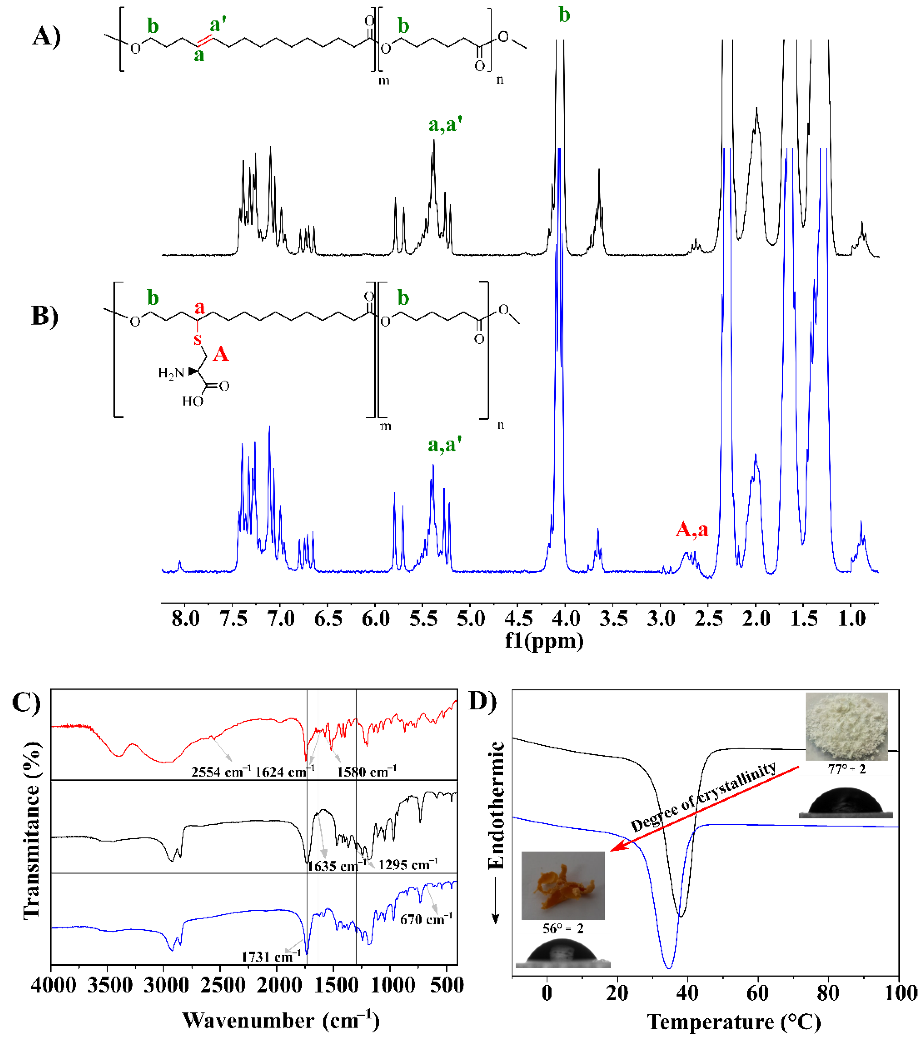
Abstract
:1. Introduction
2. Materials and Methods
2.1. Materials
2.2. Experimental Procedure
2.2.1. Enzymatic Synthesis of Poly(Globalide-co-ε-caprolactone)
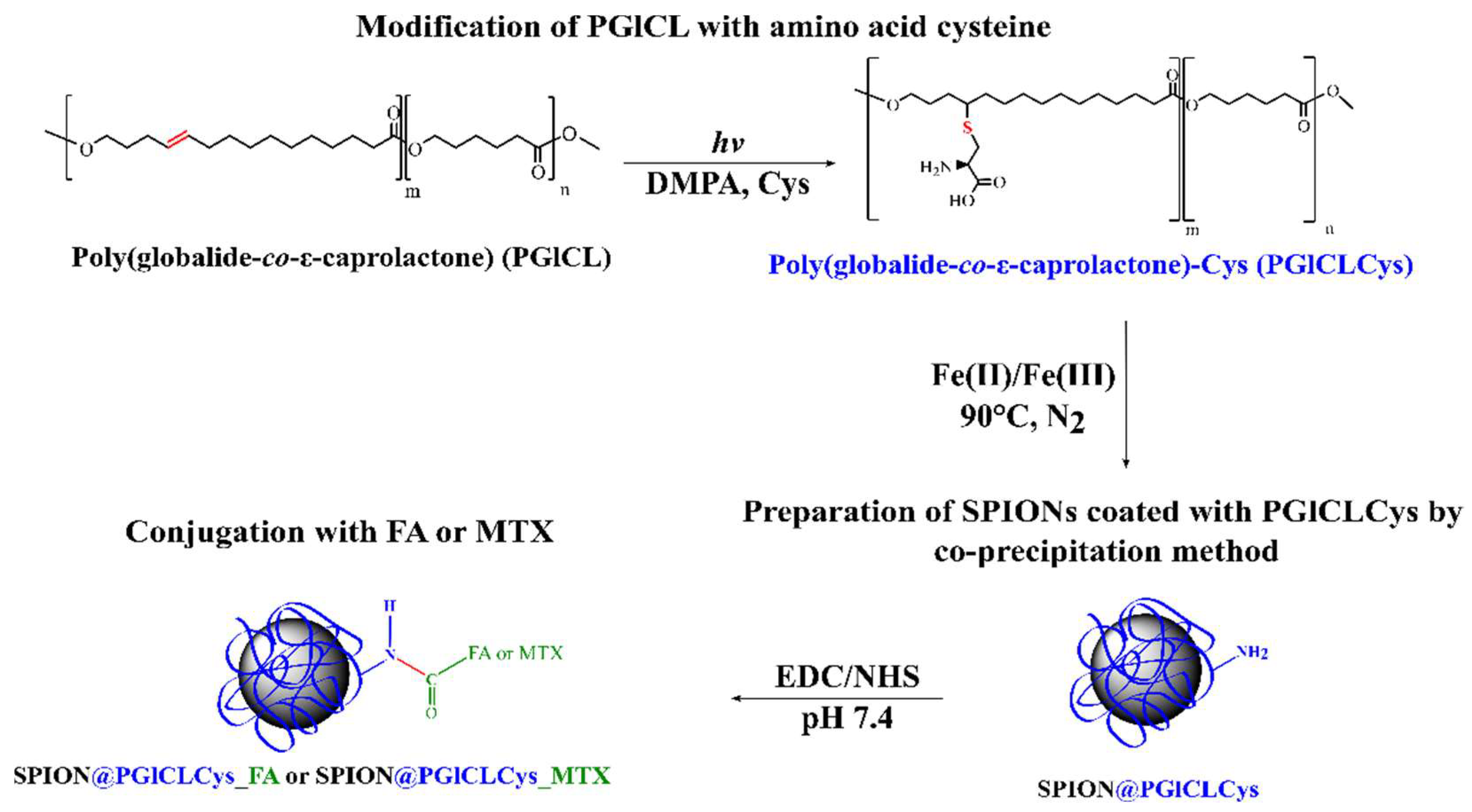
2.2.2. Modification of PGlCL with Cysteine via Thiol-Ene Reaction
2.2.3. Synthesis of Superparamagnetic Iron Oxide Nanoparticles (SPIONs) and Coating with PGlCLCys
2.2.4. Conjugation of SPION@PGlCLCys with Folic Acid
2.2.5. Conjugation of SPION@PGlCLCys with Methotrexate
2.3. Physicochemical Characterizations
2.4. Cell Culture
2.4.1. In Vitro Cell Viability of the SPION@PGlCLCys, SPION@PGlCLCys_FA, and SPION@PGlCLCys_MTX
2.4.2. Enzymatic Release of Folic Acid and Methotrexate
2.5. Computational Section
3. Results and Discussion
3.1. Synthesis, Modification, and Characterization PGlCLCys
3.2. Preparation of SPIONs Coated with PGlCLcys by Co-Precipitation Method
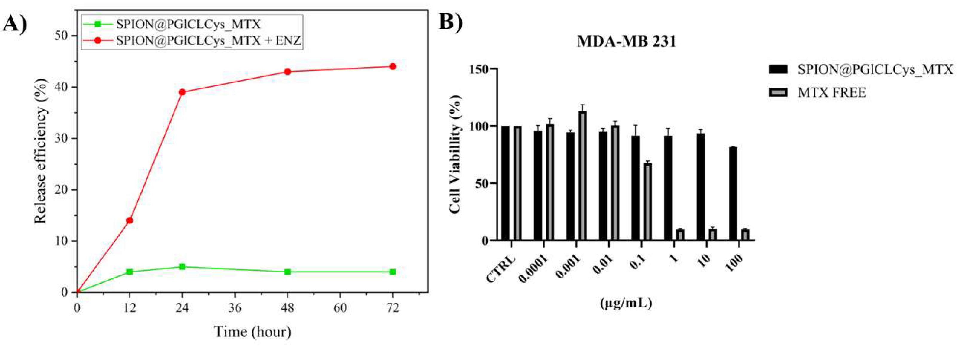
3.3. Conjugation of FA or MTX on the SPION@PGLCLCys, Enzymatic Release, and Cell Viability
4. Conclusions
Supplementary Materials
Author Contributions
Funding
Institutional Review Board Statement
Informed Consent Statement
Data Availability Statement
Acknowledgments
Conflicts of Interest
References
- Senapati, S.; Mahanta, A.K.; Kumar, S.; Maiti, P. Controlled drug delivery vehicles for cancer treatment and their performance. Signal Transduct. Target. Ther. 2018, 3, 1–19. [Google Scholar] [CrossRef] [PubMed] [Green Version]
- Grodzinski, P.; Kircher, M.; Goldberg, M.; Gabizon, A. Integrating Nanotechnology into Cancer Care. ACS Nano 2019, 13, 7370–7376. [Google Scholar] [CrossRef] [PubMed] [Green Version]
- Palanisamy, S.; Wang, Y.M. Superparamagnetic iron oxide nanoparticulate system: Synthesis, targeting, drug delivery and therapy in cancer. Dalt. Trans. 2019, 48, 9490–9515. [Google Scholar] [CrossRef] [PubMed]
- Shen, S.; Kong, F.; Guo, X.; Wu, L.; Shen, H.; Xie, M.; Wang, X.; Jin, Y.; Ge, Y. CMCTS stabilized Fe3O4 particles with extremely low toxicity as highly efficient near-infrared photothermal agents for in vivo tumor ablation. Nanoscale 2013, 5, 8056–8066. [Google Scholar] [CrossRef] [PubMed]
- Albertsson, A.C.; Varma, I.K. Recent developments in ring opening polymerization of lactones for biomedical applications. Biomacromolecules 2003, 4, 1466–1486. [Google Scholar] [CrossRef]
- Ates, Z.; Thornton, P.D.; Heise, A. Side-chain functionalisation of unsaturated polyesters from ring-opening polymerisation of macrolactones by thiol-ene click chemistry. Polym. Chem. 2011, 2, 309–312. [Google Scholar] [CrossRef]
- Dadfar, S.M.; Roemhild, K.; Drude, N.I.; von Stillfried, S.; Knüchel, R.; Kiessling, F.; Lammers, T. Iron oxide nanoparticles: Diagnostic, therapeutic and theranostic applications. Adv. Drug Deliv. Rev. 2019, 138, 302–325. [Google Scholar] [CrossRef]
- Qu, J.; Tian, Z.; Wang, Q.; Peng, S.; Luo, J.B.; Zhou, Q.H.; Lin, J. Surface design and preparation of multi-functional magnetic nanoparticles for cancer cell targeting, therapy, and imaging. RSC Adv. 2018, 8, 35437–35447. [Google Scholar] [CrossRef] [Green Version]
- Yao, X.; Mu, J.; Zeng, L.; Lin, J.; Nie, Z.; Jiang, X.; Huang, P. Stimuli-responsive cyclodextrin-based nanoplatforms for cancer treatment and theranostics. Mater. Horiz. 2019, 6, 846–870. [Google Scholar] [CrossRef]
- Van Der Meulen, I.; De Geus, M.; Antheunis, H.; Deumens, R.; Joosten, E.A.J.; Koning, C.E.; Heise, A. Polymers from functional macrolactones as potential biomaterials: Enzymatic ring opening polymerization, biodegradation, and biocompatibility. Biomacromolecules 2008, 9, 3404–3410. [Google Scholar] [CrossRef]
- Ates, Z.; Heise, A. Functional films from unsaturated poly(macrolactones) by thiol–ene cross-linking and functionalisation. Polym. Chem. 2014, 5, 2936. [Google Scholar] [CrossRef]
- de Oliveira, F.C.S.; Amaral, R.J.F.C.D.; Santos, L.E.C.D.; Cummins, C.; Morris, M.M.; Kearney, C.J.; Heise, A.J. Versatility of unsaturated polyesters from electrospun macrolactones: RGD immobilization to increase cell attachment. Biomed. Mater. Res. Part A 2021, 1–9. [Google Scholar] [CrossRef] [PubMed]
- Beltrame, J.M.; Guindani, C.; Novy, M.G.; Felipe, K.B.; Sayer, C.; Pedrosa, R.C.; De Araújo, P.H.H. Covalently Bonded N-Acetylcysteine-polyester Loaded in PCL Scaffolds for Enhanced Interactions with Fibroblasts. ACS Appl. Bio Mater. 2021, 4, 1552–1562. [Google Scholar] [CrossRef] [PubMed]
- Guindani, C.; Frey, M.-L.; Simon, J.; Koynov, K.; Schultze, J.; Ferreira, S.R.S.; Araújo, P.H.H.; Oliveira, D.; Wurm, F.R.; Mailänder, V.; et al. Covalently Binding of Bovine Serum Albumin to Unsaturated Poly(Globalide-Co-ε-Caprolactone) Nanoparticles by Thiol-Ene Reactions. Macromol. Biosci. 2019, 1900145. [Google Scholar] [CrossRef]
- Lee, N.; Schuck, P.J.; Nico, P.S.; Gilbert, B.J. Surface enhanced Raman spectroscopy of organic molecules on magnetite (Fe3O4) nanoparticles. Phys. Chem. Lett. 2015, 6, 970–974. [Google Scholar] [CrossRef] [Green Version]
- Guindani, C.; Candiotto, G.; Araújo, P.H.H.; Ferreira, S.R.S.; de Oliveira, D.; Wurm, F.R.; Landfester, K. Controlling the biodegradation rates of poly(globalide-co-ε-caprolactone) copolymers by post polymerization modification. Polym. Degrad. Stab. 2020, 179, 109287. [Google Scholar] [CrossRef]
- Pommerville, J.C. Alcamo’s Fundamentals of Microbiology: Body Systems; Jones & Bartlett Publishers: Burlington, MA, USA, 2009. [Google Scholar]
- Shetty, V.; Jakhade, A.; Shinde, K.; Chikate, R.; Kaul-Ghanekar, R. Folate mediated targeted delivery of cinnamaldehyde loaded and FITC functionalized magnetic nanoparticles in breast cancer: In vitro, in vivo and pharmacokinetic studies. New J. Chem. 2021, 45, 1500–1515. [Google Scholar] [CrossRef]
- Allard-Vannier, E.; Hervé-Aubert, K.; Kaaki, K.; Blondy, T.; Shebanova, A.; Shaitan, K.V.; Ignatova, A.A.; Saboungi, M.L.; Feofanov, A.V.; Chourpa, I. Folic acid-capped PEGylated magnetic nanoparticles enter cancer cells mostly via clathrin-dependent endocytosis. Biochim. Biophys. Acta Gen. Subj. 2017, 1861, 1578–1586. [Google Scholar] [CrossRef]
- Gonen, N.; Assaraf, Y.G. Antifolates in cancer therapy: Structure, activity and mechanisms of drug resistance. Drug Resist. Updat. 2012, 15, 183–210. [Google Scholar] [CrossRef]
- Turk, V.; Stoka, V.; Vasiljeva, O.; Renko, M.; Sun, T.; Turk, B.; Turk, D. Cysteine cathepsins: From structure, function and regulation to new frontiers. Biochim. Biophys. Acta Proteins Proteomics 2012, 1824, 68–88. [Google Scholar] [CrossRef] [PubMed] [Green Version]
- Clark, A.C. Caspase Allostery and Conformational Selection. Chem. Rev. 2016, 116, 6666–6706. [Google Scholar] [CrossRef] [PubMed]
- Guindani, C.; Dozoretz, P.; Veneral, J.G.; Silva, D.M.; Araújo, P.H.H.; Ferreira, S.R.S.; Oliveira, D.J. Enzymatic ring opening copolymerization of globalide and ε-caprolactone under supercritical conditions. Supercrit. Fluids 2017, 128, 404–411. [Google Scholar] [CrossRef]
- Polloni, A.E.; Chiaradia, V.; Amaral, R.J.F.C.D.; Kearney, C.; Gorey, B.; De Oliveira, D.; De Oliveira, J.V.; De Araújo, P.H.H.; Sayer, C.; Heise, A. Polyesters with main and side chain phosphoesters as structural motives for biocompatible electrospun fibres. Polym. Chem. 2020, 11, 2157–2165. [Google Scholar] [CrossRef]
- Zottis, A.D.A.; Beltrame, J.M.; Lara, L.R.S.; Costa, T.G.; Feldhaus, M.J.; Pedrosa, R.C.; Ourique, F.; de Campos, C.E.M.; de A Isoppo, E.; da Silva Miranda, F.; et al. Pheomelanin-coated iron oxide magnetic nanoparticles: A promising candidate for negative T 2 contrast enhancement in magnetic resonance imaging. Chem. Commun. 2015, 51, 11194–11197. [Google Scholar] [CrossRef]
- Guo, X.; Shi, C.; Wang, J.; Di, S.; Zhou, S. PH-triggered intracellular release from actively targeting polymer micelles. Biomaterials 2013, 34, 4544–4554. [Google Scholar] [CrossRef] [PubMed]
- Mosmann, T. Rapid colorimetric assay for cellular growth and survival: Application to proliferation and cytotoxicity assays. J. Immunol. Methods 1983, 65, 55–63. [Google Scholar] [CrossRef]
- Gupta, J.; Bhargava, P.; Bahadur, D.J. Methotrexate conjugated magnetic nanoparticle for targeted drug delivery and thermal therapy. Appl. Phys. 2014, 115, 2012–2015. [Google Scholar] [CrossRef]
- Chu, C.H.; Leung, C. The convolution equation of Choquet and Deny on [IN]-groups. Integr. Equations Oper. Theory 2001, 40, 391–402. [Google Scholar] [CrossRef]
- Jung, J.Y.; Park, J.H.; Jeong, Y.J.; Yang, K.H.; Choi, N.K.; Kim, S.H.; Kim, W.J. Involvement of Bcl-2 family and caspases cascade in sodium fluoride-induced apoptosis of human gingival fibroblasts. Korean J. Physiol. Pharmacol. 2006, 10, 289–295. [Google Scholar]
- Hanwell, M.D.; Curtis, D.E.; Lonie, D.C.; Vandermeersch, T.; Zurek, E.; Hutchison, G.R. Avogadro: An advanced semantic chemical editor, visualization, and analysis platform. J. Cheminform. 2012, 4, 17. [Google Scholar] [CrossRef] [Green Version]
- Garrido, N.M.; Jorge, M.; Queimada, A.J.; Macedo, E.A.; Economou, I.G. Using molecular simulation to predict solute solvation and partition coefficients in solvents of different polarity. Phys. Chem. Chem. Phys. 2011, 13, 9155. [Google Scholar] [CrossRef] [Green Version]
- Nedyalkova, M.A.; Madurga, S.; Tobiszewski, M.; Simeonov, V.J. Calculating the Partition Coefficients of Organic Solvents in Octanol/Water and Octanol/Air. Chem. Inf. Model. 2019, 59, 2257–2263. [Google Scholar] [CrossRef] [PubMed]
- Candiotto, G.; Giro, R.; Horta, B.A.C.; Rosselli, F.P.; De Cicco, M.; Achete, C.A.; Cremona, M.; Capaz, R.B. Emission redshift in DCM2-doped Alq3 caused by nonlinear Stark shifts and Förster-mediated exciton diffusion. Phys. Rev. B 2020, 102, 1–7. [Google Scholar] [CrossRef]
- Bauer, K.N.; Liu, L.; Wagner, M.; Andrienko, D.; Wurm, F.R. Mechanistic study on the hydrolytic degradation of polyphosphates. Eur. Polym. J. 2018, 108, 286–294. [Google Scholar] [CrossRef]
- Neese, F. The ORCA program system. Wiley Interdiscip. Rev. Comput. Mol. Sci. 2012, 2, 73–78. [Google Scholar] [CrossRef]
- Guindani, C.; Dozoretz, P.; Araújo, P.H.H.; Ferreira, S.R.S.; de Oliveira, D. N-acetylcysteine side-chain functionalization of poly(globalide-co-ε-caprolactone) through thiol-ene reaction. Mater. Sci. Eng. C 2019, 94, 477–483. [Google Scholar] [CrossRef]
- Pachence, J.M.; Bohrer, M.P.; Kohn, J. Principles of Tissue Engineering, 3rd ed.; Lanza, R., Langer, R., Vacanti, J., Eds.; Elsevier: Amsterdam, The Netherlands, 2007; pp. 323–339. [Google Scholar]
- Bee, A.; Massart, R.; Neveu, S.J. Synthesis of very fine maghemite particles. Magn. Magn. Mater. 1995, 149, 6–9. [Google Scholar] [CrossRef]
- Habibi, N. Preparation of biocompatible magnetite-carboxymethyl cellulose nanocomposite: Characterization of nanocomposite by FTIR, XRD, FESEM and TEM. Spectrochim. Acta Part A Mol. Biomol. Spectrosc. 2014, 131, 55–58. [Google Scholar] [CrossRef]
- Lee, N.; Hyeon, T. Designed synthesis of uniformly sized iron oxide nanoparticles for efficient magnetic resonance imaging contrast agents. Chem. Soc. Rev. 2012, 41, 2575–2589. [Google Scholar] [CrossRef]
- Hadadian, Y.; Masoomi, H.; Dinari, A.; Ryu, C.; Hwang, S.; Kim, S.; Cho, B.K.; Lee, J.Y.; Yoon, J. From Low to High Saturation Magnetization in Magnetite Nanoparticles: The Crucial Role of the Molar Ratios between the Chemicals. ACS Omega 2022, 7, 15996–16012. [Google Scholar] [CrossRef]
- Celis, J.A.; Mejía, O.F.O.; Cabral-Prieto, A.; García-Sosa, I.; Derat-Escudero, R.; Saitovitch, E.M.B.; Camarena, M.A. Alzamora Camarena, M. Synthesis and characterization of nanometric magnetite coated by oleic acid and the surfactant CTAB: Surfactant coated nanometric magnetite/maghemite. Hyperfine Interact. 2017, 238, 43. [Google Scholar] [CrossRef]
- Kumar, A.; Dixit, C.K. Methods for characterization of nanoparticles. In Advances in Nanomedicine for the Delivery of Therapeutic Nucleic Acids; Elsevier: Amsterdam, The Netherlands, 2017; pp. 43–58. [Google Scholar]
- Gunguli, N.C. Paper disk electrophoresis in the determination of isoelectric points of amino acids. Fresenius’ Zeitschrift Anal. Chem. 1956, 154, 161–167. [Google Scholar] [CrossRef]
- Yoo, J.-W.; Chambers, E.; Mitragotri, S. Factors that Control the Circulation Time of Nanoparticles in Blood: Challenges, Solutions and Future Prospects. Curr. Pharm. Des. 2010, 16, 2298–2307. [Google Scholar] [CrossRef] [PubMed]
- Brandt, J.V.; Piazza, R.D.; Santos, C.C.D.; Vega-Chacón, J.; Amantéa, B.E.; Pinto, G.C.; Jafelicci, M.; Marques, R.F.C. Synthesis of core@shell nanoparticles functionalized with folic acid-modified PCL-co-PEGMA copolymer for methotrexate delivery. Nano-Struct. Nano-Objects 2021, 25, 100675. [Google Scholar] [CrossRef]
- Piazza, R.D.; Brandt, J.V.; Gobo, G.G.; Tedesco, A.C.; Primo, F.L.; Marques, R.F.C.; Junior, M.J. mPEG-co-PCL nanoparticles: The influence of hydrophobic segment on methotrexate drug delivery. Colloids Surf. A Physicochem. Eng. Asp. 2018, 555, 142–149. [Google Scholar] [CrossRef]


Disclaimer/Publisher’s Note: The statements, opinions and data contained in all publications are solely those of the individual author(s) and contributor(s) and not of MDPI and/or the editor(s). MDPI and/or the editor(s) disclaim responsibility for any injury to people or property resulting from any ideas, methods, instructions or products referred to in the content. |
© 2023 by the authors. Licensee MDPI, Basel, Switzerland. This article is an open access article distributed under the terms and conditions of the Creative Commons Attribution (CC BY) license (https://creativecommons.org/licenses/by/4.0/).
Share and Cite
Beltrame, J.M.; Ribeiro, B.B.P.; Guindani, C.; Candiotto, G.; Felipe, K.B.; Lucas, R.; Zottis, A.D.; Isoppo, E.; Sayer, C.; de Araújo, P.H.H. Coating of SPIONs with a Cysteine-Decorated Copolyester: A Possible Novel Nanoplatform for Enzymatic Release. Pharmaceutics 2023, 15, 1000. https://doi.org/10.3390/pharmaceutics15031000
Beltrame JM, Ribeiro BBP, Guindani C, Candiotto G, Felipe KB, Lucas R, Zottis AD, Isoppo E, Sayer C, de Araújo PHH. Coating of SPIONs with a Cysteine-Decorated Copolyester: A Possible Novel Nanoplatform for Enzymatic Release. Pharmaceutics. 2023; 15(3):1000. https://doi.org/10.3390/pharmaceutics15031000
Chicago/Turabian StyleBeltrame, Jeovandro Maria, Brena Beatriz Pereira Ribeiro, Camila Guindani, Graziâni Candiotto, Karina Bettega Felipe, Rodrigo Lucas, Alexandre D’Agostini Zottis, Eduardo Isoppo, Claudia Sayer, and Pedro Henrique Hermes de Araújo. 2023. "Coating of SPIONs with a Cysteine-Decorated Copolyester: A Possible Novel Nanoplatform for Enzymatic Release" Pharmaceutics 15, no. 3: 1000. https://doi.org/10.3390/pharmaceutics15031000