
N-Benzylethanolammonium Ionic Liquids and Molten Salts in the Synthesis of 68Ga- and Al18F-Labeled Radiopharmaceuticals
 , ,
, ,
Abstract
:1. Introduction
2. Materials and Methods
2.1. Materials
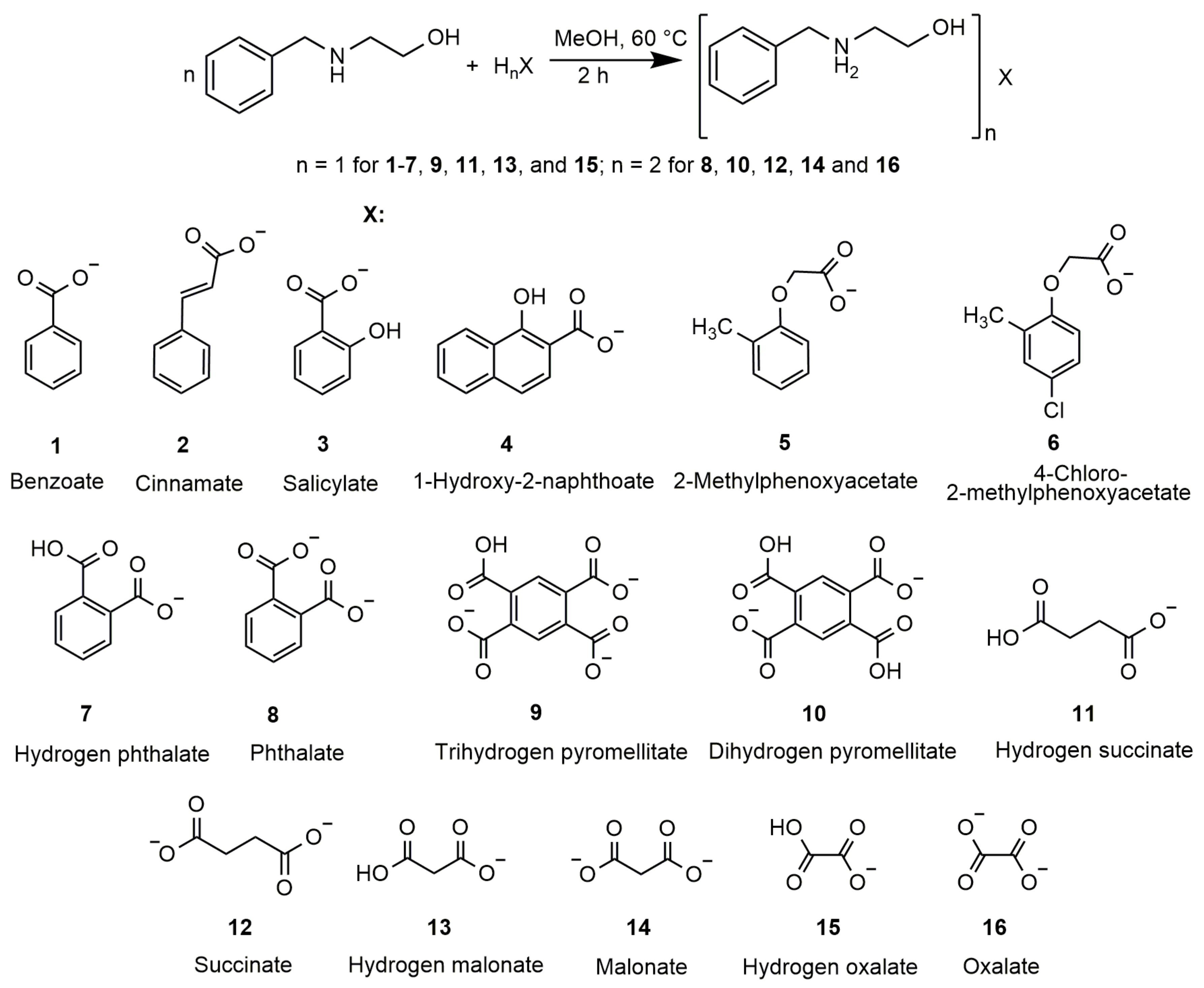
2.2. Synthesis
2.3. Methods
2.4. X-ray Structure Determination
2.5. Hirshfeld Surfaces Analysis
2.6. ADME Analysis
2.7. Buffer Activity
2.7.1. Preparation of 68Ga
2.7.2. Preparation of 18F
2.7.3. General Method of High-Temperature Radiolabeling of BCAs and Peptides with 68Ga (HT Radiolabeling)
2.7.4. General Method of Low-Temperature Radiolabeling of BCAs and Peptides with 68Ga (LT Radiolabeling)
2.7.5. General Method of Radiolabeling of BCAs and Peptides with 18F
2.7.6. Statistical Analysis
2.7.7. pH Profiles
3. Results and Discussion
3.1. Synthesis and Characterization
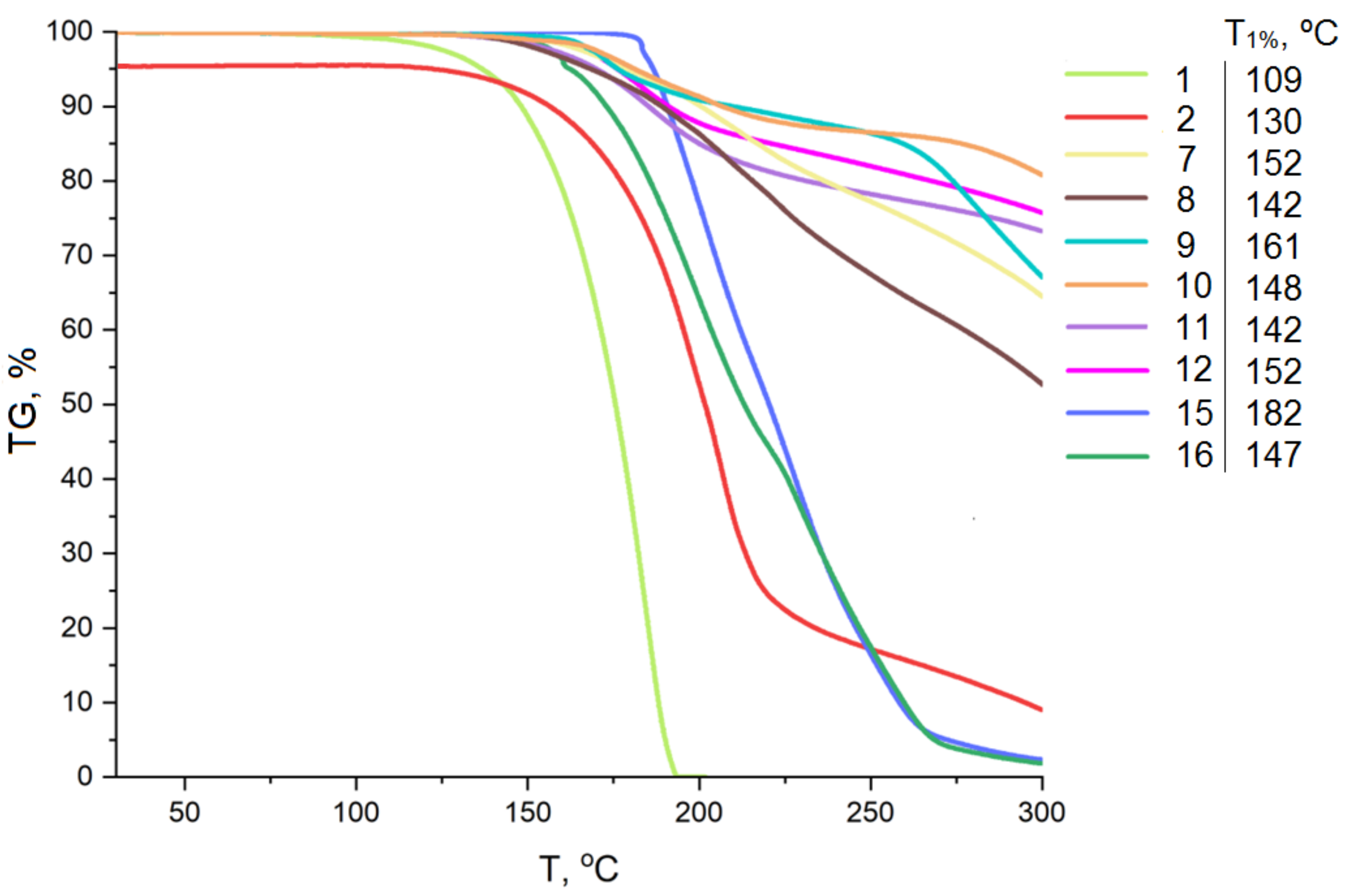
3.2. Thermogravimetric Analysis
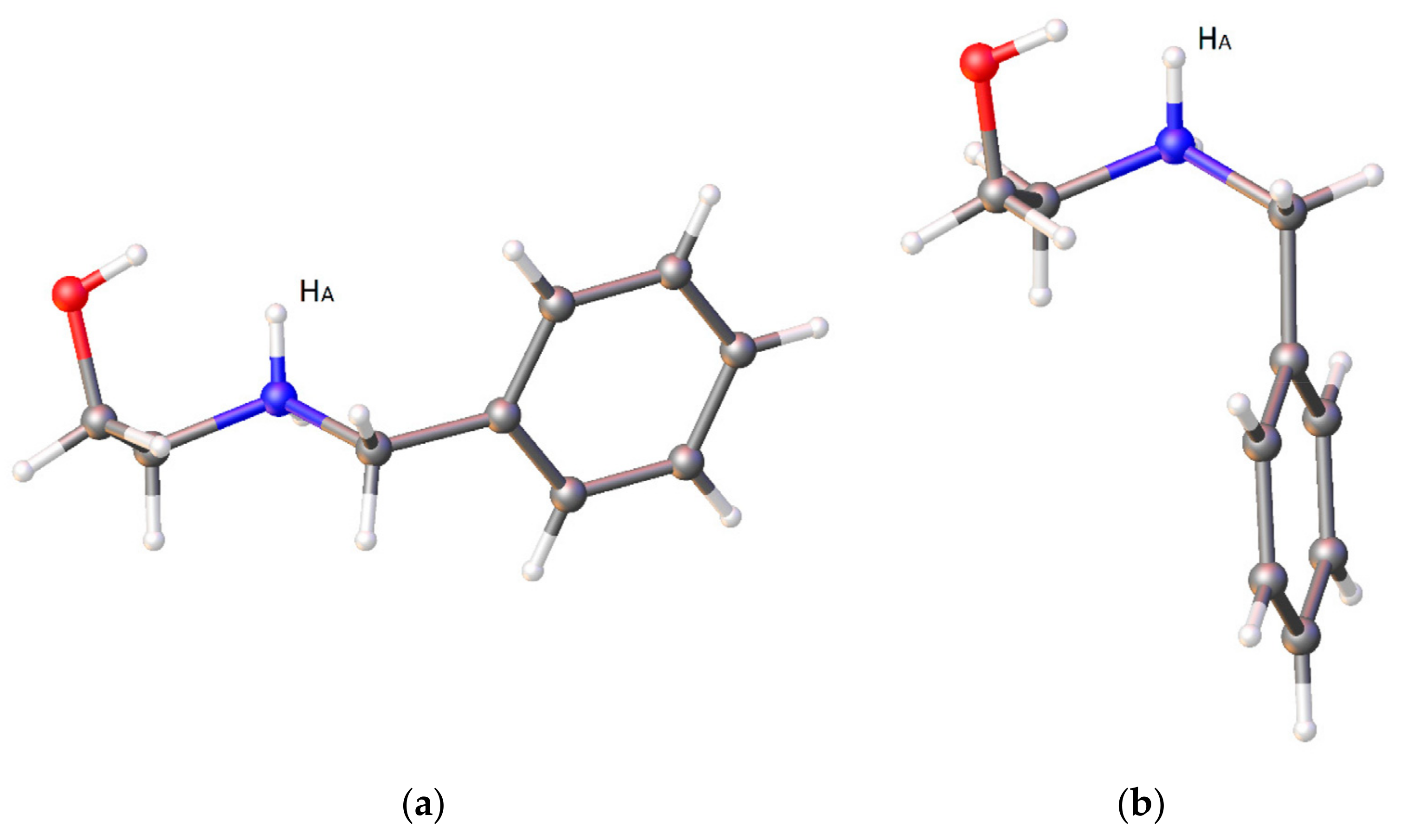
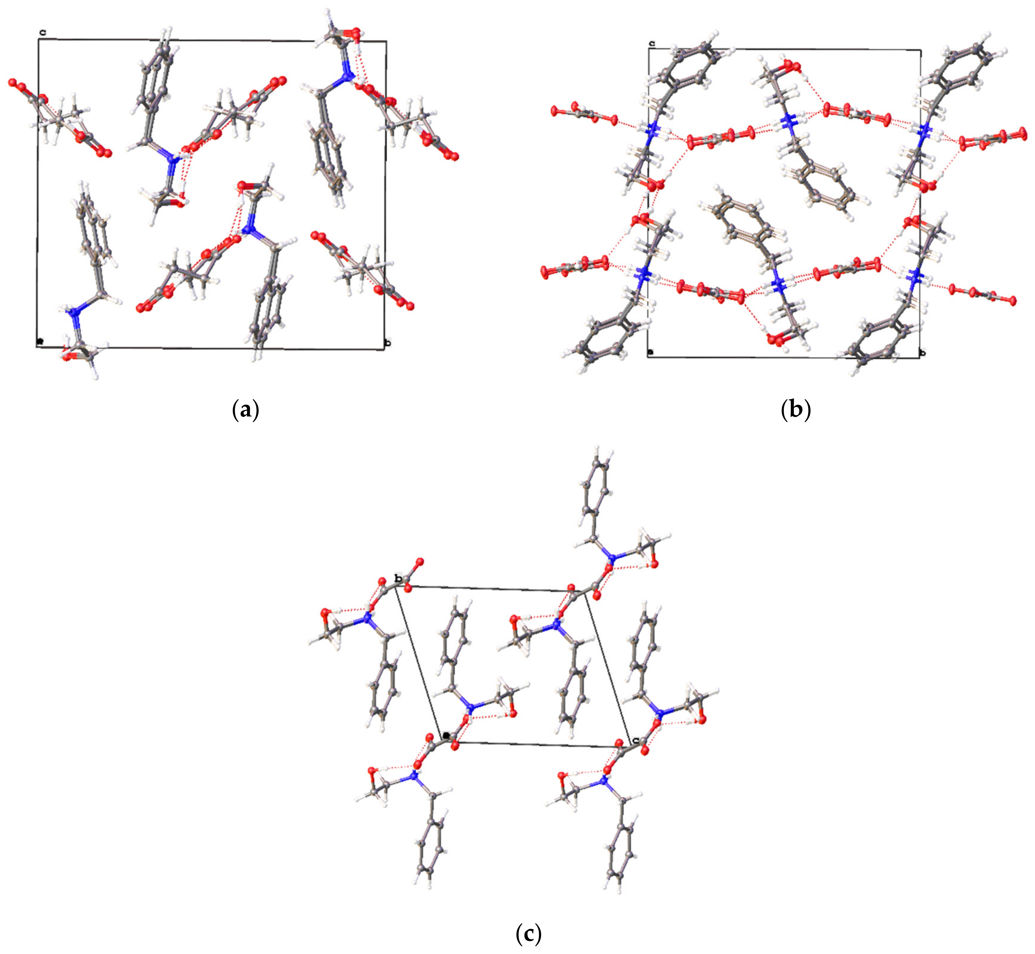
3.3. X-ray Crystal Structure
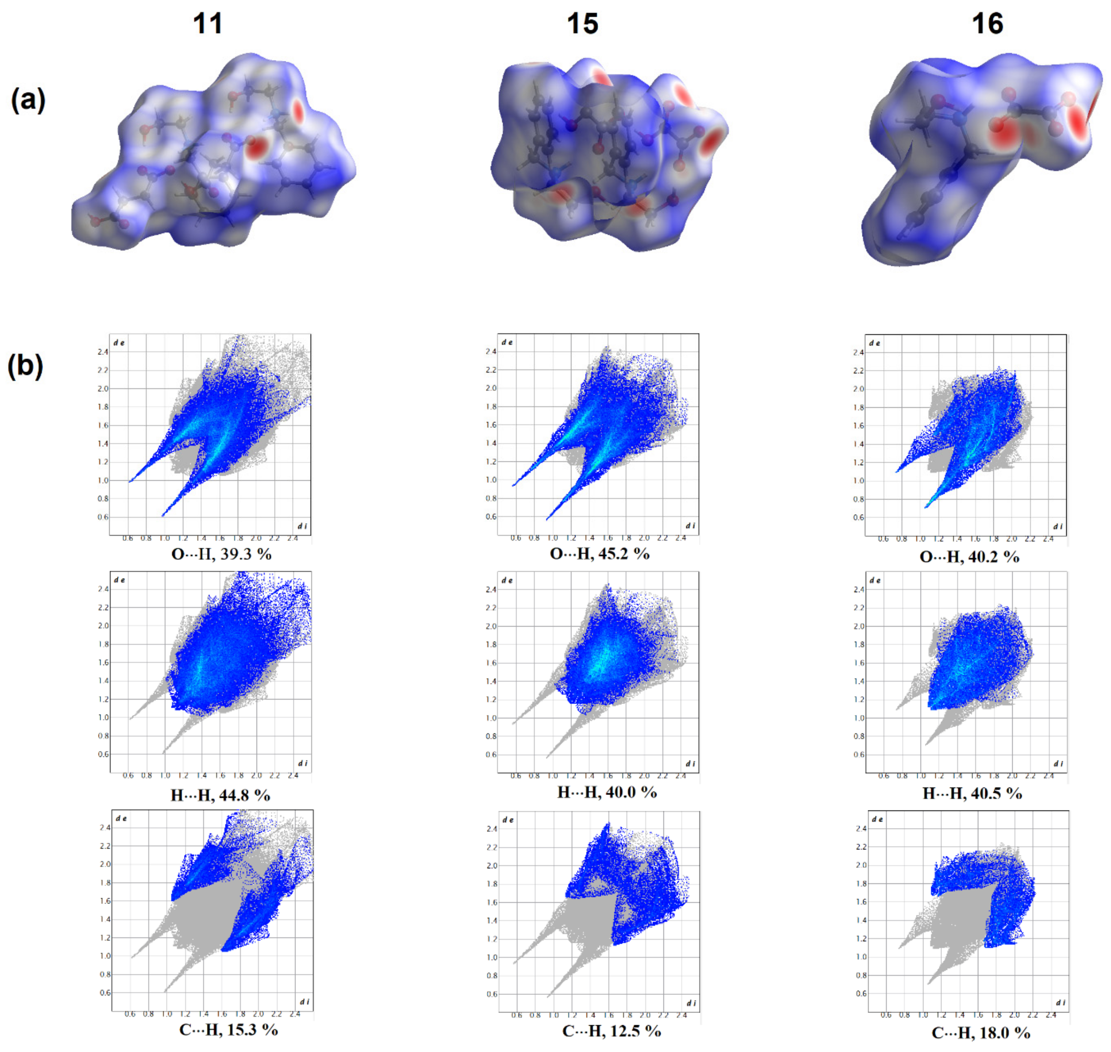
3.4. Hirshfeld Surface Analysis
3.5. In Silico ADME Prediction
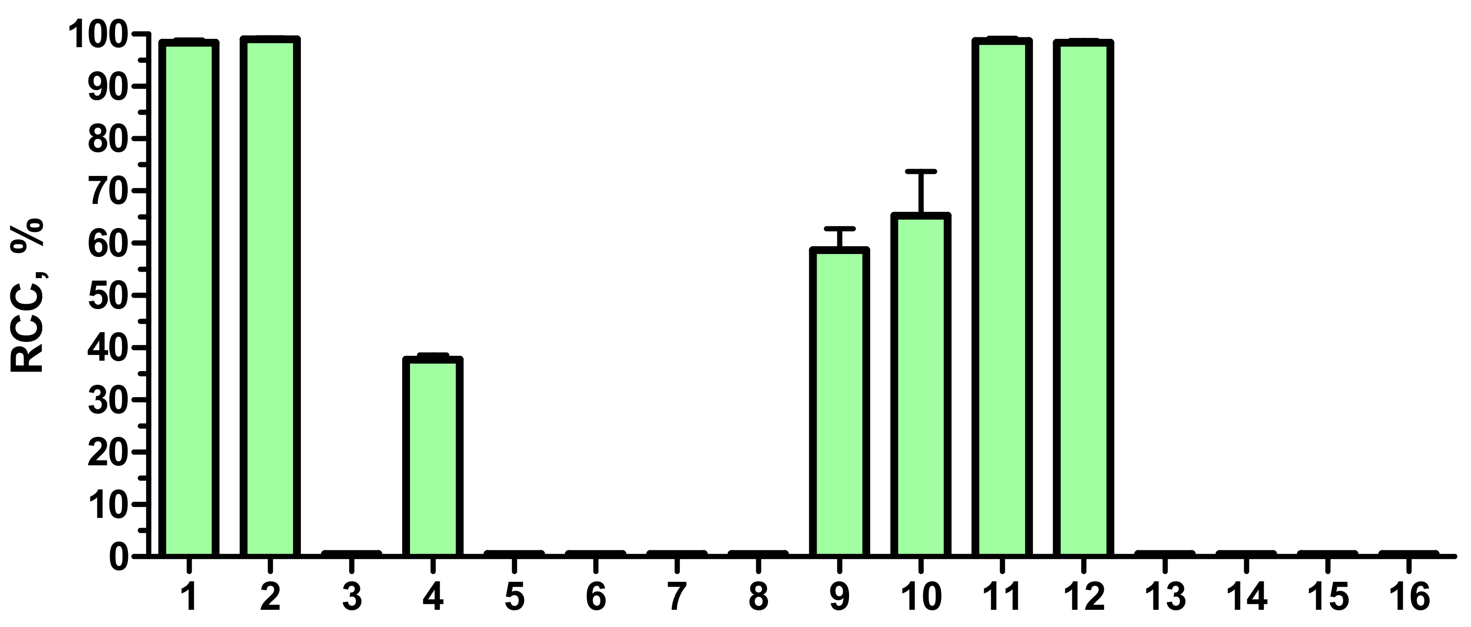
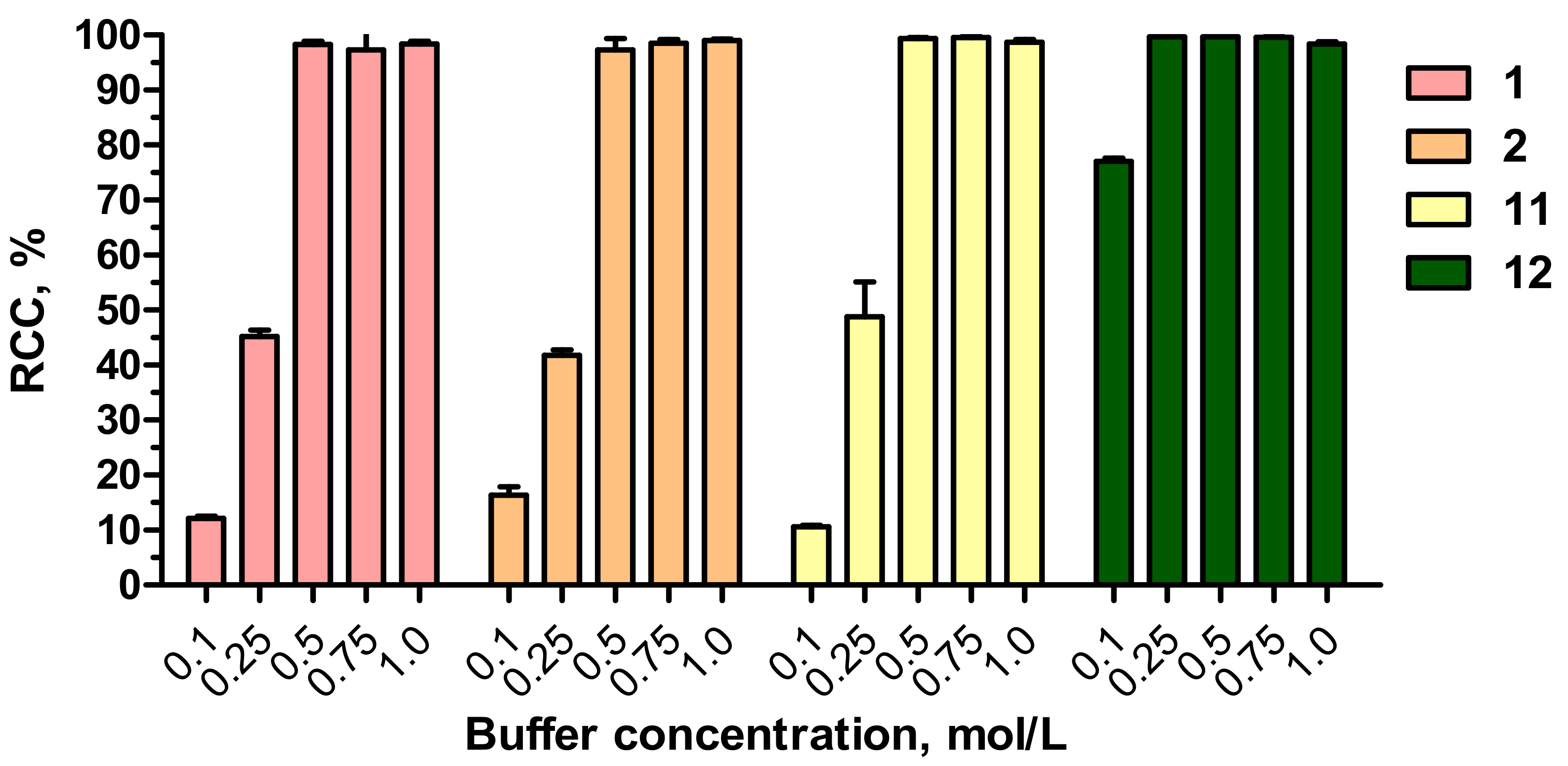
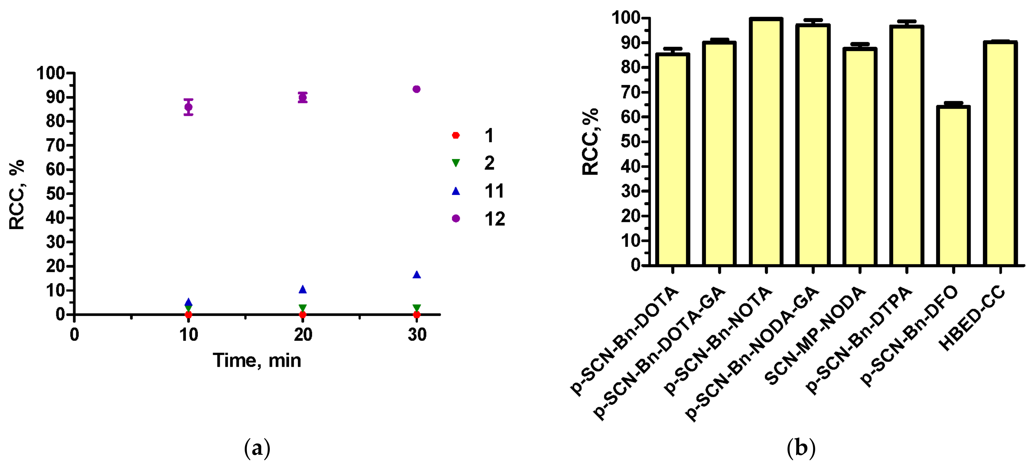
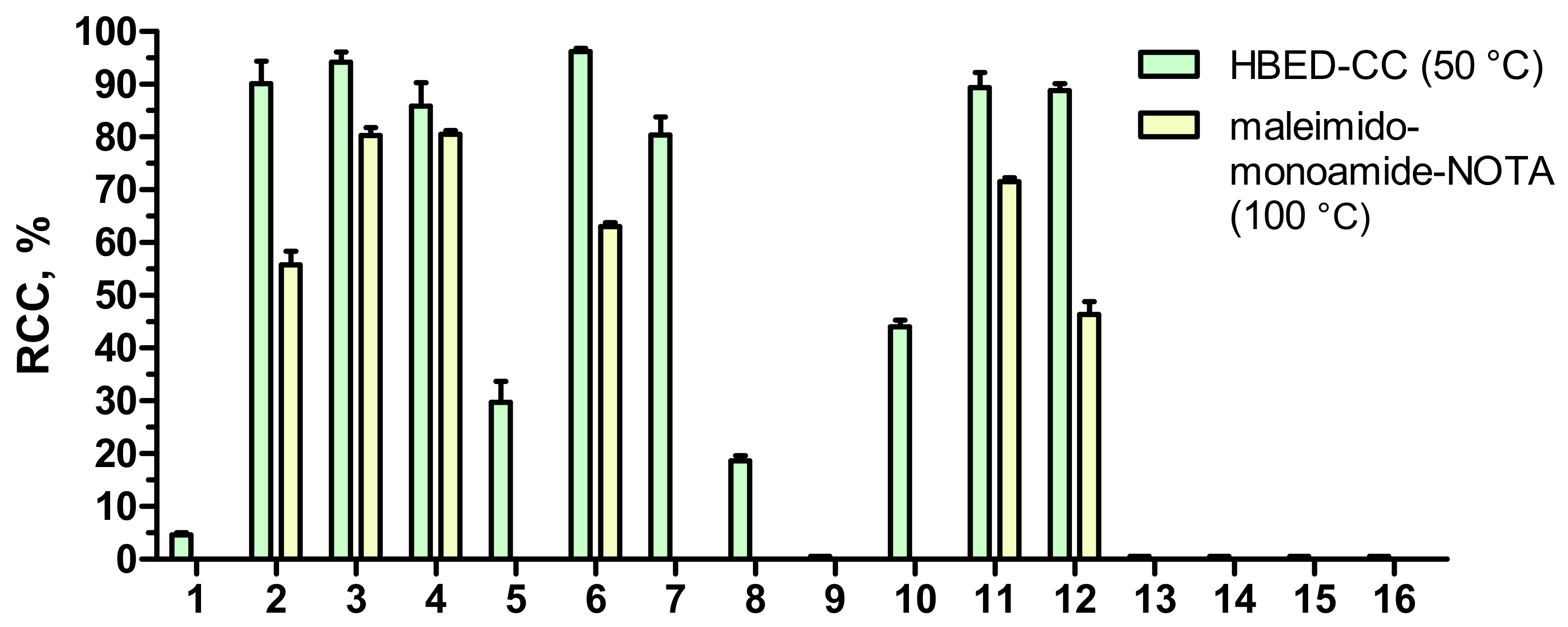
3.6. Buffer Activity in 68Ga-Radiolabeling Reactions
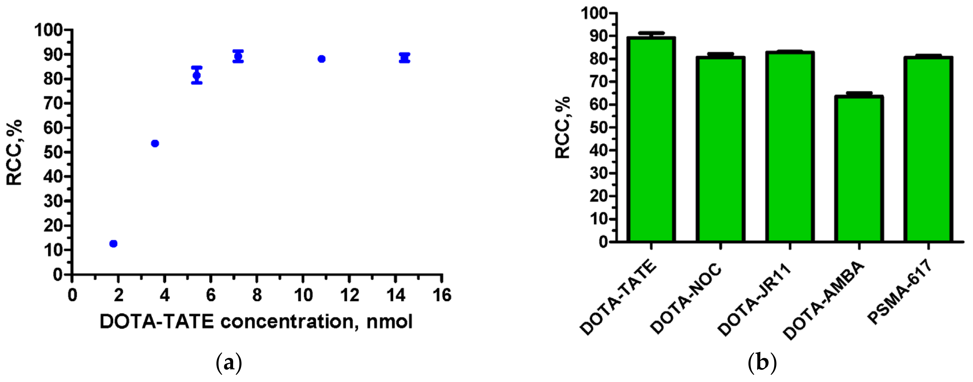
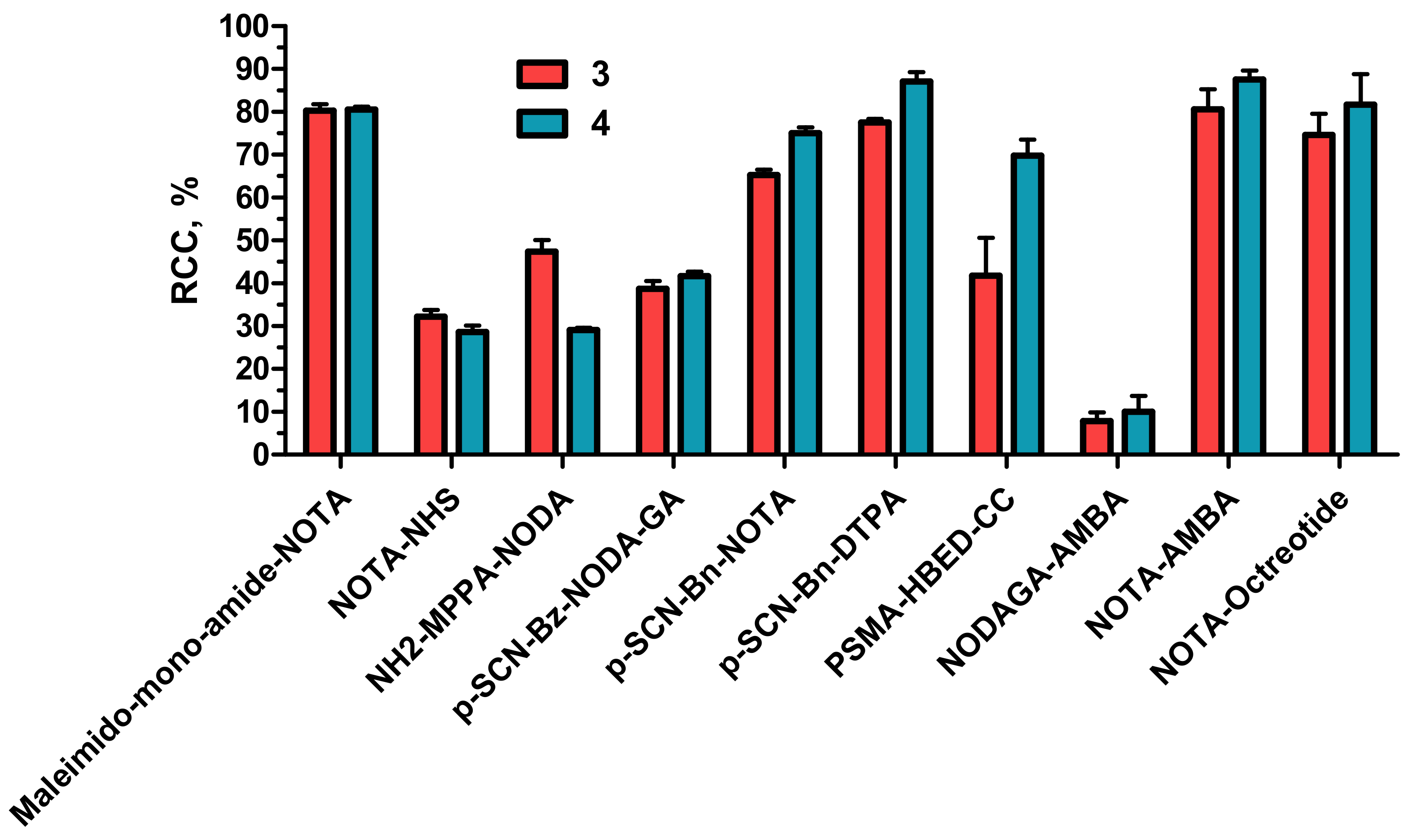
3.7. Buffer Activity in Al18F-Radiolabeling Reactions
4. Conclusions
Supplementary Materials
Author Contributions
Funding
Data Availability Statement
Acknowledgments
Conflicts of Interest
References
- Ibsen, K.N.; Ma, H.; Banerjee, A.; Tanner, E.E.L.; Nangia, S.; Mitragotri, S. Mechanism of Antibacterial Activity of Choline-Based Ionic Liquids (CAGE). ACS Biomater. Sci. Eng. 2018, 4, 2370–2379. [Google Scholar] [CrossRef]
- Moshikur, R.M.; Chowdhury, M.R.; Moniruzzaman, M.; Goto, M. Biocompatible ionic liquids and their applications in pharmaceutics. Green Chem. 2020, 22, 8116–8139. [Google Scholar] [CrossRef]
- Moshikur, R.M.; Goto, M. Ionic Liquids as Active Pharmaceutical Ingredients (APIs). In Application of Ionic Liquids in Drug Delivery; Goto, M., Moniruzzaman, M., Eds.; Springer: Singapore, 2021; pp. 13–33. [Google Scholar] [CrossRef]
- Egorova, K.S.; Gordeev, E.G.; Ananikov, V.P. Biological Activity of Ionic Liquids and Their Application in Pharmaceutics and Medicine. Chem. Rev. 2017, 117, 7132–7189. [Google Scholar] [CrossRef] [Green Version]
- Curreri, A.M.; Mitragotri, S.; Tanner, E.E.L. Recent Advances in Ionic Liquids in Biomedicine. Adv. Sci. 2021, 8, 2004819. [Google Scholar] [CrossRef]
- Shukla, S.K.; Mikkola, J.-P. Use of Ionic Liquids in Protein and DNA Chemistry. Front. Chem. 2020, 8, 598662. [Google Scholar] [CrossRef]
- Pedro, S.N.; R. Freire, C.S.; Silvestre, A.J.D.; Freire, M.G. The Role of Ionic Liquids in the Pharmaceutical Field: An Overview of Relevant Applications. Int. J. Mol. Sci. 2020, 21, 8298. [Google Scholar] [CrossRef]
- Siopa, F.; Figueiredo, T.; Frade, R.F.M.; Neto, I.; Meirinhos, A.; Reis, C.P.; Sobral, R.G.; Afonso, C.A.M.; Rijo, P. Choline-Based Ionic Liquids: Improvement of Antimicrobial Activity. ChemistrySelect 2016, 1, 5909–5916. [Google Scholar] [CrossRef]
- Pernak, J.; Syguda, A.; Mirska, I.; Pernak, A.; Nawrot, J.; Prądzyńska, A.; Griffin, S.; Rogers, R. Choline-derivative-based ionic liquids. Chem. Eur. J. 2007, 13, 6817–6827. [Google Scholar] [CrossRef]
- Pernak, J.; Chwała, P. Synthesis and anti-microbial activities of choline-like quaternary ammonium chlorides. Eur. J. Med. Chem. 2003, 38, 1035–1042. [Google Scholar] [CrossRef]
- Ahlstrom, B.; Chelminska-Bertilsson, M.; Thompson, R.A.; Edebo, L. Long-chain alkanoylcholines, a new category of soft antimicrobial agents that are enzymatically degradable. Antimicrob. Agents Chemother. 1995, 39, 50–55. [Google Scholar] [CrossRef] [Green Version]
- Zakrewsky, M.; Lovejoy, K.S.; Kern, T.L.; Miller, T.E.; Le, V.; Nagy, A.; Goumas, A.M.; Iyer, R.S.; Del Sesto, R.E.; Koppisch, A.T.; et al. Ionic liquids as a class of materials for transdermal delivery and pathogen neutralization. Proc. Natl. Acad. Sci. USA 2014, 111, 13313–13318. [Google Scholar] [CrossRef] [Green Version]
- Correia, D.M.; Fernandes, L.C.; Fernandes, M.M.; Hermenegildo, B.; Meira, R.M.; Ribeiro, C.; Ribeiro, S.; Reguera, J.; Lanceros-Méndez, S. Ionic Liquid-Based Materials for Biomedical Applications. Nanomaterials 2021, 11, 2401. [Google Scholar] [CrossRef]
- Chowdhury, M.R.; Moshikur, R.M.; Wakabayashi, R.; Tahara, Y.; Kamiya, N.; Moniruzzaman, M.; Goto, M. Ionic-liquid-based paclitaxel preparation: A new potential formulation for cancer treatment. Mol. Pharm. 2018, 15, 2484–2488. [Google Scholar] [CrossRef]
- Jadhav, N.R.; Bhosale, S.P.; Bhosale, S.S.; Mali, S.D.; Toraskar, P.B.; Kadam, T.S. Ionic liquids: Formulation avenues, drug delivery and therapeutic updates. J. Drug Deliv. Sci. Technol. 2021, 65, 102694. [Google Scholar] [CrossRef]
- Taha, M.; Silva, F.A.E.; Quental, M.V.; Ventura, S.P.M.; Freire, M.G.; Coutinho, J.A.P. Good’s buffers as a basis for developing self-buffering and biocompatible ionic liquids for biological research. Green Chem. 2014, 16, 3149–3159. [Google Scholar] [CrossRef] [Green Version]
- Good, N.E.; Winget, G.D.; Winter, W.; Connolly, T.N.; Izawa, S.; Singh, R.M.M. Hydrogen Ion Buffers for Biological Research. Biochemistry 1966, 5, 467–477. [Google Scholar] [CrossRef]
- Lee, S.Y.; Vicente, F.A.; e Silva, F.A.; Sintra, T.E.; Taha, M.; Khoiroh, I.; Coutinho, J.A.P.; Show, P.L.; Ventura, S.P.M. Evaluating Self-buffering Ionic Liquids for Biotechnological Applications. ACS Sustain. Chem. Eng. 2015, 3, 3420–3428. [Google Scholar] [CrossRef]
- Taha, M.; Quental, M.V.; Correia, I.; Freire, M.G.; Coutinho, J.A.P. Extraction and stability of bovine serum albumin (BSA) using cholinium-based Good’s buffers ionic liquids. Process Biochem. 2015, 50, 1158–1166. [Google Scholar] [CrossRef] [Green Version]
- Taha, M.; Khan, I.; Coutinho, J.A.P. Coordination abilities of Good’s buffer ionic liquids toward europium(III) ion in aqueous solution. J. Chem. Thermodyn. 2016, 94, 152–159. [Google Scholar] [CrossRef]
- Gupta, B.S.; Taha, M.; Lee, M.-J. Extraction of an active enzyme by self-buffering ionic liquids: A green medium for enzymatic research. RSC Adv. 2016, 6, 18567–18576. [Google Scholar] [CrossRef]
- Antuganov, D.; Timofeev, V.; Timofeeva, K.; Antuganova, Y.; Kondratenko, Y.A. Evaluation of Protic Ionic Liquids Based on Triethanolammonium and Tris(hydroxymethyl)methylammonium Salts as Buffers for 68Ga-Radiolabelling of PSMA-HBED-CC. ChemistrySelect 2019, 4, 12524–12527. [Google Scholar] [CrossRef]
- Kondratenko, Y.A.; Antuganov, D.O.; Zolotarev, A.A.; Nadporojskii, M.A.; Ugolkov, V.L.; Kochina, T.A. Diethanolammonium protic ionic liquids—Promising buffers for the synthesis of 68Ga-labelled radiopharmaceuticals. J. Mol. Liq. 2022, 345, 117029. [Google Scholar] [CrossRef]
- Kondratenko, Y.A.; Antuganov, D.O.; Kadnikova, O.Y.; Zolotarev, A.A.; Ugolkov, V.L.; Nadporojskii, M.A.; Kochina, T.A. Synthesis, crystal structure and properties of tris(2-hydroxypropyl)ammonium based protic ionic liquids and protic molten salts. J. Mol. Liq. 2021, 324, 114717. [Google Scholar] [CrossRef]
- Kondratenko, Y.A.; Antuganov, D.O.; Zolotarev, A.A.; Nadporojskii, M.A.; Ugolkov, V.L.; Kochina, T.A. Protic Ionic Liquids Based on BIS-TRIS Carboxylates: Synthesis, Structural Characterization and Buffer Activity. ChemistrySelect 2022, 7, e202200660. [Google Scholar] [CrossRef]
- Kondratenko, Y.A.; Makovskaya, O.N.; Antuganov, D.O.; Zolotarev, A.A.; Ugolkov, V.L.; Nadporojskii, M.A.; Kochina, T.A. Dicationic protic ionic liquids based on N,N,N′,N′-tetrakis(2-hydroxyethyl)ethylenediamine. J. Mol. Liq. 2022, 363, 119891. [Google Scholar] [CrossRef]
- Poletto, G.; Cecchin, D.; Sperti, S.; Filippi, L.; Realdon, N.; Evangelista, L. Head-to-Head Comparison between Peptide-Based Radiopharmaceutical for PET and SPECT in the Evaluation of Neuroendocrine Tumors: A Systematic Review. Curr. Issues Mol. Biol. 2022, 44, 5516–5530. [Google Scholar] [CrossRef]
- Fine, G.C.; Covington, M.F.; Koppula, B.R.; Salem, A.E.; Wiggins, R.H.; Hoffman, J.M.; Morton, K.A. PET-CT in Clinical Adult Oncology—VI. Primary Cutaneous Cancer, Sarcomas and Neuroendocrine Tumors. Cancers 2022, 14, 2835. [Google Scholar] [CrossRef]
- Caracciolo, M.; Castello, A.; Urso, L.; Borgia, F.; Ortolan, N.; Uccelli, L.; Cittanti, C.; Castellani, M.; Bartolomei, M.; Lazzeri, M.; et al. The Role of [68Ga]PSMA PET/CT for Clinical Suspicion of Prostate Cancer in Patients with or without Previous Negative Biopsy: A Systematic Review. Cancers 2022, 14, 5036. [Google Scholar] [CrossRef]
- Dondi, F.; Albano, D.; Bertagna, F.; Treglia, G. Bone Scintigraphy versus PSMA-Targeted PET/CT or PET/MRI in Prostate Cancer: Lessons Learned from Recent Systematic Reviews and Meta-Analyses. Cancers 2022, 14, 4470. [Google Scholar] [CrossRef]
- Moerlein, S.M.; Welch, M.J. The chemistry of gallium and indium as related to radiopharmaceutical production. Int. J. Nucl. Med. Biol. 1981, 8, 277–287. [Google Scholar] [CrossRef]
- Bauwens, M.; Chekol, R.; Vanbilloen, H.; Bormans, G.; Verbruggen, A. Optimal buffer choice of the radiosynthesis of 68Ga–Dotatoc for clinical application. Nucl. Med. Commun. 2010, 31, 753–758. [Google Scholar] [CrossRef]
- Martins, A.F.; Prata, M.I.M.; Rodrigues, S.P.J.; Geraldes, C.F.G.C.; Riss, P.J. Amor-Coarasa, A.; Burchardt, C.; Kroll, C.; Roesch, F. Spectroscopic, radiochemical, and theoretical studies of the Ga3+-N-2-hydroxyethyl piperazine-N′-2-ethanesulfonic acid (HEPES buffer) system: Evidence for the formation of Ga3+-HEPES complexes in 68 Ga labeling reactions. Contrast Media Mol. Imaging 2013, 8, 265–273. [Google Scholar] [CrossRef] [Green Version]
- Le Roux, J.; Kleynhans, J.; Rubow, S. The use of HEPES-buffer in the production of gallium-68 radiopharmaceuticals—Time to reconsider strict Pharmacopoeial limits? EJNMMI Radiopharm. Chem. 2021, 6, 15. [Google Scholar] [CrossRef]
- European Pharmacopoeia, 9th ed.; Council of Europe: Strasbourg, France, 2019; Volume 3, pp. 1150–1152.
- Antuganov, D.; Nadporojskii, M.; Sysoev, D.; Shatik, S.; Kondratenko, Y. Alkanolammonium Protic Ionic Liquids for Low Temperature 68Ga-Radiolabeling of DOTA-Functionalized Compounds. ChemistrySelect 2020, 5, 10953–10957. [Google Scholar] [CrossRef]
- Antuganov, D.O.; Nadporojskii, M.A.; Kondratenko, Y.A. Al [18F]F-HBED-CC-radiolabeling in a media of protic alkanolammonium ionic liquids. Mendeleev Commun. 2022, 32, 408–410. [Google Scholar] [CrossRef]
- Voronkov, M.G.; Rasulov, M.M. Trecrezan: Progenitor of a new class of adaptogens and immunomodulators. Pharm. Chem. J. 2007, 41, 1–6. [Google Scholar] [CrossRef]
- Rasulov, M.M.; Nurbekov, M.K.; Bobkova, S.N.; Belikova, O.A.; Voronkov, M.G. Trecrezan as an activator of aminoacyl-tRNA synthase mRNA. Pharm. Chem. J. 2011, 45, 381–384. [Google Scholar] [CrossRef]
- Voronkov, M.G.; Kolesnikova, O.P.; Rasulov, M.M.; Mirskova, A.N. Pharmacological activity and clinical effects of trecrezan and other tris-(2-hydroxyethyl)ammonium salts of arylheteroacetic acids (A review). Pharm. Chem. J. 2007, 41, 244–248. [Google Scholar] [CrossRef]
- Kondratenko, Y.A.; Kochina, T.A. Contribution of the Scientific School of Academician, M.G. Voronkov to the Development of the Chemistry of Biologically Active Atranes (Protatranes and Hydrometallatranes) (A Review). Russ. J. Gen. Chem. 2021, 91, 2331–2351. [Google Scholar] [CrossRef]
- Voronkov, M.G.; Sofronov, G.A.; Starchenko, D.A.; Adamovich, S.N.; Mirskova, A.N. Protective properties of chlorocresacine against adverse impact of electromagnetic radiation. Dokl. Biol. Sci. 2009, 428, 398. [Google Scholar] [CrossRef]
- Kolesnikova, O.P.; Mirskova, A.N.; Adamovich, S.N.; Mirskov, R.G.; Kudaeva, O.T.; Voronkov, M.G. Alkanolammonium salts of o-cresoxy- and p-chlorocresoxyacetic acids as immunopoiesis modulators and cytostatics. Dokl. Biol. Sci. 2009, 425, 107–111. [Google Scholar] [CrossRef]
- Mirskova, A.N.; Levkovskaya, G.G.; Kolesnikova, O.P.; Perminova, O.M.; Rudyakova, E.V.; Adamovich, S.N. Directed synthesis and immunoactive properties of (2-hydroxyethyl)ammonium salts of 1-R-indol-3-ylsulfanyl(sulfonyl)alkanecarboxylic acids. Russ. Chem. Bull. 2010, 59, 2236–2246. [Google Scholar] [CrossRef]
- Mirskova, A.N.; Mirskov, R.G.; Adamovich, S.N.; Voronkov, M.G. Indole-3-ylsulfonyl acetate tris-(2-hydroxyethyl)-ammonium is an effective antioxidant and cell membranes stabilizer. Dokl. Biol. Sci. 2010, 435, 390–392. [Google Scholar] [CrossRef]
- Sheldrick, G.M. A short history of SHELX. Acta Cryst. A 2008, 64, 112–122. [Google Scholar] [CrossRef] [Green Version]
- Dolomanov, O.V.; Bourhis, L.J.; Gildea, R.J.; Howard, J.A.K.; Puschmann, H. OLEX2: A complete structure solution, refinement and analysis program. J. Appl. Cryst. 2009, 42, 339–341. [Google Scholar] [CrossRef]
- Turner, M.J.; McKinnon, J.J.; Wolff, S.K.; Grimwood, D.J.; Spackman, P.R.; Jayatilaka, D.; Spackman, M.A. Crystal Explorer 17; University of Western Australia: Pert, Australia, 2017. [Google Scholar]
- Daina, A.; Michielin, O.; Zoete, V. SwissADME: A free web tool to evaluate pharmacokinetics, drug-likeness and medicinal chemistry friendliness of small molecules. Sci. Rep. 2017, 7, 42717. [Google Scholar] [CrossRef] [Green Version]
- Boumediene, M.; Haddad, B.; Paolone, A.; Drai, M.; Villemin, D.; Rahmouni, M.; Bresson, S.; Abbas, O. Synthesis, thermal stability, vibrational spectra and conformational studies of novel dicationic meta-xylyl linked bis-1-methylimidazolium ionic liquids. J. Mol. Struct. 2019, 1186, 68–79. [Google Scholar] [CrossRef]
- Fundamensky, V.S.; Kochina, T.A.; Kondratenko, Y.A.; Zolotarev, A.A.; Vlasov, Y.G.; Ignatyev, I.S. Ionic liquids based on triethanolammonium salts of dicarboxylic acids (oxalic, malonic, succinic). Crystal structure and cation-anion interaction. J. Mol. Liq. 2017, 230, 113–120. [Google Scholar] [CrossRef]
- Van de Waterbeemd, H. 5.28—In Silico Models to Predict Oral Absorption. In Comprehensive Medicinal Chemistry II; Testa, B., van de Waterbeemd, H., Eds.; Elsevier: Oxford, UK, 2007; pp. 669–697. [Google Scholar] [CrossRef]
- Zerroug, A.; Belaidi, S.; BenBrahim, I.; Sinha, L.; Chtita, S. Virtual screening in drug-likeness and structure/activity relationship of pyridazine derivatives as Anti-Alzheimer drugs. J. King Saud Univ. Sci. 2018, 31, 595–601. [Google Scholar] [CrossRef]
- De León-Rodríguez, L.M.; Kovacs, Z. The Synthesis and Chelation Chemistry of DOTA−Peptide Conjugates. Bioconjug. Chem. 2008, 19, 391–402. [Google Scholar] [CrossRef]
- Malik, N.; Zlatopolskiy, B.; Machulla, H.-J.; Reske, S.N.; Solbach, C. One pot radiofluorination of a new potential PSMA ligand [Al18F]NOTA-DUPA-Pep. J. Label. Compd. Radiopharm. 2012, 55, 320–325. [Google Scholar] [CrossRef]
- Cleeren, F.; Lecina, J.; Billaud, E.M.F.; Ahamed, M.; Verbruggen, A.; Bormans, G.M. New Chelators for Low Temperature Al18F-Labeling of Biomolecules. Bioconjug. Chem. 2016, 27, 790–798. [Google Scholar] [CrossRef] [Green Version]
- D’Souza, C.A.; McBride, W.J.; Sharkey, R.M.; Todaro, L.J.; Goldenberg, D.M. High-yielding aqueous 18F-labeling of peptides via Al18F chelation. Bioconjug. Chem. 2011, 22, 1793–1803. [Google Scholar] [CrossRef] [Green Version]
- McBride, W.J.; Sharkey, R.M.; Karacay, H.; D’Souza, C.A.; Rossi, E.A.; Laverman, P.; Chang, C.-H.; Boerman, O.C.; Goldenberg, D.M. A novel method of 18F radiolabeling for PET. J. Nucl. Med. 2009, 50, 991–998. [Google Scholar] [CrossRef] [Green Version]
- Liu, Y.; Hu, X.; Liu, H.; Bu, L.; Ma, X.; Cheng, K.; Li, J.; Tian, M.; Zhang, H.; Cheng, Z. A comparative study of radiolabeled bombesin analogs for the PET imaging of prostate cancer. J. Nucl. Med. 2013, 54, 2132–2138. [Google Scholar] [CrossRef] [Green Version]
- Waldmann, C.M.; Stuparu, A.D.; van Dam, R.M.; Slavik, R. The Search for an Alternative to [68Ga]Ga-DOTA-TATE in Neuroendocrine Tumor Theranostics: Current State of 18F-labeled Somatostatin Analog Development. Theranostics 2019, 9, 1336–1347. [Google Scholar] [CrossRef]
- Pauwels, E.; Cleeren, F.; Tshibangu, T.; Koole, M.; Serdons, K.; Boeckxstaens, L.; Dekervel, J.; Vandamme, T.; Lybaert, W.; Van den Broeck, B.; et al. (18)F-AlF-NOTA-octreotide outperforms (68)Ga-DOTA-TATE/-NOC PET in neuroendocrine tumor patients: Results from a prospective, multicenter study. J. Nucl. Med. 2022, 64, jnumed.122.264563. [Google Scholar] [CrossRef]


| No. | Formula | Aggregate State | Yield, % | M.P., °C | Elemental Analysis, % | |||||
|---|---|---|---|---|---|---|---|---|---|---|
| Calculated | Found | |||||||||
| C | H | N | C | H | N | |||||
| 1 | C16H19NO3 | Solid | 87 | 109 | 70.31 | 7.01 | 5.12 | 70.85 | 7.33 | 4.81 |
| 2 | C18H21NO3 | Solid | 88 | 113 | 72.22 | 7.07 | 4.68 | 72.73 | 7.26 | 4.43 |
| 3 | C16H19NO3 | Liquid | 74 | - | - | |||||
| 4 | C20H21NO4 | Liquid | 85 | - | - | |||||
| 5 | C18H23NO5 | Liquid | 69 | - | - | |||||
| 6 | C18H22ClNO5 | Liquid | 67 | - | - | |||||
| 7 | C17H19NO5 | Solid | 86 | 93 | 64.34 | 6.03 | 4.41 | 64.95 | 6.33 | 4.27 |
| 8 | C26H32N2O6 | Solid | 96 | 91 | 66.65 | 6.88 | 5.98 | 67.05 | 6.95 | 5.74 |
| 9 | C19H19NO9 | Solid | 99 | 171 | 56.30 | 4.72 | 3.46 | 56.84 | 4.96 | 3.31 |
| 10 | C28H32N2O10 | Solid | 99 | 154 | 60.42 | 5.80 | 5.03 | 60.89 | 5.99 | 4.83 |
| 11 | C13H19NO5 | Solid | 83 | 98 | 57.98 | 7.11 | 5.20 | 58.42 | 6.56 | 4.72 |
| 12 | C22H32N2O6 | Solid | 71 | 96 | 62.84 | 7.67 | 6.66 | 63.27 | 7.92 | 6.51 |
| 13 | C12H17NO5 | Liquid | 63 | - | - | |||||
| 14 | C21H30N2O6 | Liquid | 60 | - | - | |||||
| 15 | C11H15NO5 | Solid | 75 | 183 | 54.77 | 6.27 | 5.81 | 55.38 | 5.96 | 5.76 |
| 16 | C20H28N2O6 | Solid | 82 | 163 | 61.21 | 7.19 | 7.14 | 61.79 | 6.79 | 6.81 |
| BEA Salts | |||
|---|---|---|---|
| 11 | 15 | 16 | |
| Chemical formula | C13H19NO5 | C11H15NO5 | C10H14NO3 |
| Mr | 269.29 | 241.24 | 196.22 |
| Crystal system, space group | Monoclinic, P21/c | Monoclinic, P21/n | Triclinic, P-1 |
| a, b, c (Å) | 12.0752(1); 15.8387(2); 14.0684(2) | 11.0513(2); 13.5777(2); 15.7497(2) | 6.1853(3); 8.4052(3); 9.9266(3) |
| α, β, γ (°) | 90; 90.697(1); 90 | 90; 101.412(2); 90 | 108.291(3); 100.797(4); 90.629(4) |
| V (Å3) | 2690.45(6) | 2316.53(6) | 479.97(3) |
| Z | 8 | 8 | 2 |
| Radiation type | CuKα (λ = 1.54184 Å) | CuKα (λ = 1.54184 Å) | CuKα (λ = 1.54184 Å) |
| µ (mm−1) | 0.855 | 0.930 | 0.831 |
| Crystal size (mm) | 0.22 × 0.17 × 0.10 | 0.18 × 0.14 × 0.10 | 0.22 × 0.14 × 0.09 |
| Diffractometer | Rigaku «XtaLAB Synergy» | Rigaku «XtaLAB Synergy» | Rigaku «XtaLAB Synergy» |
| T, K | 100 | 100 | 100 |
| Absorption correction | Multi-scan | Multi-scan | Multi-scan |
| Tmin, Tmax | 0.968; 1.000 | 0.850; 1.000 | 0.831; 1.000 |
| No. of measured, independent and observed [I > 2σ(I)] reflections | 14,439; 5000; 4445 | 12,011; 4300; 3717 | 3485; 1775; 1608 |
| Rint | 0.0301 | 0.0302 | 0.0289 |
| θmin, θmax | 3.661; 69.096 | 4.336; 69.166 | 4.79; 69.034 |
| R[F2 > 2σ(F2)], wR(F2), S | 0.0349; 0.0922; 1.044 | 0.0375; 0.1026; 1.035 | 0.0408; 0.1127; 1.049 |
| No. of reflections | 5000 | 4300 | 1775 |
| No. of parameters | 359 | 323 | 131 |
| No. of restraints | 0 | 4 | 0 |
| H-atom treatment | mixed | mixed | mixed |
| Δρmax, Δρmin (eÅ−3) | 0.21; −0.22 | 0.36; −0.24 | 0.25; −0.24 |
| No. | ν(OH) | ν(CH), ν(CH2), ν(CH3) | ν(N+H2) | νas(COO−), νs(COO−) | ν(Ar) | ν(COOH) |
|---|---|---|---|---|---|---|
| 1 | 3190 | 3010, 2925, 2875, 2780, 2730 | 2630 (br.), 2500, 2430 | 1530, 1380 | 1640, 1590 (br.), 1460 | - |
| 2 | 3180 | 3030, 2925, 2875, 2780, 2730 | 2590, 2520, 2430 | 1540, 1380 | 1650, 1490, 1450 | - |
| 3 | 3115 (br.) | 3010, 2940, 2810, 2720 | 2600, 2450 | 1550, 1340 | 1600, 1480, 1460 | - |
| 4 | 3270 (br.w) | 3060, 2945, 2874, 2825 | 2600 (br.w), 2420 (br.w) | 1580, 1404 | 1625, 1505, 1466, 1435 | - |
| 5 | 3200 (br.) | 3020, 2920, 2790 (br.) | 2660 (br.), 2420 (br.) | 1580, 1400 | 1490, 1460 | - |
| 6 | 3200 (br.) | 3030, 2950, 2920, 2790 (br.) | 2400 (br.) | 1580, 1400 | 1490, 1460 | - |
| 7 | 3440 | 2990, 2850, 2755 | 2650, 2600, 2520 | 1624, 1380 | 1685, 1590, 1455 | 1700 |
| 8 | 3445 | 3000, 2960, 2850, 2755 | 2655, 2610, 2523 | 1620, 1400 | 1680, 1590, 1455 | - |
| 9 | 3120 | 2975, 2865, 2790 | 2600, 2470 | 1540, 1393 | 1650, 1455 | 1700 |
| 10 | 3440, 3385 | 3030, 3010, 2955, 2880, 2830 | 2490 (br.w) | 1560, 1323 | 1455 | 1700 w |
| 11 | 3250 (br.) | 3030, 2950 | 2650 (br.), 2540 (br.) | 1580, 1400 | 1500, 1460, 1430 | 1700 |
| 12 | 3220 (br.) | 3060, 3030, 2940, 2820 | 2530 (br. w), 2370, 2350 | 1555, 1390 | 1500, 1455 | - |
| 13 | 3300 (br.) | 3040, 2950, 2790 (br.) | 2670 (br.) 2540 (br.) | 1570, 1350 | 1450, 1410 | 1710 |
| 14 | 3180 (br.) | 3030, 2950, 2810 | 2670 (br.), 2540 (br.), 2400 (br.) | 1550, 1330 | 1500, 1450 | - |
| 15 | 3385 (br.) | 3036, 2970, 2900, 2860, 2830 | 2680 (br.w), 2430 (br.w) | 1540, 1420 | 1651, 1462 | 1700 |
| 16 | 3210 | 2965, 2940, 2830, 2760 | 2665, 2595, 2530 | 1580, 1380 | 1455 | - |
| D-H···A | D-H (Å) | H···A (Å) | D···A (Å) | ∠ D-H···A, ° |
|---|---|---|---|---|
| 11 | ||||
| N24-H···O9 | 0.91 | 1.90 | 2.782 (1) | 162.3 |
| N24-H···O8 | 0.91 | 1.82 | 2.717 (1) | 168.5 |
| N24-H···O8 | 0.85 | 1.90 | 2.729 (1) | 168 (2) |
| N24-H···O8 | 0.91 | 1.99 | 2.868 (1) | 161.8 |
| N24-H···O8 | 0.91 | 1.92 | 2.820 (1) | 169.4 |
| N24-H···O8 | 0.88 | 1.86 | 2.735 (1) | 172 (2) |
| N24-H···O8 | 0.92 | 1.67 | 2.595 (1) | 178 (2) |
| N24-H···O8 | 0.95 | 1.65 | 2.589 (1) | 171 (2) |
| 15 | ||||
| N20-H···O6 | 0.91 | 1.96 | 2.854 (1) | 166.9 |
| N20-H···O4 | 0.91 | 2.01 | 2.884 (1) | 160.6 |
| O23-H···O4 | 0.82 | 1.97 | 2.789 (1) | 179 (2) |
| O1-H···O10 | 0.95 | 1.50 | 2.450 (1) | 175 (2) |
| N31-H···O12 | 0.91 | 1.99 | 2.888 (1) | 167.0 |
| N31-H···O11 | 0.91 | 1.96 | 2.826 (1) | 158.9 |
| O34-H···O23 | 0.82 | 2.03 | 2.845 (1) | 169 (2) |
| O7-H···O5 | 0.93 | 1.55 | 2.481 (1) | 175 (2) |
| 16 | ||||
| N11-H···O3 | 0.91 | 1.91 | 2.762 (1) | 155.9 |
| N11-H···O1 | 0.91 | 1.93 | 2.781 (1) | 155.7 |
| O14-H···O1 | 0.89 | 1.85 | 2.739 (2) | 177 (3) |
| Salt | Mw | HBD | HBA | RB | log P | Lipinski’s Rule (Vio) | GIA | BAS | SA |
|---|---|---|---|---|---|---|---|---|---|
| Trekrezan | 315.36 | 4 | 6 | 9 | −1.53 | Yes (0) | Low | 0.55 | 2.31 |
| Chlorcrezacin | 349.81 | 4 | 6 | 9 | −0.99 | Yes (0) | Low | 0.55 | 2.48 |
| 1 | 273.33 | 2 | 3 | 5 | 0.54 | Yes (0) | High | 0.55 | 1.43 |
| 2 | 299.36 | 2 | 3 | 6 | 0.75 | Yes (0) | High | 0.55 | 2.26 |
| 3 | 289.33 | 3 | 4 | 5 | 0.48 | Yes (0) | High | 0.55 | 1.63 |
| 4 | 339.39 | 3 | 4 | 5 | 1.01 | Yes (0) | High | 0.55 | 1.89 |
| 5 | 317.38 | 2 | 4 | 7 | 0.65 | Yes (0) | High | 0.55 | 2.25 |
| 6 | 351.82 | 2 | 4 | 7 | 1.20 | Yes (0) | High | 0.55 | 2.40 |
| 7 | 317.34 | 3 | 5 | 6 | 0.11 | Yes (0) | High | 0.56 | 1.79 |
| 8 | 468.54 | 4 | 6 | 10 | −0.85 | Yes (0) | Low | 0.55 | 2.69 |
| 9 | 405.36 | 5 | 9 | 8 | −0.86 | Yes (0) | Low | 0.11 | 2.32 |
| 10 | 556.56 | 6 | 10 | 12 | −1.90 | No (3) | Low | 0.11 | 3.24 |
| 11 | 269.29 | 3 | 5 | 7 | −0.72 | Yes (0) | High | 0.56 | 1.55 |
| 12 | 420.50 | 4 | 6 | 11 | −1.70 | Yes (0) | Low | 0.55 | 2.44 |
| 13 | 255.27 | 3 | 5 | 6 | −0.88 | Yes (0) | Low | 0.56 | 1.34 |
| 14 | 406.47 | 4 | 6 | 10 | −1.81 | Yes (0) | Low | 0.55 | 2.23 |
| 15 | 241.24 | 3 | 5 | 5 | −1.11 | Yes (0) | Low | 0.56 | 1.23 |
| 16 | 392.45 | 4 | 6 | 9 | −1.87 | Yes (0) | Low | 0.55 | 2.10 |
Disclaimer/Publisher’s Note: The statements, opinions and data contained in all publications are solely those of the individual author(s) and contributor(s) and not of MDPI and/or the editor(s). MDPI and/or the editor(s) disclaim responsibility for any injury to people or property resulting from any ideas, methods, instructions or products referred to in the content. |
© 2023 by the authors. Licensee MDPI, Basel, Switzerland. This article is an open access article distributed under the terms and conditions of the Creative Commons Attribution (CC BY) license (https://creativecommons.org/licenses/by/4.0/).
Share and Cite
Kondratenko, Y.A.; Shilova, J.S.; Gavrilov, V.A.; Zolotarev, A.A.; Nadporojskii, M.A.; Kochina, T.A.; Antuganov, D.O. N-Benzylethanolammonium Ionic Liquids and Molten Salts in the Synthesis of 68Ga- and Al18F-Labeled Radiopharmaceuticals. Pharmaceutics 2023, 15, 694. https://doi.org/10.3390/pharmaceutics15020694
Kondratenko YA, Shilova JS, Gavrilov VA, Zolotarev AA, Nadporojskii MA, Kochina TA, Antuganov DO. N-Benzylethanolammonium Ionic Liquids and Molten Salts in the Synthesis of 68Ga- and Al18F-Labeled Radiopharmaceuticals. Pharmaceutics. 2023; 15(2):694. https://doi.org/10.3390/pharmaceutics15020694
Chicago/Turabian StyleKondratenko, Yulia A., Julia S. Shilova, Vladislav A. Gavrilov, Andrey A. Zolotarev, Michail A. Nadporojskii, Tatyana A. Kochina, and Dmitrii O. Antuganov. 2023. "N-Benzylethanolammonium Ionic Liquids and Molten Salts in the Synthesis of 68Ga- and Al18F-Labeled Radiopharmaceuticals" Pharmaceutics 15, no. 2: 694. https://doi.org/10.3390/pharmaceutics15020694