

Chlorambucil-Loaded Graphene-Oxide-Based Nano-Vesicles for Cancer Therapy
 and
and 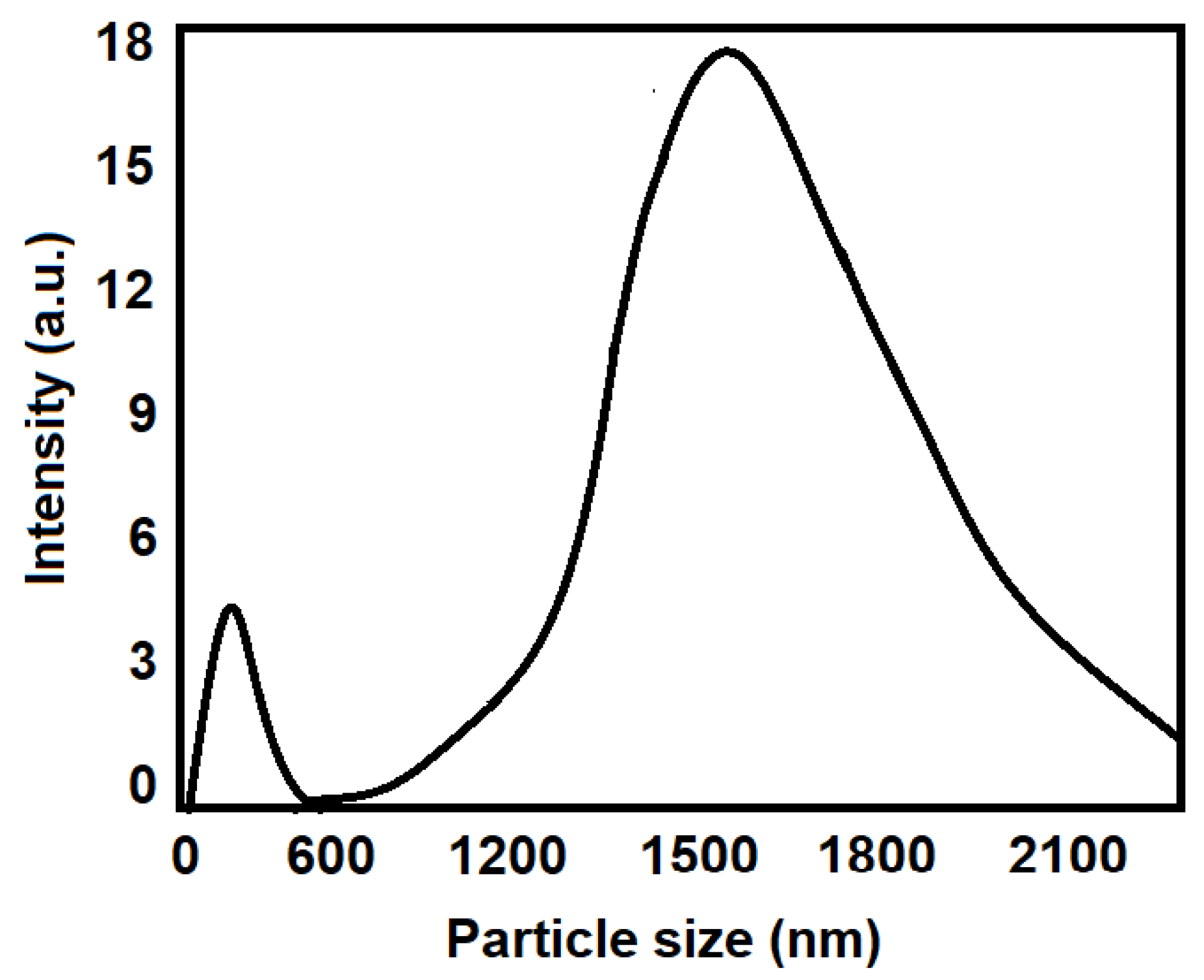
Abstract
:1. Introduction
2. Materials and Methods
2.1. Materials
2.2. GO Synthesis
2.3. Conversion of GO into GO-COOH
2.4. Sulfonation of GO
2.4.1. Synthesis of Aryl Diazonium Salt of Sulfanilic Acid
2.4.2. Sulfonation
2.5. Conjugation of FA
2.6. Characterization Studies
2.6.1. Particle Size
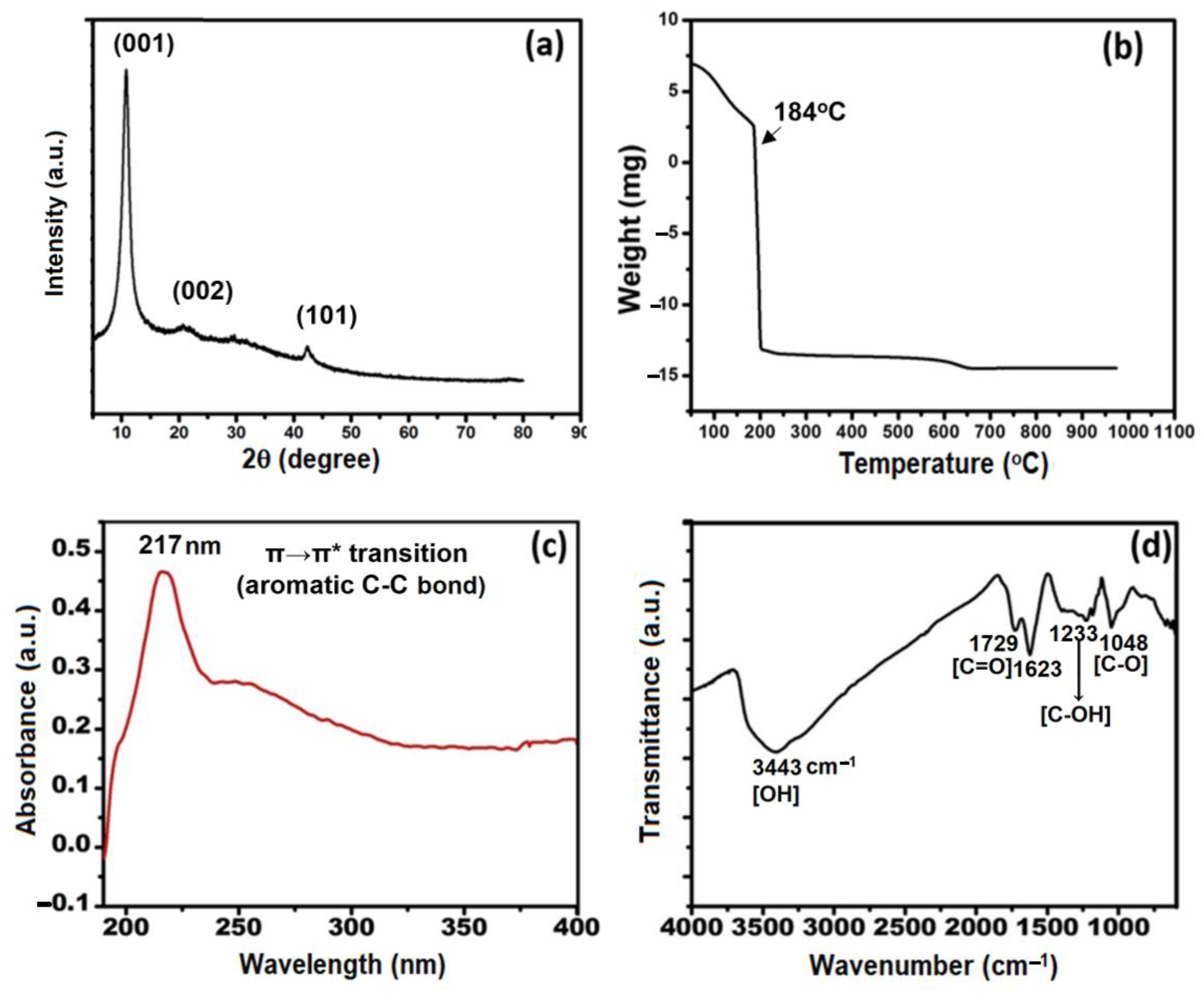
2.6.2. XRD Analysis
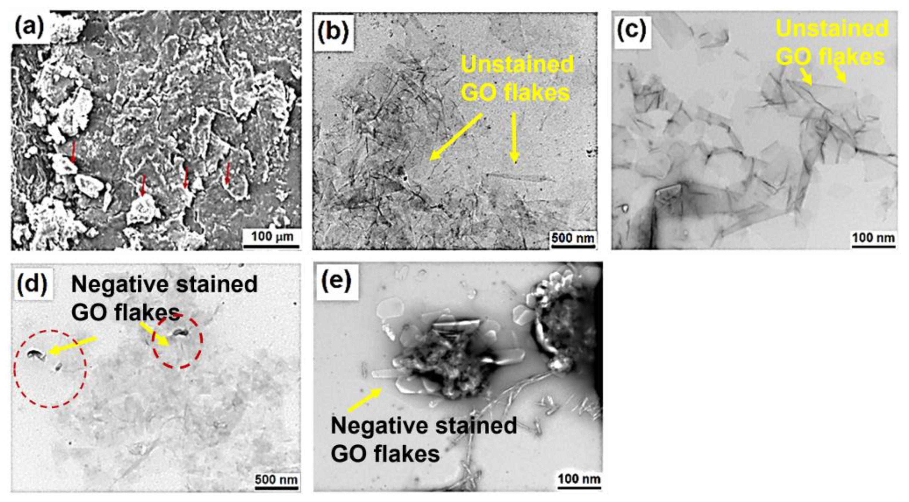
2.6.3. Surface Morphology
2.6.4. Thermogravimetric Analysis
2.6.5. FTIR and UV-Visible
2.7. Loading of Drug
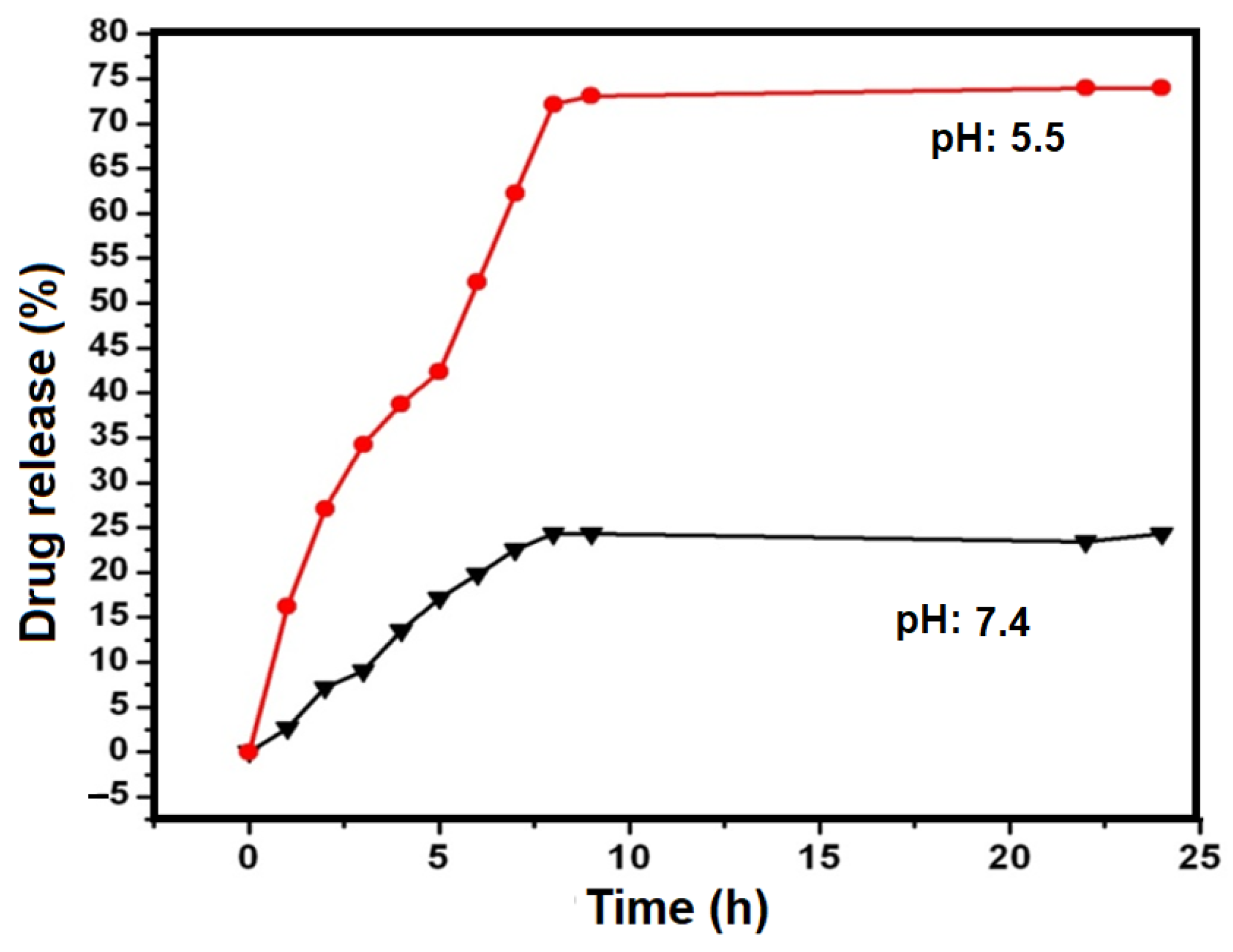
2.8. In Vitro Drug Release Study
2.9. In Vitro Cytotoxicity Studies
2.9.1. Maintenance of Cervical Cancer Cell Lines
2.9.2. Cell Growth Inhibition Assay
3. Results and Discussion
3.1. Particle Size Analysis
3.2. Surface Morphology
3.3. Structural Analysis
3.4. Chemical Bonding and Functionalization of GO
3.5. Calibration Curve of CHL Suspension and Drug Loading
3.6. In Vitro Release Study
3.7. Cytotoxicity Studies
4. Conclusions
Supplementary Materials
Author Contributions
Funding
Institutional Review Board Statement
Informed Consent Statement
Data Availability Statement
Conflicts of Interest
Abbreviations
References
- Freddie, B.; Jacques, F.M.E.; Isabelle, S.; Rebecca, L.S.; Lindsey, A.T.; Ahmedin, J. Gobal cancer statistics 2018: Globocan estimates of incidence and mortality worldwide for 36 cancer in 185 countries. CA A Cancer Found Clin. 2018, 68, 394–424. [Google Scholar]
- Cheng-Yang, Z.; Rui, C.; Yang, Z.; Zhong, M.T. Nanotechnology for cancer therapy based on chemotherapy. Molecules 2018, 23, 826. [Google Scholar]
- Onam, H.; Mihaela, T.; Cecilia, C. Implication of magnetic nanoparticles in cancer detection, screening and treatment. Magnetochemistry 2019, 5, 55. [Google Scholar]
- Gao, D.; Guo, X.; Zhang, X.; Chen, S.; Wang, Y.; Chen, T.; Huang, G.; Gao, Y.; Tian, Z.; Yang, Z. Multifunctional phototheranostic nanomedicine for cancer imaging and treatment. Mater. Today Bio 2020, 5, 100035. [Google Scholar] [CrossRef] [PubMed]
- Joan, E.; Maria, A.B. Iron oxide nanoparticles in photothermal therapy. Molecules 2018, 23, 1567. [Google Scholar]
- Gayatri, S.; Jaidip, M.J.; Abdul, K.P.; Christopher, H.P. Ionizing radiation stimulated breast cancer Nanomedicine. In External Field and Radiation Stimulated Breast Cancer Nanotheranostics; IOP Publishing: Bristol, UK, 2019; p. 8. [Google Scholar]
- Cheng-Yang, H.; Da-Tong, J.; Chih-Fen, C.; Muralidhar, P.R.; Bharath, K.V. A review on the effects of current chemotherapy drug and natural agents in treating non-small cell lung cancer. J. Biomed. 2017, 7, 23. [Google Scholar]
- Dhruba, J.B.; Marianne, K.; Mujgan, G.; Tessa, M.S.; Shaker, A.M. Nanoparticles and cancer therapy: A concise review with emphasis on dendrimers. Int. J. Nanomed. 2009, 4, 1–7. [Google Scholar]
- Maria, A.; Dictrich, B. Cisplatin as anti tumor drug cellular mechanism of activity, drug resistance and induced side effects. J. Cancers 2011, 3, 1351–1371. [Google Scholar]
- Sidipta, S.; Arun, K.M.; Sunil, K.; Pralay, M. Controlled drug delivery vehicles for cancer treatment and their performance. Singnal Transduct. Target. Ther. 2018, 3, 7. [Google Scholar]
- Han-Chung, W.; De-Kuan, C.; Chia-Ting, H. Targeted Therapy for Cancer. J. Cancer Mol. 2006, 2, 57–66. [Google Scholar]
- Bae, Y.H.; Park, K. Targeted Drug Delivery to Tumors: Myths, Reality and Possibility. J. Control. Release 2011, 153, 198–205. [Google Scholar] [CrossRef] [PubMed] [Green Version]
- Firer, M.A.; Gellerman, G. Targeted Drug Delivery for Cancer Therapy: The Other Side of Antibodies. J. Hematol. Oncol. 2012, 5, 70. [Google Scholar] [CrossRef] [PubMed] [Green Version]
- Saul, J.M.; Annapragada, A.; Natarajan, J.V.; Bellamkonda, R. V Controlled Targeting of Liposomal Doxorubicin via the Folate Receptor in Vitro. J. Control. Release 2003, 92, 49–67. [Google Scholar] [CrossRef] [PubMed]
- Singh, K.P.; Panwar, P.; Kohli, P. Sanjesh Liposome-Mesoporous Silica Nanoparticles Fused Cores: A Safer Mode of Drug Carrier. J. Biomed. Nanotechnol. 2011, 7, 60–62. [Google Scholar] [CrossRef] [PubMed]
- Quintana, A.; Raczka, E.; Piehler, L.; Lee, I.; Myc, A.; Majoros, I.; Patri, A.K.; Thomas, T.; Mulé, J.; Baker, J.R. Design and Function of a Dendrimer-Based Therapeutic Nanodevice Targeted to Tumor Cells through the Folate Receptor. Pharm. Res. 2002, 19, 1310–1316. [Google Scholar] [CrossRef] [Green Version]
- Yoo, H.S.; Park, T.G. Folate-Receptor-Targeted Delivery of Doxorubicin Nano-Aggregates Stabilized by Doxorubicin–PEG–Folate Conjugate. J. Control. Release 2004, 100, 247–256. [Google Scholar] [CrossRef]
- Panwar, P.; Pandey, B.; Lakhera, P.C.; Singh, K.P. Preparation, Characterization, and in Vitro Release Study of Albend-azole-Encapsulated Nanosize Liposomes. Int. J. Nanomed. 2010, 5, 101–108. [Google Scholar]
- Sharma, H.; Mondal, S. Functionalized Graphene Oxide for Chemotherapeutic Drug Delivery and Cancer Treatment: A Promising Material in Nanomedicine. Int. J. Mol. Sci. 2020, 21, 6280. [Google Scholar] [CrossRef]
- Singh, G.; Nenavathu, B.P.; Imtiyaz, K.; Moshahid A Rizvi, M. Fabrication of Chlorambucil Loaded Graphene- Oxide Nanocarrier and Its Application for Improved Antitumor Activity. Biomed. Pharmacother. 2020, 129, 110443. [Google Scholar] [CrossRef]
- Jampilek, J.; Kralova, K. Advances in Drug Delivery Nanosystems Using Graphene-Based Materials and Carbon Nanotubes. Materials 2021, 14, 1059. [Google Scholar] [CrossRef]
- Sun, X.; Liu, Z.; Welsher, K.; Robinson, J.T.; Goodwin, A.; Zaric, S.; Dai, H. Nano-Graphene Oxide for Cellular Imaging and Drug Delivery. Nano Res. 2008, 1, 203–212. [Google Scholar] [CrossRef] [PubMed] [Green Version]
- Lu, H.; Wang, J.; Wang, T.; Zhong, J.; Bao, Y.; Hao, H. Recent Progress on Nanostructures for Drug Delivery Applications. J. Nanomater. 2016, 2016, 5762431. [Google Scholar] [CrossRef] [Green Version]
- Nehra, A.; Singh, K. Graphene-Based Biosensing Devices for Common Bacterial Detection. Lambert Academic Publishing: Chisinau, Moldova, 2018; ISBN 9786139586158. [Google Scholar]
- Singh, K.P.; Dhek, N.S.; Nehra, A.; Ahlawat, S.; Puri, A. Applying Graphene Oxide Nano-Film over a Polycarbonate Na-noporous Membrane to Monitor E. Coli by Infrared Spectroscopy. Spectrochim. Acta Part A Mol. Biomol. Spectrosc. 2017, 170, 14–18. [Google Scholar] [CrossRef] [PubMed]
- Nehra, A.; Pal Singh, K. Current Trends in Nanomaterial Embedded Field Effect Transistor-Based Biosensor. Biosens. Bioelectron. 2015, 74, 731–743. [Google Scholar] [CrossRef] [PubMed]
- Nehra, A.; Chen, W.; Dimitrov, D.S.; Puri, A.; Singh, K.P. Graphene Oxide-Polycarbonate Track-Etched Nanosieve Platform for Sensitive Detection of Human Immunodeficiency Virus Envelope Glycoprotein. ACS Appl. Mater. Interfaces 2017, 9, 32621–32634. [Google Scholar] [CrossRef]
- Nehra, A.; Pandey, K.; Singh, K.P.; Ahalawat, S.; Joshi, R.P. Determination of E. Coli by a Graphene Oxide-Modified Quartz Crystal Microbalance. Anal. Lett. 2017, 50, 1897–1911. [Google Scholar] [CrossRef]
- Ahlawat, S.; Nehra, A.; Pandey, V.; Singh, K.P. Gold-Coated Nanoporous Polycarbonate Track–Etched Solid Platform for the Rapid Detection of Mesothelin. Ionics (Kiel) 2019, 25, 1887–1896. [Google Scholar] [CrossRef]
- Nehra, A.; Ahlawat, S.; Singh, K.P. A Biosensing Expedition of Nanopore: A Review. Sens. Actuators B Chem. 2019, 284, 595–622. [Google Scholar] [CrossRef]
- Nehra, A.; Kumar, A.; Ahlawat, S.; Kumar, V.; Singh, K.P. Substrate-Free Untagged Detection of MiR393a Using an Ultra-sensitive Electrochemical Biosensor. ACS Omega 2022, 7, 5176–5189. [Google Scholar] [CrossRef]
- Zhu, Y.; Murali, S.; Cai, W.; Li, X.; Suk, J.W.; Potts, J.R.; Ruoff, R.S. Graphene and Graphene Oxide: Synthesis, Properties, and Applications. Adv. Mater. 2010, 22, 3906–3924. [Google Scholar] [CrossRef]
- Gilje, S.; Han, S.; Wang, M.; Wang, K.L.; Kaner, R.B. A Chemical Route to Graphene for Device Applications. Nano Lett. 2007, 7, 3394–3398. [Google Scholar] [CrossRef] [PubMed]
- Schedin, F.; Geim, A.K.; Morozov, S.V.; Hill, E.W.; Blake, P.; Katsnelson, M.I.; Novoselov, K.S. Detection of Individual Gas Molecules Adsorbed on Graphene. Nat. Mater. 2007, 6, 652–655. [Google Scholar] [CrossRef]
- Liu, Z.; Robinson, J.T.; Sun, X.; Dai, H. PEGylated Nanographene Oxide for Delivery of Water-Insoluble Cancer Drugs. J. Am. Chem. Soc. 2008, 130, 10876–10877. [Google Scholar] [CrossRef] [PubMed] [Green Version]
- Singh, K.P.; Arif, H.; Ahmad, M. Evaluation of Ouabain as Blocker of Active Transport across the Pericardium. Med. Sci. Res. 1995, 23, 827. [Google Scholar]
- Bao, H.; Pan, Y.; Ping, Y.; Sahoo, N.G.; Wu, T.; Li, L.; Li, J.; Gan, L.H. Chitosan-Functionalized Graphene Oxide as a Nanocarrier for Drug and Gene Delivery. Small 2011, 7, 1569–1578. [Google Scholar] [CrossRef]
- Hu, F.; Chen, S.; Wang, C.; Yuan, R.; Yuan, D.; Wang, C. Study on the Application of Reduced Graphene Oxide and Multiwall Carbon Nanotubes Hybrid Materials for Simultaneous Determination of Catechol, Hydroquinone, p-Cresol and Nitrite. Anal. Chim. Acta 2012, 724, 40–46. [Google Scholar] [CrossRef]
- Zhang, L.; Xia, J.; Zhao, Q.; Liu, L.; Zhang, Z. Functional Graphene Oxide as a Nanocarrier for Controlled Loading and Tar-geted Delivery of Mixed Anticancer Drugs. Small 2010, 6, 537–544. [Google Scholar] [CrossRef]
- Youssef, Z.; Vanderesse, R.; Colombeau, L.; Baros, F.; Roques-Carmes, T.; Frochot, C.; Wahab, H.; Toufaily, J.; Hamieh, T.; Acherar, S.; et al. The Application of Titanium Dioxide, Zinc Oxide, Fullerene, and Graphene Nanoparticles in Photodynamic Therapy. Cancer Nanotechnol. 2017, 8, 6. [Google Scholar] [CrossRef]
- Yang, X.; Zhang, X.; Liu, Z.; Ma, Y.; Huang, Y.; Chen, Y. High-Efficiency Loading and Controlled Release of Doxorubicin Hydrochloride on Graphene Oxide. J. Phys. Chem. C 2008, 112, 17554–17558. [Google Scholar] [CrossRef]
- Du, L.; Suo, S.; Luo, D.; Jia, H.; Sha, Y.; Liu, Y. Hydroxyethylated Graphene Oxide as Potential Carriers for Methotrexate Delivery. J. Nanoparticle Res. 2013, 15, 1708. [Google Scholar] [CrossRef]
- Sun, G.; Li, X.; Qu, Y.; Wang, X.; Yan, H.; Zhang, Y. Preparation and Characterization of Graphite Nanosheets from Detonation Technique. Mater. Lett. 2008, 62, 703–706. [Google Scholar] [CrossRef]
- Zhang, R.; Olin, H. Carbon Nanomaterials as Drug Carriers: Real Time Drug Release Investigation. Mater. Sci. Eng. C 2012, 32, 1247–1252. [Google Scholar] [CrossRef]
- Hu, H.; Yu, J.; Li, Y.; Zhao, J.; Dong, H. Engineering of a Novel Pluronic F127/Graphene Nanohybrid for PH Responsive Drug Delivery. J. Biomed. Mater. Res. Part A 2012, 100A, 141–148. [Google Scholar] [CrossRef] [PubMed]
- Xiuyan, W.; Yanyan, C.; Husheng, Y. Chlorambucil loaded in mesoporous polymeric microspheres as oral sustained release formulations with enhanced hydrolytic stability. Mater. Sci. Eng. C 2018, 91, 564–569. [Google Scholar]
- Hummers, W.S.; Offeman, R.E. Preparation of Graphitic Oxide. J. Am. Chem. Soc. 1958, 80, 1339. [Google Scholar] [CrossRef]
- Ling, Y.; Huang, Y. Preparation and Release Efficiency of Poly (Lactic-Co-Glycolic) Acid Nanoparticles for Drug Loaded Paclitaxel. In 7th Asian-Pacific Conference on Medical and Biological Engineering; Springer: Berlin/Heidelberg, Germany, 2008; pp. 514–517. [Google Scholar]
- Mazzotta, E.; Tavano, L.; Muzzalupo, R. Thermo-Sensitive Vesicles in Controlled Drug Delivery for Chemotherapy. Pharmaceutics 2018, 10, 150. [Google Scholar] [CrossRef] [Green Version]
- Mozafari, M.R.; Pardakhty, A.; Azarmi, S.; Jazayeri, J.A.; Nokhodchi, A.; Omri, A. Role of nanocarrier systems in cancer nanotherapy. J. Liposome Res. 2009, 19, 310–321. [Google Scholar] [CrossRef]
- Aw-Yong, P.Y.; Gan, P.H.; Sasmita, A.O.; Mak, S.T.; Ling, A.P. Nanoparticles as carriers of phytochemicals: Recent applications against lung cancer. Int. J. Res. Biomed. Biotechnol. 2018, 7, 1–11. [Google Scholar]
- Maeda, H. Toward a full understanding of the EPR effect in primary and metastatic tumors as well as issues related to its heterogeneity. Adv. Drug Deliv. Rev. 2015, 91, 3–6. [Google Scholar] [CrossRef]
- Caracciolo, G. Clinically approved liposomal nanomedicines: Lessons learned from the biomolecular corona. Nanoscale 2018, 10, 4167–4172. [Google Scholar] [CrossRef]
- Kandasamy, V.; Naresh, K.R.; Andy, R.; Nanhakumar, E. Folate receptor targeted delivery of paclitaxel to breast cancer cells via folic acid conjugated graphene oxide grafted methyl acrylate nanocarrier. Biomed. Pharmacother. 2019, 110, 906–917. [Google Scholar]
- Li, D.; Müller, M.B.; Gilje, S.; Kaner, R.B.; Wallace, G.G. Processable Aqueous Dispersions of Graphene Nanosheets. Nat. Nanotechnol. 2008, 3, 101–105. [Google Scholar] [CrossRef] [PubMed]
- Meriga, V.; Valligatla, S.; Sundaresan, S.; Cahill, C.; Dhanak, V.R.; Chakraborty, A.K. Optical, electrical, and electrochemical properties of graphene based water soluble polyaniline composites. J. Appl. Polym. Sci. 2015, 132, 42766. [Google Scholar] [CrossRef]
- Xu, Y.; Bai, H.; Lu, G.; Li, C.; Shi, G. Flexible Graphene Films via the Filtration of Water-Soluble Noncovalent Functionalized Graphene Sheets. J. Am. Chem. Soc. 2008, 130, 5856–5857. [Google Scholar] [CrossRef] [PubMed]
- Si, Y.; Samulski, E.T. Synthesis of Water Soluble Graphene. Nano Lett. 2008, 8, 1679–1682. [Google Scholar] [CrossRef]
- Jingquan, L.; Liang, C.; Dusan, L. Graphene and graphene oxide as new nanocarriers for drug delivery application. Acta Biomater. 2013, 9, 9243–9257. [Google Scholar]
- Abedelbary, M.A.E.; Waqar, A.; Israr, U.H.; Vinod, R.D.; Antony, D.E. Carbon nanotubes in cancer therapy and drug delivery. J. Drug Deliv. 2011, 2012, 347–363. [Google Scholar]


Disclaimer/Publisher’s Note: The statements, opinions and data contained in all publications are solely those of the individual author(s) and contributor(s) and not of MDPI and/or the editor(s). MDPI and/or the editor(s) disclaim responsibility for any injury to people or property resulting from any ideas, methods, instructions or products referred to in the content. |
© 2023 by the authors. Licensee MDPI, Basel, Switzerland. This article is an open access article distributed under the terms and conditions of the Creative Commons Attribution (CC BY) license (https://creativecommons.org/licenses/by/4.0/).
Share and Cite
Kumari, S.; Nehra, A.; Gupta, K.; Puri, A.; Kumar, V.; Singh, K.P.; Kumar, M.; Sharma, A. Chlorambucil-Loaded Graphene-Oxide-Based Nano-Vesicles for Cancer Therapy. Pharmaceutics 2023, 15, 649. https://doi.org/10.3390/pharmaceutics15020649
Kumari S, Nehra A, Gupta K, Puri A, Kumar V, Singh KP, Kumar M, Sharma A. Chlorambucil-Loaded Graphene-Oxide-Based Nano-Vesicles for Cancer Therapy. Pharmaceutics. 2023; 15(2):649. https://doi.org/10.3390/pharmaceutics15020649
Chicago/Turabian StyleKumari, Surabhi, Anuj Nehra, Kshitij Gupta, Anu Puri, Vinay Kumar, Krishna Pal Singh, Mukesh Kumar, and Ashutosh Sharma. 2023. "Chlorambucil-Loaded Graphene-Oxide-Based Nano-Vesicles for Cancer Therapy" Pharmaceutics 15, no. 2: 649. https://doi.org/10.3390/pharmaceutics15020649