Article

Biological Distribution after Oral Administration of Radioiodine-Labeled Acetaminophen to Estimate Gastrointestinal Absorption Function via OATPs, OATs, and/or MRPs

1
Division of Health Sciences, Graduate School of Medical Sciences, Kanazawa University, 5-11-80 Kodatsuno, Kanazawa 920-0942, Japan
2
Faculty of Health Sciences, Institute of Medical, Pharmaceutical and Health Sciences, Kanazawa University, 5-11-80 Kodatsuno, Kanazawa 920-0942, Japan
3
Department of Molecular Imaging and Theranostics, Institute for Quantum Medical Science, Quantum Life and Medical Science Directorate, National Institutes for Quantum Science and Technology, 4-9-1 Anagawa, Inage 263-8555, Japan
4
Department of Radioisotope Medicine, Atomic Bomb Disease Institute, Nagasaki University, 1-12-4 Sakamoto, Nagasaki 852-8523, Japan
5
Faculty of Pharmacy, Takasaki University of Health and Welfare, 60 Nakaorui-machi, Takasaki 370-0033, Japan
6
Faculty of Pharmaceutical Sciences, Institute of Medical, Pharmaceutical and Health Sciences, Kanazawa University, Kakuma, Kanazawa 920-1192, Japan
7
Biomedical Imaging Research Center, University of Fukui, 23-3 Matsuokashimoaizuki, Eiheiji 910-1193, Japan
*
Author to whom correspondence should be addressed.
These authors contributed equally to this work.
Pharmaceutics 2023, 15(2), 497; https://doi.org/10.3390/pharmaceutics15020497
Submission received: 31 December 2022 / Revised: 28 January 2023 / Accepted: 30 January 2023 / Published: 2 February 2023
(This article belongs to the Special Issue Recent Advances in Radiopharmaceutics, 2nd Edition)

Abstract

:
We evaluated the whole-body distribution of orally-administered radioiodine-125 labeled acetaminophen (125I-AP) to estimate gastrointestinal absorption of anionic drugs. 125I-AP was added to human embryonic kidney (HEK)293 and Flp293 cells expressing human organic anion transporting polypeptide (OATP)1B1/3, OATP2B1, organic anion transporter (OAT)1/2/3, or carnitine/organic cation transporter (OCTN)2, with and without bromosulfalein (OATP and multidrug resistance-associated protein (MRP) inhibitor) and probenecid (OAT and MRP inhibitor). The biological distribution in mice was determined by oral administration of 125I-AP with and without bromosulfalein and by intravenous administration of 125I-AP. The uptake of 125I-AP was significantly higher in HEK293/OATP1B1, OATP1B3, OATP2B1, OAT1, and OAT2 cells than that in mock cells. Bromosulfalein and probenecid inhibited OATP- and OAT-mediated uptake, respectively. Moreover, 125I-AP was easily excreted in the urine when administered intravenously. The accumulation of 125I-AP was significantly lower in the blood and urinary bladder of mice receiving oral administration of both 125I-AP and bromosulfalein than those receiving only 125I-AP, but significantly higher in the small intestine due to inhibition of OATPs and/or MRPs. This study indicates that whole-body distribution after oral 125I-AP administration can be used to estimate gastrointestinal absorption in the small intestine via OATPs, OATs, and/or MRPs by measuring radioactivity in the urinary bladder.

1. Introduction

Many oral drugs and medicines are absorbed into the blood via drug transporters expressed on small intestinal epithelial cells and accumulate in the target tissues and organs. Drug transporters are broadly divided into the solute carrier (SLC) and ATP-binding cassette (ABC) transporters, which contribute significantly to the influx and efflux of drugs [1,2]. In the small intestine, these transporters are expressed on the villous side of the brush border membrane and the vascular side of the basolateral membrane [3] and are mainly involved in the absorption of oral drugs or medicines by the gastrointestinal tract. The SLC transporters on the brush border membrane side (Figure 1) comprise organic anion transporting polypeptide (OATP), mainly OATP2B1 (SLC21A9), and organic cation/carnitine transporter (OCTN), mainly OCTN2 (SLC22A5) and peptide transporter (PEPT)1 (SLC15A1) [4,5,6,7]. In addition, on the brush border membrane side, ABC transporters comprise P-glycoprotein and multidrug resistance-associated protein (MRP)2, whereas, on the basolateral membrane side, these receptors express MRP3 [1,2,5]. Therefore, the expression levels of these transporters play a crucial role in the absorption of oral drugs or medicines. In addition, gastrointestinal diseases that alter the expression levels of these transporters in small intestinal epithelial cells may reduce the gastrointestinal absorption of oral drugs or medicines.
The gastrointestinal absorption function is primarily measured using liquid chromatography/mass spectrometry (LC/MS) [8] or LC/MS/MS. However, this technique is limited because it requires liquid samples (e.g., blood) from the human body after the administration of oral drugs or medicines and cannot accurately reflect the gastrointestinal absorption function for specific SLC transporters on the brush border membrane in the small intestine. Therefore, there is a need for an imaging technique that can accurately estimate gastrointestinal absorption function to personalize drug administration. In our previous study, we used oral administration of iodine-123 labeled m-iodobenzylguanidine (123I-MIBG) to estimate gastrointestinal absorption of cationic anticancer drugs or medicines via OCTN and/or OCT in the small intestine by measuring radioactivity in the heart, liver, and urinary bladder [9]. However, this imaging method cannot estimate the absorption function of organic anion drugs and medicines in the gastrointestinal tract.
Acetaminophen (AP, N-acetyl-p-aminophenol, and paracetamol) is an anionic drug that is frequently used as an analog and antipyretic drug. AP is taken up by epithelial cells of the gastrointestinal tract via OATP and OAT [10]. We have previously developed radioiodine-125-labeled AP (125I-AP) for the imaging of melanoma and hypothesized that 125I-AP might also have affinities for OATP and/or OAT. Therefore, in the present study, we evaluated the whole-body distribution of orally-administered radioiodine-labeled 125I-AP to estimate gastrointestinal absorption of anionic drugs or medicines. This imaging technique may help estimate the gastrointestinal absorption of orally administered anionic drugs or medicines, thereby personalizing drug administration.

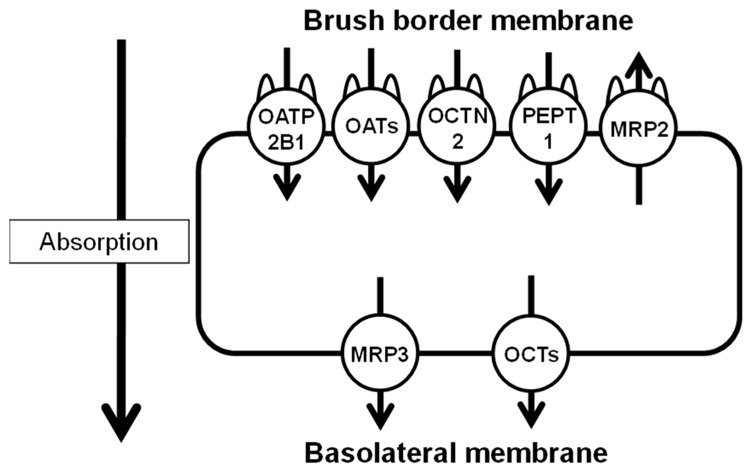
2. Materials and Methods

2.1. 125I Labeling of AP and Purification

125I-AP was synthesized according to a previously reported protocol [11]. Briefly, 125I-AP was synthesized using the chloramine T method by adding 4 mM chloramine T and Na125I to 10 mM AP. 125I-AP was purified using high-performance liquid chromatography (HPLC, Hitachi, Ibaraki, Japan). Labeling rates were determined using thin-layer chromatography (TLC, silica gel 60 F254; Millipore-Sigma, Burlington, MA, USA). Radiochemical purity was examined with HPLC.

2.2. HEK293 and Flp293 Cells Expressing High Levels of Various SLC Transporters

Human embryonic kidney (HEK)293 cells (American Type Culture Collection, Manassas, VA, USA) with high expression levels of OATP1B1 (SLC21A6, NM_006446), OATP1B3 (SLC21A8, NM_019844), OATP2B1 (NM_007256), OAT2 (SLC22A7, NM_006672), and OCTN2 (NM_003060) were used. Flp293 cells derived from HEK293 cells stably express the human α1A-adrenoreceptor, with high expression of OAT1 (SLC22A6, NM_004790) and OAT3 (SLC22A8, NM_004254), were also used. HEK293/OATP1B1, HEK293/OATP1B3, HEK293/OATP2B1, HEK293/OAT2, HEK293/OCTN2, Flp293/OAT1, Flp293/OAT3, and mock control cell lines were established by transfecting HEK293 and Flp293 cells with plasmids encoding each transporter. All cells were cultivated in Dulbecco’s Modified Eagle’s medium (FUJIFILM Wako Chemicals, Osaka, Japan) mixed with 10% fetal bovine serum (Life Technologies, Carlsbad, CA, USA), 1% sodium pyruvate, and 1% penicillin at 37 °C and 5% CO2.

2.3. Accumulation of 125I-AP in HEK293 Cells

Before the experiments with 125I-AP, OATP and OAT expressions were confirmed using p-14C-aminohippuric acid (PerkinElmer Inc., Waltham, MA, USA) in HEK293 and Flp293 cells, and OCTN2 expression was confirmed using methyl-3H-4-phenylpyridinium (American Radiolabeled Chemicals Inc., St. Louis, MO, USA) in HEK293 cells [9]. HEK293 cells expressing the respective SLC transporter were seeded at 1.0 × 10⁵ cells/well in 12-well plastic plates in DMEM and cultivated at 37 °C and 5% CO2 for 24 h. On the day of the experiments, each cell type was pre-incubated in phosphate-buffered saline (PBS) for 5 min and then incubated with 125I-AP (18.5 kBq/well) for 5 min (n = 4). After incubation, the cells were washed twice with 600 µL of PBS and lysed with 500 µL of 0.1 M NaOH. Radioactivity was measured using a gamma counter (AccuFLEX γ7000; Hitachi Aloka Medical, Tokyo, Japan). Intracellular protein levels were measured using a bicinchoninic acid protein assay kit (Thermo Fisher Scientific, Waltham, MA, USA), using bovine serum albumin as the standard.

2.4. Accumulation of 125I-AP in HEK293 Cells Treated with Inhibitors of Drug Transporters

HEK293 cells expressing the respective SLC transporter were seeded at 1.0 × 10⁵ cells/well in 12-well plastic plates in DMEM and cultivated at 37 °C and 5% CO2 for 24 h. The cells were pre-incubated in PBS for 5 min and then incubated with 125I-AP (18.5 kBq/well) and 1.0 mM bromosulfalein (Sigma-Aldrich, St. Louis, MO, USA), an inhibitor of OATPs and MRPs [12,13], or 1.0 mM probenecid (Sigma-Aldrich), an inhibitor of OATs and MRPs for 5 min (n = 4) [13,14]. After incubation, the cells were washed twice with 600 µL of PBS and lysed with 500 µL of 0.1 M NaOH. Radioactivity was measured using an auto well gamma counter. Intracellular protein levels were measured using a bicinchoninic acid protein assay kit (Thermo Fisher Scientific), using bovine serum albumin as the standard.

2.5. Biological Distribution of 125I-AP in Normal Mice

All animal experiments performed in this study were conducted in compliance with the ethical standards of Kanazawa University (Animal Care Committee of Kanazawa University, AP-194046) and the international standards for animal welfare and institutional guidelines. 125I-AP was orally and intragastrically administered to ddY mice (male, 8 weeks old; SLC Inc., Hamamatsu, Japan) that had fasted for 6 h, using Fuchigami animal feeding needles (Natsume Seisakusho Co., Ltd., Tokyo, Japan) at a dose of approximately 37 kBq/200 µL per mouse. After 5, 10, 30, and 60 min of administration (n = 4), heart blood samples were collected under isoflurane anesthesia (FUJIFILM Wako Pure Chemical Corporation), and mice were sacrificed by cervical dislocation. The organs (thyroid gland, heart, stomach, liver, small intestine, kidney, and urinary bladder) were then quickly collected. The weight of each organ was measured using an electronic balance (AUX220, Shimadzu Corporation, Kyoto, Japan), and radioactivity was measured using an auto gamma counter (AccuFLEX gamma ARC-7010, Hitachi, Ltd., Tokyo, Japan). The results are shown as the percent injected dose (%ID) for the thyroid gland, stomach, small intestine, and urinary bladder and %ID/g wet tissue (%ID/g tissue) for the other organs. As the thyroid gland of mice is quite small and the stomach and small intestine can’t completely remove the contents, it is difficult to measure the accurate weight of these organs. Thus, these organs are indicated by %ID. The urinary bladder results are expressed in %ID because it is undesirable that the radioactivity of urine is corrected by the weight of the urinary bladder. In addition, 125I-AP was diluted with saline to approximately 20 kBq/200 µL and administrated intravenously through the tail vein of the mice. After 5, 10, 30, and 60 min of administration (n = 4), heart blood samples were collected, and their weights and radioactivity were measured.

2.6. Biological Distribution of 125I-AP Administered with an OATP Inhibitor

Mice (ddY, male, 8 weeks old; SLC Inc., Hamamatsu, Japan) fasted for 6 h before using Fuchigami feeding needles for oral and intragastric administration of the drugs. 125I-AP was diluted with saline to approximately 20 kBq/100 µL, and 100 µL of 125I-AP was orally administered to mice using feeding needles after 5 min of oral administration of bromosulfalein, an inhibitor of OATP. After 5, 10, 30, and 60 min of administration (n = 4), heart blood samples were collected under isoflurane anesthesia, and the mice were sacrificed by cervical dislocation. The organs (thyroid gland, heart, stomach, liver, small intestine, kidney, and urinary bladder) were quickly collected. The weight of each organ was measured using an electronic balance, and radioactivity was measured using a gamma counter. The results are shown as %ID or %ID/g of tissue.

2.7. Statistical Analysis

P-values were analyzed using the two-tailed paired Student’s t-test for comparisons between two groups and analysis of variance and the post hoc comparisons with Dunn’s test for comparisons among three groups using GraphPad Prism 8 statistical software (GraphPad Software, Inc., La Jolla, CA, USA). Statistical significance was set as p < 0.01.

3. Results

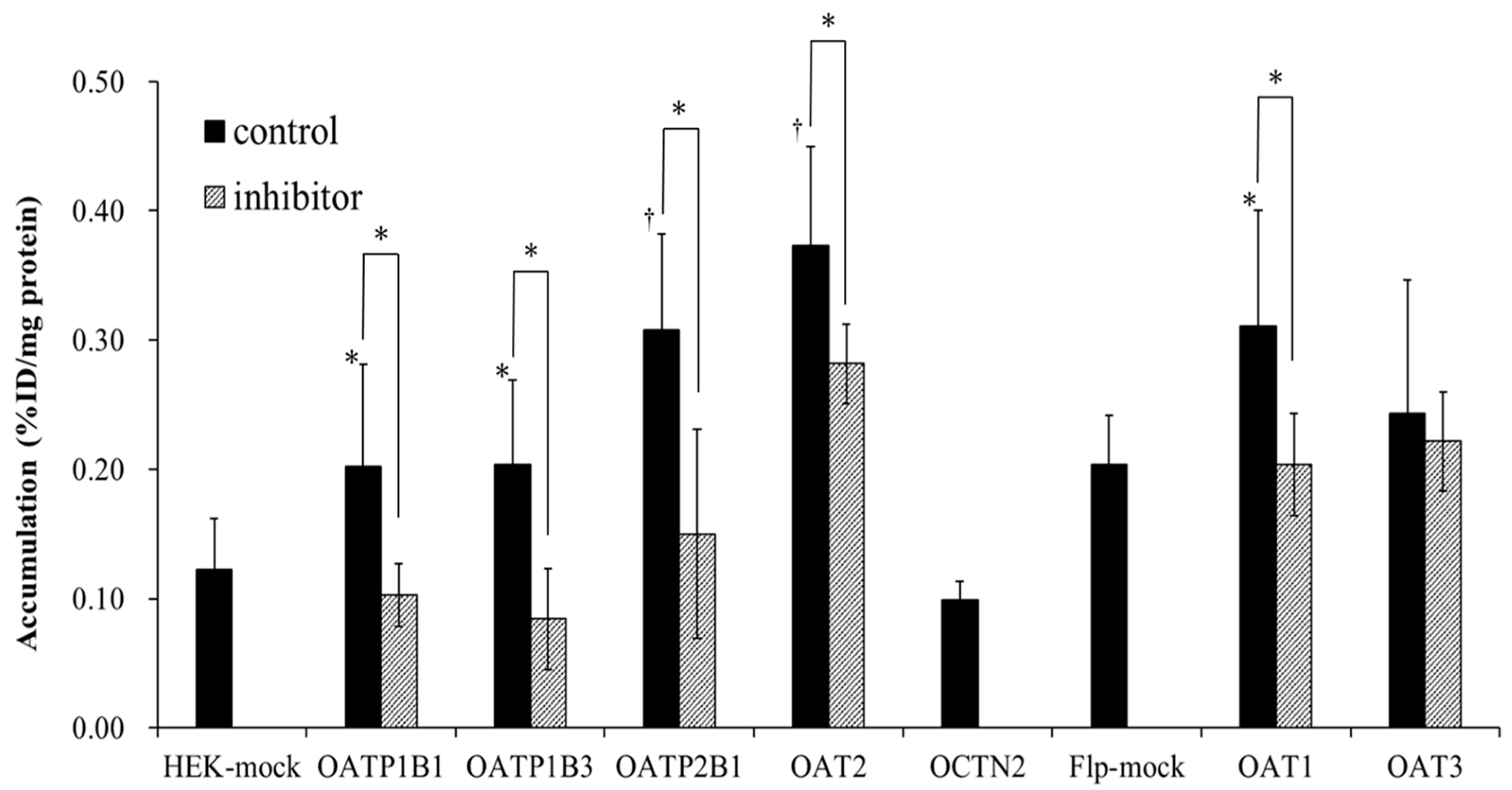
3.1. 125I-AP Accumulation in HEK293 and Flp293 Cells Expressing High Levels of Different SLC Transporters

125I-AP was labeled with an efficiency of more than 80% and was obtained with a radiochemical purity of more than 95% after HPLC analysis. Mass-spectrometry data of 127I-AP have been reported in our previous study [11]. The date of 127I-AP revealed an MH (+) of 278, which matched the calculated MH (+) of 278 C8H8INO2.
The accumulation of 125I-AP in HEK293 and Flp293 cells with high expression of an SLC transporter is shown in Figure 2. Before performing the experiments with 125I-AP, the function of the SLC transporter in HEK293 and Flp293 cells was confirmed using 3H- or 14C-labeled substrates [9]. Accumulation of the p-14C-aminohippuric acid into HEK293 mock cells is 0.19 ± 0.03%ID/mg protein for HEK mock for OATP and OAT2 and into Flp293 mock cells is 0.25 ± 0.06%ID/mg protein for Flp-mock for OAT1 and OAT3. Accumulation of the methyl-3H-4-phenylpyridinium into HEK293 mock cells is 0.09 ± 0.06%ID/mg protein for HEK mock for OCTN2. In addition, accumulation of methyl-3H-4-phenylpyridinium or p-14C-aminohippuric acid into HEK293 and Flp293 cells expressing a SLC transporter is as follows: 0.28 ± 0.06%ID/mg protein for HEK293/OATP1B1, 0.27± 0.05%ID/mg protein for HEK293/OATP1B3, 0.34 ± 0.09%ID/mg protein for HEK293/OATP2B1, 0.42 ± 0.11%ID/mg protein for HEK293/OAT2, 0.39 ± 0.08%ID/mg protein for HEK293/OCTN2, 0.46 ± 0.03%ID/mg protein for Flp293/OAT1 and 0.45 ± 0.04%ID/mg protein for Flp293/OAT3.
Compared to HEK293 mock cells, the intracellular accumulation of 125I-AP was significantly increased in HEK293/OATP1B1 (p < 0.05), HEK293/OATP1B3 (p < 0.05), HEK293/OATP2B1 (p < 0.01), and HEK293/OAT2 cells (p < 0.01). However, the accumulation of 125I-AP was not increased in HEK293/OCTN2 cells compared to that in HEK293 mock cells. Compared to Flp-mock cells, the accumulation of 125I-AP was significantly increased in Flp293/OAT1 (p < 0.05) but not in Flp293/OAT3 cells. In the presence of bromosulfalein, an inhibitor of OATP and MRP, the accumulation of 125I-AP was significantly decreased in HEK293/OATP1B1, HEK293/OATP1B3, and HEK293/OATP2B1 cells compared to that in HEK293 mock cells. In the presence of probenecid, an inhibitor of OAT and MRP, the accumulation of 125I-AP was significantly decreased in Flp293/OAT1, Flp293/OAT3, and HEK293/OAT2 compared to that in Flp293 or HEK293 mock cells; however, the accumulation of 125I-AP in HEK293/OAT2 cells did not decrease to the same level as that in HEK293 mock cells. These results suggest that the uptake of 125I-AP was significantly higher in HEK293/OATP1B1, HEK293/OATP1B3, HEK293/OATP2B1, Flp293/OAT1, and HEK293/OAT2 cells than that in mock cells, and that bromosulfalein and probenecid inhibited OATP- and OAT-mediated uptake, respectively.

3.2. Biological Distribution of Orally-Administered 125I-AP with or without Bromosulfalein and of Intravenously-Administered 125I-AP In Vivo

The in vivo stability of intravenously-administered 125I-AP has been confirmed in blood and homogenized liver of mice using TLC analysis in our previous study [11]. Although 125I-AP was slightly metabolized at 5 min after administration, it remained stable 60 min after administration. The biodistribution of orally-administered 125I-AP with or without the oral administration of bromosulfalein and intravenously-administered 125I-AP is shown in Table 1. In all the mice, the accumulation of 125I-AP in the thyroid gland was low. In the mice group receiving an intravenous administration of 125I-AP, 125I-AP was easily excreted in the urine. In contrast, the urinary excretion of 125I-AP was slower in the oral administration group than in the intravenous administration group. In addition, compared to mice receiving oral administration of 125I-AP, those receiving intravenous administration showed a higher accumulation of 125I-AP in the blood, heart, small intestine, kidney, and urinary bladder at 5 min after administration. In the group that received oral administration of 125I-AP and bromosulfalein, the accumulation of 125I-AP in the blood significantly decreased at 5 min after administration, and that in the urinary bladder significantly decreased at 5, 10, and 30 min after administration. However, in the small intestine, the accumulation of 125I-AP increased at 5 min after administration. Therefore, biological distribution after oral 125I-AP administration is used to estimate gastrointestinal absorption in the small intestine by measuring radioactivity at the early time phase in the urinary bladder.

4. Discussion

The gastrointestinal absorption function varies among individuals and in the same individual under disease states. Therefore, an imaging method that can estimate gastrointestinal absorption function would help personalize drug administration. In our previous study, we investigated the distribution of the oral administration of 123I-MIBG to estimate the gastrointestinal absorption of cationic anticancer drugs or medicines via OCTN and OCT in the small intestine [9]. This imaging technique may help investigate differences in gastrointestinal absorption, which can affect the activity of cationic anticancer drugs. In the present study, we investigated the biodistribution of orally-administered 125I-AP to estimate the absorption function of anionic anticancer drugs or medicines in the gastrointestinal tract.
AP is an anionic drug that is taken up by cells via OATP and OAT [10]. In the present study, 125I-AP had affinity for OATP1B1, OATP1B3, OATP2B1, OAT1, and OAT2. Moreover, in the presence of bromosulfalein, an OATP inhibitor, the accumulation of 125I-AP in cells with high expressions of OATPs and OAT1 decreased to the same levels as that in HEK293 or Flp293-mock cells (control). However, in the presence of probenecid, an inhibitor of OAT, the accumulation of 125I-AP in cells with high OAT2 expression levels did not decrease to the same levels as those in the control cells. These results suggest that the effect of OATPs on the accumulation of 125I-AP was more significant than that of OAT2. However, bromosulfalein inhibits not only OATPs but also OAT2 [15]. Therefore, the reduced accumulation of 125I-AP in HEK293/OATP1B1, HEK293/OATP1B3, and HEK293/OATP2B1 in the presence of bromosulfalein may also partly involve OAT2.
Among SLC transporters present on the brush border membrane side of the small intestine, OATP2B1 is the most abundant, whereas OATs, including OAT2, are slightly expressed in the small intestine [4,5,6,7]. In the present study, after intravenous and oral administration of 125I-AP in mice, the accumulation of 125I-AP in the thyroid gland was low, indicating that 125I-AP was little de-iodine in vivo. In addition, intravenously-administrated 125I-AP showed high in vivo stability of 125I-AP in the blood (94.1 ± 5.1% at 60 min of administration) and liver (88.2 ± 5.5% at 60 min of administration) of mice in our previous study [11]. Therefore, we consider that 125I-AP with oral administration also shows high in vivo stability.
Furthermore, compared to control mice, mice receiving oral administration of 125I-AP with bromosulfalein showed significantly decreased radioactivity in the blood 5 min after administration but significantly increased radioactivity in the small intestine. These results suggest that OATP2B1 is the key transporter involved in the gastrointestinal absorption of orally-administered 125I-AP because the uptake of 125I-AP from the brush border membrane to blood was inhibited by the administration of bromosulfalein.
Bromosulfalein also inhibits MRPs that are expressed on both sides of the brush border membrane and basolateral membrane in the small intestine [13]. In addition, because the chemical structure of AP contains a peptide bond in its chemical structure, we evaluated the biological distribution of the oral administration of 125I-AP with a peptide substrate, cephradine. The administration of cephradine increased the gastrointestinal absorption of 125I-AP (average 9.61, 9.86, 8.96, 2.66 %ID/g in accumulation of blood at 5, 10, 30, 60 min after oral administration with cephradine). Collectively, these results suggest that 125I-AP would be absorbed by OATPs and/or OATs on the brush border membrane and by MRP on the basolateral membrane of the small intestine.
Anionic anticancer drugs or medicines are generally excreted via MRPs of the ABC transporters. Therefore, 125I-AP also may have an affinity for MRPs. In the present study, the accumulation of 125I-AP in the urinary bladder of mice receiving oral administration of 125I-AP and bromosulfalein was significantly decreased within 30 min compared to that in mice orally administered only 125I-AP. This effect is due to the inhibition of gastrointestinal absorption by bromosulfalein. Therefore, when gastrointestinal diseases that alter the expression levels of these transporters in small intestinal epithelial cells reduce the gastrointestinal absorption of oral drugs or medicines, it is possible to estimate the gastrointestinal absorption function of the small intestine via OATPs and/or OATs using time–radioactivity curves of the urinary bladder in the early time phase of 123I-AP imaging, which was changed from 125I to 123I.
Currently, the gastrointestinal absorption function of orally administrated drugs or medicines is primarily measured using LC/MS or LC/MS/MS [8]. We can confirm pharmacokinetics with these data using the samples (blood etc.) after oral administration of a drug or medicine [16]. However, the LC/MS or LC/MS/MS data are difficult to use to evaluate the effect of the individual drug transporter in the small intestine on gastrointestinal absorption. In addition, the absorption function becomes apparent after oral drug or medicine administration. Whole-body imaging after oral 123I-AP administration cloud reflects the function of drug transporters prior to oral administration of the drug or medicine. Therefore, it may be possible to control the appropriate dose of the drug or medicine for an individual patient.
We speculate that the effect of bromosulfalein is limited because we did not observe a reduction in the accumulation of 125I-AP in the liver that has high expression levels of OATPs. When 123I-MIBG whole-body imaging could estimate the gastrointestinal absorption function via cationic transporters in our previous study, we used DSS-induced experimental colitis mouse as well as the effect of inhibitors to drug transporters in the small intestine because DSS-induced experimental colitis mouse decreased expression levels of OCTN1 [17]. However, there are no disease mouse models which decreased the expression of OATP and/or OAT. If more effective mouse models, such as OATP- or OAT-deficient mouse models, were used, the biological distribution after the oral administration of 125I-AP would be slightly different from that reported in the present study. In the future, whole-body imaging after oral 123I-AP administration should be performed to estimate gastrointestinal absorption function via OATPs, OATs, and/or MRPs in the small intestine.

5. Conclusions

The present study demonstrates that whole-body distribution after oral 125I-AP administration can be used to estimate gastrointestinal absorption of anionic drugs or medicines in the small intestine via OATPs, OATs, and/or MRPs by measuring radioactivity in the urinary bladder. Before oral administration with a drug or medicine, whole-body imaging after oral 123I-AP administration could estimate the function of OATPs, OATs, and/or MRPs in the small intestine and lead to control of the appropriate dose of orally administrated drugs or medicines for an individual patient as personalized medicine.

Author Contributions

Conceptualization, M.K.; methodology, K.S., A.M., Y.M. and K.N.; investigation, K.S., A.M., Y.M., J.Y. and M.K.; resources, K.Y. and R.N.; writing—original draft preparation, K.S.; writing—review and editing, M.K.; supervision, T.N., I.T., K.K. and M.K. All authors have read and agreed to the published version of the manuscript.

Funding

This study was funded in part by Grants-in-Aid for Scientific Research from the Japan Society for the Promotion of Science (No. 21H02865, 22K19504, 22H03016) and the Network-type Joint Usage/Research Center for Radiation Disaster Medical Science.

Institutional Review Board Statement

Not applicable.

Informed Consent Statement

Not applicable.

Data Availability Statement

The authors confirm that the data supporting the findings of this study are available within the article.

Acknowledgments

The authors would like to thank Mikie Ohtaki and other staff members of the Faculty of Health Sciences, Kanazawa University.

Conflicts of Interest

The authors declare no conflict of interest. The funders had no role in the design of the study; in the collection, analyses, or interpretation of data; in the writing of the manuscript; or in the decision to publish the results.

References

  1. Nakanishi, T. Drug transporters as targets for cancer chemotherapy. Cancer Genom. Proteom. 2007, 4, 241–254. [Google Scholar]
  2. Carmichael, N.; Day, P.J.R. Cell Surface Transporters and Novel Drug Developments. Front. Pharmacol. 2022, 13, 690. [Google Scholar] [CrossRef] [PubMed]
  3. Estudante, M.; Morais, J.G.; Soveral, G.; Benet, L.Z. Intestinal drug transporters: An overview. Adv. Drug Deliv. Rev. 2013, 65, 1340–1356. [Google Scholar] [CrossRef] [PubMed]
  4. Lee, W.; Ha, J.-M.; Sugiyama, Y. Post-translational regulation of the major drug transporters in the families of organic anion transporters and organic anion–transporting polypeptides. J. Biol. Chem. 2020, 295, 17349–17364. [Google Scholar] [CrossRef] [PubMed]
  5. Tamai, I. Oral drug delivery utilizing intestinal OATP transporters. Adv. Drug Deliv. Rev. 2012, 64, 508–514. [Google Scholar] [CrossRef] [PubMed]
  6. Oswald, S. Organic Anion Transporting Polypeptide (OATP) transporter expression, localization and function in the human intestine. Pharmacol. Ther. 2018, 195, 39–53. [Google Scholar] [CrossRef] [PubMed]
  7. Shekhawat, P.B.; Pokharkar, V.B. Understanding peroral absorption: Regulatory aspects and contemporary approaches to tackling solubility and permeability hurdles. Acta Pharm. Sin. B 2017, 7, 260–280. [Google Scholar] [CrossRef] [PubMed]
  8. Liu, X. Transporter-Mediated Drug-Drug Interactions and Their Significance. In Drug Transporters in Drug Disposition, Effects and Toxicity; Springer: Singapore, 2019; Volume 1141, pp. 241–291. [Google Scholar] [CrossRef]
  9. Kobayashi, M.; Mizutani, A.; Muranaka, Y.; Nishi, K.; Komori, H.; Nishii, R.; Shikano, N.; Nakanishi, T.; Tamai, I.; Kawai, K. Biological Distribution of Orally Administered [123I]MIBG for Estimating Gastrointestinal Tract Absorption. Pharmaceutics 2021, 14, 61. [Google Scholar] [CrossRef] [PubMed]
  10. Mazaleuskaya, L.L.; Sangkuhl, K.; Thorn, C.F.; FitzGerald, G.A.; Altman, R.B.; Klein, T.E. PharmGKB summary. Pharm. Genom. 2015, 25, 416–426. [Google Scholar] [CrossRef] [PubMed]
  11. Zhu, W.J.; Kobayashi, M.; Yamada, K.; Nishi, K.; Takahashi, K.; Mizutani, A.; Nishii, R.; Flores, L.G.; Shikano, N.; Kunishima, M.; et al. Development of radioiodine labeled acetaminophen for specific, high-contrast imaging of malignant melanoma. Nucl. Med. Biol. 2018, 59, 16–21. [Google Scholar] [CrossRef] [PubMed]
  12. Klatt, S.; Fromm, M.F.; König, J. The Influence of Oral Antidiabetic Drugs on Cellular Drug Uptake Mediated by Hepatic OATP Family Members. Basic Clin. Pharmacol. Toxicol. 2012, 112, 244–250. [Google Scholar] [CrossRef] [PubMed]
  13. Zha, W.; Wang, G.; Xu, W.; Liu, X.; Wang, Y.; Zha, B.S.; Shi, J.; Zhao, Q.; Gerk, P.M.; Studer, E.; et al. Inhibition of P-Glycoprotein by HIV Protease Inhibitors Increases Intracellular Accumulation of Berberine in Murine and Human Macrophages. PLoS ONE 2013, 8, e54349. [Google Scholar] [CrossRef] [PubMed]
  14. Monien, B.H.; Müller, C.; Bakhiya, N.; Donath, C.; Frank, H.; Seidel, A.; Glatt, H. Probenecid, an inhibitor of transmembrane organic anion transporters, alters tissue distribution of DNA adducts in 1-hydroxymethylpyrene-treated rats. Toxicology 2009, 262, 80–85. [Google Scholar] [CrossRef] [PubMed]
  15. Shen, H.; Liu, T.; Morse, B.L.; Zhao, Y.; Zhang, Y.; Qiu, X.; Chen, C.; Lewin, A.C.; Wang, X.-T.; Liu, G.; et al. Characterization of Organic Anion Transporter 2 (SLC22A7): A Highly Efficient Transporter for Creatinine and Species-Dependent Renal Tubular Expression. Drug Metab. Dispos. 2015, 43, 984–993. [Google Scholar] [CrossRef] [PubMed]
  16. Araujo-León, J.A.; Ortiz-Andrade, R.; Hernández-Baltazar, E.; Hernández-Núñez, E.; Rivera-Leyva, J.C.; Yáñez-Pérez, V.; Vazquez-Garcia, P.; Cicero-Sarmiento, C.G.; Sánchez-Salgado, J.C.; Segura-Campos, M.R. A Pharmacokinetic Study of Mix-160 by LC-MS/MS: Oral Bioavailability of a Dosage Form of Citroflavonoids Mixture. Molecules 2022, 27, 391. [Google Scholar] [CrossRef] [PubMed]
  17. Tamai, I. Pharmacological and pathophysiological roles of carnitine/organic cation transporters (OCTNs: SLC22A4, SLC22A5 and Slc22a21). Biopharm. Drug Dispos. 2012, 34, 29–44. [Google Scholar] [CrossRef] [PubMed] [Green Version]
Figure 1. Solute carrier and ATP-binding cassette transporters are mainly expressed on small intestinal epithelial cell membranes. OATP, organic anion transporting polypeptide; OCTN, organic cation/carnitine transporter; PEPT, peptide transporter; MRP, multidrug resistance-associated protein.
Pharmaceutics 15 00497 g001
Figure 2. Accumulation of 125I-AP in HEK293 and Flp293 cells with high expression of a solute carrier transporter (n = 4). Before the experiments with 125I-AP, the function of the SLC transporter in HEK293 and Flp293 cells was confirmed using 3H- or 14C-labeled substrates. Accumulation of the p-14C-aminohippuric acid into HEK293 mock cells is 0.19 ± 0.03%ID/mg protein for HEK mock for OATP and OAT2 and into Flp293 mock cells is 0.25 ± 0.06%ID/mg protein for Flp-mock for OAT1 and OAT3. Accumulation of the methyl-3H-4-phenylpyridinium into HEK293 mock cells is 0.09 ± 0.06%ID/mg protein for HEK mock for OCTN2. In addition, accumulation of methyl-3H-4-phenylpyridinium or p-14C-aminohippuric acid into HEK293 and Flp293 cells expressing a SLC transporter is as follows: 0.28 ± 0.06%ID/mg protein for HEK293/OATP1B1, 0.27± 0.05%ID/mg protein for HEK293/OATP1B3, 0.34 ± 0.09%ID/mg protein for HEK293/OATP2B1, 0.42 ± 0.11%ID/mg protein for HEK293/OAT2, 0.39 ± 0.08%ID/mg protein for HEK293/OCTN2, 0.46 ± 0.03%ID/mg protein for Flp293/OAT1 and 0.45 ± 0.04%ID/mg protein for Flp293/OAT3. 125I-AP accumulation is significantly increased in HEK293/OATP1B1, HEK293/OATP1B3, HEK293/OATP2B1, HEK293/OAT2, and Flp293/OAT1 cells compared to that in mock cells. The addition of bromosulfalein, an inhibitor of OATP and MRP, inhibits 125I-AP uptake into HEK293/OATP1B1, HEK293/OATP1B3, HEK293/OATP2B1 cells. The addition of probenecid, an inhibitor of OAT and MRP, inhibits 125I-AP uptake into HEK293/OAT2 and Flp293/OAT1 cells. p < 0.01 and * p < 0.05 vs. mock cells and cells loaded with both inhibitors. OATP, organic anion transporting polypeptide; OCTN, organic cation/carnitine transporter; PEPT, peptide transporter; MRP, multidrug resistance-associated protein.
Pharmaceutics 15 00497 g002
Table 1. Biological distribution of 125I-AP in mice by oral administration, with or without bromosulfalein, and by intravenous administration.
Time after 125I-AP Administration
MiceOrgan5 min10 min30 min60 min
Oral
administration
Blood3.97 ± 0.584.53 ± 0.712.76 ± 0.681.19 ± 0.30
Thyroid gland0.22 ± 0.080.04 ± 0.000.09 ± 0.020.13 ± 0.07
Heart1.48 ± 0.461.59 ± 0.161.15 ± 0.330.44 ± 0.15
Stomach62.7± 9.9138.5 ± 7.1722.9 ± 9.7815.9 ± 8.55
Liver2.41 ± 0.822.38 ± 0.751.42 ± 0.270.54 ± 0.13
Small intestine4.39 ± 2.4815.6 ± 5.8916.6 ± 5.7619.6 ± 7.41
Kidney4.11 ± 0.256.86 ± 2.983.53 ± 0.395.78 ± 4.38
Urinary bladder0.23 ± 0.132.22 ± 12.517.8 ± 7.2916.6 ± 10.8
Oral
administration with bromosulfalein
Blood1.10 ± 0.33 *3.38 ± 1.762.40 ± 0.781.11 ± 0.11
Thyroid gland0.18 ± 0.090.07 ± 0.030.08 ± 0.010.13 ± 0.02
Heart1.53 ± 0.631.25 ± 0.680.85 ± 0.280.43 ± 0.06
Stomach57.0 ± 35.156.3 ± 23.224.8 ± 15.011.3 ± 1.79
Liver1.81 ± 0.301.75 ± 0.681.22 ± 0.310.64 ± 0.18
Small intestine15.2 ± 6.85 * 16.4 ± 2.7919.0 ± 4.6818.4 ± 5.79
Kidney4.22 ± 1.445.50 ± 2.065.98 ± 2.422.01 ± 0.71
Urinary bladder0.06 ± 0.02 *0.20 ± 0.23 *7.20 ± 2.71 *17.8 ± 0.59
Intravenous
administration
Blood7.95 ± 2.15 3.51 ± 1.131.55 ± 0.43 0.79 ± 0.10 *
Thyroid gland0.05 ± 0.01 *0.05 ± 0.040.06 ± 0.020.10 ± 0.05
Heart2.99 ± 0.60 *1.14 ± 0.17 0.49 ± 0.12 *0.26 ± 0.06 *
Stomach1.04 ± 0.57 1.27 ± 0.30 1.01 ± 0.44 †1.05 ± 0.41 *
Liver2.81 ± 0.191.07 ± 0.08 0.56 ± 0.15 *0.32 ± 0.04 *
Small intestine8.52 ± 0.99 8.31 ± 0.88 8.56 ± 1.56 *9.21 ± 0.67
Kidney19.4 ± 3.13 *12.1 ± 7.896.85 ± 8.234.30 ± 2.84
Urinary bladder10.60 ± 7.65 * 16.6 ± 19.0 23.7 ± 22.042.7 ± 16.7
%ID indicates the percent injected dose for the thyroid gland, stomach, small intestine, and urinary bladder. %ID/g indicates the percent injected dose per gram of blood, heart, liver, and kidney. Values represent the mean ± standard deviation of four mice. * p < 0.01 and p < 0.05 compared to oral administration.
Disclaimer/Publisher’s Note: The statements, opinions and data contained in all publications are solely those of the individual author(s) and contributor(s) and not of MDPI and/or the editor(s). MDPI and/or the editor(s) disclaim responsibility for any injury to people or property resulting from any ideas, methods, instructions or products referred to in the content.

