Figure 1.
The measured reference Raman spectra of different solid phases for leucine (crystalline alpha = α-leu, crystalline beta = β-leu, and amorphous = am-leu) and trehalose (amorphous = am-tre, crystalline trehalose dihydrate = c-tre).
Figure 1.
The measured reference Raman spectra of different solid phases for leucine (crystalline alpha = α-leu, crystalline beta = β-leu, and amorphous = am-leu) and trehalose (amorphous = am-tre, crystalline trehalose dihydrate = c-tre).
Figure 2.
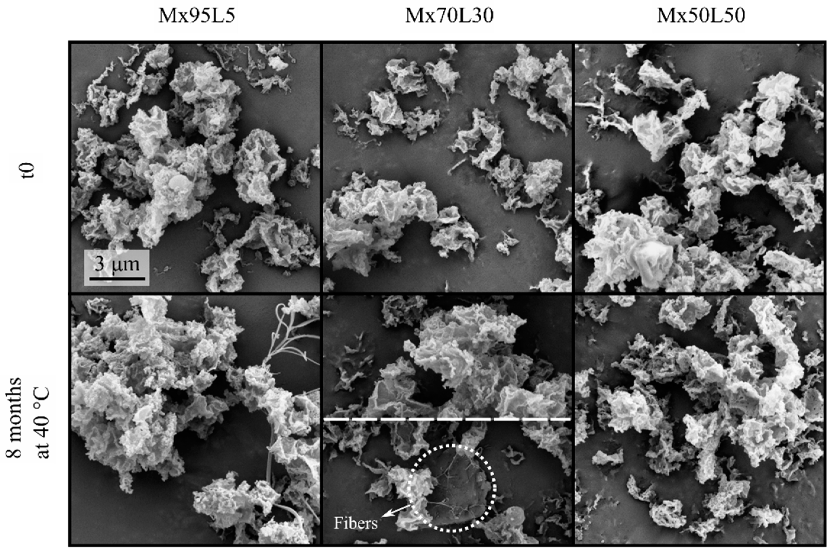
Electron micrographs of the spray-dried bevacizumab–trehalose–leucine formulations at t0 and after 8 months of storage at 40 °C. Note the appearance of a small number of thin fibers in the bottom row. The scale bars apply to all micrographs in each row.
Figure 2.
Electron micrographs of the spray-dried bevacizumab–trehalose–leucine formulations at t0 and after 8 months of storage at 40 °C. Note the appearance of a small number of thin fibers in the bottom row. The scale bars apply to all micrographs in each row.
Figure 3.
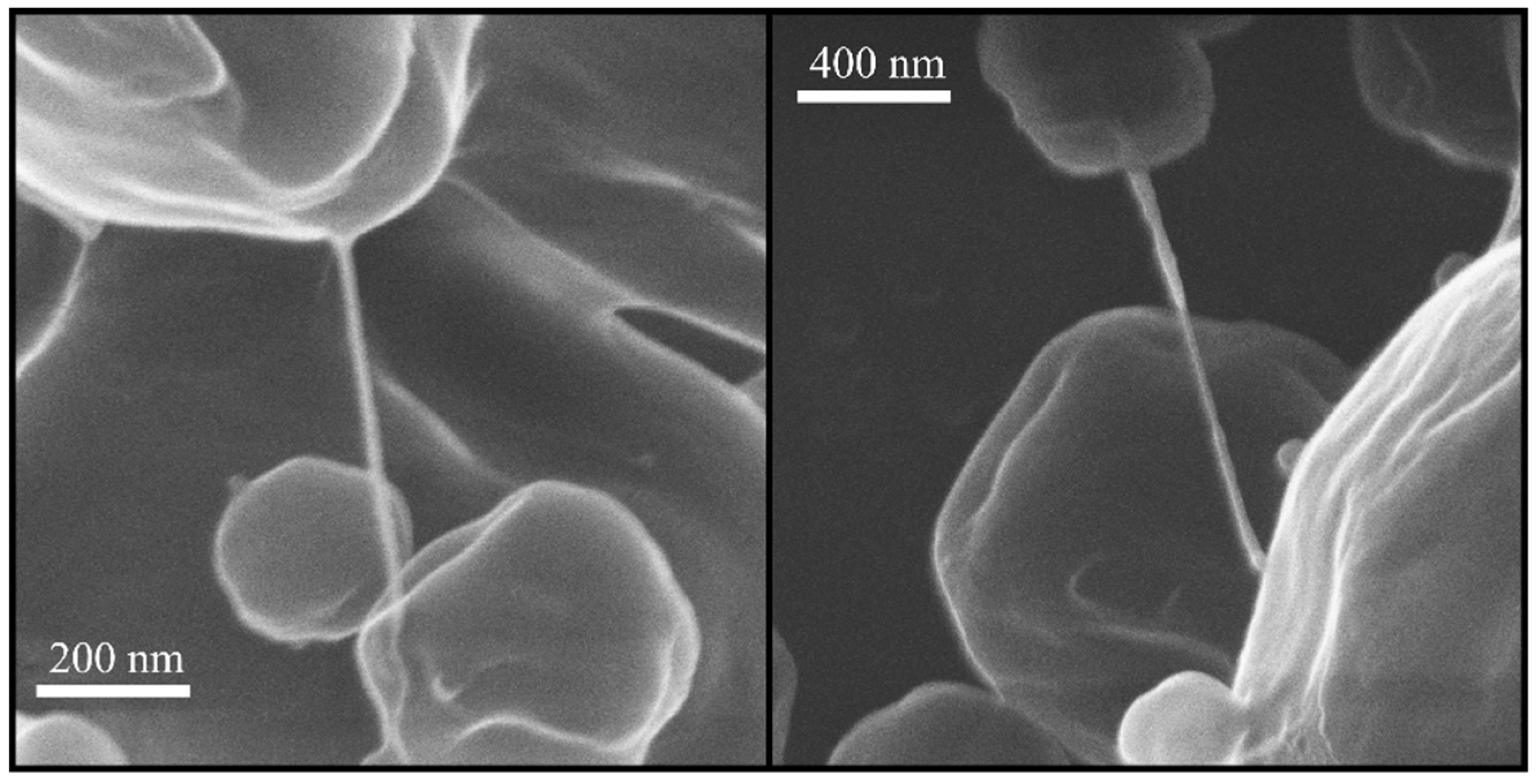
Helium ion micrographs of some of the fibers in the B18T72L10 formulation after 1 month of storage at 40 °C.
Figure 3.
Helium ion micrographs of some of the fibers in the B18T72L10 formulation after 1 month of storage at 40 °C.
Figure 4.
Electron micrographs of the spray-dried moxidectin–leucine formulations at t0 and after 8 months of storage at 40 °C. Some fibers can be seen in the left panel of the bottom row. Only a few fibers could be seen for the Mx70L30 after 8 months of storage. For this case, the top half shows a typical field of view without fibers. The bottom half shows small fibers that were occasionally seen (dotted circle). No fibers can be seen in right panel of the bottom row. The scale bar applies to all micrographs.
Figure 5.
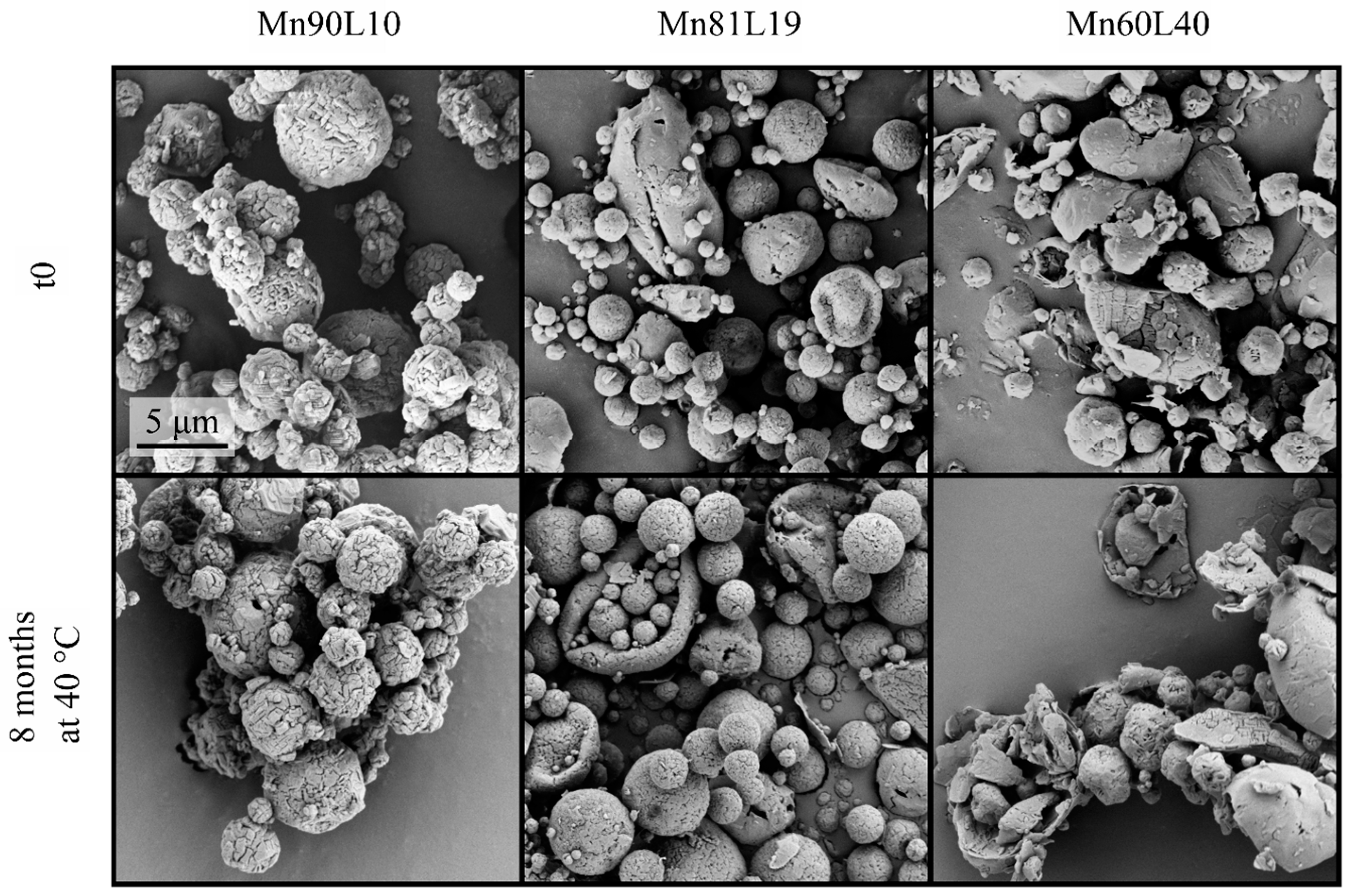
Micrographs of the spray-dried mannitol–leucine formulations at t0 and after 8 months of storage at 40 °C. The scale bar applies to all micrographs.
Figure 5.
Micrographs of the spray-dried mannitol–leucine formulations at t0 and after 8 months of storage at 40 °C. The scale bar applies to all micrographs.
Figure 6.
PXRD diffractograms of the spray-dried bevacizumab–trehalose–leucine powders at t0 and after 4 months of storage at 25 and 40 °C.
Figure 6.
PXRD diffractograms of the spray-dried bevacizumab–trehalose–leucine powders at t0 and after 4 months of storage at 25 and 40 °C.
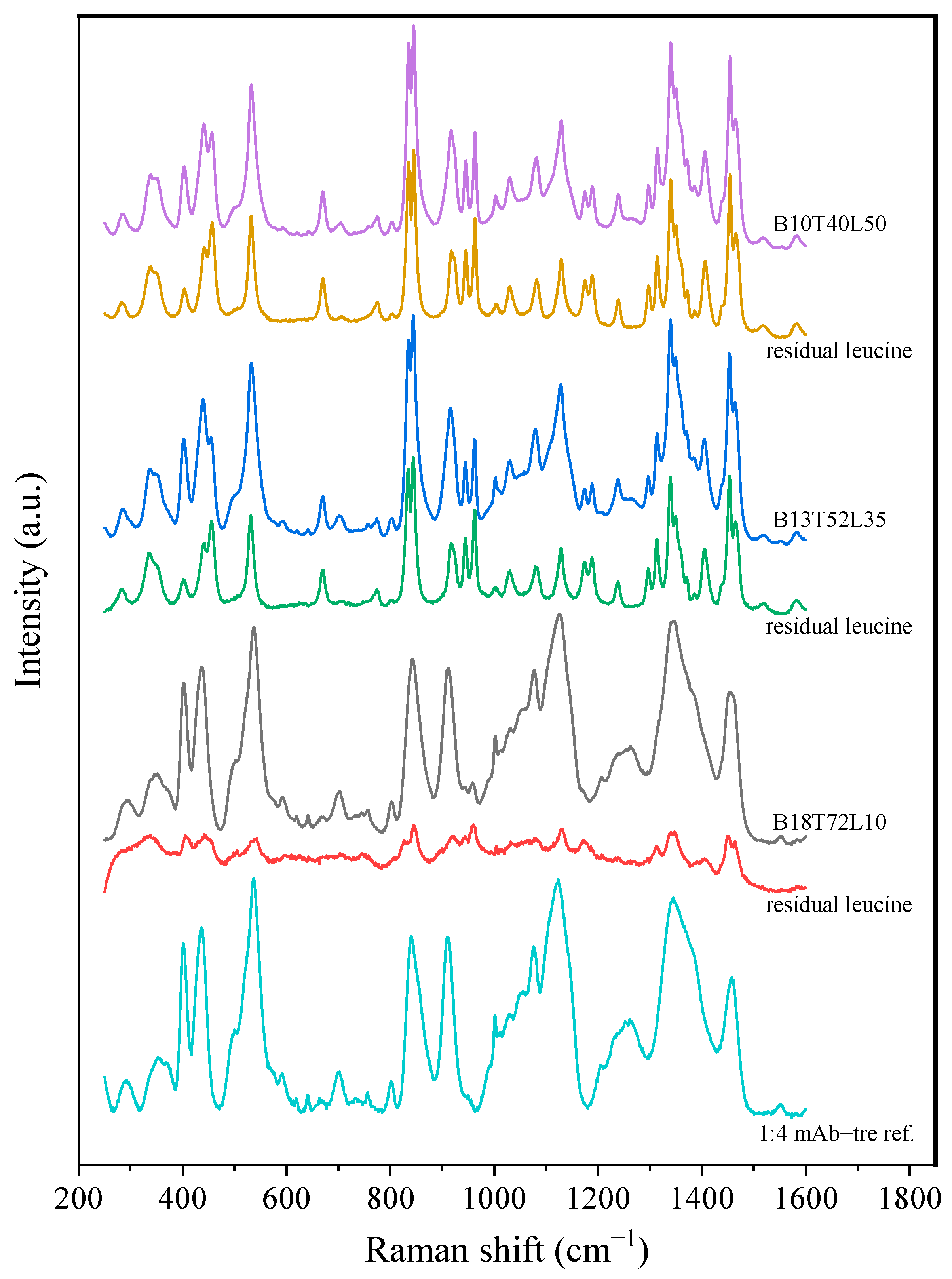
Figure 7.
The raw and residual leucine spectra of the bevacizumab formulations after 8 months of storage at 40 °C. No significant difference was detected between the t0 and 8-month measurements.
Figure 7.
The raw and residual leucine spectra of the bevacizumab formulations after 8 months of storage at 40 °C. No significant difference was detected between the t0 and 8-month measurements.
Figure 8.
PXRD diffractograms of the spray-dried moxidectin–leucine powders at t0 and after 4 months of storage at 25 and 40 °C.
Figure 8.
PXRD diffractograms of the spray-dried moxidectin–leucine powders at t0 and after 4 months of storage at 25 and 40 °C.
Figure 9.
The raw and residual leucine spectra of the moxidectin formulations after 8 months of storage at 40 °C. No significant difference was detected between the t0 and 8-month measurements.
Figure 9.
The raw and residual leucine spectra of the moxidectin formulations after 8 months of storage at 40 °C. No significant difference was detected between the t0 and 8-month measurements.
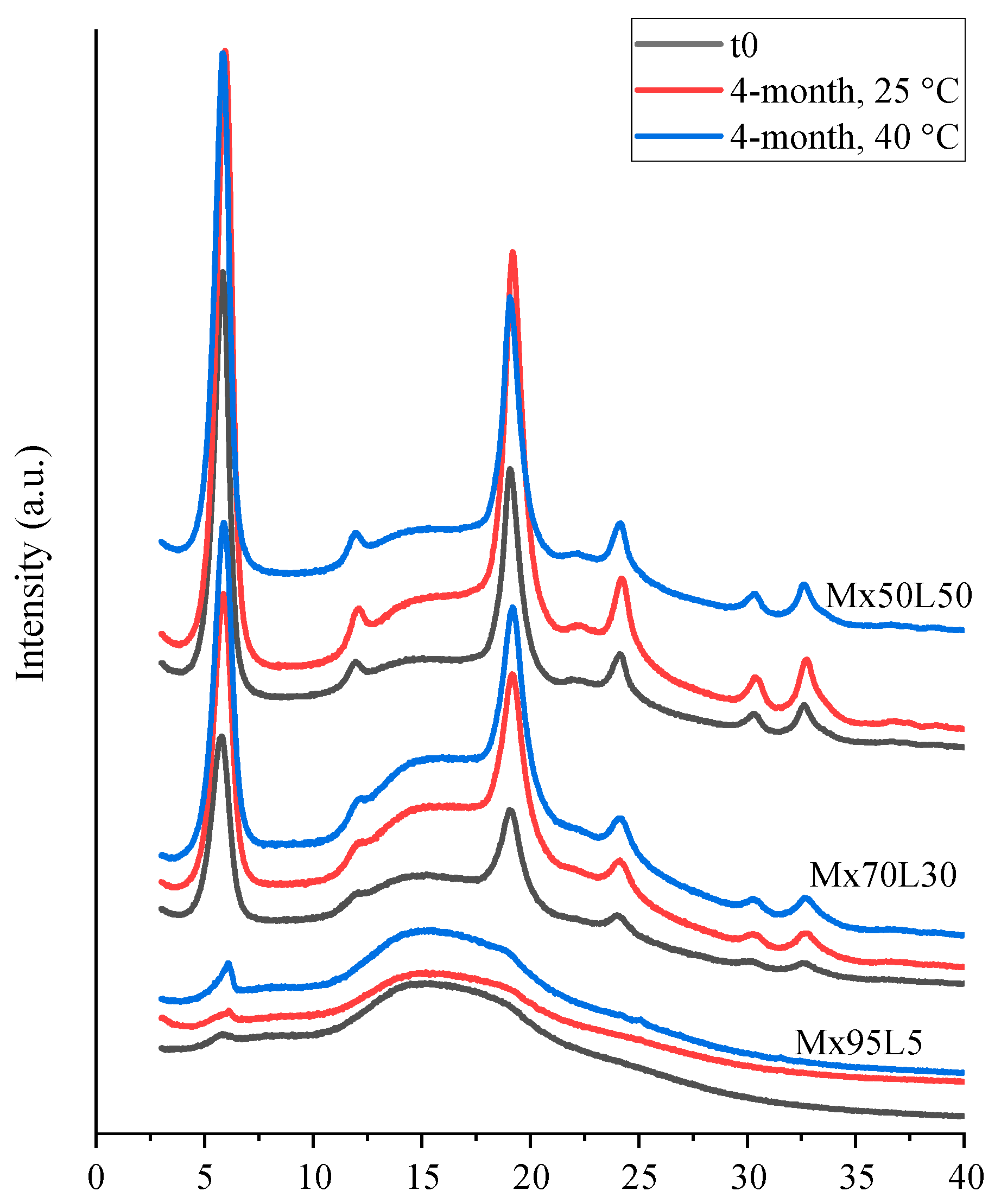
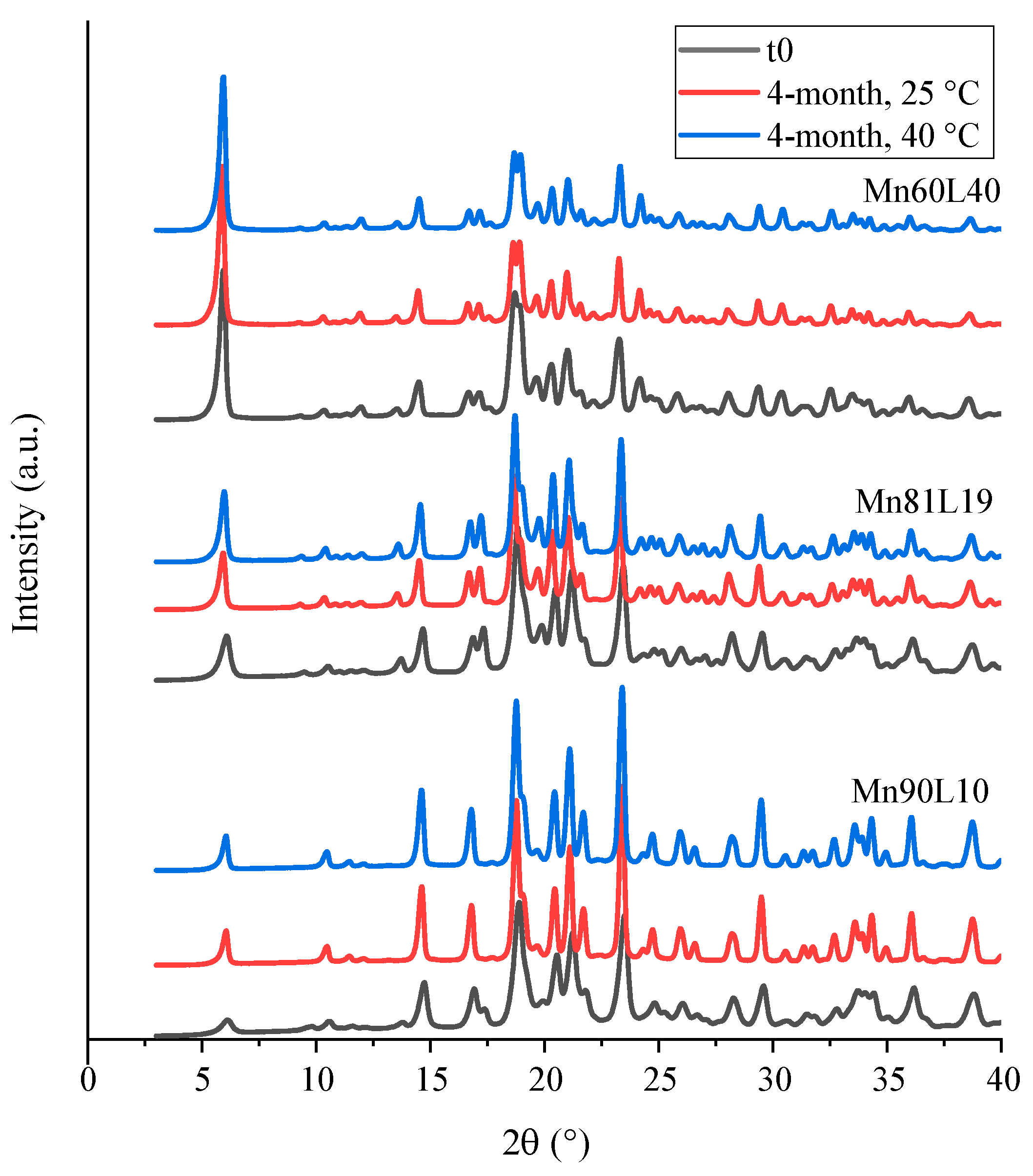
Figure 10.
PXRD diffractograms of the spray-dried mannitol–leucine powders at t0 and after 4 months of storage at 25 and 40 °C.
Figure 10.
PXRD diffractograms of the spray-dried mannitol–leucine powders at t0 and after 4 months of storage at 25 and 40 °C.
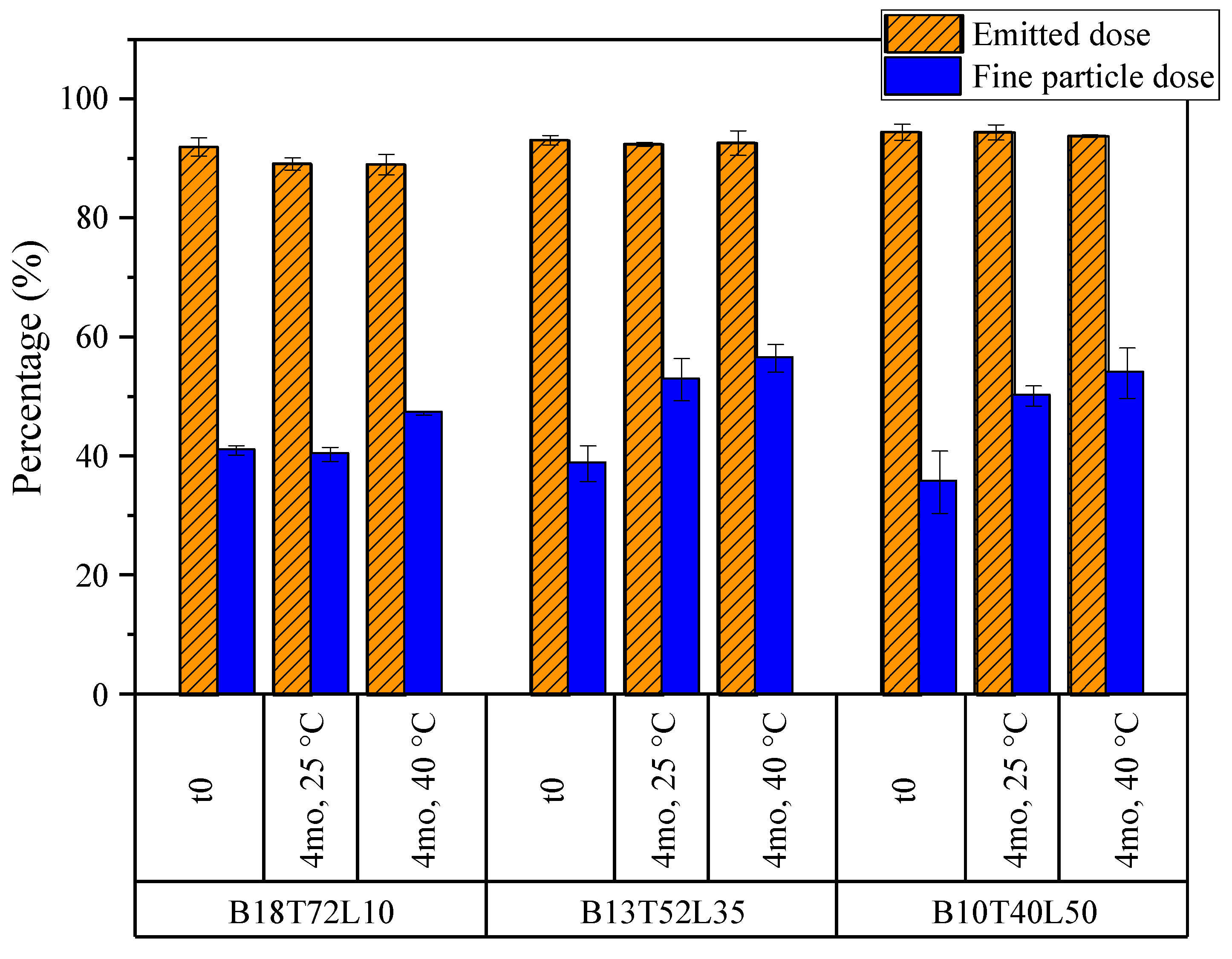
Figure 11.
The emitted doses and fine particle doses of the bevacizumab formulations obtained from NGI measurements. The fine particle dose is defined as the fraction of the emitted particles with aerodynamic diameters of less than 5 microns. The error bars correspond to one standard deviation.
Figure 11.
The emitted doses and fine particle doses of the bevacizumab formulations obtained from NGI measurements. The fine particle dose is defined as the fraction of the emitted particles with aerodynamic diameters of less than 5 microns. The error bars correspond to one standard deviation.
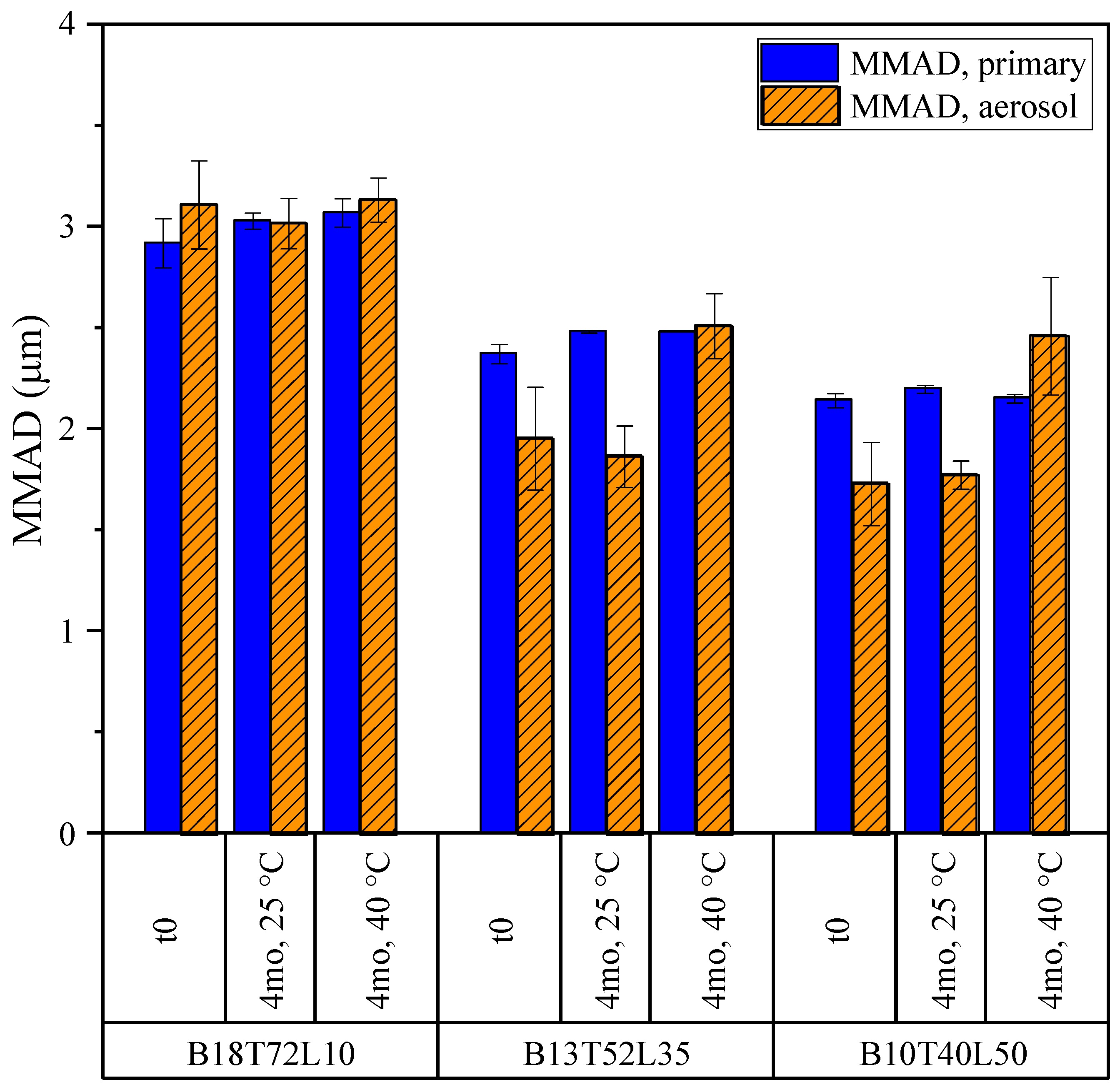
Figure 12.
The mass median aerodynamic diameters (MMADs) of the bevacizumab formulations for the primary particle size and aerosol size distribution from a DPI. The error bars correspond to one standard deviation.
Figure 12.
The mass median aerodynamic diameters (MMADs) of the bevacizumab formulations for the primary particle size and aerosol size distribution from a DPI. The error bars correspond to one standard deviation.
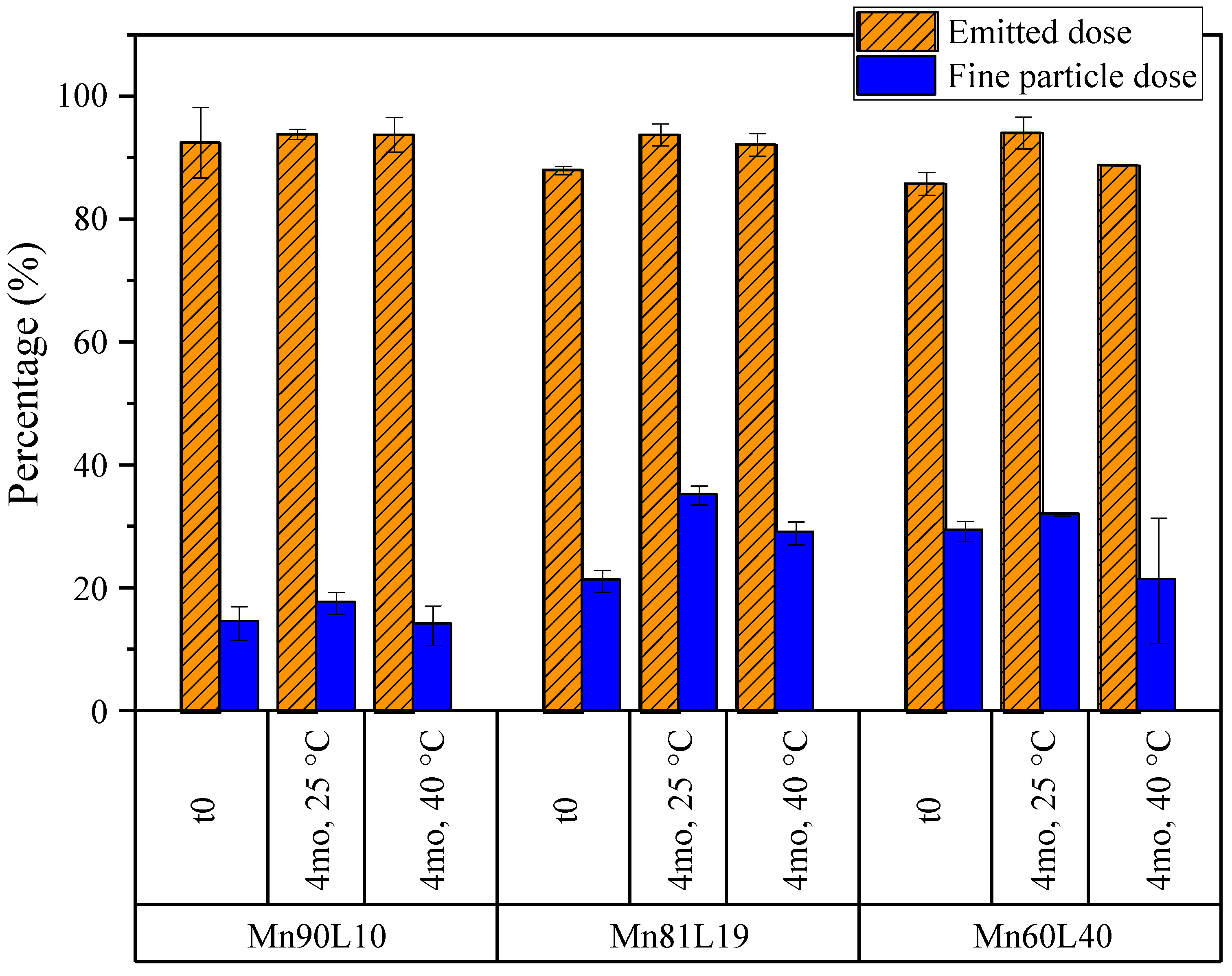
Figure 13.
The emitted doses and fine particle doses of the moxidectin formulations obtained from NGI measurements. The fine particle dose is defined as the fraction of the emitted particles with aerodynamic diameters of less than 5 microns. The error bars correspond to one standard deviation.
Figure 13.
The emitted doses and fine particle doses of the moxidectin formulations obtained from NGI measurements. The fine particle dose is defined as the fraction of the emitted particles with aerodynamic diameters of less than 5 microns. The error bars correspond to one standard deviation.
Figure 14.
The mass median aerodynamic diameters (MMADs) of the moxidectin formulations for the primary particle size and aerosol size distribution from a DPI, obtained from APS and NGI measurements, respectively. The error bars correspond to one standard deviation.
Figure 14.
The mass median aerodynamic diameters (MMADs) of the moxidectin formulations for the primary particle size and aerosol size distribution from a DPI, obtained from APS and NGI measurements, respectively. The error bars correspond to one standard deviation.
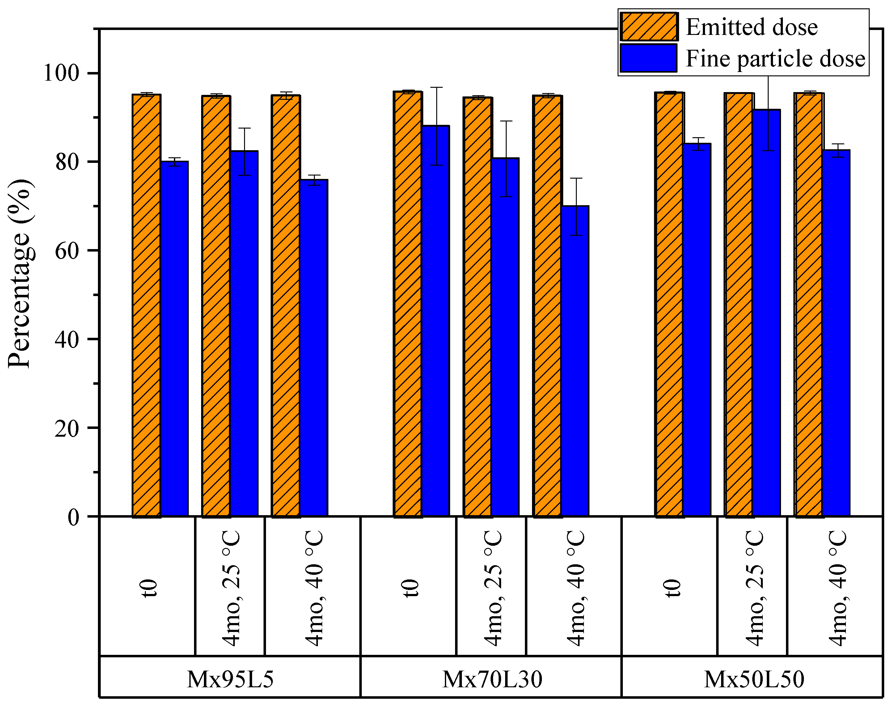
Figure 15.
The emitted doses and fine particle doses of the mannitol formulations obtained from NGI measurements. The fine particle dose is defined as the fraction of the emitted particles with aerodynamic diameters of less than 5 µm. The error bars correspond to one standard deviation.
Figure 15.
The emitted doses and fine particle doses of the mannitol formulations obtained from NGI measurements. The fine particle dose is defined as the fraction of the emitted particles with aerodynamic diameters of less than 5 µm. The error bars correspond to one standard deviation.
Figure 16.
The mass median aerodynamic diameters (MMADs) of the mannitol formulations for the primary particle size and aerosol size distribution from a DPI, obtained from APS and NGI measurements, respectively. The error bars correspond to one standard deviation.
Figure 16.
The mass median aerodynamic diameters (MMADs) of the mannitol formulations for the primary particle size and aerosol size distribution from a DPI, obtained from APS and NGI measurements, respectively. The error bars correspond to one standard deviation.
Table 1.
The selected formulations based on the results obtained from the particle formation models and the predicted outcome. is the predicted crystallization window of leucine defined as the time at which the next component reached its respective critical concentration on the droplet surface minus the time at which leucine reached the critical concentration on the surface.
Formulation
Name | Total Solids Loading (mg/mL) | Mass Fractions (-) | (ms)
| Predicted Outcome |
|---|
| First System | Bevacizumab | Trehalose | Leucine | | |
|---|
| B18T72L10 | 40 | 0.18 | 0.72 | 0.10 | −5.5 | Fully amorphous leucine |
| B13T52L35 | 0.13 | 0.52 | 0.35 | 0.4 | Partially amorphous leucine |
| B10T40L50 | 0.10 | 0.40 | 0.50 | 3.1 | Fully crystalline leucine |
| Second System | Ethanol:Water | Moxidectin | Leucine | | |
| Mx95L5 | 5 | 0.5:0.5 | 0.95 | 0.05 | - | Least leucine crystallinity |
| Mx70L30 | 10 | 0.70 | 0.30 | - | Moderate leucine crystallinity |
| Mx50L50 | 10 | 0.50 | 0.50 | - | Most leucine crystallinity |
| Third System | | Mannitol | Leucine | | |
| Mn90L10 | 40 | 0.90 | 0.10 | −1.8 | Mannitol nucleates first |
| Mn81L19 | 0.81 | 0.19 | 0.0 | Both nucleate together |
| Mn60L40 | 0.60 | 0.40 | 3.7 | Leucine nucleates first |
Table 2.
The time points and assays for the stability analysis performed on the three model systems stored at 25 and 40 °C.
Table 2.
The time points and assays for the stability analysis performed on the three model systems stored at 25 and 40 °C.
| | SEM/Raman Spectroscopy | PXRD/NGI/APS/KF/DSC |
|---|
| Beva/Trehalose/Leucine | t0, 1, and 2 weeks, 1, 2, 4, and 8 months | t0, 4 months |
| Moxidectin/Leucine | t0, 2 weeks, 1, 2, 4, and 8 months | t0, 4 months |
| Mannitol/Leucine | t0, 1, and 2 weeks, 1, 2, 4, and 8 months | t0, 4 months |
Table 3.
The solid phases of the spray-dried formulations containing bevacizumab, trehalose, and leucine obtained from the deconvolution of their respective Raman spectra. The solid phases of all powders were stable for up to 8 months of storage at temperatures as high as 40 °C.
Table 3.
The solid phases of the spray-dried formulations containing bevacizumab, trehalose, and leucine obtained from the deconvolution of their respective Raman spectra. The solid phases of all powders were stable for up to 8 months of storage at temperatures as high as 40 °C.
| Formulation | t0 | 4 and 8 Months |
|---|
| mAb + Trehalose | Leucine | mAb + Trehalose | Leucine |
|---|
| B18T72L10 | amorphous | amorphous | amorphous | amorphous |
| B13T52L35 | amorphous | β-form | amorphous | β-form |
| B10T40L50 | amorphous | β-form | amorphous | β-form |
Table 4.
The glass transition temperatures, water content, and active concentration values of the spray-dried formulations containing bevacizumab, trehalose, and leucine. The uncertainties represent one standard deviation of at least three replicates.
Table 4.
The glass transition temperatures, water content, and active concentration values of the spray-dried formulations containing bevacizumab, trehalose, and leucine. The uncertainties represent one standard deviation of at least three replicates.
| | B18T72L10 | B13T52L35 | B10T40L50 |
|---|
| t0 | 4 Months at 25 °C | 4 Months at 40 °C | t0 | 4 Months at 25 °C | 4 Months at 40 °C | t0 | 4 Months at 25 °C | 4 Months at 40 °C |
|---|
| Dry (midpoint, °C) | 118.0 ± 0.2 | 119.1 ± 0.4 | 118.3 ± 2.1 | 121.5 ± 2.0 | 123.9 ± 0.4 | 123.4 ± 0.7 | 122.0 ± 3.2 | 121.7 ± 1.4 | 121.8 ± 0.5 |
| Water Content (wt %) | 4.82 ± 0.09 | 4.23 ± 0.09 | 3.75 ± 0.04 | 3.59 ± 0.00 | 2.81 ± 0.11 | 2.86 ± 0.08 | 2.92 ± 0.01 | 2.44 ± 0.00 | 2.23 ± 0.08 |
| mAb Concentration (wt %) | 19.1 ± 0.9 | 18.1 ± 0.1 | 18.2 ± 0.3 | 13.0 ± 0.1 | 13.0 ± 0.1 | 12.7 ± 0.1 | 9.2 ± 0.2 | 9.6 ± 0.1 | 9.0 ± 0.1 |
Table 5.
The solid phases of the spray-dried formulations containing moxidectin and leucine obtained from the deconvolution of their respective Raman spectra. The solid phases of all powders were stable for up to 8 months of storage at temperatures as high as 40 °C.
Table 5.
The solid phases of the spray-dried formulations containing moxidectin and leucine obtained from the deconvolution of their respective Raman spectra. The solid phases of all powders were stable for up to 8 months of storage at temperatures as high as 40 °C.
| Formulation | t0 | 4 and 8 Months |
|---|
| Moxidectin | Leucine | Moxidectin | Leucine |
|---|
| Mx95L5 | amorphous | Crystalline * | amorphous | Crystalline * |
| Mx70L30 | amorphous | β-form | amorphous | β-form |
| Mx50L50 | amorphous | β-form | amorphous | β-form |
Table 6.
The glass transition temperatures, water content, and active concentration values of the spray-dried moxidectin–leucine formulations. The uncertainties represent one standard deviation of at least three replicates.
Table 6.
The glass transition temperatures, water content, and active concentration values of the spray-dried moxidectin–leucine formulations. The uncertainties represent one standard deviation of at least three replicates.
| | Mx95L5 | Mx70L30 | Mx50L50 |
|---|
| t0 | 4 Months at 25 °C | 4 Months at 40 °C | t0 | 4 Months at 25 °C | 4 Months at 40 °C | t0 | 4 Months at 25 °C | 4 Months at 40 °C |
|---|
| Dry (midpoint, °C) | 115.9 ± 1.9 | 114.0 ± 1.0 | 116.7 ± 0.3 | 115.2 ± 0.7 | 116.8 ± 0.3 | 115.8 ± 0.5 | 117.9 ± 2.1 | 115.4 ± 1.2 | 119.6 ± 0.1 |
| Water Content (wt %) | 0.66 ± 0.02 | 0.46 ± 0.02 | 0.42 ± 0.01 | 0.58 ± 0.01 | 0.44 ± 0.02 | 0.40 ± 0.03 | 0.49 ± 0.02 | 0.38 ± 0.05 | 0.35 ± 0.01 |
| Moxidectin Concentration (wt %) | 95.6 ± 0.8 | 95.0 ± 0.8 | 94.8 ± 0.6 | 70.7 ± 0.2 | 70.3 ± 0.2 | 69.6 ± 0.5 | 50.8 ± 0.1 | 50.4 ± 0.1 | 50.2 ± 0.1 |
Table 7.
The melting temperatures, water content, and active concentration values of the spray-dried formulations containing mannitol and leucine. The uncertainties represent one standard deviation of at least three replicates.
Table 7.
The melting temperatures, water content, and active concentration values of the spray-dried formulations containing mannitol and leucine. The uncertainties represent one standard deviation of at least three replicates.
| | Mn90L10 | Mn81L19 | Mn60L40 |
|---|
| t0 | 4 Months at 25 °C | 4 Months at 40 °C | t0 | 4 Months at 25 °C | 4 Months at 40 °C | t0 | 4 Months at 25 °C | 4 Months at 40 °C |
|---|
| (Onset, °C) | 164.5 ± 0.1 | 164.8 ± 0.1 | 164.8 ± 0.1 | 163.7 ± 1.2 | 164.3 ± 0.0 | 164.3 ± 0.1 | 164.4 ± 0.0 | 164.4 ± 0.1 | 164.4 ± 0.0 |
| Water Content (wt %) | 0.04 | 0.02 | 0.04 | 0.04 | 0.09 | 0.09 | <0.01 | 0.03 | <0.01 |
| Mannitol Concentration (wt %) | 90.9 | 90.7 | 91.6 | 83.0 | 82.5 | 83.0 | 62.5 | 63.8 | 63.4 |
Table 8.
The prevalence of leucine fibers in electron micrographs of the three model systems during 8 months of storage under two different conditions. The time points correspond to the number of months of storage.
Table 8.
The prevalence of leucine fibers in electron micrographs of the three model systems during 8 months of storage under two different conditions. The time points correspond to the number of months of storage.
| Formulation | t0 | t0.5 | t1 | t2 | t4 | t8 |
|---|
| | 25 °C | 40 °C | 25 °C | 40 °C | 25 °C | 40 °C | 25 °C | 40 °C | 25 °C | 40 °C |
|---|
| B18T72L10 | 0 | + | ++ | + | +++ | ++ | +++ | ++ | +++ | +++ | +++ |
| B13T52L35 | 0 | 0 | + | + | ++ | + | +++ | + | +++ | + | +++ |
| B10T40L50 | 0 | 0 | + | 0 | + | 0 | + | 0 | + | 0 | ++ |
| Mx95L5 | 0 | 0 | 0 | 0 | 0 | 0 | + | 0 | ++ | + | +++ |
| Mx70L30 | 0 | 0 | 0 | 0 | 0 | 0 | 0 | 0 | 0 | 0 | + |
| Mx50L50 | 0 | 0 | 0 | 0 | 0 | 0 | 0 | 0 | 0 | 0 | 0 |
| All mannitol formulations | 0 | 0 | 0 | 0 | 0 | 0 | 0 | 0 | 0 | 0 | 0 |

 ,
, 


