

Novel Tumor-Targeted Self-Nanostructured and Compartmentalized Water-in-Oil-in-Water Polyurethane-Polyurea Nanocapsules for Cancer Theragnosis
 , , , , , , and
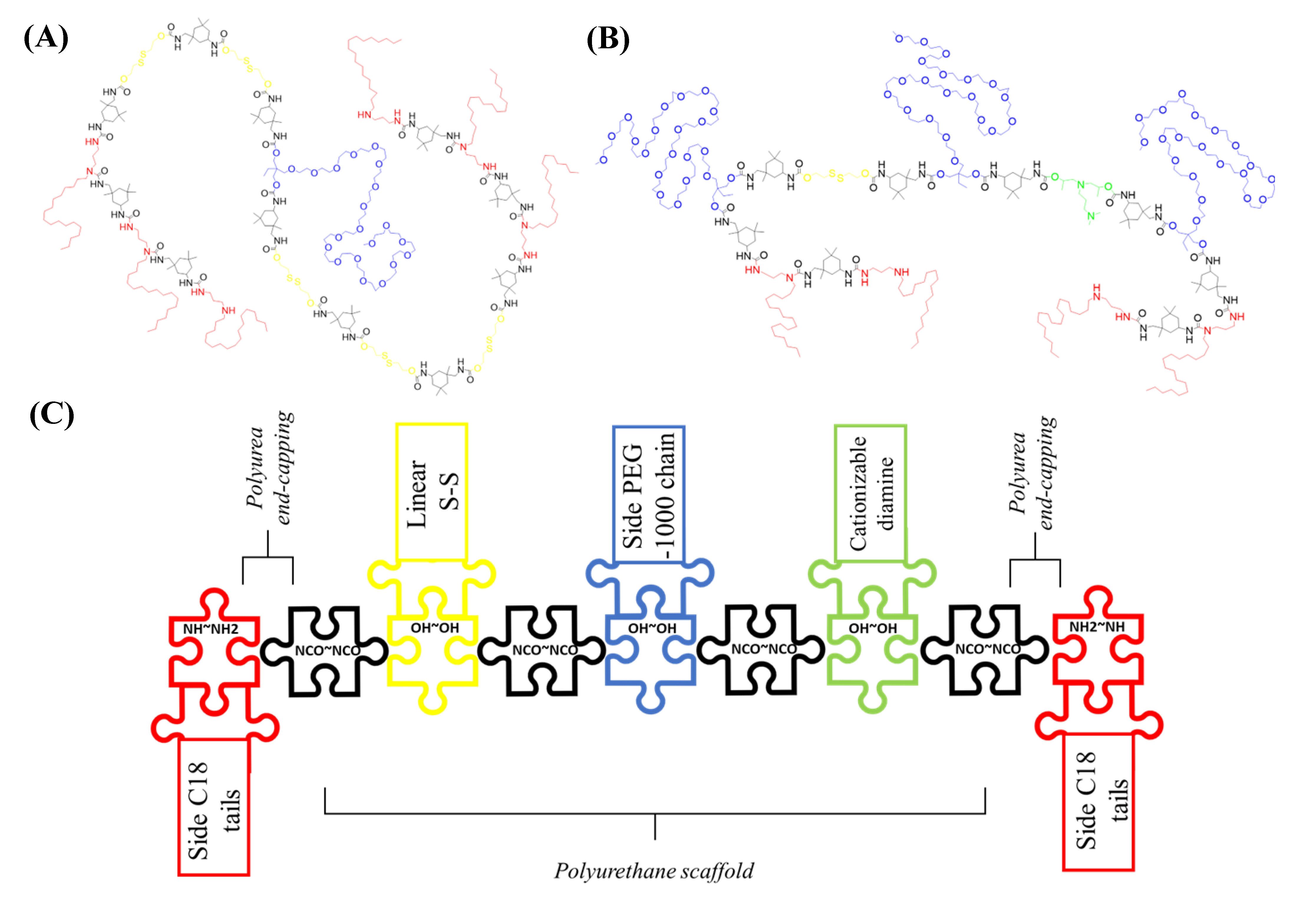
Abstract
:1. Introduction
2. Materials and Methods
2.1. Synthetic Procedures
2.1.1. Synthesis of Redox-Responsive Amphiphilic Low HLB Prepolymer (P1)
2.1.2. Synthesis of Redox Responsive Amphiphilic Cationic Prepolymer (P2)
2.1.3. Synthesis of Dye-Loaded Cationic Redox Responsive Water-in-Oil-in-Water NCs (Cationic Dye-NCs)
2.1.4. Synthesis of Dye-Loaded Amphoteric Redox Responsive Water-in-Oil-in-Water NCs (Amphoteric dye-NCs)
2.2. Biological Studies
2.2.1. Monocyte-Derived Dendritic Cells (moDCs) Generation
2.2.2. Fluorescence Imaging by Confocal Microscopy
2.2.3. Internalization Evaluation by Flow Cytometry
2.2.4. In Vivo Safety Assay of Amphoteric and Cationic Control NCs
2.2.5. In Vivo Fluorescence Imaging Biodistribution in Subcutaneous Tumor Mice Models
3. Results and Discussion
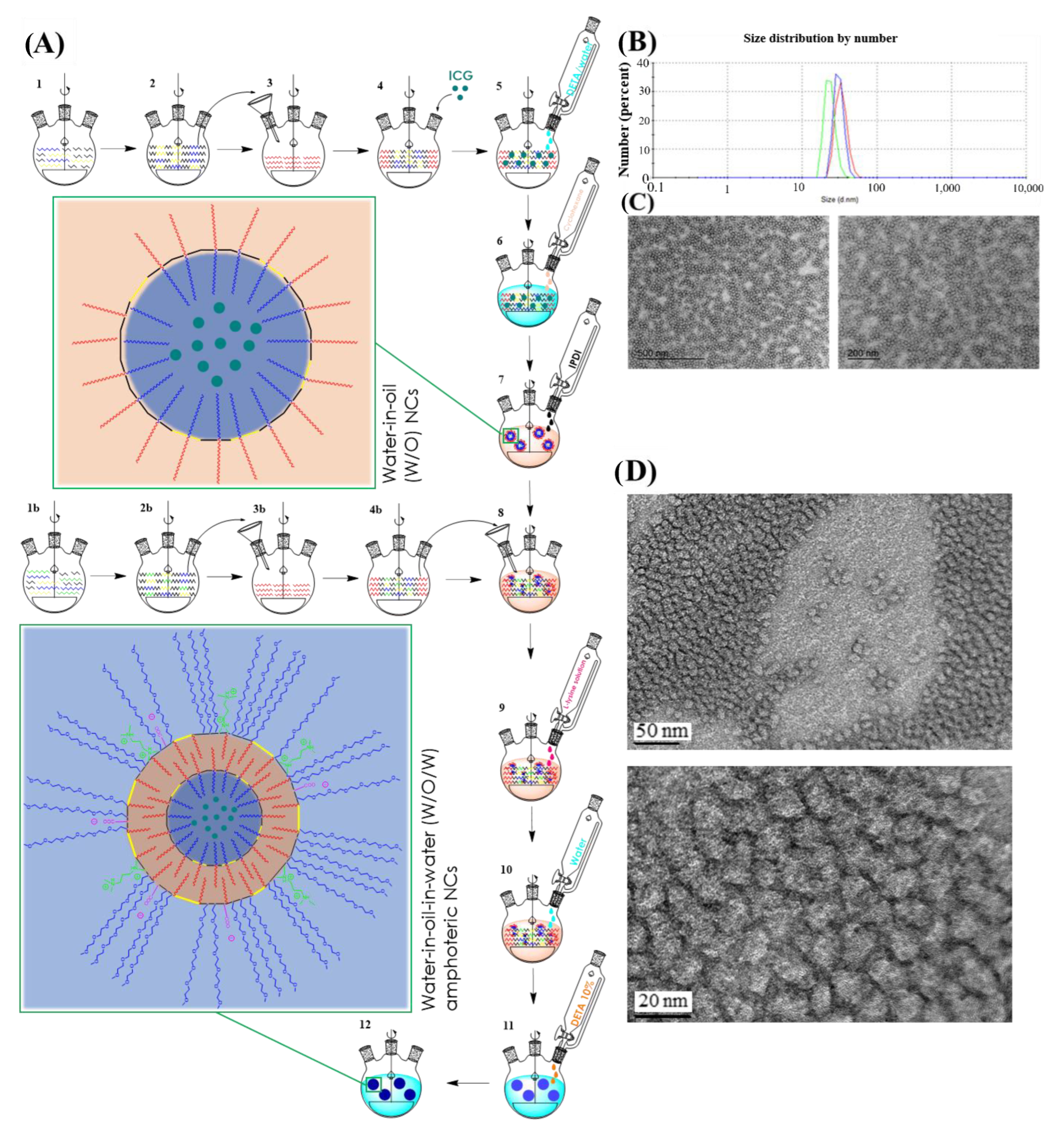
3.1. Design and Synthesis of WOW Double Walled Polyurethane-Polyurea Redox-Responsive Hybrid NCs
3.2. Synthesis and Characterization of ICG-Loaded NCs
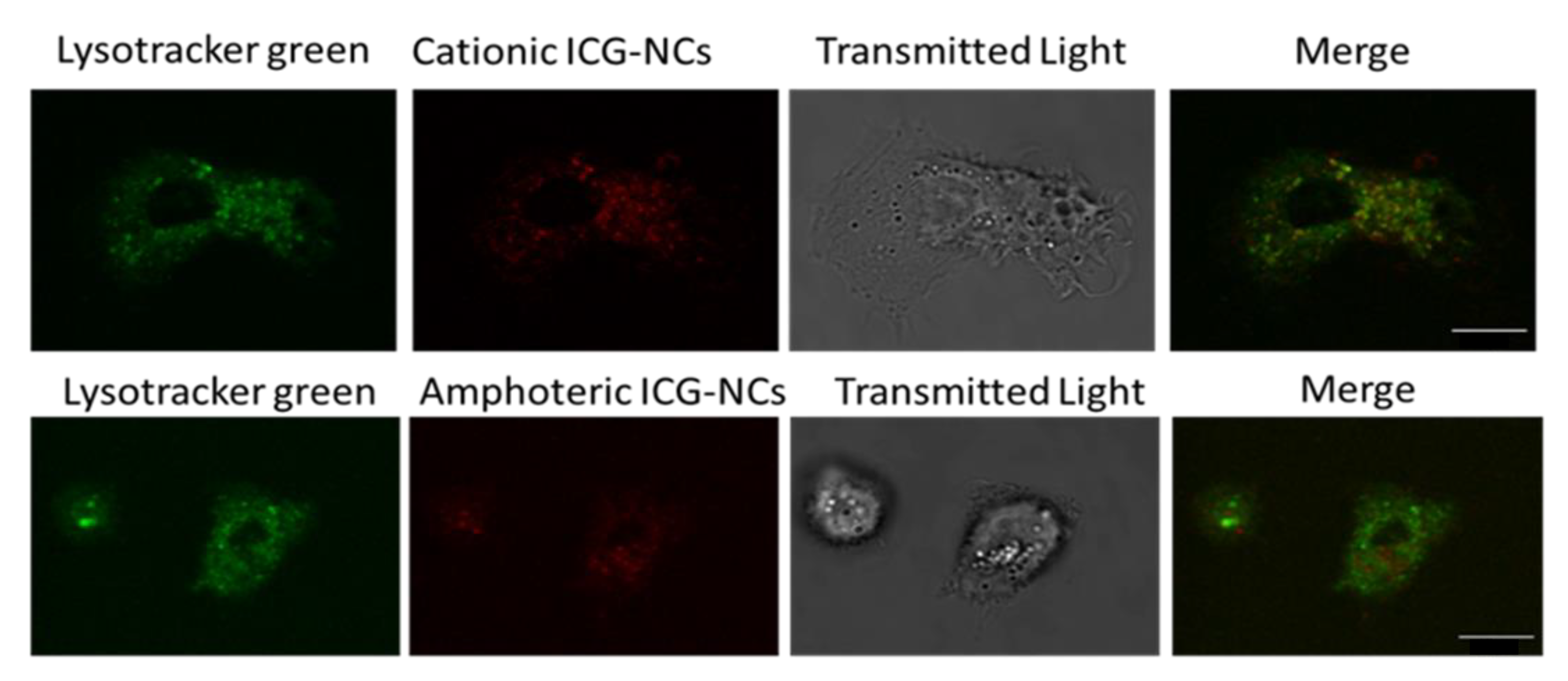
3.3. In Vitro Cellular Uptake Evaluation of the NCs
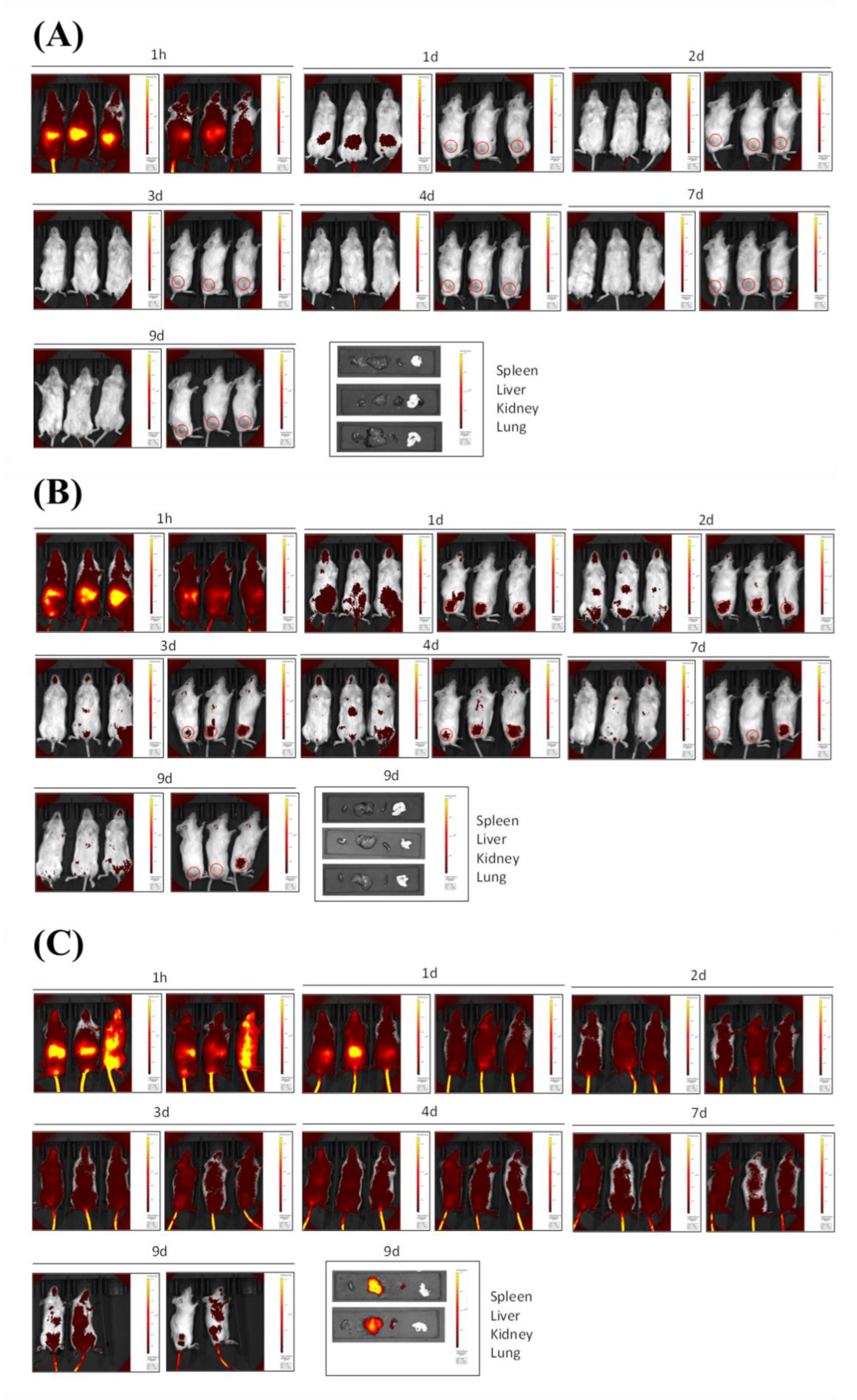
3.4. In Vivo Fluorescence Imaging Biodistribution in Healthy Mice
3.5. In Vivo Fluorescence Imaging Biodistribution in Subcutaneous Tumor Mice Model
4. Conclusions
5. Patent
Supplementary Materials
Author Contributions
Funding
Institutional Review Board Statement
Informed Consent Statement
Data Availability Statement
Acknowledgments
Conflicts of Interest
References
- Banik, B.L.; Fattahi, P.; Brown, J.L. Polymeric nanoparticles: The future of nanomedicine. Nanomed. Nanobiotechnol. 2016, 8, 271–299. [Google Scholar] [CrossRef] [PubMed]
- Alexis, F.; Pridgen, E.; Molnar, L.K.; Farokhzad, O.C. Factors Affecting the Clearance and Biodistribution of Polymeric Nanoparticles. Mol. Pharm. 2008, 5, 505–515. [Google Scholar] [CrossRef] [PubMed] [Green Version]
- Wu, W.; Pu, Y.; Shi, J. Nanomedicine-enabled chemotherapy-based synergetic cancer treatments. J. Nanobiotechnol. 2022, 20, 4. [Google Scholar] [CrossRef] [PubMed]
- Santerre, J.P.; Woodhouse, K.; Laroche, G.; Labow, R.S. Understanding the biodegradation of polyurethanes: From classical implants to tissue engineering materials. Biomaterials 2005, 26, 7457–7470. [Google Scholar] [CrossRef] [PubMed]
- Rusu, L.C.; Ardelean, L.C.; Jitariu, A.A.; Miu, C.A.; Streian, C.G. An Insight into the Structural Diversity and Clinical Applicability of Polyurethanes in Biomedicine. Polymers 2020, 12, 1197. [Google Scholar] [CrossRef] [PubMed]
- Wang, Y.; Zhu, L.; Wang, Y.; Li, L.; Lu, Y.; Shen, L.; Zhang, L.W. Ultrasensitive GSH-Responsive Ditelluride-Containing Poly(ether-urethane) Nanoparticles for Controlled Drug Release. ACS Appl. Mater. Interfaces 2016, 8, 35106–35113. [Google Scholar] [CrossRef]
- Rosenbauer, E.M.; Landfester, K.; Musyanovych, A. Surface-Active Monomer as a Stabilizer for Polyurea Nanocapsules Synthesized via Interfacial Polyaddition in Inverse Miniemulsion. Langmuir 2009, 25, 12084–12091. [Google Scholar] [CrossRef]
- Schoth, A.; Landfester, K.; Muñoz-Espí, R. Surfactant-Free Polyurethane Nanocapsules via Inverse Pickering Miniemulsion. Langmuir 2015, 31, 3784–3788. [Google Scholar] [CrossRef]
- Morral-Ruíz, G.; Melgar-Lesmes, P.; Solans, C.; García-Celma, M.J. Multifunctional polyurethane-urea nanoparticles to target and arrest inflamed vascular environment: A potential tool for cancer therapy and diagnosis. J. Control. Release 2013, 171, 163–171. [Google Scholar] [CrossRef]
- Pramanik, S.K.; Sreedharan, S.; Singh, H.; Khan, M.; Tiwari, K.; Shiras, A.; Smythe, C.; Thomas, J.A.; Das, A. Mitochondria Targeting Non-Isocyanate-Based Polyurethane Nanocapsules for Enzyme-Triggered Drug Release. Bioconjugate Chem. 2018, 29, 3532–3543. [Google Scholar] [CrossRef]
- Rosenbauer, E.M.; Wagner, M.; Musyanovych, A.; Landfester, K. Controlled Release from Polyurethane Nanocapsules via pH-, UV-Light- or Temperature-Induced Stimuli. Macromolecules 2010, 43, 5083–5093. [Google Scholar] [CrossRef]
- Huang, D.; Zhou, Y.; Xiang, Y.; Shu, M.; Chen, H.; Yang, B.; Liao, X. Polyurethane/doxorubicin nanoparticles based on electrostatic interactions as pH-sensitive drug delivery carriers. Polym. Int. 2018, 67, 1186–1193. [Google Scholar] [CrossRef]
- Tsvetkova, Y.; Beztsinna, N.; Baues, M.; Klein, D.; Rix, A.; Golombek, S.K.; al Rawashdeh, W.; Gremse, F.; Barz, M.; Koynov, K.; et al. Balancing Passive and Active Targeting to Different Tumor Compartments Using Riboflavin-Functionalized Polymeric Nanocarriers. Nano Lett. 2017, 17, 4665–4674. [Google Scholar] [CrossRef]
- Jiang, B. Aerobic glycolysis and high level of lactate in cancer metabolism and microenvironment. Genes Dis. 2017, 4, 25–27. [Google Scholar] [CrossRef]
- Cuscó, C.; Garcia, J.; Nicolás, E.; Rocas, P.; Rocas, J. Multisensitive drug-loaded polyurethane/polyurea nanocapsules with pH-synchronized shell cationization and redox-triggered release. Polym. Chem. 2016, 7, 6457–6466. [Google Scholar] [CrossRef]
- Pérez-Hernández, M.; Cuscó, C.; Benítez-García, C.; Bonelli, J.; Nuevo-Fonoll, M.; Soriano, A.; Martínez-García, D.; Arias-Betancur, A.; García-Valverde, M.; Segura, M.F.; et al. Multi-Smart and Scalable Bioligands-Free Nanomedical Platform for Intratumorally Targeted Tambjamine Delivery, a Difficult to Administrate Highly Cytotoxic Drug. Biomedicines 2021, 9, 508. [Google Scholar] [CrossRef]
- Fu, X.; Hosta-Rigau, L.; Chandrawati, R.; Cui, J. Multi-Stimuli-Responsive Polymer Particles, Films, and Hydrogels for Drug Delivery. Chem 2018, 4, 2084–2107. [Google Scholar] [CrossRef] [Green Version]
- Ruiz, A.L.; Ramirez, A.; McEnnis, K. Single and Multiple Stimuli-Responsive Polymer Particles for Controlled Drug Delivery. Pharmaceutics 2022, 14, 421. [Google Scholar] [CrossRef]
- Li, Z.; Huang, J.; Wu, J. pH-sensitive nanogels for drug delivery in cancer therapy. Biomater. Sci. 2021, 9, 574–589. [Google Scholar] [CrossRef]
- Khan, M.W.; Zou, C.; Hassan, S.; Din, F.U.; Razak, M.Y.A.; Nawaz, A.; Zeb, A.; Wahabh, A.; Bangash, S.A. Cisplatin and oleanolic acid Co-loaded pH-sensitive CaCO3 nanoparticles for synergistic chemotherapy. RSC Adv. 2022, 12, 14808–14818. [Google Scholar] [CrossRef]
- Rocas, P.; Fernández, Y.; Schwartz, S.; Abasolo, I.; Rocas, J.; Albericio, F. Multifunctionalized polyurethane-polyurea nanoparticles: Hydrophobically driven self-stratification at the o/w interface modulates encapsulation stability. J. Mater. Chem. B 2015, 3, 7604–7613. [Google Scholar] [CrossRef]
- Bonelli, J.; Ortega-Forte, E.; Vigueras, G.; Bosch, M.; Cutillas, N.; Rocas, J.; Ruiz, J.; Marchán, V. Polyurethane–polyurea hybrid nanocapsules as efficient delivery systems of anticancer Ir(III) metallodrugs. Inorg. Chem. Front. 2022, 9, 2123–2138. [Google Scholar] [CrossRef]
- Flórez-Grau, G.; Rocas, P.; Cabezón, R.; España, C.; Panés, J.; Rocas, J.; Albericio, F.; Benítez-Ribas, D. Nanoencapsulated budesonide in self-stratified polyurethane-polyurea nanoparticles is highly effective in inducing human tolerogenic dendritic cells. Int. J. Pharm. 2016, 511, 785–793. [Google Scholar] [CrossRef] [PubMed]
- Bonelli, J.; Ortega-Forte, E.; Rovira, A.; Bosch, M.; Torres, O.; Cuscó, C.; Rocas, J.; Ruiz, J.; Marchán, V. Improving Photodynamic Therapy Anticancer Activity of a Mitochondria-Targeted Coumarin Photosensitizer Using a Polyurethane-Polyurea Hybrid Nanocarrier. Biomacromolecules 2022, 23, 2900–2913. [Google Scholar] [CrossRef] [PubMed]
- Li, Q.; Li, X.; Zhao, C. Strategies to Obtain Encapsulation and Controlled Release of Small Hydrophilic Molecules. Front. Bioeng. Biotechnol. 2020, 8, 437. [Google Scholar] [CrossRef] [PubMed]
- Russell, L.M.; Hultz, M.; Searson, P.C. Leakage kinetics of the liposomal chemotherapeutic agent Doxil: The role of dissolution, protonation, and passive transport, and implications for mechanism of action. J. Control. Release 2018, 269, 171–176. [Google Scholar] [CrossRef]
- Luo, L.; Zhang, Q.; Luo, Y.; He, Z.; Tian, X.; Battaglia, G. Thermosensitive nanocomposite gel for intra-tumoral two-photon photodynamic therapy. J. Control. Release 2019, 298, 99–109. [Google Scholar] [CrossRef]
- Kunjachan, S.; Gremse, F.; Theek, B.; Koczera, P.; Pola, R.; Pechar, M.; Etrych, T.; Ulbrich, K.; Storm, G.; Kiessling, F.; et al. Noninvasive Optical Imaging of Nanomedicine Biodistribution. ACS Nano 2013, 7, 252–262. [Google Scholar] [CrossRef]
- Hill, T.K.; Abdulahad, A.; Kelkar, S.S.; Marini, F.C.; Long, T.E.; Provenzale, J.M.; Mohs, A.M. Indocyanine Green-Loaded Nanoparticles for Image-Guided Tumor Surgery. Bioconjugate Chem. 2015, 26, 294–303. [Google Scholar] [CrossRef] [Green Version]
- Charalampaki, P.; Proskynitopoulos, P.J.; Heimann, A.; Nakamura, M. 5-Aminolevulinic Acid Multispectral Imaging for the Fluorescence-Guided Resection of Brain Tumors: A Prospective Observational Stud. Front. Oncol. 2020, 10, 1069. [Google Scholar] [CrossRef]
- Zhang, R.R.; Schroeder, A.B.; Grudzinski, J.J.; Rosenthal, E.L.; Warram, J.M.; Pinchuk, A.N.; Eliceiri, K.W.; Kuo, J.S.; Weichert, J.P. Beyond the margins: Real-time detection of cancer using targeted fluorophores. Nat. Rev. Clin. Oncol. 2017, 14, 347–364. [Google Scholar] [CrossRef] [Green Version]
- Theek, B.; Rizzo, L.Y.; Ehling, J.; Kiessling, F.; Lammers, T. The Theranostic Path to Personalized Nanomedicine. Clin. Transl. Imaging 2014, 2, 67–76. [Google Scholar] [CrossRef] [Green Version]
- Boni, L.; David, G.; Mangano, A.; Dionigi, G.; Rausei, S.; Spampatti, S.; Cassinotti, E.; Fingerhut, A. Clinical applications of indocyanine green (ICG) enhanced fluorescence in laparoscopic surgery. Surg. Endosc. 2015, 29, 2046–2055. [Google Scholar] [CrossRef] [Green Version]
- Sabapathy, V.; Mentam, J.; Jacob, P.M.; Kumar, S. Noninvasive Optical Imaging and In Vivo Cell Tracking of Indocyanine Green Labeled Human Stem Cells Transplanted at Superficial or In-Depth Tissue of SCID Mice. Stem Cells Int. 2015, 2015, 606415. [Google Scholar] [CrossRef] [Green Version]
- Gowsalya, K.; Yasothamani, V.; Vivek, R. Emerging indocyanine green-integrated nanocarriers for multimodal cancer therapy: A review. Nanoscale Adv. 2021, 3, 3332–3352. [Google Scholar] [CrossRef]
- Mindt, S.; Karampinis, I.; John, M.; Neumaier, M.; Nowak, K. Stability and degradation of indocyanine green in plasma, aqueous solution and whole blood. Photochem. Photobiol. Sci. 2018, 17, 1189–1196. [Google Scholar] [CrossRef]
- Gandioso, A.; Bresolí-Obach, R.; Nin-Hill, A.; Bosch, M.; Palau, M.; Galindo, A.; Contreras, S.; Rovira, A.; Rovira, C.; Nonell, S.; et al. Redesigning the Coumarin Scaffold into Small Bright Fluorophores with Far-Red to Near-Infrared Emission and Large Stokes Shifts Useful for Cell Imaging. J. Org. Chem. 2018, 83, 1185–1195. [Google Scholar] [CrossRef]
- Rovira, A.; Pujals, M.; Gandioso, A.; López-Corrales, M.; Bosch, M.; Marchán, V. Modulating Photostability and Mitochondria Selectivity in Far-Red/NIR Emitting Coumarin Fluorophores through Replacement of Pyridinium by Pyrimidinium. J. Org. Chem. 2020, 85, 6086–6097. [Google Scholar] [CrossRef]
- Rovira, A.; Gandioso, A.; Gonalons, M.; Galindo, A.; Massaguer, A.; Manel, M.; Marchán, V. Solid-Phase Approaches for Labeling Targeting Peptides with Far-Red Emitting Coumarin Fluorophores. J. Org. Chem. 2019, 84, 1808–1817. [Google Scholar] [CrossRef] [Green Version]
- De Vries, I.J.M.; Lesterhuis, W.J.; Scharenborg, N.M.; Engelen, L.P.H.; Ruiter, D.J.; Gerritsen, M.-J.P.; Croockewit, S.; Britten, C.M.; Torensma, R.; Adema, G.J.; et al. Maturation of Dendritic Cells Is a Prerequisite for Inducing Immune Responses in Advanced Melanoma Patients. Clin. Cancer Res. 2003, 9, 5091–5100. [Google Scholar]
- Bolte, S.; Cordelières, F.P. A guided tour into subcellular colocalization in light microscopy. J. Microsc. 2007, 224, 213–232. [Google Scholar] [CrossRef] [PubMed]
- Rocas, P.; Hoyos-Nogués, M.; Rocas, J.; Manero, J.M.; Gil, J.; Albericio, F.; Mas-Moruno, C. Installing Multifunctionality on Titanium with RGD-Decorated Polyurethane-Polyurea Roxithromycin Loaded Nanoparticles: Toward New Osseointegrative Therapies. Adv. Healthc. Mater. 2015, 4, 1956–1960. [Google Scholar] [CrossRef] [PubMed] [Green Version]
- Wicki, A.; Witzigmann, D.; Balasubramanian, V.; Huwyler, J. Nanomedicine in cancer therapy: Challenges, opportunities, and clinical applications. J. Control. Release 2015, 200, 138–157. [Google Scholar] [CrossRef] [PubMed]
- Chauhan, V.P.; Stylianopoulos, T.; Martin, J.D.; Popović, Z.; Chen, O.; Kamoun, W.S.; Bawendi, M.G.; Fukumura, D.; Jain, R.K. Normalization of tumour blood vessels improves the delivery of nanomedicines in a size-dependent manner. Nat. Nanotechnol. 2012, 7, 383–388. [Google Scholar] [CrossRef] [PubMed]


Disclaimer/Publisher’s Note: The statements, opinions and data contained in all publications are solely those of the individual author(s) and contributor(s) and not of MDPI and/or the editor(s). MDPI and/or the editor(s) disclaim responsibility for any injury to people or property resulting from any ideas, methods, instructions or products referred to in the content. |
© 2022 by the authors. Licensee MDPI, Basel, Switzerland. This article is an open access article distributed under the terms and conditions of the Creative Commons Attribution (CC BY) license (https://creativecommons.org/licenses/by/4.0/).
Share and Cite
Bonelli, J.; Velasco-de Andrés, M.; Isidro, N.; Bayó, C.; Chumillas, S.; Carrillo-Serradell, L.; Casadó-Llombart, S.; Mok, C.; Benítez-Ribas, D.; Lozano, F.; et al. Novel Tumor-Targeted Self-Nanostructured and Compartmentalized Water-in-Oil-in-Water Polyurethane-Polyurea Nanocapsules for Cancer Theragnosis. Pharmaceutics 2023, 15, 58. https://doi.org/10.3390/pharmaceutics15010058
Bonelli J, Velasco-de Andrés M, Isidro N, Bayó C, Chumillas S, Carrillo-Serradell L, Casadó-Llombart S, Mok C, Benítez-Ribas D, Lozano F, et al. Novel Tumor-Targeted Self-Nanostructured and Compartmentalized Water-in-Oil-in-Water Polyurethane-Polyurea Nanocapsules for Cancer Theragnosis. Pharmaceutics. 2023; 15(1):58. https://doi.org/10.3390/pharmaceutics15010058
Chicago/Turabian StyleBonelli, Joaquín, María Velasco-de Andrés, Neus Isidro, Cristina Bayó, Sergi Chumillas, Laura Carrillo-Serradell, Sergi Casadó-Llombart, Cheryl Mok, Daniel Benítez-Ribas, Francisco Lozano, and et al. 2023. "Novel Tumor-Targeted Self-Nanostructured and Compartmentalized Water-in-Oil-in-Water Polyurethane-Polyurea Nanocapsules for Cancer Theragnosis" Pharmaceutics 15, no. 1: 58. https://doi.org/10.3390/pharmaceutics15010058