
The Combined Anti-Tumor Efficacy of Bioactive Hydroxyapatite Nanoparticles Loaded with Altretamine
 ,
,  , , and
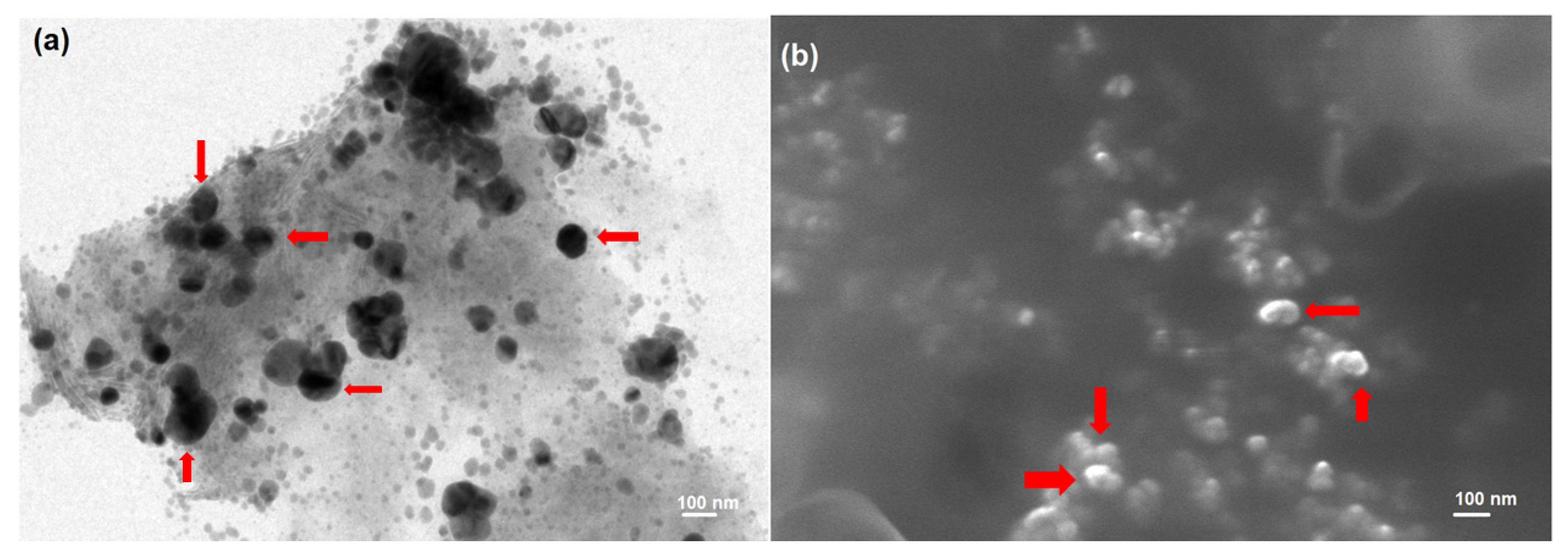
Abstract
:1. Introduction
2. Materials and Methods
2.1. Materials
2.2. Synthesis ALT-Loaded HA-NPs (ALT-HA-NPs)
2.3. Determination of the Encapsulation Efficiency % and Loading Capacity % of ALT-HA-NPs
2.4. Particle Size Analysis of ALT-HA-NPs
2.5. Determination of Surface Morphology of ALT-HA-NPs
2.5.1. Transmission Electron Microscopy (TEM)
2.5.2. Scanning Electron Microscopy (SEM)
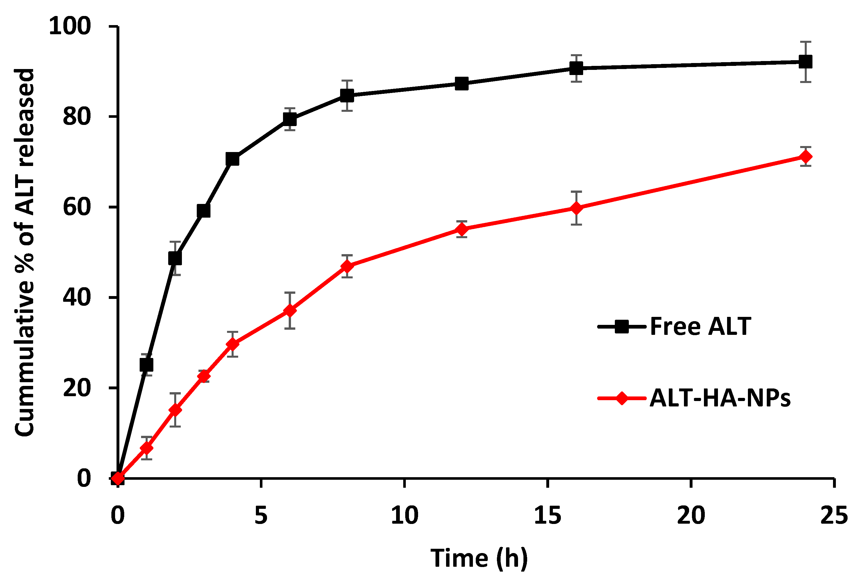
2.6. In Vitro Release Studies
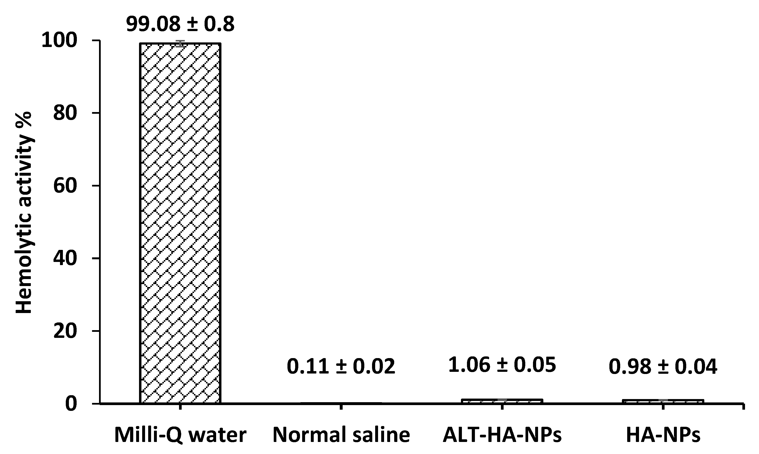
2.7. Hemolytic Activity Determination
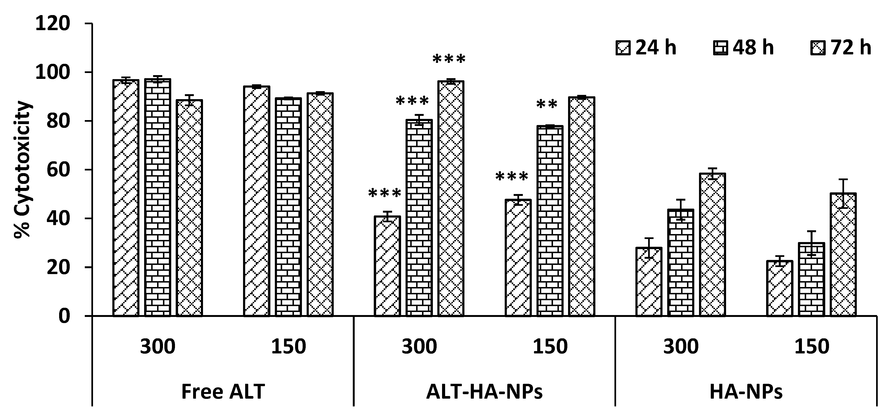
2.8. In Vitro Cell Line Studies
Cytotoxicity Assessment
2.9. In Vivo Studies
2.9.1. Animals
2.9.2. Pharmacokinetic Evaluation
2.9.3. Tumor Regression Evaluation
2.10. Statistical Analysis
3. Results and Discussion
3.1. In Vitro Release Outcomes
3.2. Hemolysis Outcomes
3.3. Cell Line Studies
MTT Assay
3.4. In Vivo Animal Model
3.4.1. Pharmacokinetic Parameters
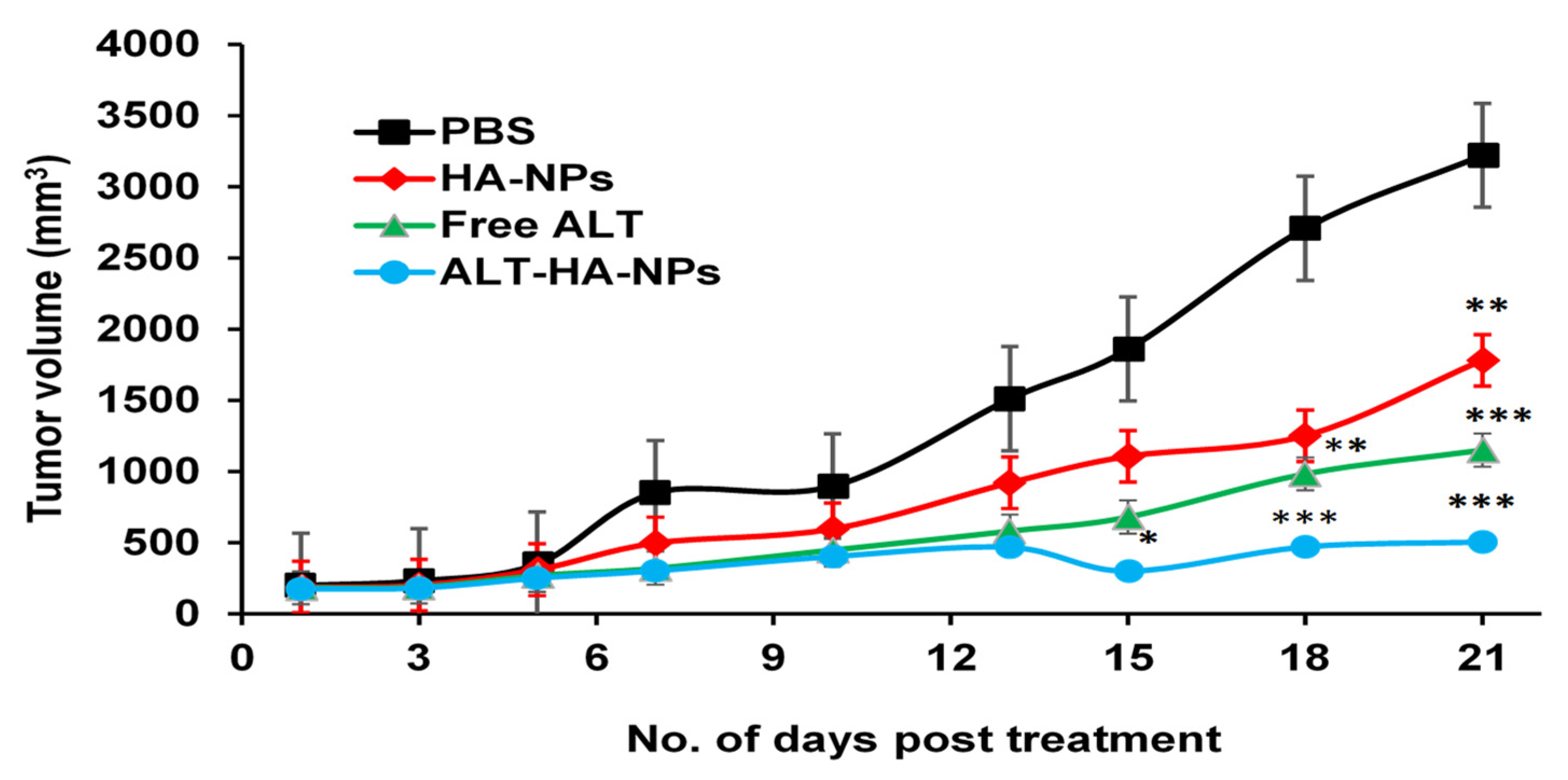
3.4.2. Tumor Regression Studies
4. Conclusions
Author Contributions
Funding
Institutional Review Board Statement
Informed Consent Statement
Data Availability Statement
Acknowledgments
Conflicts of Interest
References
- Hassan, M.; Watari, H.; AbuAlmaaty, A.; Ohba, Y.; Sakuragi, N. Apoptosis and Molecular Targeting Therapy in Cancer. BioMed Res. Int. 2014, 2014, 150845. [Google Scholar] [CrossRef] [PubMed] [Green Version]
- Kim, H.S.; Lim, J.M.; Kim, J.Y.; Kim, Y.; Park, S.; Sohn, J. Panaxydol, a Component of P Anax Ginseng, Induces Apoptosis in Cancer Cells through EGFR Activation and ER Stress and Inhibits Tumor Growth in Mouse Models. Int. J. Cancer 2016, 138, 1432–1441. [Google Scholar] [CrossRef] [Green Version]
- Nagoor Meeran, M.F.; Javed, H.; Al Taee, H.; Azimullah, S.; Ojha, S.K. Pharmacological Properties and Molecular Mechanisms of Thymol: Prospects for Its Therapeutic Potential and Pharmaceutical Development. Front. Pharmacol. 2017, 8, 380. [Google Scholar] [CrossRef] [PubMed] [Green Version]
- Levingstone, T.J.; Herbaj, S.; Dunne, N.J. Calcium Phosphate Nanoparticles for Therapeutic Applications in Bone Regeneration. Nanomaterials 2019, 9, 1570. [Google Scholar] [CrossRef] [Green Version]
- Kargozar, S.; Mollazadeh, S.; Kermani, F.; Webster, T.J.; Nazarnezhad, S.; Hamzehlou, S.; Baino, F. Hydroxyapatite Nanoparticles for Improved Cancer Theranostics. J. Funct. Biomater. 2022, 13, 100. [Google Scholar] [CrossRef] [PubMed]
- Ghate, P.; Prabhu, D.; Murugesan, G.; Goveas, L.C.; Varadavenkatesan, T.; Vinayagam, R.; Chi, N.T.L.; Pugazhendhi, A.; Selvaraj, R. Synthesis of Hydroxyapatite Nanoparticles Using Acacia Falcata Leaf Extract and Study of Their Anti-Cancerous Activity against Cancerous Mammalian Cell Lines. Environ. Res. 2022, 214, 113917. [Google Scholar] [CrossRef] [PubMed]
- Zhang, K.; Zhou, Y.; Xiao, C.; Zhao, W.; Wu, H.; Tang, J.; Li, Z.; Yu, S.; Li, X.; Min, L. Application of Hydroxyapatite Nanoparticles in Tumor-Associated Bone Segmental Defect. Sci. Adv. 2019, 5, eaax6946. [Google Scholar] [CrossRef] [Green Version]
- Tang, W.; Yuan, Y.; Liu, C.; Wu, Y.; Lu, X.; Qian, J. Differential Cytotoxicity and Particle Action of Hydroxyapatite Nanoparticles in Human Cancer Cells. Nanomedicine 2014, 9, 397–412. [Google Scholar] [CrossRef]
- Singh, R.K.; Kumar, S.; Prasad, D.N.; Bhardwaj, T.R. Therapeutic Journery of Nitrogen Mustard as Alkylating Anticancer Agents: Historic to Future Perspectives. Eur. J. Med. Chem. 2018, 151, 401–433. [Google Scholar] [CrossRef]
- Dabral, D.; Van den Bogaart, G. The Roles of Phospholipase A2 in Phagocytes. Front. Cell Dev. Biol. 2021, 9, 673502. [Google Scholar] [CrossRef]
- Mohammadinejad, R.; Moosavi, M.A.; Tavakol, S.; Vardar, D.Ö.; Hosseini, A.; Rahmati, M.; Dini, L.; Hussain, S.; Mandegary, A.; Klionsky, D.J. Necrotic, Apoptotic and Autophagic Cell Fates Triggered by Nanoparticles. Autophagy 2019, 15, 4–33. [Google Scholar] [CrossRef] [Green Version]
- Dmitry, B.; Juhaszova, M.; Sollot, S. Mitochondrial ROS-Induced ROS Release: An Update and Review. Physiol. Rev. 2014, 94, 909–950. [Google Scholar]
- Swulius, M.T.; Waxham, M.N. Ca2+/Calmodulin-Dependent Protein Kinases. Cell. Mol. Life Sci. 2008, 65, 2637–2657. [Google Scholar] [CrossRef] [PubMed] [Green Version]
- Ribeiro, T.P.; Monteiro, F.J.; Laranjeira, M.S. Duality of Iron (III) Doped Nano Hydroxyapatite in Triple Negative Breast Cancer Monitoring and as a Drug-Free Therapeutic Agent. Ceram. Int. 2020, 46, 16590–16597. [Google Scholar] [CrossRef]
- Yelten-Yilmaz, A.; Yilmaz, S. Wet Chemical Precipitation Synthesis of Hydroxyapatite (HA) Powders. Ceram. Int. 2018, 44, 9703–9710. [Google Scholar] [CrossRef]
- El-Bassyouni, G.T.; Eldera, S.S.; Kenawy, S.H.; Hamzawy, E.M. Hydroxyapatite Nanoparticles Derived from Mussel Shells for in Vitro Cytotoxicity Test and Cell Viability. Heliyon 2020, 6, e04085. [Google Scholar] [CrossRef] [PubMed]
- Pandey, S.; Rai, N.; Mahtab, A.; Mittal, D.; Ahmad, F.J.; Sandal, N.; Neupane, Y.R.; Verma, A.K.; Talegaonkar, S. Hyaluronate-Functionalized Hydroxyapatite Nanoparticles Laden with Methotrexate and Teriflunomide for the Treatment of Rheumatoid Arthritis. Int. J. Biol. Macromol. 2021, 171, 502–513. [Google Scholar] [CrossRef] [PubMed]
- AbouAitah, K.; Stefanek, A.; Higazy, I.M.; Janczewska, M.; Swiderska-Sroda, A.; Chodara, A.; Wojnarowicz, J.; Szałaj, U.; Shahein, S.A.; Aboul-Enein, A.M. Effective Targeting of Colon Cancer Cells with Piperine Natural Anticancer Prodrug Using Functionalized Clusters of Hydroxyapatite Nanoparticles. Pharmaceutics 2020, 12, 70. [Google Scholar] [CrossRef] [Green Version]
- Sun, Y.; Devore, D.; Ma, X.; Yuan, Y.; Kohn, J.; Qian, J. Promotion of Dispersion and Anticancer Efficacy of Hydroxyapatite Nanoparticles by the Adsorption of Fetal Bovine Serum. J. Nanoparticle Res. 2019, 21, 1–12. [Google Scholar] [CrossRef]
- Ansari, L.; Derakhshi, M.; Bagheri, E.; Shahtahmassebi, N.; Malaekeh-Nikouei, B. Folate Conjugation Improved Uptake and Targeting of Porous Hydroxyapatite Nanoparticles Containing Epirubicin to Cancer Cells. Pharm. Dev. Technol. 2020, 25, 601–609. [Google Scholar] [CrossRef] [PubMed]
- Yedekci, Y.; Gedik, E.; Evis, Z.; Dogan, L.; Özyigit, G.; Gürkaynak, M. Radiosensitization Induced by Zinc-Doped Hydroxyapatite Nanoparticles in Breast Cancer Cells. Int. J. Appl. Ceram. Technol. 2021, 18, 563–572. [Google Scholar] [CrossRef]
- Xu, Y.; Zhang, Z.; Wang, H.; Zhong, W.; Sun, C.; Sun, W.; Wu, H. Zoledronic Acid-Loaded Hybrid Hyaluronic Acid/Polyethylene Glycol/Nano-Hydroxyapatite Nanoparticle: Novel Fabrication and Safety Verification. Front. Bioeng. Biotechnol. 2021, 9, 629928. [Google Scholar] [CrossRef] [PubMed]
- Dong, X.; Sun, Y.; Li, Y.; Ma, X.; Zhang, S.; Yuan, Y.; Kohn, J.; Liu, C.; Qian, J. Synergistic Combination of Bioactive Hydroxyapatite Nanoparticles and the Chemotherapeutic Doxorubicin to Overcome Tumor Multidrug Resistance. Small 2021, 17, 2007672. [Google Scholar] [CrossRef]
- Yadav, K.; Yadav, D.; Kumar, S.; Narra, K.; El-Sherbiny, M.; Al-Serwi, R.H.; Othman, G.; Sendy, J.S.; Mohamed, J.M.M. Natural Biodegradable and Polymeric Nanoparticles for the Delivery of Noscapine for Cancer Treatment. Biomass Convers. Biorefin. 2022, 1–13. [Google Scholar] [CrossRef]
- Jamal Moideen, M.M.; Alqahtani, A.; Venkatesan, K.; Ahmad, F.; Krisharaju, K.; Gayasuddin, M.; Shaik, R.A.; Ibraheem, K.M.M.; Salama, M.E.M.; Abed, S.Y. Application of the Box–Behnken Design for the Production of Soluble Curcumin: Skimmed Milk Powder Inclusion Complex for Improving the Treatment of Colorectal Cancer. Food Sci. Nutr. 2020, 8, 6643–6659. [Google Scholar] [CrossRef] [PubMed]
- Mohamed, J.M.; Alqahtani, A.; Ahmad, F.; Krishnaraju, V.; Kalpana, K. Pectin Co-Functionalized Dual Layered Solid Lipid Nanoparticle Made by Soluble Curcumin for the Targeted Potential Treatment of Colorectal Cancer. Carbohydr. Polym. 2021, 252, 117180. [Google Scholar] [CrossRef] [PubMed]
- Mishra, S.; Tamta, A.K.; Sarikhani, M.; Desingu, P.A.; Kizkekra, S.M.; Pandit, A.S.; Kumar, S.; Khan, D.; Raghavan, S.C.; Sundaresan, N.R. Subcutaneous Ehrlich Ascites Carcinoma Mice Model for Studying Cancer-Induced Cardiomyopathy. Sci. Rep. 2018, 8, 5599. [Google Scholar] [CrossRef] [Green Version]
- Soleimani, M.; Elmi, F.; Anijdan, S.H.M.; Elmi, M.M. Evaluating the Radiosensitization Effect of Hydroxyapatite Nanoparticles on Human Breast Adenocarcinoma Cell Line and Fibroblast. Iran. J. Med. Sci. 2020, 45, 368. [Google Scholar]
- Cai, Y.; Gao, T.; Fu, S.; Sun, P. Development of Zoledronic Acid Functionalized Hydroxyapatite Loaded Polymeric Nanoparticles for the Treatment of Osteoporosis. Exp. Ther. Med. 2018, 16, 704–710. [Google Scholar] [CrossRef] [Green Version]
- Luo, H.; Zhang, Y.; Gan, D.; Yang, Z.; Ao, H.; Zhang, Q.; Yao, F.; Wan, Y. Incorporation of Hydroxyapatite into Nanofibrous PLGA Scaffold towards Improved Breast Cancer Cell Behavior. Mater. Chem. Phys. 2019, 226, 177–183. [Google Scholar] [CrossRef]
- Kumar, P.; Ganure, A.L.; Subudhi, B.B.; Shukla, S. Design and Comparative Evaluation of In-Vitro Drug Release, Pharmacokinetics and Gamma Scintigraphic Analysis of Controlled Release Tablets Using Novel PH Sensitive Starch and Modified Starch-Acrylate Graft Copolymer Matrices. Iran. J. Pharm. Res. IJPR 2015, 14, 677. [Google Scholar] [PubMed]
- Khan, S.; Ansari, A.A.; Rolfo, C.; Coelho, A.; Abdulla, M.; Al-Khayal, K.; Ahmad, R. Evaluation of in Vitro Cytotoxicity, Biocompatibility, and Changes in the Expression of Apoptosis Regulatory Proteins Induced by Cerium Oxide Nanocrystals. Sci. Technol. Adv. Mater. 2017, 18, 364–373. [Google Scholar] [CrossRef] [PubMed]
- Yang, V.; Arumugam, S.R.; Pan, J.; Rajeshkumar, T.; Sun, Y.; Liu, X. Metal Oxide Nanoparticles as Biomedical Materials. J. Colloid Interface Sci. 2020, 5, 27. [Google Scholar]
- Jones, C.A.; Hazlehurst, L.A. Role of Calcium Homeostasis in Modulating EMT in Cancer. Biomedicines 2021, 9, 1200. [Google Scholar] [CrossRef]
- Weber, G.F. Molecular Therapies of Cancer; Springer: Berlin/Heidelberg, Germany, 2015. [Google Scholar]
- Navarro-Ocón, A.; Blaya-Cánovas, J.L.; López-Tejada, A.; Blancas, I.; Sánchez-Martín, R.M.; Garrido, M.J.; Griñán-Lisón, C.; Calahorra, J.; Cara, F.E.; Ruiz-Cabello, F. Nanomedicine as a Promising Tool to Overcome Immune Escape in Breast Cancer. Pharmaceutics 2022, 14, 505. [Google Scholar] [CrossRef]


Disclaimer/Publisher’s Note: The statements, opinions and data contained in all publications are solely those of the individual author(s) and contributor(s) and not of MDPI and/or the editor(s). MDPI and/or the editor(s) disclaim responsibility for any injury to people or property resulting from any ideas, methods, instructions or products referred to in the content. |
© 2023 by the authors. Licensee MDPI, Basel, Switzerland. This article is an open access article distributed under the terms and conditions of the Creative Commons Attribution (CC BY) license (https://creativecommons.org/licenses/by/4.0/).
Share and Cite
Alghazwani, Y.; Venkatesan, K.; Prabahar, K.; El-Sherbiny, M.; Elsherbiny, N.; Qushawy, M. The Combined Anti-Tumor Efficacy of Bioactive Hydroxyapatite Nanoparticles Loaded with Altretamine. Pharmaceutics 2023, 15, 302. https://doi.org/10.3390/pharmaceutics15010302
Alghazwani Y, Venkatesan K, Prabahar K, El-Sherbiny M, Elsherbiny N, Qushawy M. The Combined Anti-Tumor Efficacy of Bioactive Hydroxyapatite Nanoparticles Loaded with Altretamine. Pharmaceutics. 2023; 15(1):302. https://doi.org/10.3390/pharmaceutics15010302
Chicago/Turabian StyleAlghazwani, Yahia, Krishnaraju Venkatesan, Kousalya Prabahar, Mohamed El-Sherbiny, Nehal Elsherbiny, and Mona Qushawy. 2023. "The Combined Anti-Tumor Efficacy of Bioactive Hydroxyapatite Nanoparticles Loaded with Altretamine" Pharmaceutics 15, no. 1: 302. https://doi.org/10.3390/pharmaceutics15010302