
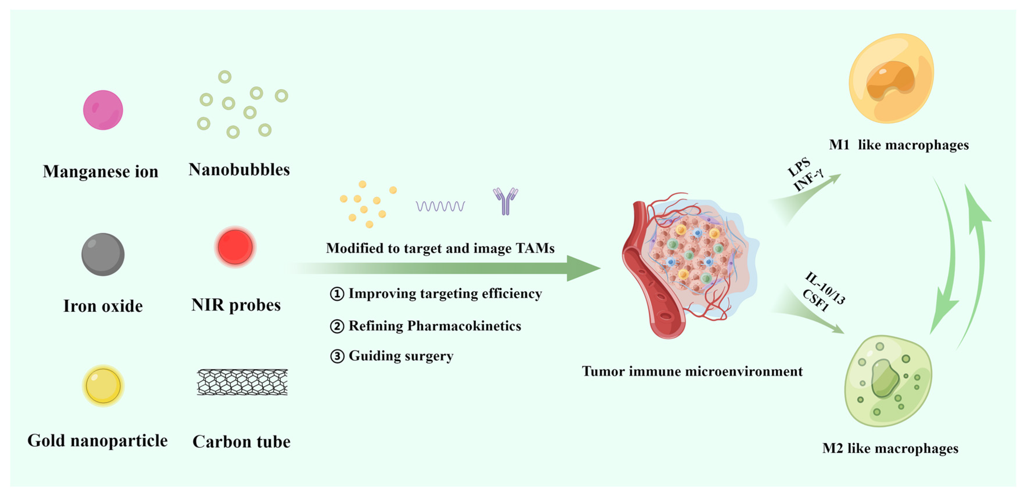
Targeting Tumor-Associated Macrophages for Imaging
Abstract
:1. Introduction
2. Nanoprobes for TAM Imaging and Therapy
2.1. Metal-Based Nanoprobes
2.1.1. Iron-Based Nanoprobes
2.1.2. Manganese-Based Nanoprobes
2.1.3. Gold-Based Nanoprobes
2.1.4. Silver-Based Nanoprobes
2.2. Fluorine-19-Based Nanoprobes
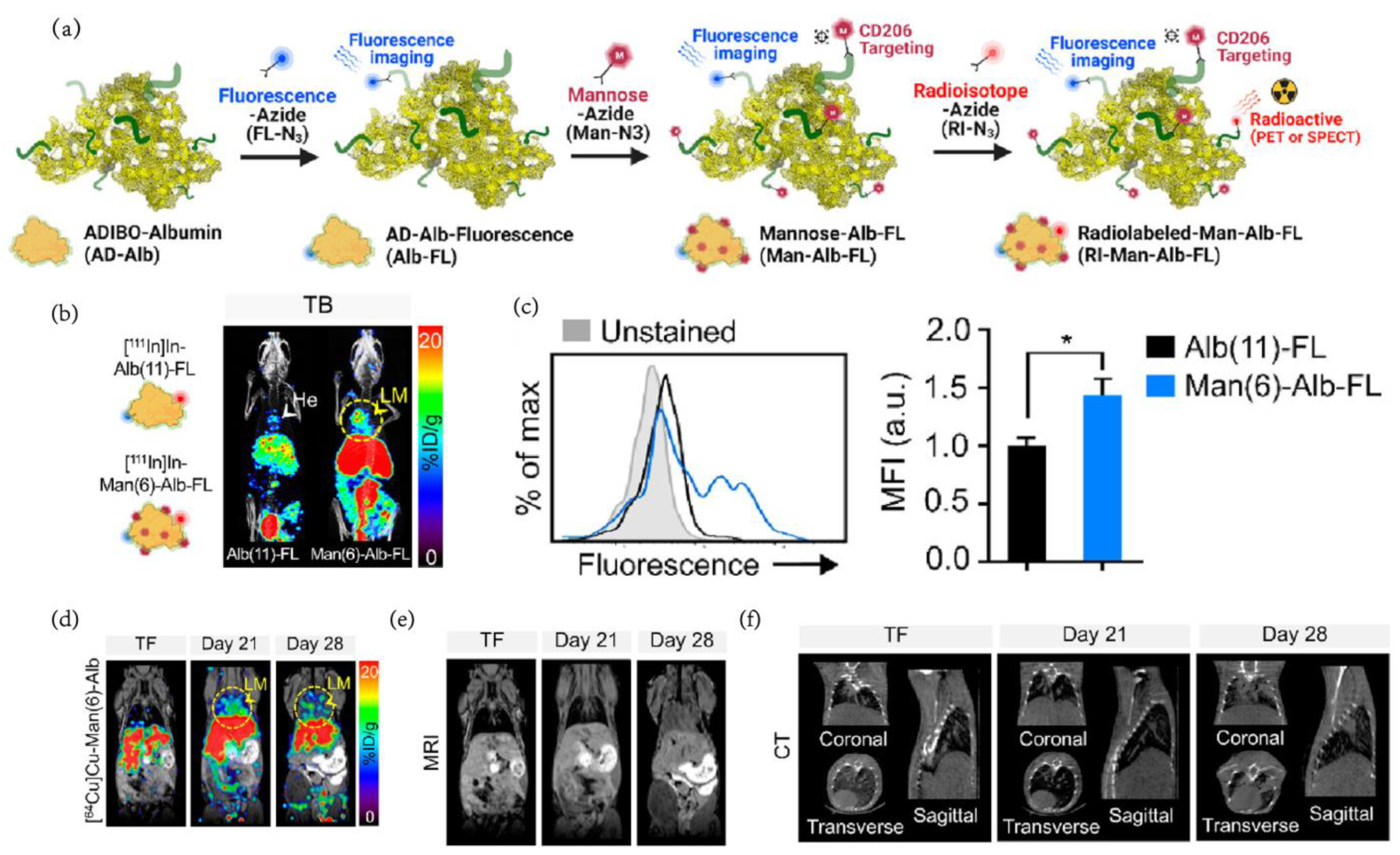
2.3. Radiolabeled Agents
2.4. Near Infrared Fluorescence Dye
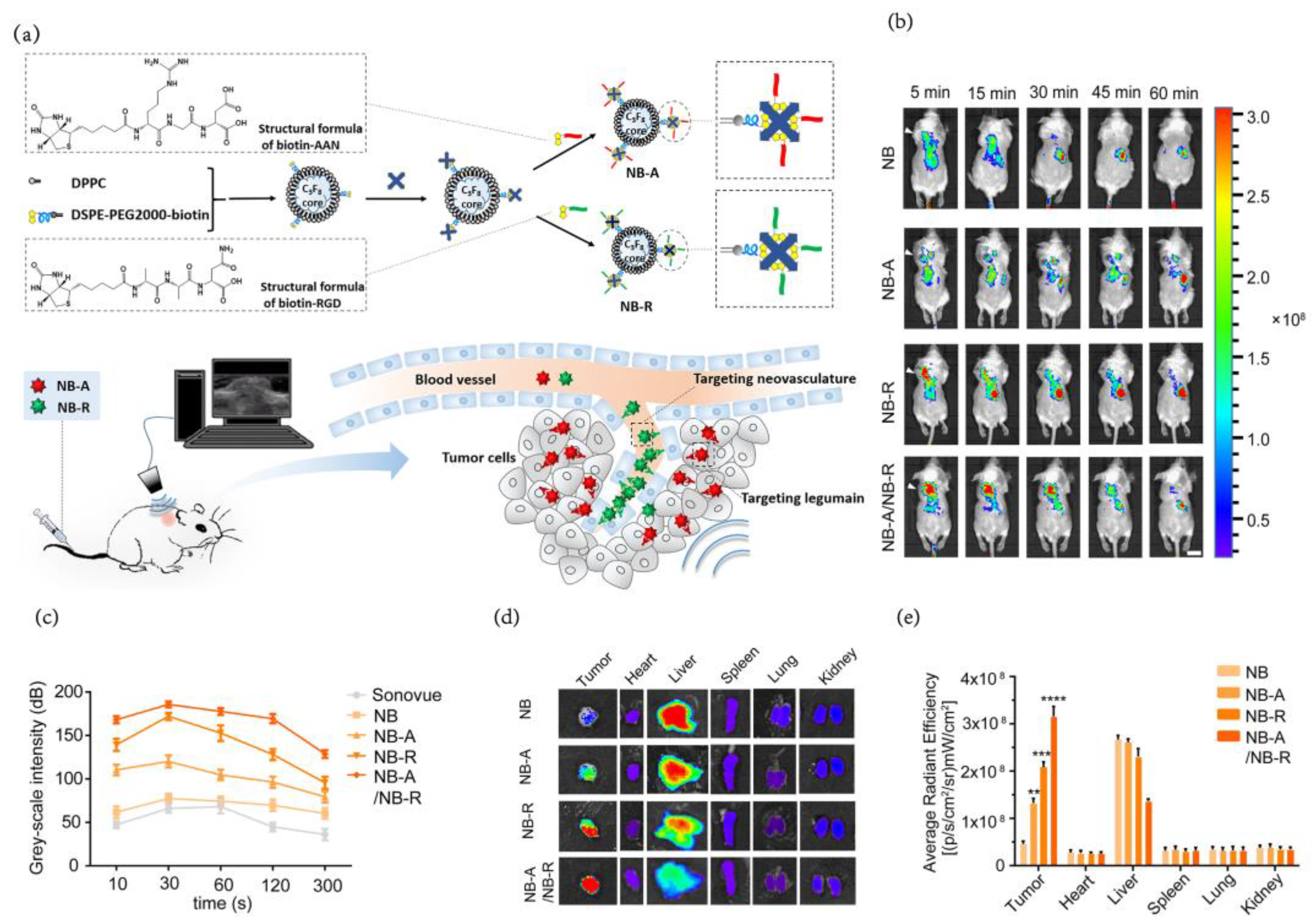
2.5. Ultrasonic Nanobubbles
2.6. Multimodal Imaging
3. Principlesof Designing TAMs Imaging Nanoprobes
3.1. Targeting Efficiency
3.2. Pharmacokinetics
3.3. Surgical Operation Guidance
4. Summary and Future Directions
Author Contributions
Funding
Institutional Review Board Statement
Informed Consent Statement
Data Availability Statement
Conflicts of Interest
References
- Deo, S.V.S.; Sharma, J.; Kumar, S. GLOBOCAN 2020 Report on Global Cancer Burden: Challenges and Opportunities for Surgical Oncologists. Ann. Surg. Oncol. 2022, 29, 6497–6500. [Google Scholar] [CrossRef] [PubMed]
- Geoghegan, R.; ter Haar, G.; Nightingale, K.; Marks, L.; Natarajan, S. Methods of monitoring thermal ablation of soft tissue tumors—A comprehensive review. Med. Phys. 2022, 49, 769–791. [Google Scholar] [CrossRef] [PubMed]
- Akakuru, O.U.; Zhang, Z.; Iqbal, M.Z.; Zhu, C.; Zhang, Y.; Wu, A. Chemotherapeutic nanomaterials in tumor boundary delineation: Prospects for effective tumor treatment. Acta Pharm. Sin. B 2022, 12, 2640–2657. [Google Scholar] [CrossRef] [PubMed]
- Burnet, N.G.; Thomas, S.J.; Burton, K.E.; Jefferies, S.J. Defining the tumour and target volumes for radiotherapy. Cancer Imaging Off. Publ. Int. Cancer Imaging Soc. 2004, 4, 153–161. [Google Scholar] [CrossRef] [PubMed] [Green Version]
- Tohme, S.; Simmons, R.L.; Tsung, A. Surgery for Cancer: A Trigger for Metastases. Cancer Res. 2017, 77, 1548–1552. [Google Scholar] [CrossRef] [PubMed] [Green Version]
- Binnewies, M.; Roberts, E.W.; Kersten, K.; Chan, V.; Fearon, D.F.; Merad, M.; Coussens, L.M.; Gabrilovich, D.I.; Ostrand-Rosenberg, S.; Hedrick, C.C.; et al. Understanding the tumor immune microenvironment (TIME) for effective therapy. Nat. Med. 2018, 24, 541–550. [Google Scholar] [CrossRef] [PubMed]
- Anderson, N.M.; Simon, M.C. The tumor microenvironment. Curr. Biol. 2020, 30, R921–R925. [Google Scholar] [CrossRef]
- Balta, E.; Wabnitz, G.H.; Samstag, Y. Hijacked Immune Cells in the Tumor Microenvironment: Molecular Mechanisms of Immunosuppression and Cues to Improve T Cell-Based Immunotherapy of Solid Tumors. Int. J. Mol. Sci. 2021, 22, 5736. [Google Scholar] [CrossRef]
- Chen, Y.; Song, Y.; Du, W.; Gong, L.; Chang, H.; Zou, Z. Tumor-associated macrophages: An accomplice in solid tumor progression. J. Biomed. Sci. 2019, 26, 78. [Google Scholar] [CrossRef] [Green Version]
- Yan, S.; Wan, G. Tumor-associated macrophages in immunotherapy. FEBS J. 2021, 288, 6174–6186. [Google Scholar] [CrossRef]
- Wang, Y.; Lyu, Z.; Qin, Y.; Wang, X.; Sun, L.; Zhang, Y.; Gong, L.; Wu, S.; Han, S.; Tang, Y.; et al. FOXO1 promotes tumor progression by increased M2 macrophage infiltration in esophageal squamous cell carcinoma. Theranostics 2020, 10, 11535–11548. [Google Scholar] [CrossRef]
- Quail, D.F.; Joyce, J.A. Microenvironmental regulation of tumor progression and metastasis. Nat. Med. 2013, 19, 1423–1437. [Google Scholar] [CrossRef]
- Qi, J.; Sun, H.; Zhang, Y.; Wang, Z.; Xun, Z.; Li, Z.; Ding, X.; Bao, R.; Hong, L.; Jia, W. Single-cell and spatial analysis reveal interaction of FAP+ fibroblasts and SPP1+ macrophages in colorectal cancer. Nat. Commun. 2022, 13, 1742. [Google Scholar] [CrossRef]
- Casanova-Acebes, M.; Dalla, E.; Leader, A.M.; LeBerichel, J.; Nikolic, J.; Morales, B.M.; Brown, M.; Chang, C.; Troncoso, L.; Chen, S.T. Tissue-resident macrophages provide a pro-tumorigenic niche to early NSCLC cells. Nature 2021, 595, 578–584. [Google Scholar] [CrossRef]
- Wei, X.; Wang, J.; Liang, M.; Song, M. Development of functional nanomedicines for tumor associated macrophages-focused cancer immunotherapy. Theranostics 2022, 12, 7821–7852. [Google Scholar] [CrossRef]
- Li, X.; Wang, R.; Zhang, Y.; Han, S.; Gan, Y.; Liang, Q.; Ma, X.; Rong, P.; Wang, W.; Li, W. Molecular imaging of tumor-associated macrophages in cancer immunotherapy. Ther. Adv. Med. Oncol. 2022, 14, 17588359221076194. [Google Scholar] [CrossRef]
- Yang, R.; Hamilton, A.M.; Sun, H.; Rawji, K.S.; Sarkar, S.; Mirzaei, R.; Pike, G.B.; Yong, V.W.; Dunn, J.F. Detecting monocyte trafficking in an animal model of glioblastoma using R-2* and quantitative susceptibility mapping. Cancer Immunol. Immunother. Online ahead of print. 2022. [Google Scholar] [CrossRef]
- Li, Y.; Wu, H.; Ji, B.; Qian, W.; Xia, S.; Wang, L.; Xu, Y.; Chen, J.; Yang, L.; Mao, H. Targeted Imaging of CD206 Expressing Tumor-Associated M2-like Macrophages Using Mannose-Conjugated Antibiofouling Magnetic Iron Oxide Nanoparticles. ACS Appl. Bio Mater. 2020, 3, 4335–4347. [Google Scholar] [CrossRef]
- Wang, W.; Li, F.; Li, S.; Hu, Y.; Xu, M.; Zhang, Y.; Khan, M.I.; Wang, S.; Wu, M.; Ding, W.; et al. M2 macrophage-targeted iron oxide nanoparticles for magnetic resonance image-guided magnetic hyperthermia therapy. J. Mater. Sci. Technol. 2021, 81, 77–87. [Google Scholar] [CrossRef]
- Makela, A.V.; Gaudet, J.M.; Schott, M.A.; Sehl, O.C.; Contag, C.H.; Foster, P.J. Magnetic Particle Imaging of Macrophages Associated with Cancer: Filling the Voids Left by Iron-Based Magnetic Resonance Imaging. Mol. Imaging Biol. 2020, 22, 958–968. [Google Scholar] [CrossRef]
- Zhou, Z.; Xu, B.; Hu, N.; Guo, Z.; Bao, W.; Shao, B.; Yang, W. Targeting the Macrophage-Ferroptosis Crosstalk: A Novel Insight into Tumor Immunotherapy. Front. Biosci.-Landmark 2022, 27, 203. [Google Scholar] [CrossRef] [PubMed]
- Chen, G.; Cai, Y.; Li, B.; Lin, M.; Wang, X.; Wang, Z.; Shuai, X. Theranostic nanosystem mediating cascade catalytic reactions for effective immunotherapy of highly immunosuppressive and poorly penetrable pancreatic tumor. Sci. China-Chem. 2022, 65, 1383–1400. [Google Scholar] [CrossRef]
- Zhou, J.; Meli, V.S.; Yu-Tin Chen, E.; Kapre, R.; Nagalla, R.; Xiao, W.; Borowsky, A.D.; Lam, K.S.; Liu, W.F.; Louie, A.Y. Magnetic resonance imaging of tumor-associated-macrophages (TAMs) with a nanoparticle contrast agent. RSC Adv. 2022, 12, 7742–7756. [Google Scholar] [CrossRef] [PubMed]
- Cai, X.; Zhu, Q.; Zeng, Y.; Zeng, Q.; Chen, X.; Zhan, Y. Manganese Oxide Nanoparticles as MRI Contrast Agents In Tumor Multimodal Imaging And Therapy. Int. J. Nanomed. 2019, 14, 8321–8344. [Google Scholar] [CrossRef] [PubMed] [Green Version]
- Hsu, B.Y.W.; Kirby, G.; Tan, A.; Seifalian, A.M.; Li, X.; Wang, J. Relaxivity and toxicological properties of manganese oxide nanoparticles for MRI applications. RSC Adv. 2016, 6, 45462–45474. [Google Scholar] [CrossRef] [Green Version]
- Luo, L.-J.; Liu, X.-M.; Zhang, X.; Liu, J.; Gao, Y.; Sun, T.-Y.; Li, L.-L. Quantitative Detection of In Vivo Aggregation Degree for Enhanced M2 Macrophage MR Imaging. Nano Lett. 2022, 22, 1694–1702. [Google Scholar] [CrossRef]
- Bonet-Aleta, J.; Calzada-Funes, J.; Hueso, J.L. Manganese oxide nano-platforms in cancer therapy: Recent advances on the development of synergistic strategies targeting the tumor microenvironment. Appl. Mater. Today 2022, 29, 101628. [Google Scholar] [CrossRef]
- Song, M.; Liu, T.; Shi, C.; Zhang, X.; Chen, X. Bioconjugated Manganese Dioxide Nanoparticles Enhance Chemotherapy Response by Priming Tumor-Associated Macrophages toward M1-like Phenotype and Attenuating Tumor Hypoxia. ACS Nano 2016, 10, 633–647. [Google Scholar] [CrossRef] [Green Version]
- Lim, J.-W.; Son, H.Y.; Huh, Y.-M.; Haam, S. Cationic poly(amino acid) surface functionalized manganese nanoparticles for nitric oxide-based immunotherapy and magnetic resonance imaging. J. Mater. Chem. B 2022, 10, 5402–5409. [Google Scholar] [CrossRef]
- Song, R.; Zhang, M.; Liu, Y.; Cui, Z.; Zhang, H.; Tang, Z.; Chen, X.; Wu, H.; Yao, Z.; He, M.; et al. A multifunctional nanotheranostic for the intelligent MRI diagnosis and synergistic treatment of hypoxic tumor. Biomaterials 2018, 175, 123–133. [Google Scholar] [CrossRef]
- Luo, D.; Wang, X.; Burda, C.; Basilion, J.P. Recent Development of Gold Nanoparticles as Contrast Agents for Cancer Diagnosis. Cancers 2021, 13, 1825. [Google Scholar] [CrossRef]
- Lv, W.; Xu, C.; Wu, H.; Zhu, Y.; Akakuru, O.U.; Du, H.; Nie, F.; Wu, A.; Li, J. Ultrasound-visualized nanocarriers with siRNA for targeted inhibition of M2-like TAM polarization to enhance photothermal therapy in NSCLC. Nano Res. 2022, 1–12. [Google Scholar] [CrossRef]
- Sun, I.-C.; Jo, S.; Dumani, D.; Yun, W.S.; Yoon, H.Y.; Lim, D.-K.; Ahn, C.-H.; Emelianov, S.; Kim, K. Theragnostic Glycol Chitosan-Conjugated Gold Nanoparticles for Photoacoustic Imaging of Regional Lymph Nodes and Delivering Tumor Antigen to Lymph Nodes. Nanomaterials 2021, 11, 1700. [Google Scholar] [CrossRef]
- Gomes, H.I.O.; Martins, C.S.M.; Prior, J.A.V. Silver Nanoparticles as Carriers of Anticancer Drugs for Efficient Target Treatment of Cancer Cells. Nanomaterials 2021, 11, 964. [Google Scholar] [CrossRef]
- Yan, X.; Qi, Y.; Ren, L.; Ma, J.; Xu, M.; Xia, T.; Liu, S. Silver nanoclusters show advantages in macrophage tracing in vivo and modulation of anti-tumor immuno-microenvironment. J. Control. Release 2022, 348, 470–482. [Google Scholar] [CrossRef]
- Pal, R.; Chakraborty, B.; Nath, A.; Singh, L.M.; Ali, M.; Rahman, D.S.; Ghosh, S.K.; Basu, A.; Bhattacharya, S.; Baral, R.; et al. Noble metal nanoparticle-induced oxidative stress modulates tumor associated macrophages (TAMs) from an M2 to M1 phenotype: An in vitro approach. Int. Immunopharmacol. 2016, 38, 332–341. [Google Scholar] [CrossRef]
- Floegel, U.; Ding, Z.; Hardung, H.; Jander, S.; Reichmann, G.; Jacoby, C.; Schubert, R.; Schrader, J. In vivo monitoring of inflammation after cardiac and cerebral ischemia by fluorine magnetic resonance imaging. Circulation 2008, 118, 140–148. [Google Scholar] [CrossRef] [Green Version]
- Makela, A.V.; Gaudet, J.M.; Foster, P.J. Quantifying tumor associated macrophages in breast cancer: A comparison of iron and fluorinebased MRI cell tracking. Sci. Rep. 2017, 7, 42109. [Google Scholar] [CrossRef] [Green Version]
- Zambito, G.; Deng, S.; Haeck, J.; Gaspar, N.; Himmelreich, U.; Censi, R.; Lowik, C.; Di Martino, P.; Mezzanotte, L. Fluorinated PLGA-PEG-Mannose Nanoparticles for Tumor-Associated Macrophage Detection by Optical Imaging and MRI. Front. Med. 2021, 8, 1374. [Google Scholar] [CrossRef]
- Ramamonjisoa, N.; Ackerstaff, E. Characterization of the Tumor Microenvironment and Tumor-Stroma interaction by Non-invasive Preclinical imaging. Front. Oncol. 2017, 7, 3. [Google Scholar] [CrossRef]
- Wang, M.; Zhao, J.; Zhang, L.; Wei, F.; Lian, Y.; Wu, Y.; Gong, Z.; Zhang, S.; Zhou, J.; Cao, K.; et al. Role of tumor microenvironment in tumorigenesis. J. Cancer 2017, 8, 761–773. [Google Scholar] [CrossRef] [PubMed] [Green Version]
- Croci, D.; Santalla Mendez, R.; Temme, S.; Soukup, K.; Fournier, N.; Zomer, A.; Colotti, R.; Wischnewski, V.; Flogel, U.; van Heeswijk, R.B.; et al. Multispectral fluorine-19 MRI enables longitudinal and noninvasive monitoring of tumor-associated macrophages. Sci. Transl. Med. 2022, 14, eabo2952. [Google Scholar] [CrossRef] [PubMed]
- Ametamey, S.M. Molecular imaging with PET. Chem. Rev. 2008, 108, 4036. [Google Scholar] [CrossRef] [Green Version]
- Tarkin, J.M.; Joshi, F.R.; Rudd, J.H.F. PET imaging of inflammation in atherosclerosis. Nat. Rev. Cardiol. 2014, 11, 443–457. [Google Scholar] [CrossRef] [PubMed]
- Leuzy, A.; Chiotis, K.; Lemoine, L.; Gillberg, P.-G.; Almkvist, O.; Rodriguez-Vieitez, E.; Nordberg, A. Tau PET imaging in neurodegenerative tauopathies-still a challenge. Mol. Psychiatry 2019, 24, 1112–1134. [Google Scholar] [CrossRef] [Green Version]
- Lee, H.J.; Ehlerding, E.B.; Cai, W. Antibody-Based Tracers for PET/SPECT Imaging of Chronic Inflammatory Diseases. Chembiochem 2019, 20, 422–436. [Google Scholar] [CrossRef] [Green Version]
- Mukherjee, S.; Sonanini, D.; Maurer, A.; Daldrup-Link, H.E. The yin and yang of imaging tumor associated macrophages with PET and MRI. Theranostics 2019, 9, 7730–7748. [Google Scholar] [CrossRef]
- Fernandes, B.; Feltes, P.K.; Luft, C.; Nazario, L.R.; Jeckel, C.M.M.; Antunes, I.F.; Elsinga, P.H.; de Vries, E.F.J. Potential PET tracers for imaging of tumor-associated macrophages. Ejnmmi Radiopharm. Chem. 2022, 7, 11. [Google Scholar] [CrossRef]
- Chung, H.; Park, J.Y.; Kim, K.; Yoo, R.J.; Suh, M.; Gu, G.J.; Kim, J.S.; Choi, T.H.; Byun, J.W.; Ju, Y.W.; et al. Circulation Time-Optimized Albumin Nanoplatform for Quantitative Visualization of Lung Metastasis via Targeting of Macrophages. ACS Nano 2022, 16, 12262–12275. [Google Scholar] [CrossRef]
- Shi, D.; Si, Z.; Xu, Z.; Cheng, Y.; Lin, Q.; Fu, Z.; Fu, W.; Yang, T.; Shi, H.; Cheng, D. Synthesis and Evaluation of Ga-68-NOTA-COG1410 Targeting to TREM2 of TAMs as a Specific PET Probe for Digestive Tumor Diagnosis. Anal. Chem. 2022, 94, 3819–3830. [Google Scholar] [CrossRef]
- Kim, H.-Y.; Li, R.; Ng, T.S.C.; Courties, G.; Rodell, C.B.; Prytyskach, M.; Kohler, R.H.; Pittet, M.J.; Nahrendorf, M.; Weissleder, R.; et al. Quantitative Imaging of Tumor-Associated Macrophages and Their Response to Therapy Using Cu-64-Labeled Macrin. ACS Nano 2018, 12, 12015–12029. [Google Scholar] [CrossRef]
- Brandt, M.; Cardinale, J.; Aulsebrook, M.L.; Gasser, G.; Mindt, T.L. An Overview of PET Radiochemistry, Part 2: Radiometals. J. Nucl. Med. 2018, 59, 1500–1506. [Google Scholar] [CrossRef] [Green Version]
- Aluicio-Sarduy, E.; Ellison, P.A.; Barnhart, T.E.; Cai, W.; Nickles, R.J.; Engle, J.W. PET radiometals for antibody labeling. J. Label. Compd. Radiopharm. 2018, 61, 636–651. [Google Scholar] [CrossRef]
- Zhu, H.; Ren, F.; Wang, T.; Jiang, Z.; Sun, Q.; Li, Z. Targeted Immunoimaging of Tumor-Associated Macrophages in Orthotopic Glioblastoma by the NIR-IIb Nanoprobes. Small 2022, 18, 2202201. [Google Scholar] [CrossRef]
- Jiang, C.; Cai, H.; Peng, X.; Zhang, P.; Wu, X.; Tian, R. Targeted Imaging of Tumor-Associated Macrophages by Cyanine 7-Labeled Mannose in Xenograft Tumors. Mol. Imaging 2017, 16, 1–10. [Google Scholar] [CrossRef] [Green Version]
- Barth, N.D.; Van Dalen, F.J.; Karmakar, U.; Bertolini, M.; Mendive-Tapia, L.; Kitamura, T.; Verdoes, M.; Vendrell, M. Enzyme-Activatable Chemokine Conjugates for In Vivo Targeting of Tumor-Associated Macrophages. Angew. Chem.-Int. Ed. 2022, 61, e202207508. [Google Scholar] [CrossRef]
- Xu, Y.; Zeng, Y.; Xiao, X.; Liu, H.; Zhou, B.; Luo, B.; Er Saw, P.; Jiang, Q. Targeted Imaging of Tumor Associated Macrophages in Breast Cancer. BIO Integr. 2022, 3, 20220010. [Google Scholar] [CrossRef]
- Wan, D.-H.; Ma, X.-Y.; Lin, C.; Zhu, D.-H.; Li, X.; Zheng, B.-Y.; Li, J.; Ke, M.-R.; Huang, J.-D. Noncovalent Indocyanine Green Conjugate of C-Phycocyanin: Preparation and Tumor-Associated Macrophages-Targeted Photothermal Therapeutics. Bioconjugate Chem. 2020, 31, 1438–1448. [Google Scholar] [CrossRef]
- Ramesh, A.; Kumar, S.; Brouillard, A.; Nandi, D.; Kulkarni, A. A Nitric Oxide (NO) Nanoreporter for Noninvasive Real-Time Imaging of Macrophage Immunotherapy. Adv. Mater. 2020, 32, 2000648. [Google Scholar] [CrossRef]
- Onoda, A.; Umezawa, M. Carbon Nanotubes—Potential of Use for Deep Bioimaging. In Transparency in Biology; Springer: Singapore, 2021; pp. 85–107. [Google Scholar] [CrossRef]
- Iizumi, Y.; Yudasaka, M.; Kim, J.; Sakakita, H.; Takeuchi, T.; Okazaki, T. Oxygen-doped carbon nanotubes for near-infrared fluorescent labels and imaging probes. Sci. Rep. 2018, 8, 6272. [Google Scholar] [CrossRef] [Green Version]
- O’Connell, M.J.; Bachilo, S.M.; Huffman, C.B.; Moore, V.C.; Strano, M.S.; Haroz, E.H.; Rialon, K.L.; Boul, P.J.; Noon, W.H.; Kittrell, C.; et al. Band gap fluorescence from individual single-walled carbon nanotubes. Science 2002, 297, 593–596. [Google Scholar] [CrossRef] [PubMed] [Green Version]
- Wang, F.; Dukovic, G.; Brus, L.E.; Heinz, T.F. The optical resonances in carbon nanotubes arise from excitons. Science 2005, 308, 838–841. [Google Scholar] [CrossRef] [PubMed] [Green Version]
- Morimoto, T.; Joung, S.-K.; Saito, T.; Futaba, D.N.; Hata, K.; Okazaki, T. Length-Dependent Plasmon Resonance in Single-Walled Carbon Nanotubes. ACS Nano 2014, 8, 9897–9904. [Google Scholar] [CrossRef]
- Iverson, N.M.; Barone, P.W.; Shandell, M.; Trudel, L.J.; Sen, S.; Sen, F.; Ivanov, V.; Atolia, E.; Farias, E.; McNicholas, T.P.; et al. In vivo biosensing via tissue-localizable near-infrared-fluorescent single-walled carbon nanotubes. Nat. Nanotechnol. 2013, 8, 873–880. [Google Scholar] [CrossRef] [PubMed] [Green Version]
- Ghosh, D.; Bagley, A.F.; Na, Y.J.; Birrer, M.J.; Bhatia, S.N.; Belcher, A.M. Deep, noninvasive imaging and surgical guidance of submillimeter tumors using targeted M13-stabilized single-walled carbon nanotubes. Proc. Natl. Acad. Sci. USA 2014, 111, 13948–13953. [Google Scholar] [CrossRef] [Green Version]
- Ceppi, L.; Bardhan, N.M.; Na, Y.; Siegel, A.; Rajan, N.; Fruscio, R.; Del Carmen, M.G.; Belcher, A.M.; Birrer, M.J. Real-Time Single-Walled Carbon Nanotube-Based Fluorescence Imaging Improves Survival after Debulking Surgery in an Ovarian Cancer Model. ACS Nano 2019, 13, 5356–5365. [Google Scholar] [CrossRef]
- Liu, Z.; Cai, W.; He, L.; Nakayama, N.; Chen, K.; Sun, X.; Chen, X.; Dai, H. In vivo biodistribution and highly efficient tumour targeting of carbon nanotubes in mice. Nat. Nanotechnol. 2007, 2, 47–52. [Google Scholar] [CrossRef]
- Smith, B.R.; Ghosn, E.E.B.; Rallapalli, H.; Prescher, J.A.; Larson, T.; Herzenberg, L.A.; Gambhir, S.S. Selective uptake of single-walled carbon nanotubes by circulating monocytes for enhanced tumour delivery. Nat. Nanotechnol. 2014, 9, 481–487. [Google Scholar] [CrossRef] [Green Version]
- VanHandel, M.; Alizadeh, D.; Zhang, L.; Kateb, B.; Bronikowski, M.; Manohara, H.; Badie, B. Selective uptake of multi-walled carbon nanotubes by tumor macrophages in a murine glioma model. J. Neuroimmunol. 2009, 208, 3–9. [Google Scholar] [CrossRef]
- Kateb, B.; Van Handel, M.; Zhang, L.; Bronikowski, M.J.; Manohara, H.; Badie, B. Internalization of MWCNTs by microglia: Possible application in immunotherapy of brain tumors. Neuroimage 2007, 37, S9–S17. [Google Scholar] [CrossRef]
- Zhang, M.; Yang, M.; Morimoto, T.; Tajima, N.; Ichiraku, K.; Fujita, K.; Iijima, S.; Yudasaka, M.; Okazaki, T. Size-dependent cell uptake of carbon nanotubes by macrophages: A comparative and quantitative study. Carbon 2018, 127, 93–101. [Google Scholar] [CrossRef]
- Turabekova, M.; Rasulev, B.; Theodore, M.; Jackman, J.; Leszczynska, D.; Leszczynski, J. Immunotoxicity of nanoparticles: A computational study suggests that CNTs and C-60 fullerenes might be recognized as pathogens by Toll-like receptors. Nanoscale 2014, 6, 3488–3495. [Google Scholar] [CrossRef]
- Mukherjee, S.P.; Bondarenko, O.; Kohonen, P.; Andon, F.T.; Brzicova, T.; Gessner, I.; Mathur, S.; Bottini, M.; Calligari, P.; Stella, L.; et al. Macrophage sensing of single-walled carbon nanotubes via Toll-like receptors. Sci. Rep. 2018, 8, 1115. [Google Scholar] [CrossRef] [Green Version]
- Zhao, D.; Alizadeh, D.; Zhang, L.; Liu, W.; Farrukh, O.; Manuel, E.; Diamond, D.J.; Badie, B. Carbon Nanotubes Enhance CpG Uptake and Potentiate Antiglioma Immunity. Clin. Cancer Res. 2011, 17, 771–782. [Google Scholar] [CrossRef] [Green Version]
- Zanganeh, S.; Li, H.; Kumavor, P.D.; Alqasemi, U.; Aguirre, A.; Mohammad, I.; Stanford, C.; Smith, M.B.; Zhu, Q. Single wall carbon nanotube/bis carboxylic acid-ICG as a sensitive contrast agent for in vivo tumor imaging in photoacoustic tomography. In Proceedings of the Conference on Photons Plus Ultrasound-Imaging and Sensing, San Francisco, CA, USA, 2–7 February 2013; pp. 826–832. [Google Scholar] [CrossRef]
- Gao, C.; Jian, J.; Luo, L.; Liang, J.; Li, Z.; Pang, M.; Cai, H.; Shen, X.C. Single-walled carbon nanohorns-based smart nanotheranostic: From phototherapy to enzyme-activated fluorescence imaging-guided photodynamic therapy. J. Colloid Interface Sci. 2022, 628, 273–286. [Google Scholar] [CrossRef]
- Lu, G.-H.; Shang, W.-T.; Deng, H.; Han, Z.-Y.; Hu, M.; Liang, X.-Y.; Fang, C.-H.; Zhu, X.-H.; Fan, Y.-F.; Tian, J. Targeting carbon nanotubes based on IGF-1R for photothermal therapy of orthotopic pancreatic cancer guided by optical imaging. Biomaterials 2019, 195, 13–22. [Google Scholar] [CrossRef]
- Duan, L.; Yang, L.; Jin, J.; Yang, F.; Liu, D.; Hu, K.; Wang, Q.; Yue, Y.; Gu, N. Micro/nano-bubble-assisted ultrasound to enhance the EPR effect and potential theranostic applications. Theranostics 2020, 10, 462–483. [Google Scholar] [CrossRef]
- Wu, R.; Yang, X.; Li, X.e.; Dong, N.; Liu, Y.; Zhang, P. Nanobubbles for tumors: Imaging and drug carriers. J. Drug Deliv. Sci. Technol. 2021, 65, 102749. [Google Scholar] [CrossRef]
- Jiang, Q.; Zeng, Y.; Xu, Y.; Xiao, X.; Liu, H.; Zhou, B.; Kong, Y.; Saw, P.E.; Luo, B. Ultrasound Molecular Imaging as a Potential Non-invasive Diagnosis to Detect the Margin of Hepatocarcinoma via CSF-1R Targeting. Front. Bioeng. Biotechnol. 2020, 8, 783. [Google Scholar] [CrossRef]
- Sun, X.; Guo, L.; Shang, M.; Shi, D.; Liang, P.; Jing, X.; Meng, D.; Liu, X.; Zhou, X.; Zhao, Y.; et al. Ultrasound Mediated Destruction of LMW-HA-Loaded and Folate-Conjugated Nanobubble for TAM Targeting and Reeducation. Int. J. Nanomed. 2020, 15, 1967–1981. [Google Scholar] [CrossRef] [Green Version]
- Mi, X.; Guo, X.; Du, H.; Han, M.; Liu, H.; Luo, Y.; Wang, D.; Xiang, R.; Yue, S.; Zhang, Y.; et al. Combined legumain- and integrin-targeted nanobubbles for molecular ultrasound imaging of breast cancer. Nanomed. -Nanotechnol. Biol. Med. 2022, 42, 102533. [Google Scholar] [CrossRef] [PubMed]
- Moseley, M.; Donnan, G. Multimodality imaging—Introduction. Stroke 2004, 35, 2632–2634. [Google Scholar] [CrossRef] [Green Version]
- Marti-Bonmati, L.; Sopena, R.; Bartumeus, P.; Sopena, P. Multimodality imaging techniques. Contrast Media Mol. Imaging 2010, 5, 180–189. [Google Scholar] [CrossRef] [PubMed]
- Choi, Y.J.; Oh, S.-G.; Singh, T.D.; Ha, J.-H.; Kim, D.W.; Lee, S.W.; Jeong, S.Y.; Ahn, B.-C.; Lee, J.; Jeon, Y.H. Visualization of the Biological Behavior of Tumor-Associated Macrophages in Living Mice with Colon Cancer Using Multimodal Optical Reporter Gene Imaging. Neoplasia 2016, 18, 133–141. [Google Scholar] [CrossRef] [PubMed] [Green Version]
- Bohn, T.; Rapp, S.; Luther, N.; Klein, M.; Bruehl, T.-J.; Kojima, N.; Lopez, P.A.; Hahlbrock, J.; Muth, S.; Endo, S.; et al. Tumor immunoevasion via acidosis-dependent induction of regulatory tumor-associated macrophages. Nat. Immunol. 2018, 19, 1319–1329. [Google Scholar] [CrossRef]
- Foss, C.; Jacob, D.; Wildes, F.; Penet, M.-F. PET-MRI imaging of reactive macrophages in an orthotopic model of ovarian cancer. Cancer Res. 2021, 81, 2809. [Google Scholar] [CrossRef]
- Huang, M.; Wang, R.; Li, M.; Cai, H.; Tian, R. Peptide-Based 68Ga Ga Labeled PET Tracer for Tumor Imaging by Targeting Tumor-Associated Macrophages. Pharmaceutics 2022, 14, 2511. [Google Scholar] [CrossRef]
- Luo, X.; Hu, D.; Gao, D.; Wang, Y.; Chen, X.; Liu, X.; Zheng, H.; Sun, M.; Sheng, Z. Metabolizable Near-Infrared-II Nanoprobes for Dynamic Imaging of Deep-Seated Tumor-Associated Macrophages in Pancreatic Cancer. ACS Nano 2021, 15, 10010–10024. [Google Scholar] [CrossRef]
- Zhao, D.; Xu, M.; Yang, S.; Ma, H.; Li, H.; Wu, R.; He, Y.; Wang, S.; Liang, X. Specific diagnosis of lymph node micrometastasis in breast cancer by targeting activatable near-infrared fluorescence imaging. Biomaterials 2022, 282, 121388. [Google Scholar] [CrossRef]
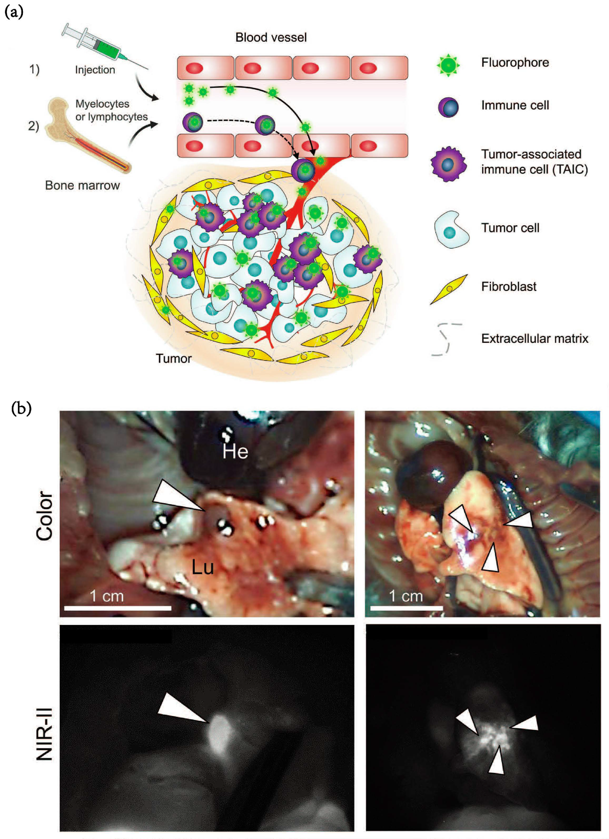
- Kang, H.; Shamim, M.; Yin, X.; Adluru, E.; Fukuda, T.; Yokomizo, S.; Chang, H.; Park, S.H.; Cui, Y.; Moy, A.J.; et al. Tumor-Associated Immune-Cell-Mediated Tumor-Targeting Mechanism with NIR-II Fluorescence Imaging. Adv. Mater. 2022, 34, 2106500. [Google Scholar] [CrossRef]
- Lee, C.; Kim, G.R.; Yoon, J.; Kim, S.E.; Yoo, J.S.; Piao, Y. In vivo delineation of glioblastoma by targeting tumor-associated macrophages with near-infrared fluorescent silica coated iron oxide nanoparticles in orthotopic xenografts for surgical guidance. Sci. Rep. 2018, 8, 11122. [Google Scholar] [CrossRef] [Green Version]
- Marotta, C.B.; Haber, T.; Berlin, J.M.; Grubbs, R.H. Surgery-Guided Removal of Ovarian Cancer Using Up-Converting Nanoparticles. ACS Appl. Mater. Interfaces 2020, 12, 48371–48379. [Google Scholar] [CrossRef]
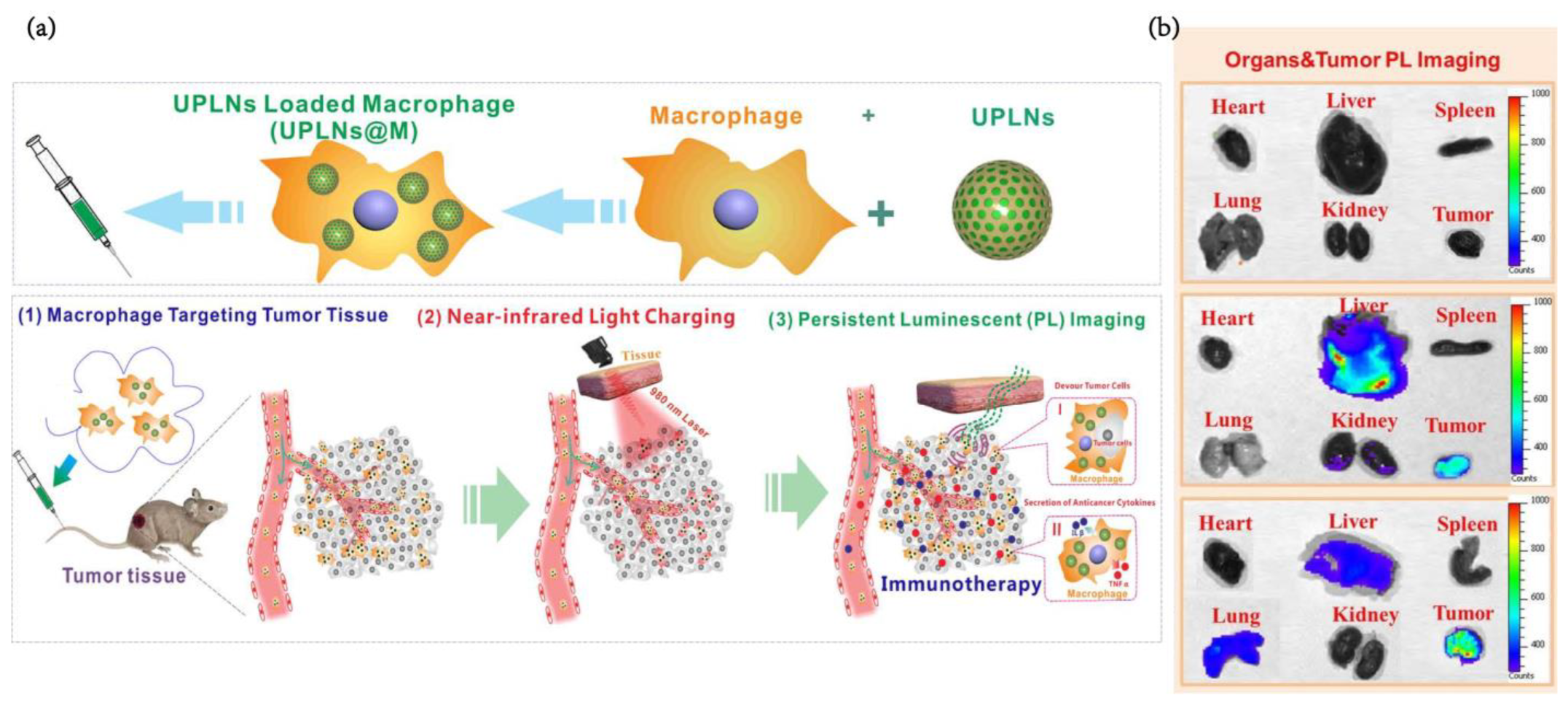
- Zheng, B.; Bai, Y.; Chen, H.; Pan, H.; Ji, W.; Gong, X.; Wu, X.; Wang, H.; Chang, J. Near-Infrared Light-Excited Upconverting Persistent Nanophosphors in Vivo for Imaging-Guided Cell Therapy. ACS Appl. Mater. Interfaces 2018, 10, 19514–19522. [Google Scholar] [CrossRef]
- Zhou, X.; Liu, Q.; Wang, X.; Yao, X.; Zhang, B.; Wu, J.; Sun, C. Exosomal ncRNAs facilitate interactive ‘dialogue’ between tumor cells and tumor-associated macrophages. Cancer Lett. 2022, 552, 215975. [Google Scholar] [CrossRef]
- Moradi-Chaleshtori, M.; Bandehpour, M.; Soudi, S.; Mohammadi-Yeganeh, S.; Hashemi, S.M. In vitro and in vivo evaluation of anti-tumoral effect of M1 phenotype induction in macrophages by miR-130 and miR-33 containing exosomes. Cancer Immunol. Immunother. 2021, 70, 1323–1339. [Google Scholar] [CrossRef]
- Gerloff, D.; Luetzkendorf, J.; Moritz, R.K.C.; Wersig, T.; Maeder, K.; Mueller, L.P.; Sunderkoetter, C. Melanoma-Derived Exosomal miR-125b-5p Educates Tumor Associated Macrophages (TAMs) by Targeting Lysosomal Acid Lipase A (LIPA). Cancers 2020, 12, 464. [Google Scholar] [CrossRef] [Green Version]
- Gunassekaran, G.R.; Vadevoo, S.M.P.; Baek, M.-C.; Lee, B. M1 macrophage exosomes engineered to foster M1 polarization and target the IL-4 receptor inhibit tumor growth by reprogramming tumor-associated macrophages into M1-like macrophages. Biomaterials 2021, 278, 121137. [Google Scholar] [CrossRef]
- Kamerkar, S.; Leng, C.; Burenkova, O.; Jang, S.C.; McCoy, C.; Zhang, K.; Dooley, K.; Kasera, S.; Zi, T.; Siso, S.; et al. Exosome-mediated genetic reprogramming of tumor-associated macrophages by exoASO-STAT6 leads to potent monotherapy antitumor activity. Sci. Adv. 2022, 8, 7002. [Google Scholar] [CrossRef]
- Liu, L.; Wang, Y.; Guo, X.; Zhao, J.; Zhou, S. A Biomimetic Polymer Magnetic Nanocarrier Polarizing Tumor-Associated Macrophages for Potentiating Immunotherapy. Small 2020, 16, 2003543. [Google Scholar] [CrossRef]
- Wang, Y.; Yu, J.; Luo, Z.; Shi, Q.; Liu, G.; Wu, F.; Wang, Z.; Huang, Y.; Zhou, D. Engineering Endogenous Tumor-Associated Macrophage-Targeted Biomimetic Nano-RBC to Reprogram Tumor Immunosuppressive Microenvironment for Enhanced Chemo-Immunotherapy. Adv. Mater. 2021, 33, 2103497. [Google Scholar] [CrossRef]
- Wang, S.; Shen, H.; Mao, Q.; Tao, Q.; Yuan, G.; Zeng, L.; Chen, Z.; Zhang, Y.; Cheng, L.; Zhang, J.; et al. Macrophage-Mediated Porous Magnetic Nanoparticles for Multimodal Imaging and Postoperative Photothermal Therapy of Gliomas. ACS Appl. Mater. Interfaces 2021, 13, 56825–56837. [Google Scholar] [CrossRef] [PubMed]
- Forte, E.; Fiorenza, D.; Torino, E.; Costagliola di Polidoro, A.; Cavaliere, C.; Netti, P.A.; Salvatore, M.; Aiello, M. Radiolabeled PET/MRI nanoparticles for tumor imaging. J. Clin. Med. 2019, 9, 89. [Google Scholar] [CrossRef] [PubMed] [Green Version]
- Rao, L.; He, Z.; Meng, Q.-F.; Zhou, Z.; Bu, L.-L.; Guo, S.-S.; Liu, W.; Zhao, X.-Z. Effective cancer targeting and imaging using macrophage membrane-camouflaged upconversion nanoparticles. J. Biomed. Mater. Res. Part A 2017, 105, 521–530. [Google Scholar] [CrossRef] [PubMed]
- Chen, J.; Tan, Q.; Yang, Z.; Jin, Y. Engineered extracellular vesicles: Potentials in cancer combination therapy. J. Nanobiotechnology 2022, 20, 132. [Google Scholar] [CrossRef] [PubMed]
- Fan, Z.; Xiao, K.; Lin, J.; Liao, Y.; Huang, X. Functionalized DNA Enables Programming Exosomes/Vesicles for Tumor Imaging and Therapy. Small 2019, 15, 1903761. [Google Scholar] [CrossRef]
- Yu, L.; Zhu, S.; Qin, K.; Fan, X.; An, L. Macrophages Loaded with Fe Nanoparticles for Enhanced Photothermal Ablation of Tumors. J. Funct. Biomater. 2022, 13, 94. [Google Scholar] [CrossRef]
- Xue, F.; Zhu, S.; Tian, Q.; Qin, R.; Wang, Z.; Huang, G.; Yang, S. Macrophage-mediated delivery of magnetic nanoparticles for enhanced magnetic resonance imaging and magnetothermal therapy of solid tumors. J. Colloid Interface Sci. 2023, 629, 554–562. [Google Scholar] [CrossRef]
- An, L.; Wang, Y.; Lin, J.; Tian, Q.; Xie, Y.; Hu, J.; Yang, S. Macrophages-Mediated Delivery of Small Gold Nanorods for Tumor Hypoxia Photoacoustic Imaging and Enhanced Photothermal Therapy. ACS Appl. Mater. Interfaces 2019, 11, 15251–15261. [Google Scholar] [CrossRef]
- Yin, F.; Fan, Y.; Xu, L.; Yin, F.; He, M.; Xiao, T.; Shi, X.; Wang, H. Macrophages loaded with dendrimer-entrapped gold nanoparticles as a theranostic platform for CT imaging-guided combinational therapy of orthotopic osteosarcoma. Chem. Eng. J. 2021, 417, 129273. [Google Scholar] [CrossRef]
- Peng, Y.; Wang, X.; Wang, Y.; Gao, Y.; Guo, R.; Shi, X.; Cao, X. Macrophage-Laden Gold Nanoflowers Embedded with Ultrasmall Iron Oxide Nanoparticles for Enhanced Dual-Mode CT/MR Imaging of Tumors. Pharmaceutics 2021, 13, 995. [Google Scholar] [CrossRef]
- Vahrmeijer, A.L.; Hutteman, M.; van der Vorst, J.R.; van de Velde, C.J.H.; Frangioni, J.V. Image-guided cancer surgery using near-infrared fluorescence. Nat. Rev. Clin. Oncol. 2013, 10, 507–518. [Google Scholar] [CrossRef] [Green Version]
- Hyun, H.; Henary, M.; Gao, T.; Narayana, L.; Owens, E.A.; Lee, J.H.; Park, G.; Wada, H.; Ashitate, Y.; Frangioni, J.V.; et al. 700-nm Zwitterionic Near-Infrared Fluorophores for Dual-Channel Image-Guided Surgery. Mol. Imaging Biol. 2016, 18, 52–61. [Google Scholar] [CrossRef] [Green Version]
- Haber, T.; Cornejo, Y.R.; Aramburo, S.; Flores, L.; Cao, P.; Liu, A.; Mooney, R.; Gilchrist, M.; Tirughana, R.; Nwokafor, U.; et al. Specific targeting of ovarian tumor-associated macrophages by large, anionic nanoparticles. Proc. Natl. Acad. Sci. USA 2020, 117, 19737–19745. [Google Scholar] [CrossRef]
- Zhou, B.; Shi, B.; Jin, D.; Liu, X. Controlling upconversion nanocrystals for emerging applications. Nat. Nanotechnol. 2015, 10, 924–936. [Google Scholar] [CrossRef]
- Starosolski, Z.; Courtney, A.N.; Srivastava, M.; Guo, L.; Stupin, I.; Metelitsa, L.S.; Annapragada, A.; Ghaghada, K.B. A Nanoradiomics Approach for Differentiation of Tumors Based on Tumor-Associated Macrophage Burden. Contrast Media Mol. Imaging 2021, 2021, 6641384. [Google Scholar] [CrossRef]
- Thust, S.C.; van den Bent, M.J.; Smits, M. Pseudoprogression of brain tumors. J. Magn. Reson. Imaging 2018, 48, 571–589. [Google Scholar] [CrossRef] [Green Version]
- Deng, X.; Liang, H.; Yang, W.; Shao, Z. Polarization and function of tumor-associated macrophages mediate graphene oxide-induced photothermal cancer therapy. J. Photochem. Photobiol. B-Biol. 2020, 208, 111913. [Google Scholar] [CrossRef]


| Number | Material | Type of NPs | Disease | Cell line | Immune Background | Animal Model | Administration | Dose | Imaging Methods | Targeting Approaches | Model | Reference |
|---|---|---|---|---|---|---|---|---|---|---|---|---|
| 1 | Iron oxide | Iron oxide nanoparticle | Glioblastoma | MbTIC0309 brain tumor cells | immunocompetent | 8–10 weeks Female C57BL/6 mice | i.v. | 30 mg/kg | MRI | Phagocytosis | in vivo | [17] |
| 2 | Iron oxide | Mannose-PEG(Poly(ethylene glycol))-b-AGE polymer coated iron oxide nanoparticles | Breast cancer | 4T1 tumor cells | immunocompetent | 6–8 weeks old female Balb/C mice | i.v. | 5 mg Fe/kg | MRI | Mannose modified | in vivo | [18] |
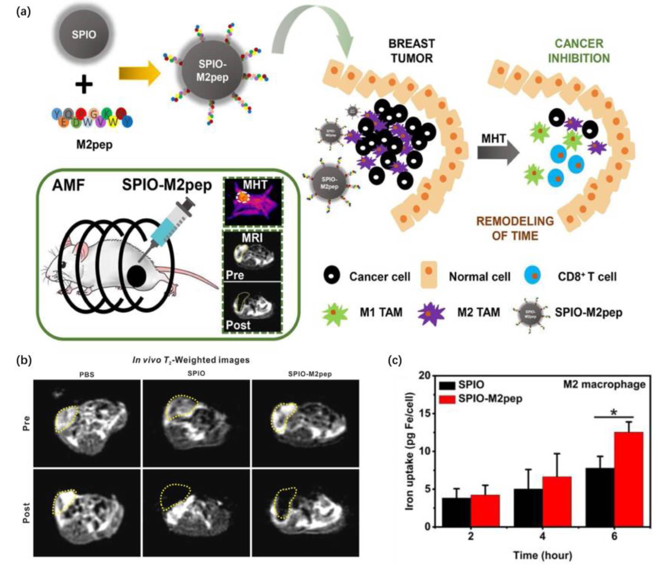
| 3 | Iron oxide | M2 macrophage targeted peptide functionalized superparamagnetic iron oxide | Breast cancer | 4T1 tumor cells | immunocompetent | female BALB/c mice | i.t. (in situ) | 0.8 uL mm−3 of tumor volume, 10 UG uL−1 * | MRI | M2 macrophage targeted peptide | in vivo | [19] |
| 4 | Iron oxide | Comprising superparamagnetic iron oxide nanocrystals and nitric oxide (NO) donors | Pancreatic tumor | Panc02 tumore cells | immunocompetent | 4–5 weeks old, female C57BL/ 6J mice | i.v. | 2 MG/mL,100μL | MRI | Phagocytosis | in vivo | [22] |
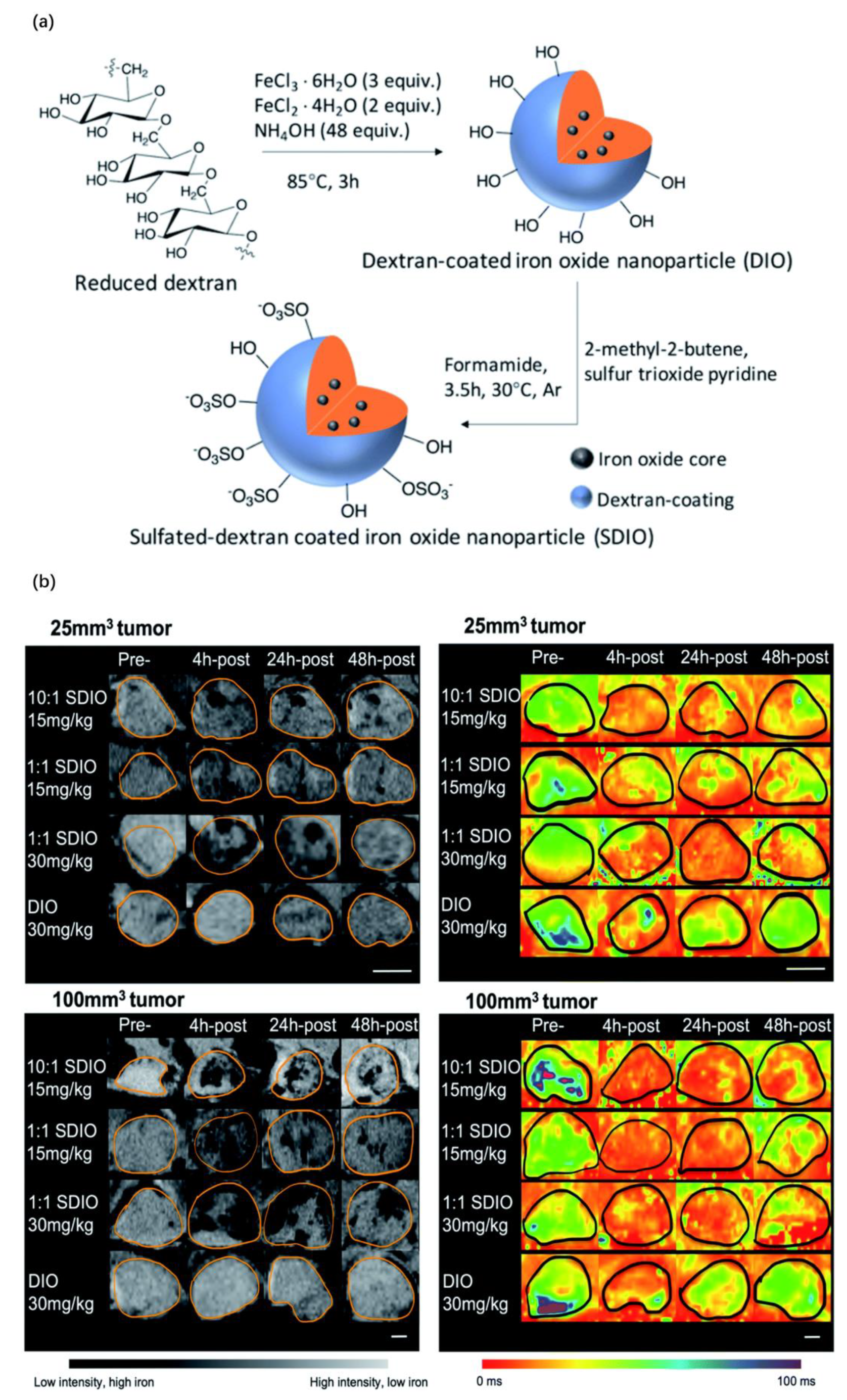
| 5 | Iron oxide | Sulfated-dextran coated iron oxide nanoparticles | Breast cancer | 4T1 tumor cells | immunocompetent | 6–12 weeks old female C57BL/6 mice | i.v. | 15 mg Fe/kg 30 mg Fe/kg | MRI | Dextran | in vivo | [23] |
| 6 | Mn2+ | Bioactivated in vivo assembly magnetic resonance probe | Breast cancer | MDA-MB-231 tumor cells | immunosuppression | Female BALB/C nude mouse | i.v. | 20 mg/kg | MRI | Mannose modified | in vivo | [26] |
| 7 | MnO2 | HA-coated, mannan-conjugated MnO2 particle | Breast cancer | 4T1 tumor cells | immunocompetent | Female Balb/c mice | i.v. | Man-HA-MnO2 NP (13.2 mg/kg) Dox (5 mg/kg) | MRI | Mannose modified and Hyaluronic acid coated | in vivo | [28] |
| 8 | MnO2 | Hyaluronic acid (HA) and PLR-coated manganese dioxide (MnO2) nanoparticles | Sarcoma | S-180 murine sarcoma cancer cell line | / | / | / | / | MRI | Hyaluronic acid and poly(L-arginine) modified | in vitro | [29] |
| 9 | MnO2 | An upconversion nanoparticle (UCNP) as the core, hollow mesoporous silica wrapped on the outside of the UCNP with DOX filled within the cavity, MnO2 nanosheets modified in mesopores as hypoxia-sensitive agents | Cervical carcinoma | Hella cells | immunosuppression | BALB/C nude mice | i.v. and i.t. | 8 MG/mL, 100 μL | MRI(T1) | Phagocytosis | in vivo | [30] |
| 10 | 111In | Optimization of mannosylated serum albumin (MSA) | Breast cancer/Melanoma | 4T1/B16F10/LLC celmmuneuno-competent | 7–8 weeks old female BALB/c mice and C57BL/6 mice | i.v. | 11.1 MBq (megabecquerel) | PET | Mannose modified | in vivo | [49] | |
| 11 | 68Ga | 68Ga-NOTA-COG1410 | Colon cancer | PCM/MGC-803/AGS/CT26.WT celmmuneuno-competent and immunosuppression | 8 weeks old male BALB/c nude mice (18–20 g) and male BALB/c mice | i.v. | 1 μg, 100 μL | PET | COG1410 for TREM2 targeting | in vivo | [50] | |
| 12 | 68Ga | [68Ga]Ga-DOTA-M2pep | Melanoma | B10-F10 cells | immunocompetent | 6–8 weeks old male C57BL/6 mice | i.v. | 3.7 MBq per mice | PET | M2pep modified | in vivo | [89] |
| 13 | 64Cu | 64Cu-labeled polyglucose nanoparticle | Breast cancer/Lung tumor/Colon | 4T1/KP1.9/MC38 cells | immunocompetent | 7–12 weeks old C57BL/6 mice and 6–8 week old BALB/c mice | i.v. | 8.5 ± 2.4 MBq in 150 ± 10 μL PBS | PET | Polyglucose modified | in vivo | [51] |
| 14 | Er-based probe | M2-targeting Er-based NIR-IIb nanoprobes | Glioblastoma | U87MG glioma cells | immunosuppression | 6–8 weeks old specific pathogen-free (SPF) grade female nude mice | i.v. | 2.4 MG/mL, 200 μL | NIR | M2pep modified | in vivo | [54] |
| 15 | Cyanine 7 | Deoxymannose labeled cyanine 7 | Hepatoma | SMMC-7721 human hepatoma cells | immunosuppression | Nude mice | i.v. | 2 nmol, 100 μL | NIR | Deoxymannose | in vivo | [55] |
| 16 | Sulfo-Cyanine 5 | Enzyme-activatable chemokine conjugates nanoprobe (chemo-cat NIR) | Breast cancer | E0771-LG celmmuneuno-competent | C57BL/6 mice | i.v. | 0.3 nmol per mouse | NIR | Enzyme-activatable chemokine conjugates | in vivo | [56] | |
| 17 | Indocyanine green(ICG) | Noncovalent indocyanine green conjugate of C-phycocyanin (CPC@ICG(Indocyanine green)) | Cervical carcinoma | H22 cells | immuno-competent | Kunming (KM) mice | i.v. | 1.25 mg ICG/kg | NIR | C-phycocyanin modified | in vivo | [58] |
| 18 | Diaminofluorescein-2-diacetate (DAF-2-DA) | NIR-NO nanoprobe combined with CSF1R inhibiting amphiphile | Breast cancer | 4T1 cells | immuno-competent | BALB/c mice | i.v. | 2 mg NO-NR/kg | NIR | CSF1R inhibiting amphiphile | in vivo | [59] |
| 19 | Nanobubble | CSF-1R targeted nanobubble | Hepatocarcinoma | SMMC-7721 cells | immunosup-pression | 5–6-week-old male BALB/c nude mice | i.v. | 4 × 10⁷ NB CSF-1R per mice | Ultrasound imaging | CSF-1R-conjugated | in vivo | [81] |
| 20 | Nanobubble | Folate-conjugated ultrasonic nanobubble (HA-FOL-NB) | Lung carcinoma | Lewis lung cells | / | / | / | / | Ultrasound imaging | Hyaluronic acid modified | in vitro | [82] |
| 21 | Nanobubble | AAN peptide and RGD peptide modified nanobubbles | Breast cancer | 4T1 | immuno-competent | 4–5 weeks old BALB/c mice | i.v. | 200 μL NPs per mice | Ultrasound imaging | AAN peptide and RGD peptide | in vivo | [83] |
| 22 | Gold nanoparticle | PGMP-small interfering RNA (siRNA) nanoparticles | Non-small cell lung cancer | A549 cells | immunosup-pression | BALB/c nude mice | i.v. | 3 mg/mL, 100 μL | Ultrasound imaging | Phagocytosis | in vivo | [32] |
| 23 | Gold nanoparticle | Glycol-chitosan-coated gold nanoparticles (GC-AuNPs) | Delivering Tumor antigen to lymph nodes | / | immuno-competent | 5 weeks old female healthy nu/nu mice | injected through the right side of the tongue | 2.5 mg Au/mL, 80 μL | PA | Phagocytosis | in vivo | [33] |
| 24 | Cyanine 5/Iron oxide | Mannose-targeted liposomes (MAN-lipo-AAG) liposomes (lipo-AAG) encapsulating Ac4GalNAz | Breast cancer | 4T1 cells | immuno-competent | 6 weeks old female BALB/c mice | i.v. | 50 mg/kg | NIR | Mannose modified | in vivo | [57] |
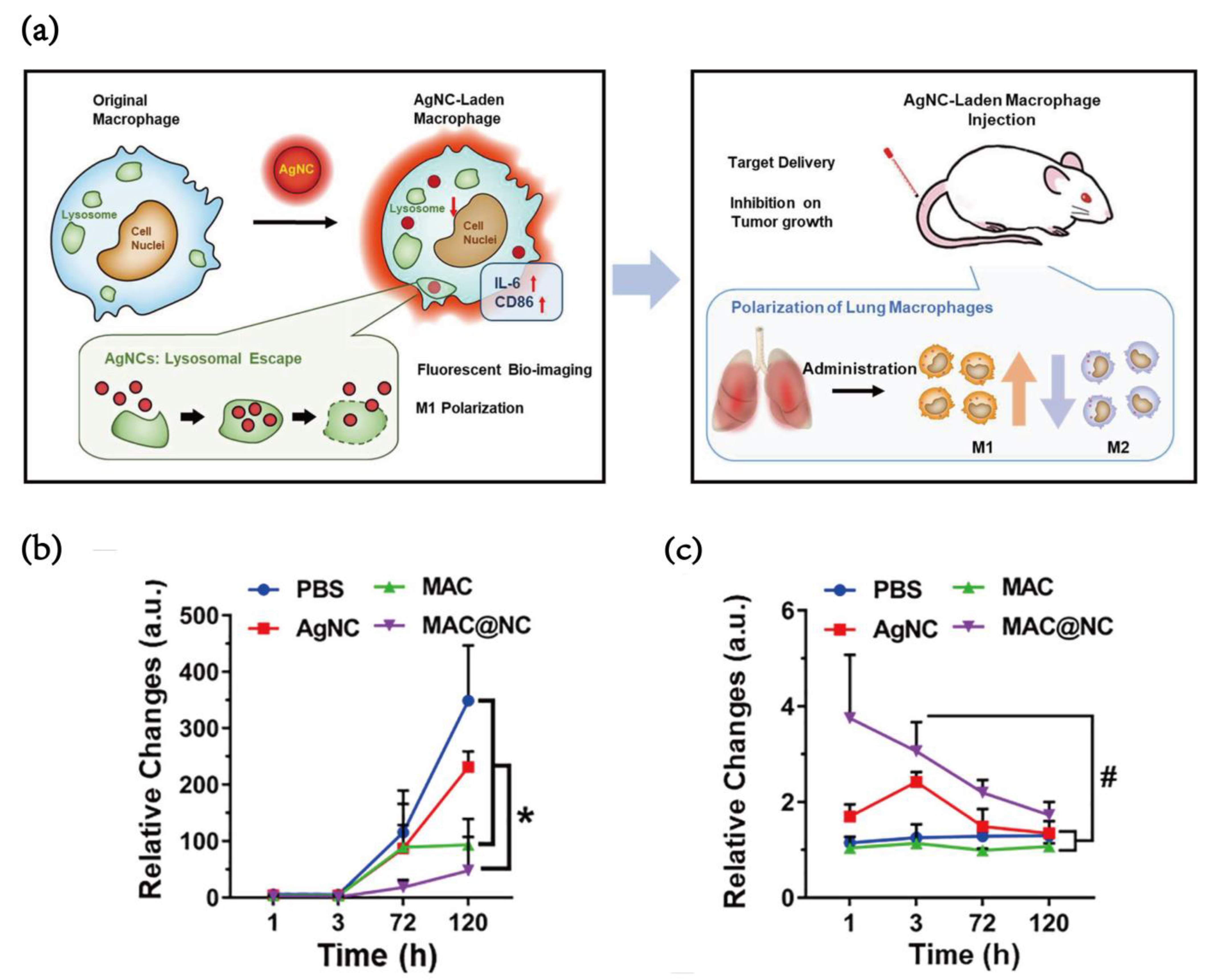
| 25 | Silver nanoclusters | AgNCs in combination with M1-like macrophages (namely the MAC@NC complex) | Lung metastasis breast cancer | 4T1-LG12 cells | immuno-competent | 7–8 weeks old female BALB/c mice | i.v. | 2.0 × 10⁶ MAC@NC per mice | NIR | Macrophage vehicle | in vivo | [35] |
| 26 | Fluorine-19 | Fluorine-19(19F)@PLGA-PEG-Man@perfluoro-15-crown-5-ether (PFCE) | Breast cancer | 4T1 cells | immuno-competent | 6–8 weeks old BALB/c mice | i.v. | 7.01 × 10¹⁹ 19F per mice | MRI | Mannose modified | in vivo | [39] |
| 27 | Fluorine-19 | Perfluorocarbon-containing nanoparticles (PFC-NP) | Gliomagenesis, breast-to-brain metastasis, and breast cancer | RCAS-PDGFB-HA and PDGFB-HA-SV40-GFP DF1 cells | immuno-competent | 6–7 weeks old C57BL/6J mice | i.v. | 10 Gy per mice | MRI | Phagocytosis | in vivo | [42] |
| 28 | Indocyanine green | Dextran-indocyanine green | Pancreatic cancer | SW1990 pancreatic cancer cells | immunosup-pression | Female BALB/c nude mice | i.v. | 0.5 ICG mg/kg | NIR | Dextran | in vivo | [90] |
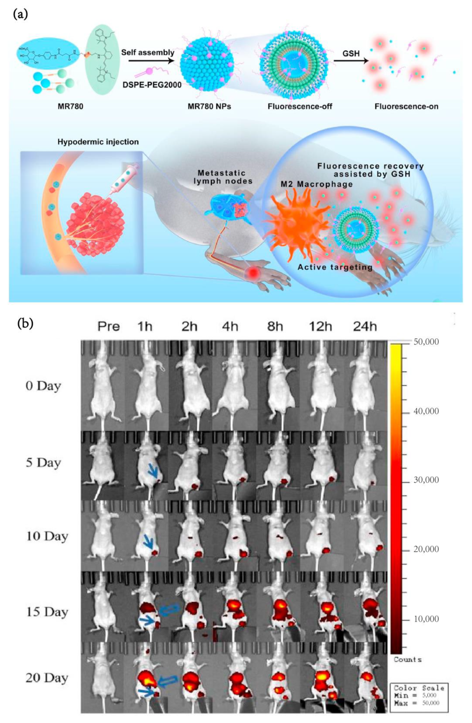
| 29 | IR780 | Self-assembled IR780 conjugated with mannose | Lymph node metastasis breast cancer | 4T1 cells | immuno-competent | 5 weeks old female BALB/c mice | subcutaneous injection (s.c.) | 2 mg/kg | NIR | Mannose modified | in vivo | [91] |
| 30 | Heptamethine cyanine-based fluorophores | TAIC targeting heptamethine cyanine-based fluorophores | Pancreatic, breast, and lung cancer | Pan02,E0771 and LLC cells | immunosup-pression and immunocompetent | 8 weeks old C57 BL/6 mice | i.v. | 1 μmol/kg | NIR | Phagocytosis | in vivo | [92] |
| 31 | Cyanine-5.5 | Near-infrared fluorescent silica coated iron oxide nanoparticles | Glioblastoma multiforme | U87-MG cells | immuno-competent | 6 weeks old male BALB/c nude mice | i.v. | 200 μg Fe | NIR | Fluorescent silica coated | in vivo | [93] |
| 32 | UCNP | Combined up-converting nanoparticles(UCNPs) with silica nanoparticles | Ovarian cancer | OVCAR8-GFP cells | immunosup-pression | 7 weeks old athymic nude mice | i.p. | 1.37 × 10¹⁰ UCNPs in 1 mL PBS | Visible fluorescence | Phagocytosis | in vivo | [94] |
| 33 | UCNP | Based on (Zn2SiO4:Mn):Y3+, Yb3+, Tm3+ upconverting persistent luminescent nanophosphors | Melanoma | B16 cells | immuno-competent | 8 weeks old C57/B6 mice | i.v. | 0.5 mg/mL, 100 μL | Visible fluorescence | Phagocytosis | in vivo | [95] |
| Imaging Material | Tumor Type | Imaging Methods | TAMs Targeting Approaches | Targeting Effect | Reference |
|---|---|---|---|---|---|
| Iron oxide | Breast cancer | MRI | Mannose modified | Colocalization degree of target protein 6 h: Targeting: non-targeting = 93.5%: 57.3% | [18] |
| Iron oxide | Breast cancer, 4t1 model | MRI | M2 targeting peptide | In vitro targeting 6 h: Targeting: non-targeting = 185% | [19] |
| Iron oxide | Pancreatic cancer | MRI | / | Tumor signal in 48 h: Targeting (5 × 10³ a.u.) Non-targeting (2 × 10³ a.u.) | [22] |
| Iron oxide | Breast cancer, 4t1 model | MRI | Mannose modified | Tumor signal in 24 h: Targeting (3 × 10⁸ p/s/cm2/sr) Non-targeting (1.3 × 10⁸ p/s/cm2/sr) | [28] |
| Near-infrared dye cyanine 7 (Cy7) | Hepatoma cell | NIR | Deoxymannose | Tumor signal in 8 h: Targeting (42 × 108 p/s/cm2/sr) | [55] |
| 111In | Breast cancer, 4t1 model | PET | Mannose | Lung metastasis %ID/g in 24 h: Targeting (5) Non-targeting (2) | [49] |
| 68Ga | Gastric cancer | PET | COG1410, as a ligand of TREM2 | Tumor %ID/g in 2 h Targeting (6) | [50] |
| 64Cu | Breast cancer 4T1 mice model | PET | Polyglucose | Tumor %ID/g in 24 h Targeting (10) | [51] |
| Er-based rare-earth nanoparticles | Orthotopic Glioblastoma | NIR | M2pep polypeptide | In vitro targeting: Targeting: non-targeting = 207% | [54] |
| Indocyanine green | Hela human cervical cells | NIR | C-phycocyanin | In vitro targeting: Targeting: non-targeting = 330% | [58] |
| Nano bubble | Breast cancer 4t1 mice model | ultrasound imaging | AAN peptide and RGD peptide | Tumor signal in 0.5 h: Targeting (3.2 × 10⁸ p/s/cm2/sr) Non-targeting(0.5 × 10⁸ p/s/cm2/sr) | [83] |
| Indocyanine green | Pancreatic Cancer | NIR | Dextran | Tumor signal in 24 h NIR-I (90 a.u.) NIR-II (130 a.u.) | [90] |
| Heptamethine-cyanine-based fluorophores | Lung carcinoma, pancreatic ductal adenocarcinoma (PDAC), and triple negative breast adenocarcinoma | NIR | / | Fluorescence positive rate of cells co-cultured with preparation: SH1:free ICG = (45.7%: 1.2%) | [92] |
| Cyanine-5.5 | Glioblastoma | NIR | Fluorescent silica coated | Tumor signal in 24 h: Targeting (21,000 a.u.) | [93] |
| Cyanine-5.5/Iron oxide | Glioblastoma | NIR/MRI | Macrophages membrane coating | Tumor %ID/g in 24 h: Targeting (8) Tumor %ID/g in 7 days: Targeting (14) | [103] |
Disclaimer/Publisher’s Note: The statements, opinions and data contained in all publications are solely those of the individual author(s) and contributor(s) and not of MDPI and/or the editor(s). MDPI and/or the editor(s) disclaim responsibility for any injury to people or property resulting from any ideas, methods, instructions or products referred to in the content. |
© 2022 by the authors. Licensee MDPI, Basel, Switzerland. This article is an open access article distributed under the terms and conditions of the Creative Commons Attribution (CC BY) license (https://creativecommons.org/licenses/by/4.0/).
Share and Cite
Hu, J.; Xu, X.; Du, Y. Targeting Tumor-Associated Macrophages for Imaging. Pharmaceutics 2023, 15, 144. https://doi.org/10.3390/pharmaceutics15010144
Hu J, Xu X, Du Y. Targeting Tumor-Associated Macrophages for Imaging. Pharmaceutics. 2023; 15(1):144. https://doi.org/10.3390/pharmaceutics15010144
Chicago/Turabian StyleHu, Jiahao, Xiaoling Xu, and Yongzhong Du. 2023. "Targeting Tumor-Associated Macrophages for Imaging" Pharmaceutics 15, no. 1: 144. https://doi.org/10.3390/pharmaceutics15010144