

Chiral Biomaterials for Nanomedicines: From Molecules to Supraparticles
Abstract
:1. Introduction
2. Chiral Nanomaterials for Nanomedicine
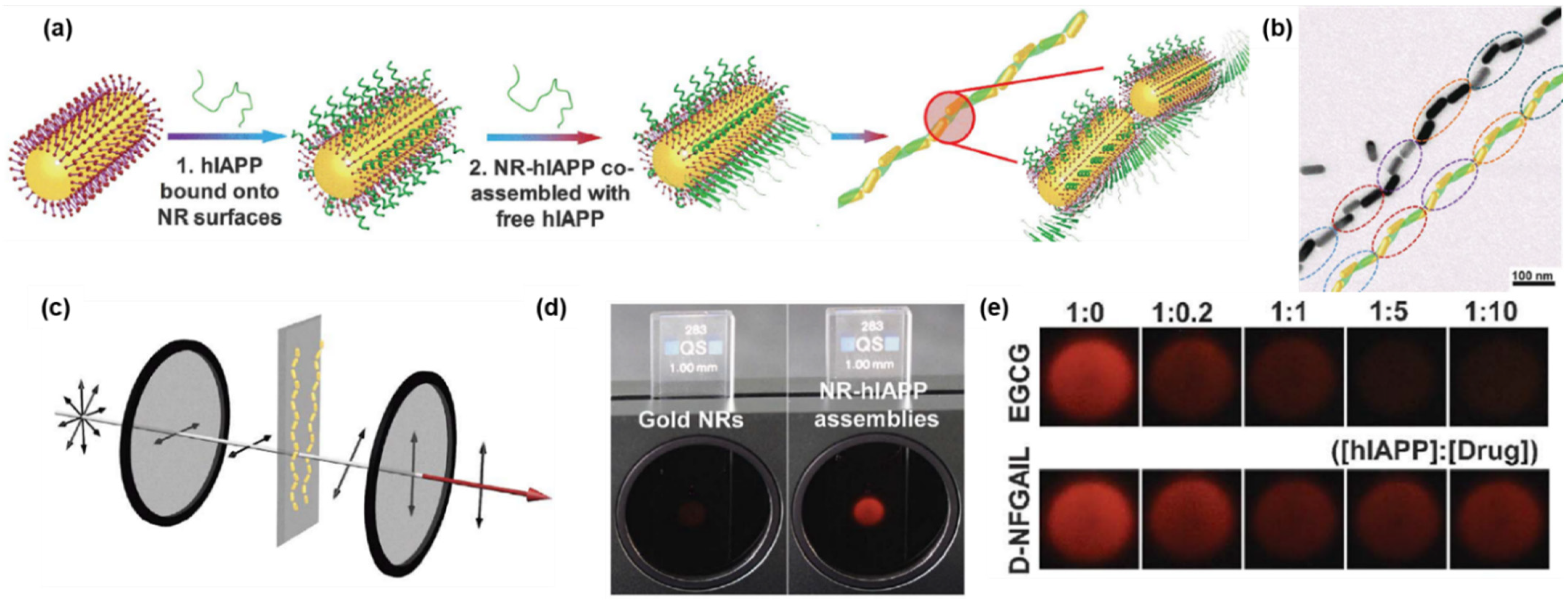
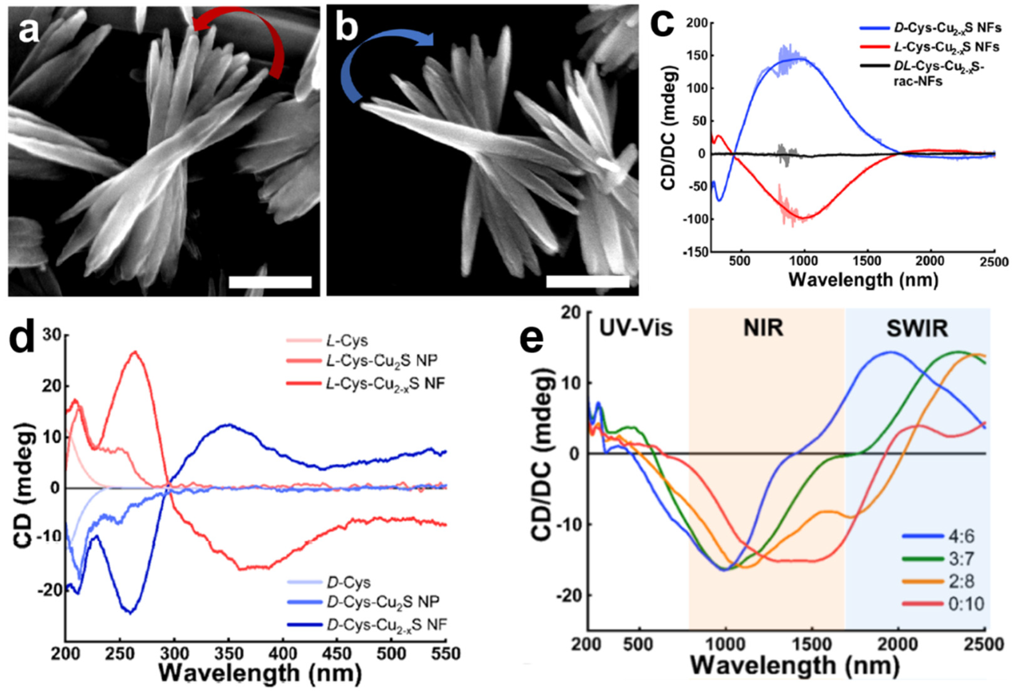
3. Chiral Biomaterials with Supramolecular Structures
4. Conclusions and Outlook
Funding
Institutional Review Board Statement
Informed Consent Statement
Conflicts of Interest
References
- Liu, M.; Zhang, L.; Wang, T. Supramolecular chirality in self-assembled systems. Chem. Rev. 2015, 115, 7304–7397. [Google Scholar] [CrossRef]
- Yeom, J.; Santos, U.S.; Chekini, M.; Cha, M.; de Moura, A.F.; Kotov, N.A. Chiromagnetic nanoparticles and gels. Science 2018, 359, 309–314. [Google Scholar] [CrossRef]
- Ciriminna, R.; Lomeli-Rodriguez, M.; Cara, P.D.; Lopez-Sanchez, J.A.; Pagliaro, M. Limonene: A versatile chemical of the bioeconomy. Chem. Commun. 2014, 50, 15288–15296. [Google Scholar] [CrossRef]
- Kawasaki, H.; Kasamatsu, C.; Nonaka, M. Cognitive structures based on culinary success factors in the development of new dishes by Japanese chefs at fine dining restaurants. Flavour 2015, 4, 1. [Google Scholar] [CrossRef]
- Temussi, P.A. Sweet, bitter and umami receptors: A complex relationship. Trends Biochem. Sci. 2009, 34, 296–302. [Google Scholar] [CrossRef]
- Ebrahimi, F.A.; Chess, A. Olfactory G proteins: Simple and complex signal transduction. Curr. Biol. 1998, 8, R431–R433. [Google Scholar] [CrossRef]
- Agranat, I.; Caner, H.; Caldwell, J. Putting chirality to work: The strategy of chiral switches. Nat. Rev. Drug Discov. 2002, 1, 753–768. [Google Scholar] [CrossRef]
- Hutt, A.; Tan, S. Drug chirality and its clinical significance. Drugs 1996, 52, 1–12. [Google Scholar] [CrossRef]
- Franks, M.E.; Macpherson, G.R.; Figg, W.D. Thalidomide. Lancet 2004, 363, 1802–1811. [Google Scholar] [CrossRef]
- D’Amato, R.J.; Loughnan, M.S.; Flynn, E.; Folkman, J. Thalidomide is an inhibitor of angiogenesis. Proc. Natl. Acad. Sci. USA 1994, 91, 4082–4085. [Google Scholar] [CrossRef] [Green Version]
- Fischer, E.S.; Böhm, K.; Lydeard, J.R.; Yang, H.; Stadler, M.B.; Cavadini, S.; Nagel, J.; Serluca, F.; Acker, V.; Lingaraju, G.M. Structure of the DDB1–CRBN E3 ubiquitin ligase in complex with thalidomide. Nature 2014, 512, 49–53. [Google Scholar] [CrossRef] [PubMed]
- Collet, A. Problems and Wonders of Chiral Molecules; Akadémiai Kiadó: Budpaest, Hungary, 1990. [Google Scholar]
- PhD, J.C. The importance of stereochemistry in drug action and disposition. J. Clin. Pharmacol. 1992, 32, 925–929. [Google Scholar] [CrossRef]
- Wermuth, C.G.; Ganellin, C.R.; Lindberg, P.; Mitscher, L.A. Glossary of terms used in medicinal chemistry (IUPAC Recommendations 1998). Pure Appl. Chem. 1998, 70, 1129–1143. [Google Scholar] [CrossRef]
- Santos, R.; Ursu, O.; Gaulton, A.; Bento, A.P.; Donadi, R.S.; Bologa, C.G.; Karlsson, A.; Al-Lazikani, B.; Hersey, A.; Oprea, T.I. A comprehensive map of molecular drug targets. Nat. Rev. Drug Discov. 2017, 16, 19–34. [Google Scholar] [CrossRef] [PubMed]
- Kim, B.Y.; Rutka, J.T.; Chan, W.C. Nanomedicine. New Engl. J. Med. 2010, 363, 2434–2443. [Google Scholar] [CrossRef] [PubMed]
- Bhatia, S.N.; Chen, X.; Dobrovolskaia, M.A.; Lammers, T. Cancer nanomedicine. Nat. Rev. Cancer 2022, 1–7. [Google Scholar] [CrossRef] [PubMed]
- Pelaz, B.; Alexiou, C.; Alvarez-Puebla, R.A.; Alves, F.; Andrews, A.M.; Ashraf, S.; Balogh, L.P.; Ballerini, L.; Bestetti, A.; Brendel, C. Diverse applications of nanomedicine. ACS Nano 2017, 11, 2313–2381. [Google Scholar] [CrossRef]
- Yeom, J.; Guimaraes, P.P.; Ahn, H.M.; Jung, B.K.; Hu, Q.; McHugh, K.; Mitchell, M.J.; Yun, C.O.; Langer, R.; Jaklenec, A. Chiral supraparticles for controllable nanomedicine. Adv. Mater. 2020, 32, 1903878. [Google Scholar] [CrossRef]
- Li, S.; Sun, M.; Hao, C.; Qu, A.; Wu, X.; Xu, L.; Xu, C.; Kuang, H. Chiral CuxCoyS nanoparticles under magnetic field and NIR light to eliminate senescent cells. Angew. Chem. Int. Ed. 2020, 59, 13915–13922. [Google Scholar] [CrossRef]
- Xin, Q.; Liu, Q.; Geng, L.; Fang, Q.; Gong, J.R. Chiral nanoparticle as a new efficient antimicrobial nanoagent. Adv. Healthc. Mater. 2017, 6, 1601011. [Google Scholar] [CrossRef]
- Malishev, R.; Arad, E.; Bhunia, S.K.; Shaham-Niv, S.; Kolusheva, S.; Gazit, E.; Jelinek, R. Chiral modulation of amyloid beta fibrillation and cytotoxicity by enantiomeric carbon dots. Chem. Commun. 2018, 54, 7762–7765. [Google Scholar] [CrossRef] [PubMed]
- Sun, M.; Xu, L.; Qu, A.; Zhao, P.; Hao, T.; Ma, W.; Hao, C.; Wen, X.; Colombari, F.M.; de Moura, A.F. Site-selective photoinduced cleavage and profiling of DNA by chiral semiconductor nanoparticles. Nat. Chem. 2018, 10, 821–830. [Google Scholar] [CrossRef] [PubMed]
- Li, Y.; Miao, Z.; Shang, Z.; Cai, Y.; Cheng, J.; Xu, X. A Visible-and NIR-Light Responsive Photothermal Therapy Agent by Chirality-Dependent MoO3−x Nanoparticles. Adv. Funct. Mater. 2020, 30, 1906311. [Google Scholar] [CrossRef]
- Avalos-Ovando, O.; Besteiro, L.V.; Movsesyan, A.; Markovich, G.; Liedl, T.; Martens, K.; Wang, Z.; Correa-Duarte, M.A.; Govorov, A.O. Chiral photomelting of dna-nanocrystal assemblies utilizing plasmonic photoheating. Nano Lett. 2021, 21, 7298–7308. [Google Scholar] [CrossRef] [PubMed]
- Zhang, H.; Hao, C.; Qu, A.; Sun, M.; Xu, L.; Xu, C.; Kuang, H. Light-Induced Chiral Iron Copper Selenide Nanoparticles Prevent β-Amyloidopathy In Vivo. Angew. Chem. Int. Ed. 2020, 59, 7131–7138. [Google Scholar] [CrossRef] [PubMed]
- Zhang, M.; Zhang, H.; Feng, J.; Zhou, Y.; Wang, B. Synergistic chemotherapy, physiotherapy and photothermal therapy against bacterial and biofilms infections through construction of chiral glutamic acid functionalized gold nanobipyramids. Chem. Eng. J. 2020, 393, 124778. [Google Scholar] [CrossRef]
- Park, K.H.; Kwon, J.; Jeong, U.; Kim, J.-Y.; Kotov, N.A.; Yeom, J. Broad Chiroptical Activity from Ultraviolet to Short-Wave Infrared by Chirality Transfer from Molecular to Micrometer Scale. ACS Nano 2021, 15, 15229–15237. [Google Scholar] [CrossRef]
- Lu, J.; Xue, Y.; Bernardino, K.; Zhang, N.-N.; Gomes, W.R.; Ramesar, N.S.; Liu, S.; Hu, Z.; Sun, T.; de Moura, A.F. Enhanced optical asymmetry in supramolecular chiroplasmonic assemblies with long-range order. Science 2021, 371, 1368–1374. [Google Scholar] [CrossRef]
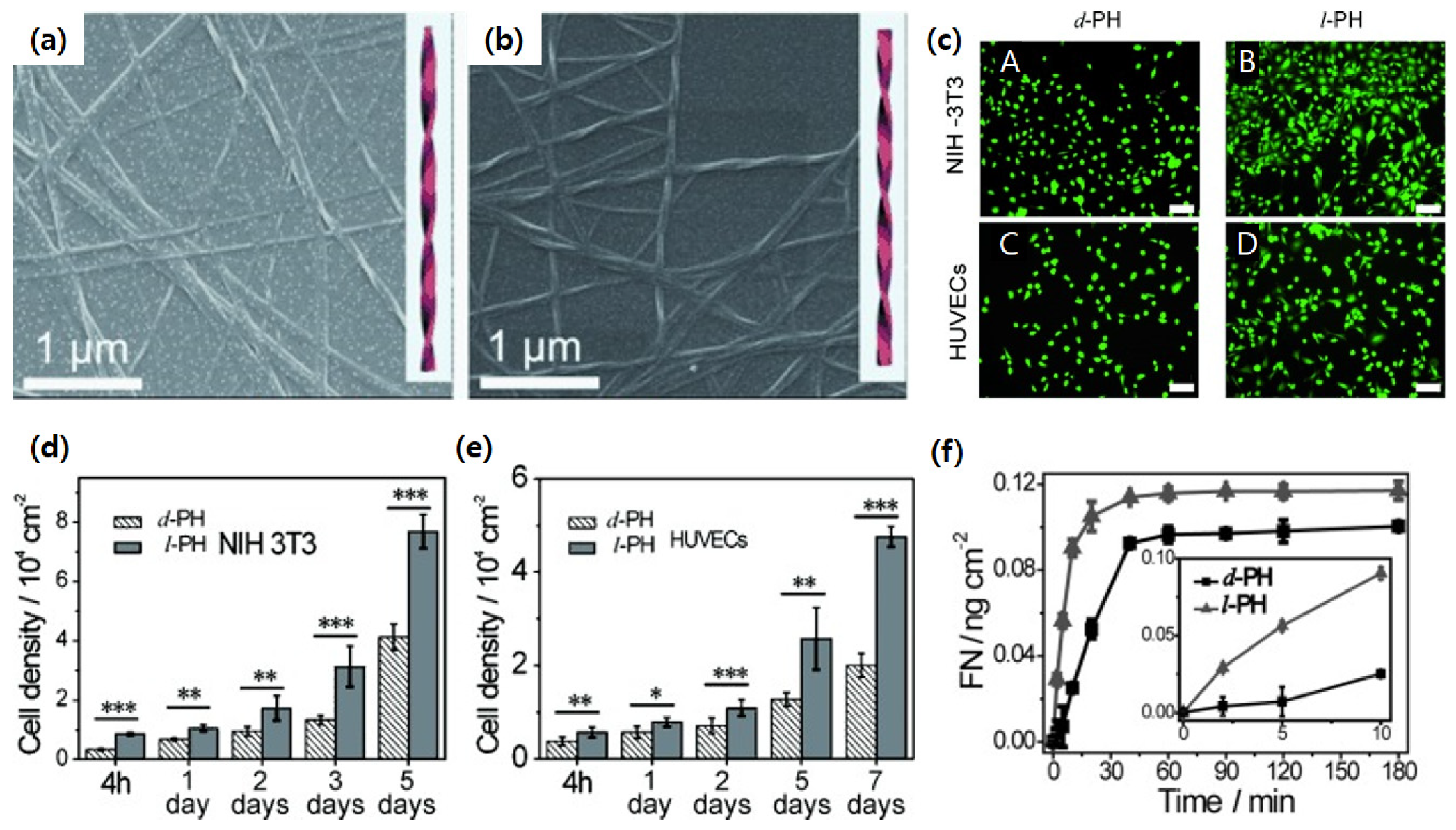
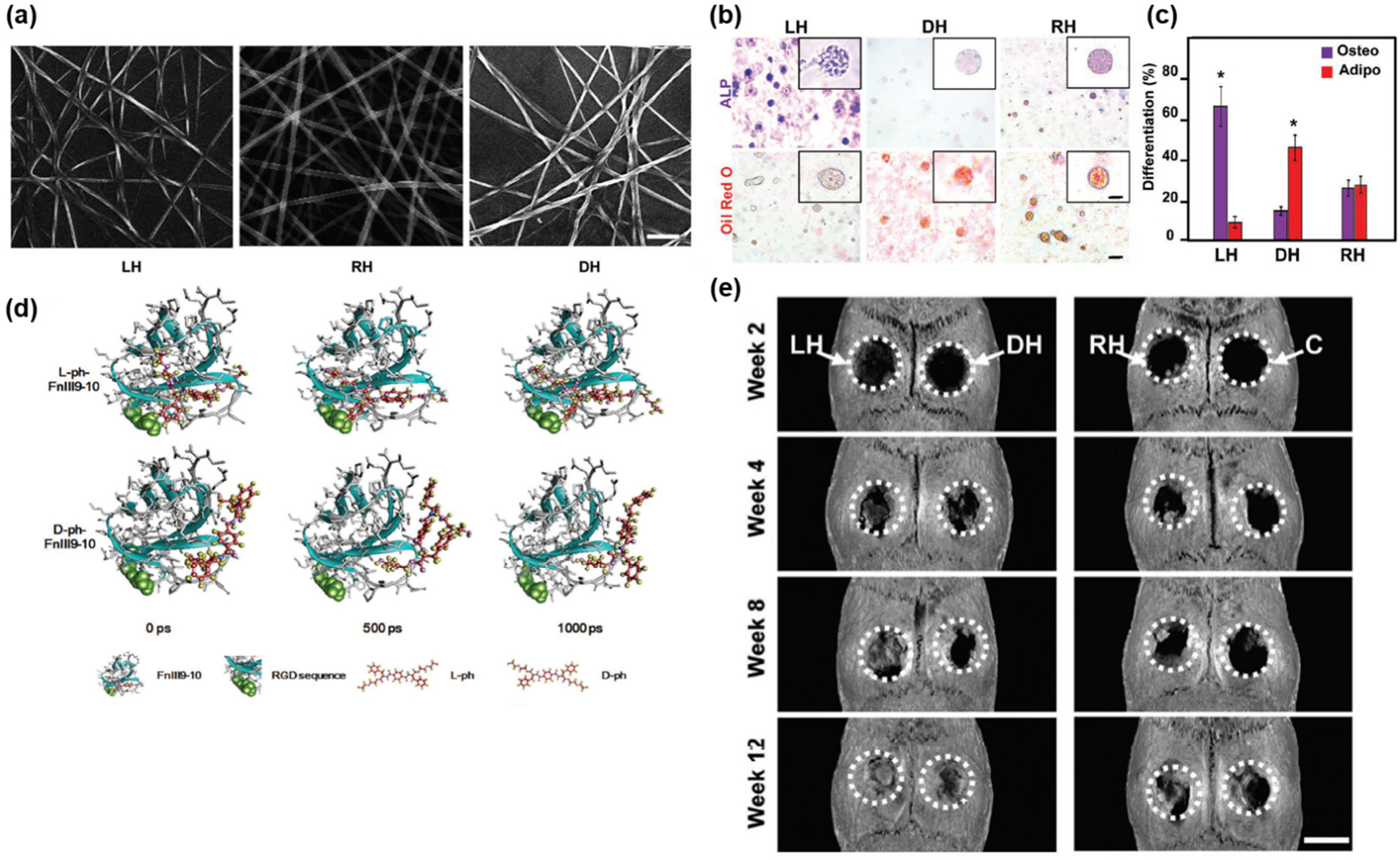
- Liu, G.F.; Zhang, D.; Feng, C.L. Control of three-dimensional cell adhesion by the chirality of nanofibers in hydrogels. Angew. Chem. Int. Ed. 2014, 53, 7789–7793. [Google Scholar] [CrossRef]
- Wei, Y.; Jiang, S.; Si, M.; Zhang, X.; Liu, J.; Wang, Z.; Cao, C.; Huang, J.; Huang, H.; Chen, L. Chirality controls mesenchymal stem cell lineage diversification through mechanoresponses. Adv. Mater. 2019, 31, 1900582. [Google Scholar] [CrossRef]
- Leyssens, L.; Vinck, B.; Van Der Straeten, C.; Wuyts, F.; Maes, L. Cobalt toxicity in humans—A review of the potential sources and systemic health effects. Toxicology 2017, 387, 43–56. [Google Scholar] [CrossRef]
- Leung, C.H.; Chan, D.S.H.; Ma, V.P.Y.; Ma, D.L. DNA-binding small molecules as inhibitors of transcription factors. Med. Res. Rev. 2013, 33, 823–846. [Google Scholar] [CrossRef]
- Zihlif, M.; Catchpoole, D.R.; Stewart, B.W.; Wakelin, L.P. Effects of DNA minor groove binding agents on global gene expression. Cancer Genom. Proteom. 2010, 7, 323–330. [Google Scholar]
- Yu, Z.; Lou, R.; Pan, W.; Li, N.; Tang, B. Nanoenzymes in disease diagnosis and therapy. Chem. Commun. 2020, 56, 15513–15524. [Google Scholar] [CrossRef] [PubMed]
- Muller, P. Glossary of terms used in physical organic chemistry (IUPAC Recommendations 1994). Pure Appl. Chem. 1994, 66, 1077–1184. [Google Scholar] [CrossRef]
- Wintzheimer, S.; Granath, T.; Oppmann, M.; Kister, T.; Thai, T.; Kraus, T.; Vogel, N.; Mandel, K. Supraparticles: Functionality from uniform structural motifs. ACS Nano 2018, 12, 5093–5120. [Google Scholar]
- Shoulders, M.D.; Raines, R.T. Collagen structure and stability. Annu. Rev. Biochem. 2009, 78, 929. [Google Scholar] [CrossRef]
- Jokinen, J.; Dadu, E.; Nykvist, P.; Käpylä, J.; White, D.J.; Ivaska, J.; Vehviläinen, P.; Reunanen, H.; Larjava, H.; Häkkinen, L. Integrin-mediated cell adhesion to type I collagen fibrils. J. Biol. Chem. 2004, 279, 31956–31963. [Google Scholar] [CrossRef]
- Liu, J.; Yuan, F.; Ma, X.; Auphedeous, D.i.Y.; Zhao, C.; Liu, C.; Shen, C.; Feng, C. The cooperative effect of both molecular and supramolecular chirality on cell adhesion. Angew. Chem. 2018, 130, 6585–6589. [Google Scholar] [CrossRef]
- Das, T.; Häring, M.; Haldar, D.; Díaz, D.D. Phenylalanine and derivatives as versatile low-molecular-weight gelators: Design, structure and tailored function. Biomater. Sci. 2018, 6, 38–59. [Google Scholar] [CrossRef]
- Wozniak, M.A.; Modzelewska, K.; Kwong, L.; Keely, P.J. Focal adhesion regulation of cell behavior. Biochim. Biophys. Acta-Mol. Cell Res. 2004, 1692, 103–119. [Google Scholar] [CrossRef]
- Hamidouche, Z.; Fromigué, O.; Ringe, J.; Häupl, T.; Vaudin, P.; Pagès, J.-C.; Srouji, S.; Livne, E.; Marie, P.J. Priming integrin α5 promotes human mesenchymal stromal cell osteoblast differentiation and osteogenesis. Proc. Natl. Acad. Sci. USA 2009, 106, 18587–18591. [Google Scholar] [CrossRef] [PubMed] [Green Version]
- Sordillo, D.C.; Sordillo, L.A.; Sordillo, P.P.; Shi, L.; Alfano, R.R. Short wavelength infrared optical windows for evaluation of benign and malignant tissues. J. Biomed. Opt. 2017, 22, 045002. [Google Scholar] [CrossRef]
- Yan, B.; Boyer, J.-C.; Branda, N.R.; Zhao, Y. Near-infrared light-triggered dissociation of block copolymer micelles using upconverting nanoparticles. J. Am. Chem. Soc. 2011, 133, 19714–19717. [Google Scholar] [CrossRef] [PubMed]
- Jung, H.S.; Han, J.; Lee, J.-H.; Lee, J.H.; Choi, J.-M.; Kweon, H.-S.; Han, J.H.; Kim, J.-H.; Byun, K.M.; Jung, J.H. Enhanced NIR radiation-triggered hyperthermia by mitochondrial targeting. J. Am. Chem. Soc. 2015, 137, 3017–3023. [Google Scholar] [CrossRef] [PubMed]
- Severoni, E.; Maniappan, S.; Liz-Marzan, L.M.; Kumar, J.; Garcia de Abajo, F.J.; Galantini, L. Plasmon-enhanced Optical Chirality through hotspot formation in surfactant-directed self-assembly of gold nanorods. ACS Nano 2020, 14, 16712–16722. [Google Scholar] [CrossRef]
- Pal, S.; Dutta, A.; Paul, M.; Chattopadhyay, A. Plasmon-Enhanced Chemical Reaction at the Hot Spots of End-to-End Assembled Gold Nanorods. J. Phys. Chem. C 2020, 124, 3204–3210. [Google Scholar] [CrossRef]
- Doberenz, F.; Zeng, K.; Willems, C.; Zhang, K.; Groth, T. Thermoresponsive polymers and their biomedical application in tissue engineering–a review. J. Mater. Chem. B 2020, 8, 607–628. [Google Scholar] [CrossRef]
- Moffitt, W.; Moscowitz, A. Optical activity in absorbing media. J. Chem. Phys. 1959, 30, 648–660. [Google Scholar] [CrossRef]
- Franko, A.; Rodriguez Camargo, D.C.; Böddrich, A.; Garg, D.; Rodriguez Camargo, A.; Rathkolb, B.; Janik, D.; Aichler, M.; Feuchtinger, A.; Neff, F. Epigallocatechin gallate (EGCG) reduces the intensity of pancreatic amyloid fibrils in human islet amyloid polypeptide (hIAPP) transgenic mice. Sci. Rep. 2018, 8, 1116. [Google Scholar] [CrossRef]
- Smith, A.M.; Mancini, M.C.; Nie, S. Second window for in vivo imaging. Nat. Nanotechnol. 2009, 4, 710–711. [Google Scholar] [CrossRef] [PubMed]
- Darnell, M.; Mooney, D.J. Leveraging advances in biology to design biomaterials. Nat. Mater. 2017, 16, 1178–1185. [Google Scholar] [CrossRef]
- Lundstrom, K. An overview on GPCRs and drug discovery: Structure-based drug design and structural biology on GPCRs. G Protein-Coupled Recept. Drug Discov. 2009, 552, 51–66. [Google Scholar]
- Ren, J.; Cai, R.; Wang, J.; Daniyal, M.; Baimanov, D.; Liu, Y.; Yin, D.; Liu, Y.; Miao, Q.; Zhao, Y. Precision nanomedicine development based on specific opsonization of human cancer patient-personalized protein coronas. Nano Lett. 2019, 19, 4692–4701. [Google Scholar] [CrossRef] [PubMed]
- Anchordoquy, T.J.; Barenholz, Y.; Boraschi, D.; Chorny, M.; Decuzzi, P.; Dobrovolskaia, M.A.; Farhangrazi, Z.S.; Farrell, D.; Gabizon, A.; Ghandehari, H. Mechanisms and barriers in cancer nanomedicine: Addressing challenges, looking for solutions. ACS Nano 2017, 11, 12–18. [Google Scholar] [CrossRef] [PubMed]
- Kumar, S.; Maurya, V.K.; Chitti, S.V.; Kabir, R.; Shanker, K.; Nayak, D.; Khurana, A.; Manchanda, R.K.; Gadugu, S.; Kumar, V. Wound Healing Activity of a Novel Formulation SKRIN via Induction of Cell Cycle Progression and Inhibition of PCNA–p21 Complex Interaction Leading to Cell Survival and Proliferation. ACS Pharmacol. Transl. Sci. 2021, 4, 352–364. [Google Scholar] [CrossRef]


| Base Materials | Chiral Agents | Size (nm) | Possible Applications | Ref. |
|---|---|---|---|---|
| Co3O4 nanoparticles (NPs) | L-/D-cysteine | 2–3 | Drug delivery system | [2,19] |
| CoxCuyS NPs | L-/D-penicillamine | 2–3 | Selective senescent cell elimination | [20] |
| Graphene quantum dots | L-/D-glutamic acid | 3 | Anti-microbial activity | [21] |
| Carbon dots | L-/D-lysine | 4 | Reducing toxicity of β-amyloid fibril | [22] |
| CdTe NPs | L-/D-cysteine | 4–5 | Site-selective DNA photocleavage | [23] |
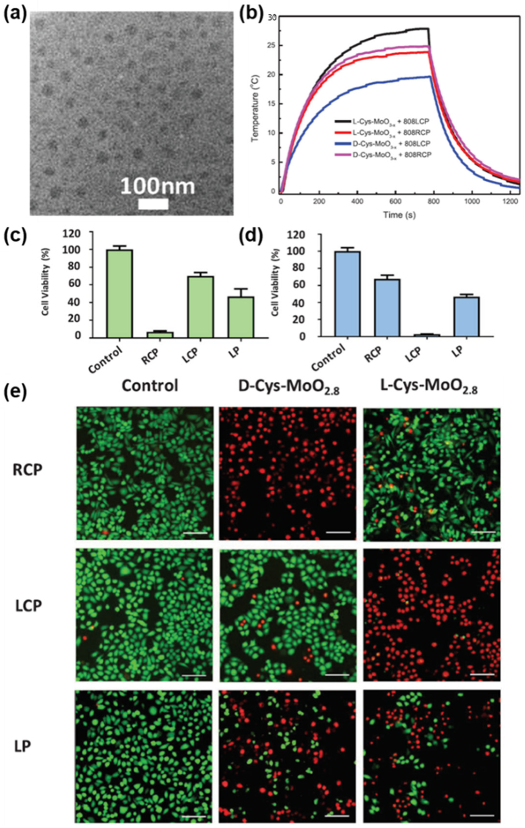
| MoO3−x NPs | L-/D-cysteine | 21–22 | Photothermal therapy | [24] |
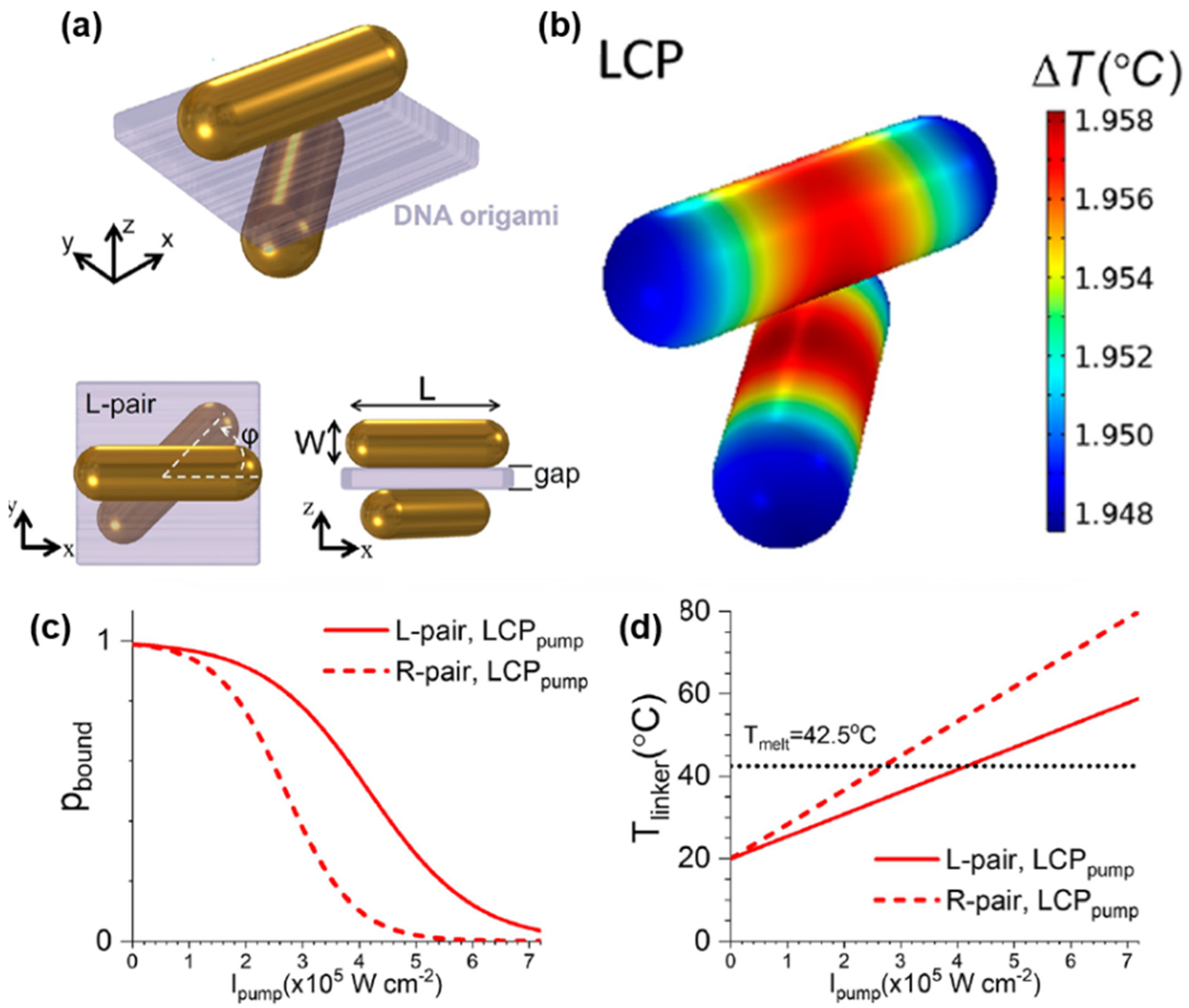
| Au nanorod dimers | DNA origami | 40 | Controlling drug release | [25] |
| FexCuySe NPs | L-/D-penicillamine | 40–50 | β-amyloid fibril elimination | [26] |
| Au bipyramid NPs | L-/D-glutamic acid | 110 (length) 35 (width) | Anti-microbial activity | [27] |
| Cu2−xS nanoflowers | L-/D-cysteine | 1500–2000 | Multi-channel bioimaging | [28] |
| Au nanorod assemblies | Human islet amyloid polypeptides | Several μm (length) 50 (width) | Drug screening | [29] |
| 1,4-benzenedicarboxamide phenylalanine hydrogel | L-/D-1,4-benzenedicarboxamide phenylalanine derivative | Several μm (length) 50–60 (width) | Scaffolds for wound healing | [30,31] |
Publisher’s Note: MDPI stays neutral with regard to jurisdictional claims in published maps and institutional affiliations. |
© 2022 by the authors. Licensee MDPI, Basel, Switzerland. This article is an open access article distributed under the terms and conditions of the Creative Commons Attribution (CC BY) license (https://creativecommons.org/licenses/by/4.0/).
Share and Cite
Jung, W.; Kwon, J.; Cho, W.; Yeom, J. Chiral Biomaterials for Nanomedicines: From Molecules to Supraparticles. Pharmaceutics 2022, 14, 1951. https://doi.org/10.3390/pharmaceutics14091951
Jung W, Kwon J, Cho W, Yeom J. Chiral Biomaterials for Nanomedicines: From Molecules to Supraparticles. Pharmaceutics. 2022; 14(9):1951. https://doi.org/10.3390/pharmaceutics14091951
Chicago/Turabian StyleJung, Wookjin, Junyoung Kwon, Wonjoon Cho, and Jihyeon Yeom. 2022. "Chiral Biomaterials for Nanomedicines: From Molecules to Supraparticles" Pharmaceutics 14, no. 9: 1951. https://doi.org/10.3390/pharmaceutics14091951