
Encapsulation of miRNA and siRNA into Nanomaterials for Cancer Therapeutics

 ,
,  , ,
, ,  ,
, 
Abstract
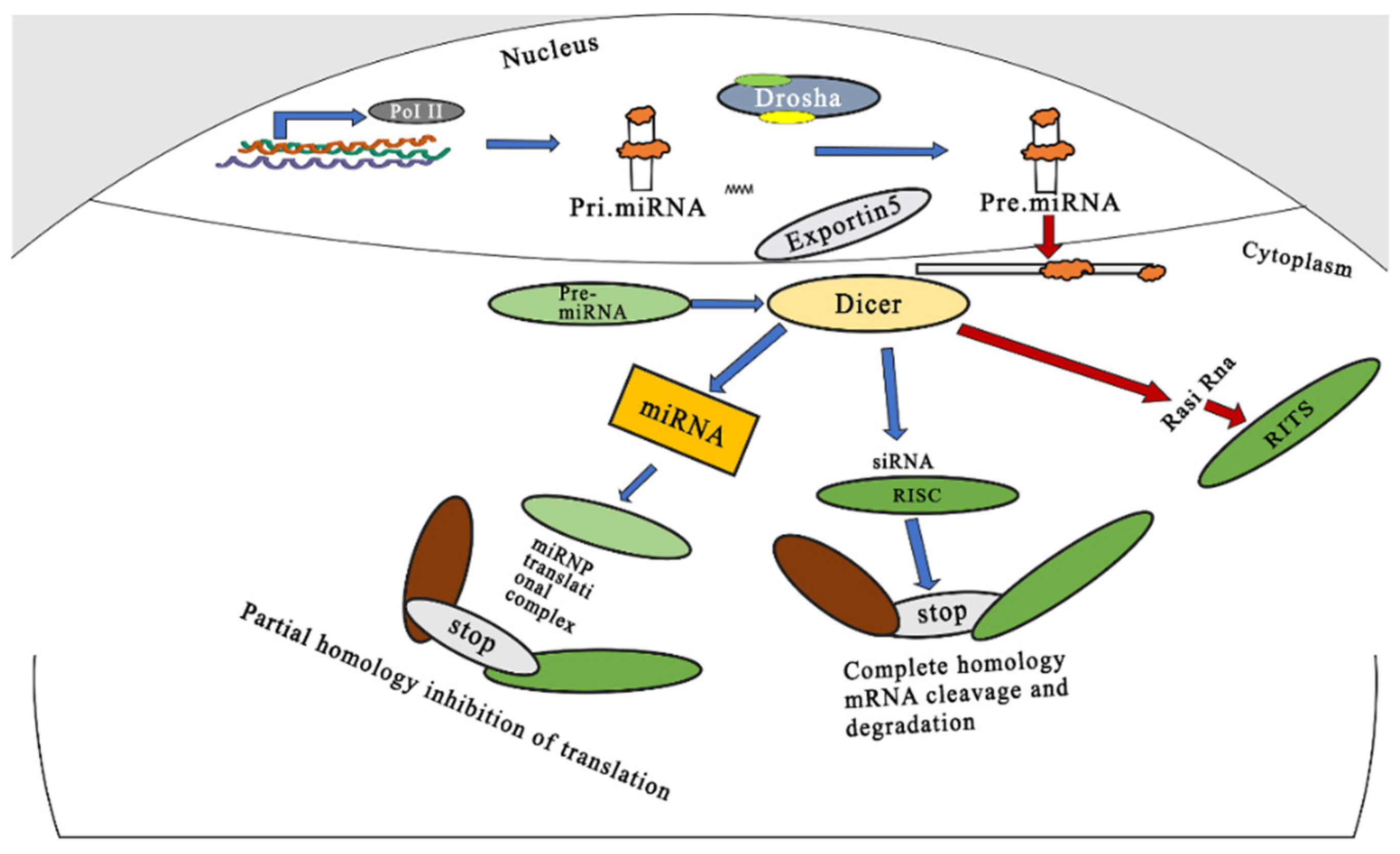
:1. Introduction
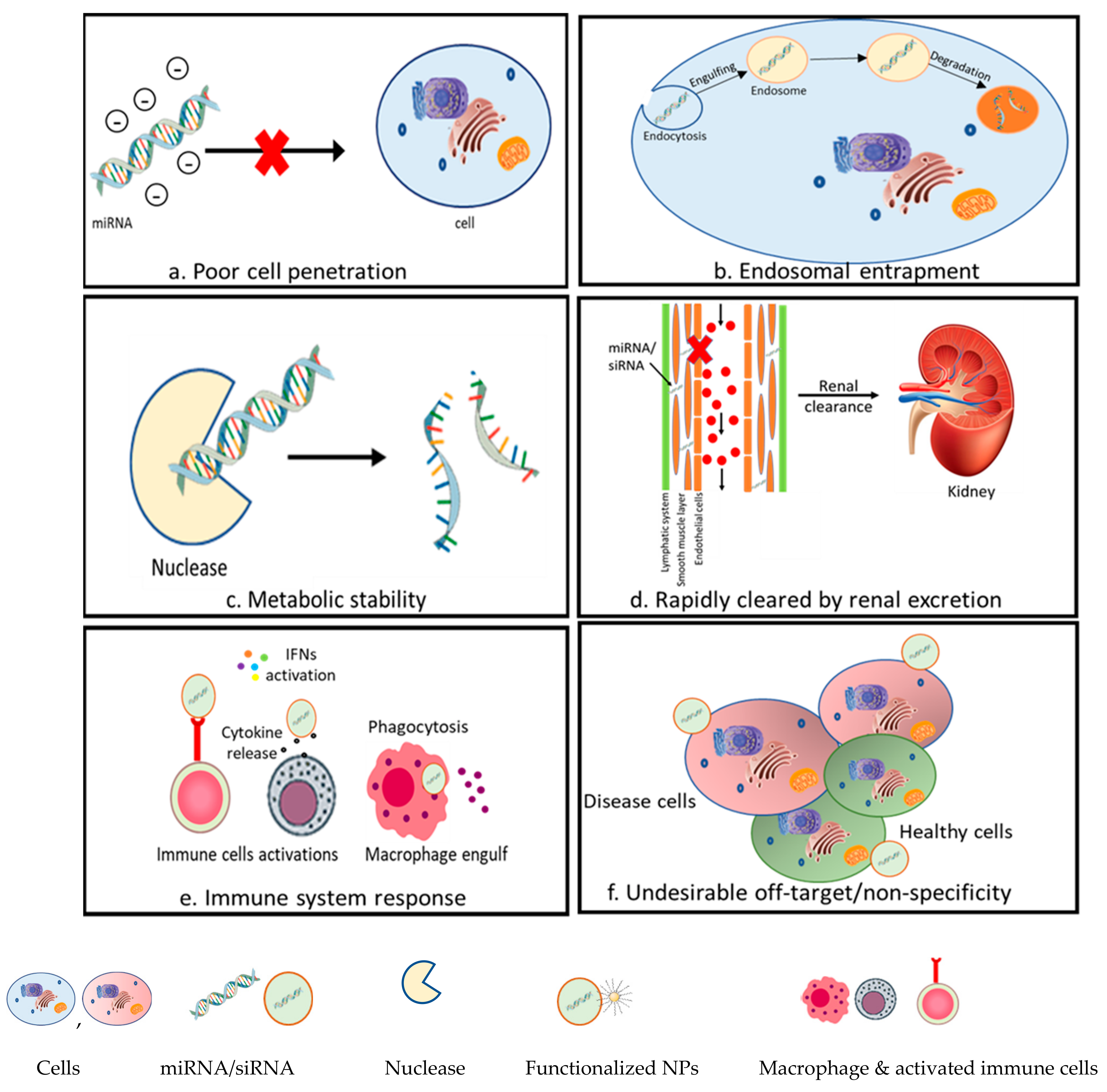
2. Challenges Associated with RNAi Therapeutics
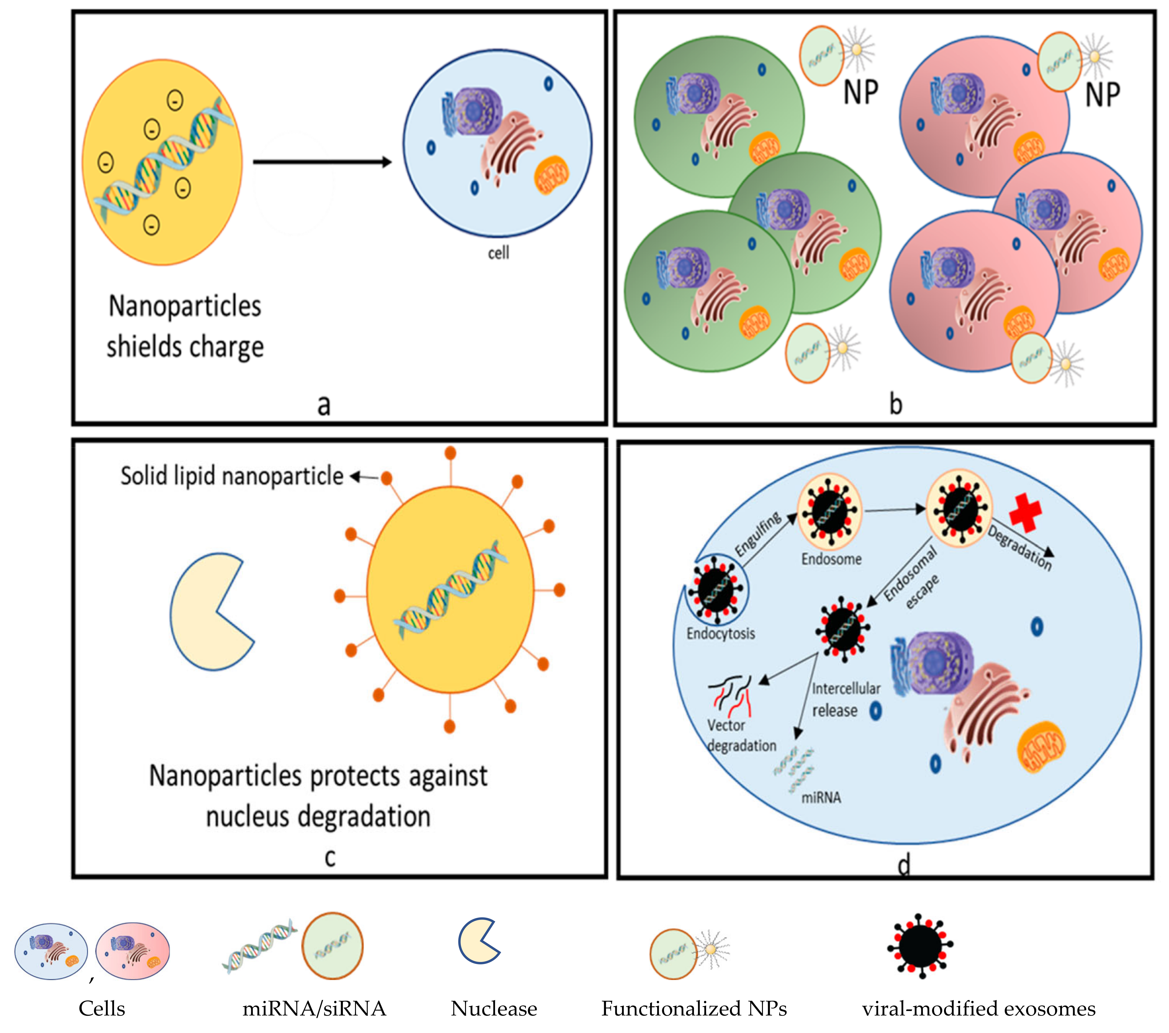
2.1. Poor Cell Membrane Penetration
2.2. Off-Target Effects
2.3. Metabolic Stability and Bioavailability
2.4. Innate Immune System Activation
3. Modification Strategies to Overcome the Challenges of Nanocarriers
4. The Importance of Using Nanoparticles for siRNA and miRNA Delivery
5. Important Classes of Nanocarriers Used for siRNA and miRNA Delivery
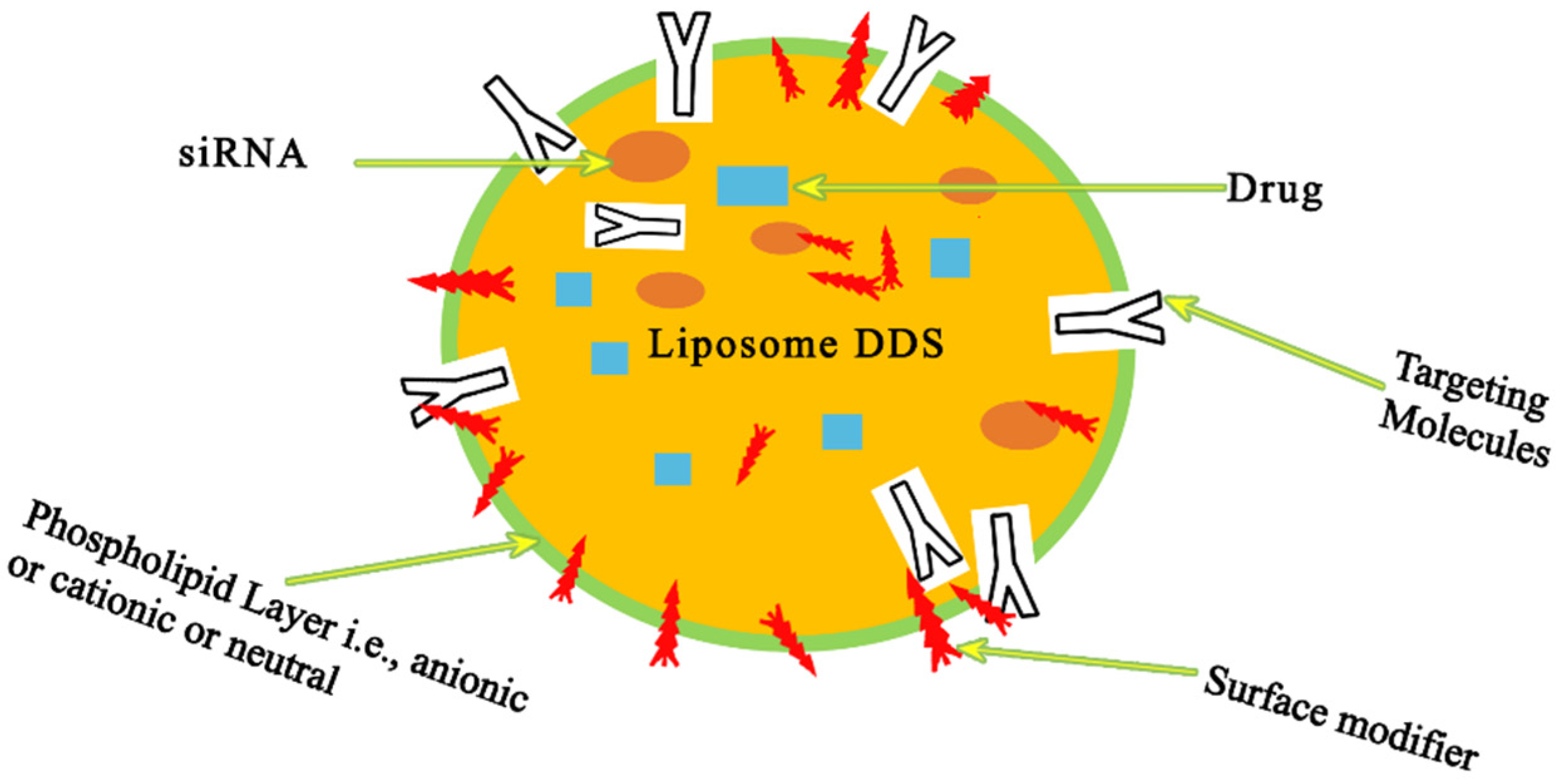
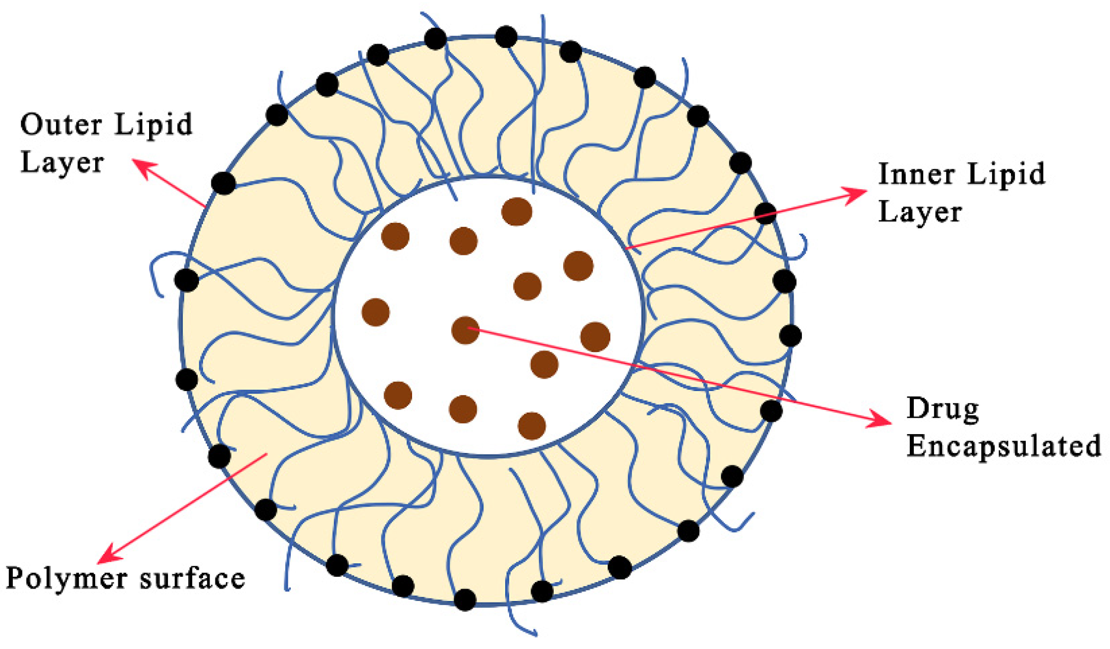
5.1. Liposome-Based Nanocarriers
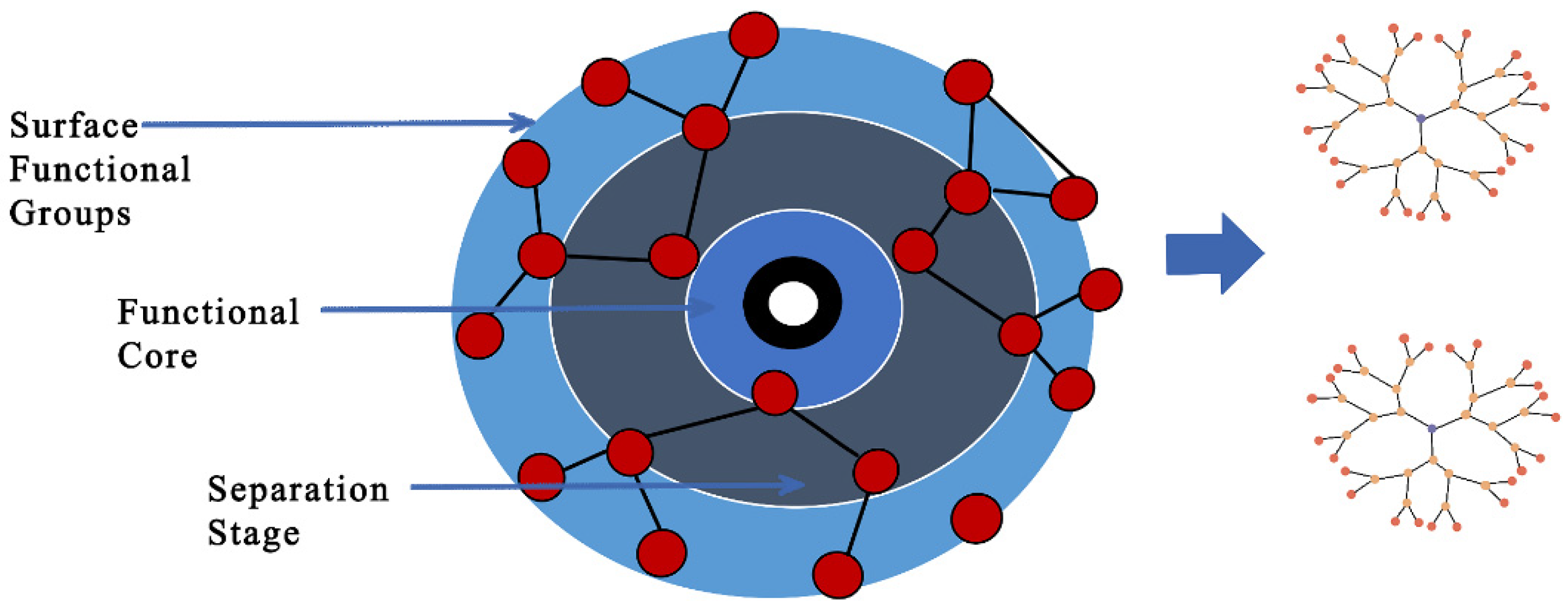
5.2. Dendrimer-Based Nanocarriers
5.3. Micelle-Based Nanocarriers and Other Biodegradable Polymers
6. Advantages of Nanocarriers in miRNA Therapeutics
7. Nanocarrier Customizations for the Delivery of Gene Agents for Cancer Therapy
8. Application of Nanocarriers
9. Opportunities in miRNA Therapeutics
10. Conclusions and Future Perspectives
Author Contributions
Funding
Institutional Review Board Statement
Informed Consent Statement
Data Availability Statement
Conflicts of Interest
References
- Djojosubroto, M.W.; Choi, Y.S.; Lee, H.W.; Rudolph, K.L. Telomeres and Telomerase in Aging, Regeneration and Cancer. Mol. Cells 2003, 15, 164–175. [Google Scholar] [PubMed]
- Heath, J.R.; Davis, M.E. Nanotechnology and Cancer. Annu. Rev. Med. 2008, 59, 251–265. [Google Scholar] [CrossRef] [PubMed]
- Hirsch, L.R.; Stafford, R.J.; Bankson, J.A.; Sershen, S.R.; Rivera, B.; Price, R.E.; Hazle, J.D.; Halas, N.J.; West, J.L. Nanoshell-Mediated near-Infrared Thermal Therapy of Tumors under Magnetic Resonance Guidance. Proc. Natl. Acad. Sci. USA 2003, 100, 13549–13554. [Google Scholar] [CrossRef] [PubMed]
- Wagner, A.; Hempel, G.; Boos, J. Trofosfamide: A Review of Its Pharmacodynamic and Pharmacokinetic Properties and Therapeutic Potential in the Oral Treatment of Cancer. Anti-Cancer Drugs 1997, 8, 419–431. [Google Scholar] [CrossRef] [PubMed]
- Bae, K.H.; Chung, H.J.; Park, T.G. Nanomaterials for Cancer Therapy and Imaging. Mol. Cells 2011, 31, 295–302. [Google Scholar] [CrossRef]
- Syn, N.L.; Wang, L.; Chow, E.K.H.; Lim, C.T.; Goh, B.C. Exosomes in Cancer Nanomedicine and Immunotherapy: Prospects and Challenges. Trends Biotechnol. 2017, 35, 665–676. [Google Scholar] [CrossRef]
- Raza, A.; Hayat, U.; Rasheed, T.; Bilal, M.; Iqbal, H.M.N. “smart” Materials-Based near-Infrared Light-Responsive Drug Delivery Systems for Cancer Treatment: A Review. J. Mater. Res. Technol. 2019, 8, 1497–1509. [Google Scholar] [CrossRef]
- Matteucci, F.; Giannantonio, R.; Calabi, F.; Agostiano, A.; Gigli, G.; Rossi, M. Deployment and Exploitation of Nanotechnology Nanomaterials and Nanomedicine. AIP Conf. Proc. 2018, 1990, 020001. [Google Scholar] [CrossRef]
- Pérez-Herrero, E.; Fernández-Medarde, A. Advanced Targeted Therapies in Cancer: Drug Nanocarriers, the Future of Chemotherapy. Eur. J. Pharm. Biopharm. 2015, 93, 52–79. [Google Scholar] [CrossRef]
- Saad, M.; Garbuzenko, O.B.; Minko, T. Co-Delivery of SiRNA and an Anticancer Drug for Treatment of Multidrug-Resistant Cancer. Nanomedicine 2008, 3, 761–776. [Google Scholar] [CrossRef]
- Ball, R.L.; Hajj, K.A.; Vizelman, J.; Bajaj, P.; Whitehead, K.A. Lipid Nanoparticle Formulations for Enhanced Co-Delivery of SiRNA and MRNA. Nano Lett. 2018, 18, 3814–3822. [Google Scholar] [CrossRef] [PubMed]
- Venturini, L.; Eder, M.; Scherr, M. RNA-Mediated Gene Silencing in Hematopoietic Cells. J. Biomed. Biotechnol. 2006, 2006, 087340. [Google Scholar] [CrossRef] [PubMed]
- Khan, D.R.; Rezler, E.M.; Lauer-Fields, J.; Fields, G.B. Effects of Drug Hydrophobicity on Liposomal Stability. Chem. Biol. Drug Des. 2008, 71, 3–7. [Google Scholar] [CrossRef]
- Li, S.D.; Chen, Y.C.; Hackett, M.J.; Huang, L. Tumor-Targeted Delivery of SiRNA by Self-Assembled Nanoparticles. Mol. Ther. 2008, 16, 163–169. [Google Scholar] [CrossRef] [PubMed]
- Kang, S.H.; Cho, H.J.; Shim, G.; Lee, S.; Kim, S.H.; Choi, H.G.; Kim, C.W.; Oh, Y.K. Cationic Liposomal Co-Delivery of Small Interfering RNA and a MEK Inhibitor for Enhanced Anticancer Efficacy. Pharm. Res. 2011, 28, 3069–3078. [Google Scholar] [CrossRef] [PubMed]
- Gandhi, N.S.; Tekade, R.K.; Chougule, M.B. Nanocarrier Mediated Delivery of SiRNA/MiRNA in Combination with Chemotherapeutic Agents for Cancer Therapy: Current Progress and Advances. J. Control. Release 2014, 194, 238–256. [Google Scholar] [CrossRef] [PubMed]
- Bosman, A.W.; Janssen, H.M.; Meijer, E.W. About Dendrimers: Structure, Physical Properties, and Applications. Chem. Rev. 1999, 99, 1665–1688. [Google Scholar] [CrossRef]
- Crooks, R.M.; Zhao, M.; Sun, L.; Chechik, V.; Yeung, L.K. Dendrimer-Encapsulated Metal Nanoparticles: Synthesis, Characterization, and Applications to Catalysis. Acc. Chem. Res. 2001, 34, 181–190. [Google Scholar] [CrossRef]
- Mammen, M.; Choi, S.K.; Whitesides, G.M. Polyvalent Interactions in Biological Systems: Implications for Design and Use of Multivalent Ligands and Inhibitors. Angew. Chem. Int. Ed. 1998, 37, 2754–2794. [Google Scholar] [CrossRef]
- Crooke, S.T.; Wang, S.; Vickers, T.A.; Shen, W.; Liang, X.H. Cellular Uptake and Trafficking of Antisense Oligonucleotides. Nat. Biotechnol. 2017, 35, 230–237. [Google Scholar] [CrossRef]
- Leung, A.K.K.; Tam, Y.Y.C.; Cullis, P.R. Lipid Nanoparticles for Short Interfering RNA Delivery; Elsevier: Amsterdam, The Netherlands, 2014; Volume 88, ISBN 9780128001486. [Google Scholar]
- Suk, J.S.; Xu, Q.; Kim, N.; Hanes, J.; Ensign, L.M. PEGylation as a Strategy for Improving Nanoparticle-Based Drug and Gene Delivery. Adv. Drug Deliv. Rev. 2016, 99, 28–51. [Google Scholar] [CrossRef]
- Sato, Y.; Hatakeyama, H.; Sakurai, Y.; Hyodo, M.; Akita, H.; Harashima, H. A PH-Sensitive Cationic Lipid Facilitates the Delivery of Liposomal SiRNA and Gene Silencing Activity in Vitro and in Vivo. J. Control. Release 2012, 163, 267–276. [Google Scholar] [CrossRef]
- Bus, T.; Traeger, A.; Schubert, U.S. The Great Escape: How Cationic Polyplexes Overcome the Endosomal Barrier. J. Mater. Chem. B 2018, 6, 6904–6918. [Google Scholar] [CrossRef] [PubMed]
- Lee, J.; Sands, I.; Zhang, W.; Zhou, L.; Chen, Y. DNA-Inspired Nanomaterials for Enhanced Endosomal Escape. Proc. Natl. Acad. Sci. USA 2021, 118, 3–5. [Google Scholar] [CrossRef] [PubMed]
- Jackson, A.L.; Bartz, S.R.; Schelter, J.; Kobayashi, S.V.; Burchard, J.; Mao, M.; Li, B.; Cavet, G.; Linsley, P.S. Expression Profiling Reveals Off-Target Gene Regulation by RNAi. Nat. Biotechnol. 2003, 21, 635–637. [Google Scholar] [CrossRef] [PubMed]
- Suter, S.R.; Ball-Jones, A.; Mumbleau, M.M.; Valenzuela, R.; Ibarra-Soza, J.; Owens, H.; Fisher, A.J.; Beal, P.A. Controlling MiRNA-like off-Target Effects of an SiRNA with Nucleobase Modifications. Org. Biomol. Chem. 2017, 15, 10029–10036. [Google Scholar] [CrossRef]
- Janas, M.M.; Schlegel, M.K.; Harbison, C.E.; Yilmaz, V.O.; Jiang, Y.; Parmar, R.; Zlatev, I.; Castoreno, A.; Xu, H.; Shulga-Morskaya, S.; et al. Selection of GalNAc-Conjugated SiRNAs with Limited off-Target-Driven Rat Hepatotoxicity. Nat. Commun. 2018, 9, 723. [Google Scholar] [CrossRef]
- Ivanovska, I. Combinatorial MicroRNAs. Cell Cycle 2008, 7, 3137–3142. [Google Scholar] [CrossRef]
- Bandi, N.; Vassella, E. MiR-34a and MiR-15a/16 Are Co-Regulated in Non-Small Cell Lung Cancer and Control Cell Cycle Progression in a Synergistic and Rb-Dependent Manner. Mol. Cancer 2011, 10, 55. [Google Scholar] [CrossRef]
- Shegokar, R.; Shaal, L.A.L.; Mishra, P.R. SiRNA Delivery: Challenges and Role of Carrier Systems. Pharmazie 2011, 66, 313–318. [Google Scholar] [CrossRef] [PubMed]
- Kwekkeboom, R.F.J.; Lei, Z.; Doevendans, P.A.; Musters, R.J.P.; Sluijter, J.P.G. Targeted Delivery of MiRNA Therapeutics for Cardiovascular Diseases: Opportunities and Challenges. Clin. Sci. 2014, 127, 351–365. [Google Scholar] [CrossRef] [PubMed]
- Segal, M.; Slack, F.J. Challenges Identifying Efficacious MiRNA Therapeutics for Cancer. Expert Opin. Drug Discov. 2020, 15, 987–992. [Google Scholar] [CrossRef] [PubMed]
- Fernandez-Valverde, S.L.; Taft, R.J.; Mattick, J.S. Dynamic IsomiR Regulation in Drosophila Development. RNA 2010, 16, 1881–1888. [Google Scholar] [CrossRef] [PubMed]
- Mil, A.; Doevendans, P.; Sluijter, J. The Potential of Modulating Small RNA Activity In Vivo. Mini-Rev. Med. Chem. 2009, 9, 235–248. [Google Scholar] [CrossRef]
- Breslow, R.; Chapman, W.H. On the Mechanism of Action of Ribonuclease A: Relevance of Enzymatic Studies with a p-Nitrophenylphosphate Ester and a Thiophosphate Ester. Proc. Natl. Acad. Sci. USA 1996, 93, 10018–10021. [Google Scholar] [CrossRef]
- Overhoff, M.; Sczakiel, G. Phosphorothioate-Stimulated Uptake of Short Interfering RNA by Human Cells. EMBO Rep. 2005, 6, 1176–1181. [Google Scholar] [CrossRef]
- Chernikov, I.V.; Gladkikh, D.V.; Meschaninova, M.I.; Ven’yaminova, A.G.; Zenkova, M.A.; Vlassov, V.V.; Chernolovskaya, E.L. Cholesterol-Containing Nuclease-Resistant SiRNA Accumulates in Tumors in a Carrier-Free Mode and Silences MDR1 Gene. Mol. Ther.-Nucleic Acids 2017, 6, 209–220. [Google Scholar] [CrossRef]
- Qian, X.; Long, L.; Shi, Z.; Liu, C.; Qiu, M.; Sheng, J.; Pu, P.; Yuan, X.; Ren, Y.; Kang, C. Star-Branched Amphiphilic PLA-b-PDMAEMA Copolymers for Co-Delivery of MiR-21 Inhibitor and Doxorubicin to Treat Glioma. Biomaterials 2014, 35, 2322–2335. [Google Scholar] [CrossRef]
- Lamb, Y.N. Inclisiran: First Approval. Drugs 2021, 81, 389–395. [Google Scholar] [CrossRef] [PubMed]
- Karikó, K.; Bhuyan, P.; Capodici, J.; Weissman, D. Small Interfering RNAs Mediate Sequence-Independent Gene Suppression and Induce Immune Activation by Signaling through Toll-Like Receptor 3. J. Immunol. 2004, 172, 6545–6549. [Google Scholar] [CrossRef]
- Pichlmair, A.; Schulz, O.; Tan, C.P.; Näslund, T.I.; Liljeström, P.; Weber, F.; Reis, E.; Sousa, C. RIG-I-Mediated Antiviral Responses to Single-Stranded RNA Bearing 5′-Phosphates. Science 2006, 314, 997–1001. [Google Scholar] [CrossRef] [PubMed]
- Li, S.; Peters, G.A.; Ding, K.; Zhang, X.; Qin, J.; Sen, G.C. Molecular Basis for PKR Activation by PACT or DsRNA. Proc. Natl. Acad. Sci. USA 2006, 103, 10005–10010. [Google Scholar] [CrossRef] [PubMed]
- Biswas, S.; Deshpande, P.P.; Navarro, G.; Dodwadkar, N.S.; Torchilin, V.P. Lipid Modified Triblock PAMAM-Based Nanocarriers for SiRNA Drug Co-Delivery. Biomaterials 2013, 34, 1289–1301. [Google Scholar] [CrossRef] [PubMed]
- Stepanov, G.; Zhuravlev, E.; Shender, V.; Nushtaeva, A.; Balakhonova, E.; Mozhaeva, E.; Kasakin, M.; Koval, V.; Lomzov, A.; Pavlyukov, M.; et al. Nucleotide Modifications Decrease Innate Immune Response Induced by Synthetic Analogs of SnRNAs and SnoRNAs. Genes 2018, 9, 531. [Google Scholar] [CrossRef] [PubMed]
- Wu, S.Y.; Chen, T.M.; Gmeiner, W.H.; Chu, E.; Schmitz, J.C. Development of Modified SiRNA Molecules Incorporating 5-Fluoro-2′-Deoxyuridine Residues to Enhance Cytotoxicity. Nucleic Acids Res. 2013, 41, 4650–4659. [Google Scholar] [CrossRef] [PubMed]
- Xia, J.; Noronha, A.; Toudjarska, I.; Li, F.; Akinc, A.; Braich, R.; Frank-Kamenetsky, M.; Rajeev, K.G.; Egli, M.; Manoharan, M. Gene Silencing Activity of SiRNAs with a Ribo-Difluorotoluyl Nucleotide. ACS Chem. Biol. 2006, 1, 176–183. [Google Scholar] [CrossRef]
- Esposito, C.L.; Catuogno, S.; De Franciscis, V. Aptamer-MiRNA Conjugates for Cancer Cell-Targeted Delivery. Methods Mol. Biol. 2016, 1364, 197–208. [Google Scholar] [CrossRef]
- Bolhassani, A.; Javanzad, S.; Saleh, T.; Hashemi, M.; Aghasadeghi, M.R.; Sadat, S.M. Polymeric Nanoparticles Potent Vectors for Vaccine Delivery Targeting Cancer and Infectious Diseases. Hum. Vaccines Immunother. 2014, 10, 321–332. [Google Scholar] [CrossRef]
- Fu, Y.; Chen, J.; Huang, Z. Recent Progress in Microrna-Based Delivery Systems for the Treatment of Human Disease. ExRNA 2019, 1, 24. [Google Scholar] [CrossRef]
- Crew, E.; Rahman, S.; Razzak-Jaffar, A.; Mott, D.; Kamundi, M.; Yu, G.; Tchah, N.; Lee, J.; Bellavia, M.; Zhong, C.J. MicroRNA Conjugated Gold Nanoparticles and Cell Transfection. Anal. Chem. 2012, 84, 26–29. [Google Scholar] [CrossRef]
- Lee, S.W.L.; Paoletti, C.; Campisi, M.; Osaki, T.; Adriani, G.; Kamm, R.D.; Mattu, C.; Chiono, V. MicroRNA Delivery through Nanoparticles. J. Control. Release 2019, 313, 80–95. [Google Scholar] [CrossRef] [PubMed]
- Akbarzadeh, A.; Rezaei-sadabady, R.; Davaran, S.; Joo, S.W.; Zarghami, N. Liposome: Classification, Preparation, and Applications. Nanoscale Res. Lett. 2013, 8, 102. [Google Scholar] [CrossRef] [PubMed]
- Gasparello, J.; Manicardi, A.; Casnati, A.; Corradini, R.; Gambari, R.; Finotti, A.; Sansone, F. Efficient Cell Penetration and Delivery of Peptide Nucleic Acids by an Argininocalix [4]Arene. Sci. Rep. 2019, 9, 3036. [Google Scholar] [CrossRef] [PubMed]
- Tomar, R.S.; Matta, H.; Chaudhary, P.M. Use of Adeno-Associated Viral Vector for Delivery of Small Interfering RNA. Oncogene 2003, 22, 5712–5715. [Google Scholar] [CrossRef] [PubMed]
- Hu, Y.; Jiang, K.; Wang, D.; Yao, S.; Lu, L.; Wang, H.; Song, J.; Zhou, J.; Fan, X.; Wang, Y.; et al. Core-Shell Lipoplexes Inducing Active Macropinocytosis Promote Intranasal Delivery of c-Myc SiRNA for Treatment of Glioblastoma. Acta Biomater. 2022, 138, 478–490. [Google Scholar] [CrossRef]
- Moitra, P.; Misra, S.K.; Kumar, K.; Kondaiah, P.; Tran, P.; Duan, W.; Bhattacharya, S. Cancer Stem Cell-Targeted Gene Delivery Mediated by Aptamer-Decorated PH-Sensitive Nanoliposomes. ACS Biomater. Sci. Eng. 2021, 7, 2508–2519. [Google Scholar] [CrossRef]
- Zhang, Y.; Qin, Y.; Li, H.; Peng, Q.; Wang, P.; Yang, L.; Chen, S.; Li, M.; Fu, J.; Yu, X.; et al. Artificial Platelets for Efficient SiRNA Delivery to Clear “Bad Cholesterol”. ACS Appl. Mater. Interfaces 2020, 12, 28034–28046. [Google Scholar] [CrossRef] [PubMed]
- Perisé-Barrios, A.J.; Jiménez, J.L.; D’Omínguez-Soto, A.; De La Mata, F.J.; Corbí, A.L.; Gomez, R.; Muñoz-Fernandez, M.Á. Carbosilane Dendrimers as Gene Delivery Agents for the Treatment of HIV Infection. J. Control. Release 2014, 184, 51–57. [Google Scholar] [CrossRef]
- Zhang, M.; Lin, J.; Jin, J.; Yu, W.; Qi, Y.; Tao, H. Delivery of SiRNA Using Functionalized Gold Nanorods Enhances Anti-Osteosarcoma Efficacy. Front. Pharmacol. 2021, 12, 1–14. [Google Scholar] [CrossRef] [PubMed]
- Rai, K.; Takigawa, N.; Ito, S.; Kashihara, H.; Ichihara, E.; Yasuda, T.; Shimizu, K.; Tanimoto, M.; Kiura, K. Liposomal Delivery of MicroRNA-7-Expressing Plasmid Overcomes Epidermal Growth Factor Receptor Tyrosine Kinase Inhibitor-Resistance in Lung Cancer Cells. Mol. Cancer Ther. 2011, 10, 1720–1727. [Google Scholar] [CrossRef] [PubMed]
- Chen, Y.; Bathula, S.R.; Li, J.; Huang, L. Multifunctional Nanoparticles Delivering Small Interfering RNA and Doxorubicin Overcome Drug Resistance in Cancer. J. Biol. Chem. 2010, 285, 22639–22650. [Google Scholar] [CrossRef] [PubMed]
- Tominaga, N.; Kosaka, N.; Ono, M.; Katsuda, T.; Yoshioka, Y.; Tamura, K.; Lötvall, J.; Nakagama, H.; Ochiya, T. Brain Metastatic Cancer Cells Release MicroRNA-181c-Containing Extracellular Vesicles Capable of Destructing Blood-Brain Barrier. Nat. Commun. 2015, 6, 6716. [Google Scholar] [CrossRef] [PubMed]
- Kapadia, C.H.; Ioele, S.A.; Day, E.S. Layer-by-Layer Assembled PLGA Nanoparticles Carrying MiR-34a Cargo Inhibit the Proliferation and Cell Cycle Progression of Triple-Negative Breast Cancer Cells. J. Biomed. Mater. Res. Part A 2020, 108, 601–613. [Google Scholar] [CrossRef]
- Li, Y.; Duo, Y.; Bi, J.; Zeng, X.; Mei, L.; Bao, S.; He, L.; Shan, A.; Zhang, Y.; Yu, X. Targeted delivery of anti-miR-155 by functionalized mesoporous silica nanoparticles for colorectal cancer therapy. Int. J. Nanomed. 2018, 13, 1241. [Google Scholar] [CrossRef] [PubMed]
- Zhang, Q.; Ran, R.; Zhang, L.; Liu, Y.; Mei, L.; Zhang, Z.; Gao, H.; He, Q. Simultaneous Delivery of Therapeutic Antagomirs with Paclitaxel for the Management of Metastatic Tumors by a PH-Responsive Anti-Microbial Peptide-Mediated Liposomal Delivery System; Elsevier: Amsterdam, The Netherlands, 2015; Volume 197, ISBN 8628855025. [Google Scholar]
- Jin, M.; Jin, G.; Kang, L.; Chen, L.; Gao, Z.; Huang, W. Smart Polymeric Nanoparticles with PH-Responsive and PEG-Detachable Properties for Co-Delivering Paclitaxel and Survivin SiRNA to Enhance Antitumor Outcomes. Int. J. Nanomed. 2018, 13, 2405–2426. [Google Scholar] [CrossRef]
- Avci, Ç.B.; Özcan, I.; Balci, T.; Özer, Ö.; Gündüz, C. Design of Polyethylene Glycol-Polyethylenimine Nanocomplexes as Non-Viral Carriers: Mir-150 Delivery to Chronic Myeloid Leukemia Cells. Cell Biol. Int. 2013, 37, 1205–1214. [Google Scholar] [CrossRef]
- Begines, B.; Ortiz, T.; Pérez-Aranda, M.; Martínez, G.; Merinero, M.; Argüelles-Arias, F.; Alcudia, A. Polymeric Nanoparticles for Drug Delivery: Recent Developments and Future Prospects. Nanomaterials 2020, 10, 1403. [Google Scholar] [CrossRef] [PubMed]
- Li, J.; Liang, H.; Liu, J.; Wang, Z. Poly (Amidoamine) (PAMAM) Dendrimer Mediated Delivery of Drug and PDNA/SiRNA for Cancer Therapy. Int. J. Pharm. 2018, 546, 215–225. [Google Scholar] [CrossRef]
- Oner, E.; Kotmakci, M.; Baird, A.M.; Gray, S.G.; Debelec Butuner, B.; Bozkurt, E.; Kantarci, A.G.; Finn, S.P. Development of EphA2 SiRNA-Loaded Lipid Nanoparticles and Combination with a Small-molecule Histone Demethylase Inhibitor in Prostate Cancer Cells and Tumor Spheroids. J. Nanobiotechnol. 2021, 19, 71. [Google Scholar] [CrossRef]
- Mousazadeh, H.; Pilehvar-Soltanahmadi, Y.; Dadashpour, M.; Zarghami, N. Cyclodextrin Based Natural Nanostructured Carbohydrate Polymers as Effective Non-Viral SiRNA Delivery Systems for Cancer Gene Therapy. J. Control. Release 2021, 330, 1046–1070. [Google Scholar] [CrossRef] [PubMed]
- Naito, M.; Chaya, H.; Toh, K.; Kim, B.S.; Hayashi, K.; Fukushima, S.; Nagata, T.; Yokota, T.; Kataoka, K.; Miyata, K. Structural Tuning of Oligonucleotides for Enhanced Blood Circulation Properties of Unit Polyion Complexes Prepared from Two-Branched Poly(Ethylene Glycol)-Block-Poly(L-Lysine). J. Control. Release 2021, 330, 812–820. [Google Scholar] [CrossRef]
- Sun, Q.; Kang, Z.; Xue, L.; Shang, Y.; Su, Z.; Sun, H.; Ping, Q.; Mo, R.; Zhang, C. A Collaborative Assembly Strategy for Tumor-Targeted SiRNA Delivery. J. Am. Chem. Soc. 2015, 137, 6000–6010. [Google Scholar] [CrossRef] [PubMed]
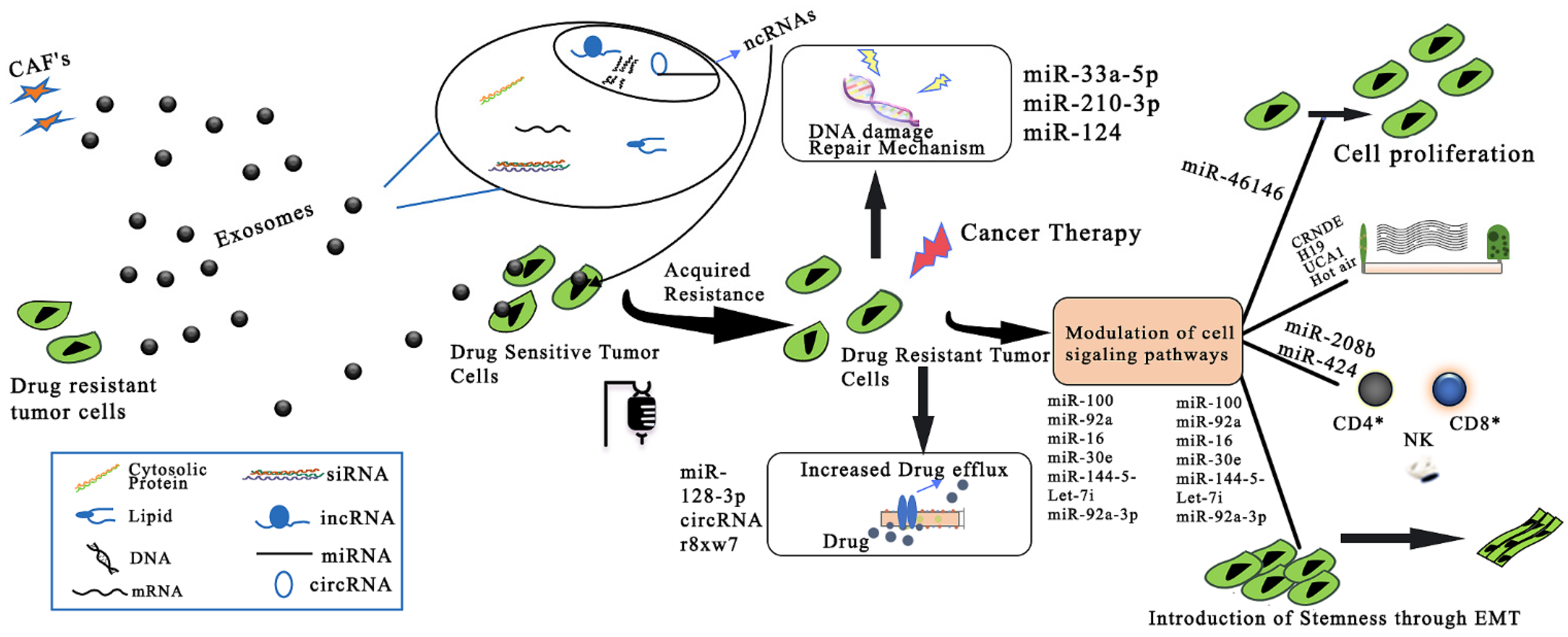
- Lampropoulou, D.I.; Pliakou, E.; Aravantinos, G.; Filippou, D.; Gazouli, M. The Role of Exosomal Non-Coding RNAs in Colorectal Cancer Drug Resistance. Int. J. Mol. Sci. 2022, 23, 1473. [Google Scholar] [CrossRef] [PubMed]
- Crombez, L.; Aldrian-Herrada, G.; Konate, K.; Nguyen, Q.N.; McMaster, G.K.; Brasseur, R.; Heitz, F.; Divita, G. A New Potent Secondary Amphipathic Cell-Penetrating Peptide for SiRNA Delivery into Mammalian Cells. Mol. Ther. 2009, 17, 95–103. [Google Scholar] [CrossRef] [PubMed]
- Sanchez, C.; Arribart, H.; Guille, M.M.G. Biomimetism and Bioinspiration as Tools for the Design of Innovative Materials and Systems. Nat. Mater. 2005, 4, 277–288. [Google Scholar] [CrossRef] [PubMed]
- Brown, S.; Khan, D.R. The Treatment of Breast Cancer Using Liposome Technology. J. Drug Deliv. 2012, 2012, 212965. [Google Scholar] [CrossRef]
- Ma, D.; Lin, Q.M.; Zhang, L.M.; Liang, Y.Y.; Xue, W. A Star-Shaped Porphyrin-Arginine Functionalized Poly(l-Lysine) Copolymer for Photo-Enhanced Drug and Gene Co-Delivery. Biomaterials 2014, 35, 4357–4367. [Google Scholar] [CrossRef] [PubMed]
- Ozpolat, B.; Sood, A.K.; Lopez-Berestein, G. Liposomal SiRNA Nanocarriers for Cancer Therapy. Adv. Drug Deliv. Rev. 2014, 66, 110–116. [Google Scholar] [CrossRef]
- Buya, A.B.; Witika, B.A.; Bapolisi, A.M.; Mwila, C.; Mukubwa, G.K.; Memvanga, P.B.; Makoni, P.A.; Nkanga, C.I. Application of Lipid-Based Nanocarriers for Antitubercular Drug Delivery: A Review. Pharmaceutics 2021, 13, 2041. [Google Scholar] [CrossRef]
- Lipinski, C.A. Avoiding Investment in Doomed Drugs Is Poor Solubility an Industry Wide Problem? Curr. Drug Discov. 2001, 1, 17–19. [Google Scholar]
- Kim, S.H.; Jeong, J.H.; Lee, S.H.; Kim, S.W.; Park, T.G. Local and Systemic Delivery of VEGF SiRNA Using Polyelectrolyte Complex Micelles for Effective Treatment of Cancer. J. Control. Release 2008, 129, 107–116. [Google Scholar] [CrossRef]
- Itaka, K.; Kataoka, K. Progress and Prospects of Polyplex Nanomicelles for Plasmid DNA Delivery. Curr. Gene Ther. 2011, 11, 457–465. [Google Scholar] [CrossRef] [PubMed]
- Aliabadi, H.M.; Lavasanifar, A. Polymeric Micelles for Drug Delivery. Expert Opin. Drug Deliv. 2006, 3, 139–162. [Google Scholar] [CrossRef] [PubMed]
- Jeong, J.H.; Park, T.G.; Kim, S.H. Self-Assembled and Nanostructured SiRNA Delivery Systems. Pharm. Res. 2011, 28, 2072–2085. [Google Scholar] [CrossRef] [PubMed]
- Castro, R.I.; Forero-Doria, O.; Guzmán, L. Perspectives of Dendrimer-Based Nanoparticles in Cancer Therapy. An. Acad. Bras. Cienc. 2018, 90, 2331–2346. [Google Scholar] [CrossRef] [PubMed]
- Chae, S.Y.; Kim, H.J.; Lee, M.S.; Jang, Y.L.; Lee, Y.; Lee, S.H.; Lee, K.; Kim, S.H.; Kim, H.T.; Chi, S.C.; et al. Energy-Independent Intracellular Gene Delivery Mediated by Polymeric Biomimetics of Cell-Penetrating Peptides. Macromol. Biosci. 2011, 11, 1169–1174. [Google Scholar] [CrossRef]
- Chen, B.; Dai, W.; He, B.; Zhang, H.; Wang, X.; Wang, Y.; Zhang, Q. Current Multistage Drug Delivery Systems Based on the Tumor Microenvironment. Theranostics 2017, 7, 538–558. [Google Scholar] [CrossRef] [PubMed]
- König, N.; Willner, L.; Carlström, G.; Zinn, T.; Knudsen, K.D.; Rise, F.; Topgaard, D.; Lund, R. Spherical Micelles with Nonspherical Cores: Effect of Chain Packing on the Micellar Shape. Macromolecules 2020, 53, 10686–10698. [Google Scholar] [CrossRef] [PubMed]
- Martínez-Ballesta, M.; Gil-Izquierdo, Á.; García-Viguera, C.; Domínguez-Perles, R. Nanoparticles and Controlled Delivery for Bioactive Compounds: Outlining Challenges for New “Smart-Foods” for Health. Foods 2018, 7, 72. [Google Scholar] [CrossRef] [PubMed]
- Gonçalves, G.A.R.; de Melo Alves Paiva, R. Gene Therapy: Advances, Challenges and Perspectives. Einstein 2017, 15, 369–375. [Google Scholar] [CrossRef] [PubMed]
- Cao, N.; Cheng, D.; Zou, S.; Ai, H.; Gao, J.; Shuai, X. The Synergistic Effect of Hierarchical Assemblies of SiRNA and Chemotherapeutic Drugs Co-Delivered into Hepatic Cancer Cells. Biomaterials 2011, 32, 2222–2232. [Google Scholar] [CrossRef]
- Zhu, C.; Jung, S.; Luo, S.; Meng, F.; Zhu, X.; Park, T.G.; Zhong, Z. Co-Delivery of SiRNA and Paclitaxel into Cancer Cells by Biodegradable Cationic Micelles Based on PDMAEMA-PCL-PDMAEMA Triblock Copolymers. Biomaterials 2010, 31, 2408–2416. [Google Scholar] [CrossRef] [PubMed]
- Zhang, J.; Fang, D.; Ma, Q.; He, Z.; Ren, K.; Zhou, R.; Zeng, S.; Li, B.; He, L.; He, G.; et al. Dual-Functional PEI–Poly (γ-Cholesterol-L-Glutamate) Copolymer for Drug/Gene Co-Delivery. Macromol. Chem. Phys. 2014, 215, 163–170. [Google Scholar] [CrossRef]
- Grossen, P.; Witzigmann, D.; Sieber, S.; Huwyler, J. PEG-PCL-Based Nanomedicines: A Biodegradable Drug Delivery System and Its Application. J. Control. Release 2017, 260, 46–60. [Google Scholar] [CrossRef]
- Liu, Q.; Li, R.T.; Qian, H.Q.; Wei, J.; Xie, L.; Shen, J.; Yang, M.; Qian, X.P.; Yu, L.X.; Jiang, X.Q.; et al. Targeted Delivery of MiR-200c/DOC to Inhibit Cancer Stem Cells and Cancer Cells by the Gelatinases-Stimuli Nanoparticles. Biomaterials 2013, 34, 7191–7203. [Google Scholar] [CrossRef] [PubMed]
- Yang, C.; Chan, K.K.; Lin, W.J.; Soehartono, A.M.; Lin, G.; Toh, H.; Yoon, H.S.; Chen, C.K.; Yong, K.T. Biodegradable Nanocarriers for Small Interfering Ribonucleic Acid (SiRNA) Co-Delivery Strategy Increase the Chemosensitivity of Pancreatic Cancer Cells to Gemcitabine. Nano Res. 2017, 10, 3049–3067. [Google Scholar] [CrossRef]
- Wu, Y.; Crawford, M.; Mao, Y.; Lee, R.J.; Davis, I.C.; Elton, T.S.; Lee, L.J.; Nana-Sinkam, S.P. Therapeutic Delivery of MicroRNA-29b by Cationic Lipoplexes for Lung Cancer. Mol. Ther. Nucleic Acids 2013, 2, e84. [Google Scholar] [CrossRef]
- Mendes, L.P.; Pan, J.; Torchilin, V.P. Dendrimers as Nanocarriers for Nucleic Acid and Drug Delivery in Cancer Therapy. Molecules 2017, 22, 1401. [Google Scholar] [CrossRef]
- Svenson, S. Dendrimers as Versatile Platform in Drug Delivery Applications. Eur. J. Pharm. Biopharm. 2009, 71, 445–462. [Google Scholar] [CrossRef]
- Hanafy, M.S.; Hufnagel, S.; Trementozzi, A.N.; Sakran, W.; Stachowiak, J.C.; Koleng, J.J.; Cui, Z. PD-1 SiRNA-Encapsulated Solid Lipid Nanoparticles Downregulate PD-1 Expression by Macrophages and Inhibit Tumor Growth: PD-1 SiRNA-Encapsulated Solid Lipid Nanoparticles. AAPS PharmSciTech 2021, 22, 60. [Google Scholar] [CrossRef]
- Loh, K.P.; Bao, Q.; Eda, G.; Chhowalla, M. Graphene Oxide as a Chemically Tunable Platform for Optical Applications. Nat. Chem. 2010, 2, 1015–1024. [Google Scholar] [CrossRef]
- Mamaeva, V.; Sahlgren, C.; Lindén, M. Mesoporous Silica Nanoparticles in Medicine-Recent Advances. Adv. Drug Deliv. Rev. 2013, 65, 689–702. [Google Scholar] [CrossRef]
- Gu, L.; Deng, Z.J.; Roy, S.; Hammond, P.T. A Combination RNAi-Chemotherapy Layer-by-Layer Nanoparticle for Systemic Targeting of KRAS/P53 with Cisplatin to Treat Non–Small Cell Lung Cancer. Clin. Cancer Res. 2017, 23, 7312–7323. [Google Scholar] [CrossRef]
- Sepantafar, M.; Maheronnaghsh, R.; Mohammadi, H.; Radmanesh, F.; Hasani-sadrabadi, M.M.; Ebrahimi, M.; Baharvand, H. Engineered Hydrogels in Cancer Therapy and Diagnosis. Trends Biotechnol. 2017, 35, 1074–1087. [Google Scholar] [CrossRef]
- Ozlu, B.; Kabay, G.; Bocek, I.; Yilmaz, M.; Piskin, A.K.; Shim, B.S.; Mutlu, M. Controlled Release of Doxorubicin from Polyethylene Glycol Functionalized Melanin Nanoparticles for Breast Cancer Therapy: Part I. Production and Drug Release Performance of the Melanin Nanoparticles. Int. J. Pharm. 2019, 570, 118613. [Google Scholar] [CrossRef]
- Zhen, X.; Xie, C.; Jiang, Y.; Ai, X.; Xing, B.; Pu, K. Semiconducting Photothermal Nanoagonist for Remote-Controlled Specific Cancer Therapy. Nano Lett. 2018, 18, 1498–1505. [Google Scholar] [CrossRef]
- Su, Y.; Zhang, X.; Ren, G.; Zhang, Z.; Liang, Y.; Wu, S.; Shen, J. In Situ Implantable Three-Dimensional Extracellular Matrix Bioactive Composite Scaffold for Postoperative Skin Cancer Therapy. Chem. Eng. J. 2020, 400, 125949. [Google Scholar] [CrossRef]
- Paulino, A.T.; Pereira, A.G.B.; Fajardo, A.R.; Erickson, K.; Kipper, M.J.; Muniz, E.C.; Belfiore, L.A.; Tambourgi, E.B. Natural Polymer-Based Magnetic Hydrogels: Potential Vectors for Remote-Controlled Drug Release. Carbohydr. Polym. 2012, 90, 1216–1225. [Google Scholar] [CrossRef]
- Jahanban-Esfahlan, R.; Derakhshankhah, H.; Haghshenas, B.; Massoumi, B.; Abbasian, M.; Jaymand, M. A Bio-Inspired Magnetic Natural Hydrogel Containing Gelatin and Alginate as a Drug Delivery System for Cancer Chemotherapy. Int. J. Biol. Macromol. 2020, 156, 438–445. [Google Scholar] [CrossRef] [PubMed]
- Lu, M.; Huang, Y. Bioinspired Exosome-like Therapeutics and Delivery Nanoplatforms. Biomaterials 2020, 242, 119925. [Google Scholar] [CrossRef]
- Yan, F.; Zhong, Z.; Wang, Y.; Feng, Y.; Mei, Z.; Li, H.; Chen, X.; Cai, L.; Li, C. Exosome-Based Biomimetic Nanoparticles Targeted to Inflamed Joints for Enhanced Treatment of Rheumatoid Arthritis. J. Nanobiotechnol. 2020, 18, 115. [Google Scholar] [CrossRef]
- Kim, M.S.; Haney, M.J.; Zhao, Y.; Mahajan, V.; Deygen, I.; Klyachko, N.L.; Inskoe, E.; Piroyan, A.; Sokolsky, M.; Okolie, O.; et al. Development of Exosome-Encapsulated Paclitaxel to Overcome MDR in Cancer Cells. Nanomed. Nanotechnol. Biol. Med. 2016, 12, 655–664. [Google Scholar] [CrossRef] [PubMed]
- Maeda, H.; Wu, J.; Sawa, T.; Matsumura, Y.; Hori, K. Tumor Vascular Permeability and the EPR Effect in Macromolecular Therapeutics: A Review. J. Control. Release 2000, 65, 271–284. [Google Scholar] [CrossRef]
- Xiao, K.; Li, Y.; Luo, J.; Lee, J.S.; Xiao, W.; Gonik, A.M.; Agarwal, R.G.; Lam, K.S. The Effect of Surface Charge on in Vivo Biodistribution of PEG-Oligocholic Acid Based Micellar Nanoparticles. Biomaterials 2011, 32, 3435–3446. [Google Scholar] [CrossRef]
- Khvorova, A. Oligonucleotide Therapeutics—A New Class of Cholesterol-Lowering Drugs. N. Engl. J. Med. 2017, 376, 4–7. [Google Scholar] [CrossRef]
- Tadin-Strapps, M.; Peterson, L.B.; Cumiskey, A.M.; Rosa, R.L.; Mendoza, V.H.; Castro-Perez, J.; Puig, O.; Zhang, L.; Strapps, W.R.; Yendluri, S.; et al. SiRNA-Induced Liver ApoB Knockdown Lowers Serum LDL-Cholesterol in a Mouse Model with Human-like Serum Lipids. J. Lipid Res. 2011, 52, 1084–1097. [Google Scholar] [CrossRef]
- Li, L.; Shen, Y. Overcoming Obstacles to Develop Effective and Safe SiRNA Therapeutics. Expert Opin. Biol. Ther. 2009, 9, 609–619. [Google Scholar] [CrossRef]
- Thun, M.J.; DeLancey, J.O.; Center, M.M.; Jemal, A.; Ward, E.M. The Global Burden of Cancer: Priorities for Prevention. Carcinogenesis 2009, 31, 100–110. [Google Scholar] [CrossRef]
- Revia, R.A.; Zhang, M. Magnetite Nanoparticles for Cancer Diagnosis, Treatment, and Treatment Monitoring: Recent Advances. Mater. Today 2016, 19, 157–168. [Google Scholar] [CrossRef]
- Fang, R.H.; Aryal, S.; Hu, C.M.J.; Zhang, L. Quick Synthesis of Lipid-Polymer Hybrid Nanoparticles with Low Polydispersity Using a Single-Step Sonication Method. Langmuir 2010, 26, 16958–16962. [Google Scholar] [CrossRef]
- Valencia, P.M.; Basto, P.A.; Zhang, L.; Rhee, M.; Langer, R.; Farokhzad, O.C.; Karnik, R. Single-Step Assembly of Homogenous Lipid-Polymeric and Lipid-Quantum Dot Nanoparticles Enabled by Microfluidic Rapid Mixing. ACS Nano 2010, 4, 1671–1679. [Google Scholar] [CrossRef] [PubMed]
- Li, Y.; Duo, Y.; Zhai, P.; He, L.; Zhong, K.; Zhang, Y.; Huang, K.; Luo, J.; Zhang, H.; Yu, X. Dual Targeting Delivery of MiR-328 by Functionalized Mesoporous Silica Nanoparticles for Colorectal Cancer Therapy. Nanomedicine 2018, 13, 1753–1772. [Google Scholar] [CrossRef] [PubMed]
- Endo-Takahashi, Y.; Negishi, Y.; Nakamura, A.; Ukai, S.; Ooaku, K.; Oda, Y.; Sugimoto, K.; Moriyasu, F.; Takagi, N.; Suzuki, R.; et al. Systemic Delivery of MiR-126 by MiRNA-Loaded Bubble Liposomes for the Treatment of Hindlimb Ischemia. Sci. Rep. 2014, 4, 3883. [Google Scholar] [CrossRef] [PubMed]
- Kim, J.H.; Yeom, J.H.; Ko, J.J.; Han, M.S.; Lee, K.; Na, S.Y.; Bae, J. Effective Delivery of Anti-MiRNA DNA Oligonucleotides by Functionalized Gold Nanoparticles. J. Biotechnol. 2011, 155, 287–292. [Google Scholar] [CrossRef]
- Kollinerová, S.; Dostál, Z.; Modrianský, M. MicroRNA Hsa-MiR-29b Potentiates Etoposide Toxicity in HeLa Cells via down-Regulation of Mcl-1. Toxicol. Vitr. 2017, 40, 289–296. [Google Scholar] [CrossRef]
- Xue, C.; Hu, S.; Gao, Z.H.; Wang, L.; Luo, M.X.; Yu, X.; Li, B.F.; Shen, Z.; Wu, Z.S. Programmably Tiling Rigidified DNA Brick on Gold Nanoparticle as Multi-Functional Shell for Cancer-Targeted Delivery of SiRNAs. Nat. Commun. 2021, 12, 2928. [Google Scholar] [CrossRef]
- Tivnan, A.; Orr, W.S.; Gubala, V.; Nooney, R.; Williams, D.E.; McDonagh, C.; Prenter, S.; Harvey, H.; Domingo-Fernández, R.; Bray, I.M.; et al. Inhibition of Neuroblastoma Tumor Growth by Targeted Delivery of MicroRNA-34a Using Anti-Disialoganglioside GD2 Coated Nanoparticles. PLoS ONE 2012, 7, e38129. [Google Scholar] [CrossRef]
- Heidari, R.; Khosravian, P.; Mirzaei, S.A.; Elahian, F. SiRNA Delivery Using Intelligent Chitosan-Capped Mesoporous Silica Nanoparticles for Overcoming Multidrug Resistance in Malignant Carcinoma Cells. Sci. Rep. 2021, 11, 20531. [Google Scholar] [CrossRef]
- Ren, Y.; Kang, C.S.; Yuan, X.B.; Zhou, X.; Xu, P.; Han, L.; Wang, G.X.; Jia, Z.; Zhong, Y.; Yu, S.; et al. Co-Delivery of as-MiR-21 and 5-FU by Poly(Amidoamine) Dendrimer Attenuates Human Glioma Cell Growth in Vitro. J. Biomater. Sci. Polym. Ed. 2010, 21, 303–314. [Google Scholar] [CrossRef]
- Ren, Y.; Zhou, X.; Mei, M.; Yuan, X.B.; Han, L.; Wang, G.X.; Jia, Z.F.; Xu, P.; Pu, P.Y.; Kang, C.S. MicroRNA-21 Inhibitor Sensitizes Human Glioblastoma Cells U251 (PTEN-Mutant) and LN229 (PTEN-Wild Type) to Taxol. BMC Cancer 2010, 10, 27. [Google Scholar] [CrossRef]
- Liu, X.X.; Rocchi, P.; Qu, F.Q.; Zheng, S.Q.; Liang, Z.C.; Gleave, M.; Iovanna, J.; Peng, L. PAMAM Dendrimers Mediate SiRNA Delivery to Target Hsp27 and Produce Potent Antiproliferative Effects on Prostate Cancer Cells. ChemMedChem 2009, 4, 1302–1310. [Google Scholar] [CrossRef] [PubMed]
- Babar, I.A.; Cheng, C.J.; Booth, C.J.; Liang, X.; Weidhaas, J.B.; Saltzman, W.M.; Slack, F.J. Nanoparticle-Based Therapy in an in Vivo MicroRNA-155 (MiR-155)-Dependent Mouse Model of Lymphoma. Proc. Natl. Acad. Sci. USA 2012, 109, E1695–E1704. [Google Scholar] [CrossRef] [PubMed]
- Risnayanti, C.; Jang, Y.S.; Lee, J.; Ahn, H.J. PLGA Nanoparticles Co-Delivering MDR1 and BCL2 SiRNA for Overcoming Resistance of Paclitaxel and Cisplatin in Recurrent or Advanced Ovarian Cancer. Sci. Rep. 2018, 8, 7498. [Google Scholar] [CrossRef] [PubMed]
- Sun, P.; Huang, W.; Jin, M.; Wang, Q.; Fan, B.; Kang, L.; Gao, Z. Chitosan-Based Nanoparticles for Survivin Targeted SiRNA Delivery in Breast Tumor Therapy and Preventing Its Metastasis. Int. J. Nanomed. 2016, 11, 4931–4945. [Google Scholar] [CrossRef]
- Piao, L.; Zhang, M.; Datta, J.; Xie, X.; Su, T.; Li, H.; Teknos, T.N.; Pan, Q. Lipid-Based Nanoparticle Delivery of Pre-MiR-107 Inhibits the Tumorigenicity of Head and Neck Squamous Cell Carcinoma. Mol. Ther. 2012, 20, 1261–1269. [Google Scholar] [CrossRef]
- Wei, L.; Guo, X.Y.; Yang, T.; Yu, M.Z.; Chen, D.W.; Wang, J.C. Brain Tumor-Targeted Therapy by Systemic Delivery of SiRNA with Transferrin Receptor-Mediated Core-Shell Nanoparticles. Int. J. Pharm. 2016, 510, 394–405. [Google Scholar] [CrossRef]
- Yan, Y.; Li, X.Q.; Duan, J.L.; Bao, C.J.; Cui, Y.N.; Su, Z.B.; Xu, J.R.; Luo, Q.; Chen, M.; Xie, Y.; et al. Nanosized Functional MiRNA Liposomes and Application in the Treatment of TNBC by Silencing Slug Gene. Int. J. Nanomed. 2019, 14, 3645–3667. [Google Scholar] [CrossRef] [PubMed]
- Kabilova, T.O.; Shmendel, E.V.; Gladkikh, D.V.; Chernolovskaya, E.L.; Markov, O.V.; Morozova, N.G.; Maslov, M.A.; Zenkova, M.A. Targeted Delivery of Nucleic Acids into Xenograft Tumors Mediated by Novel Folate-Equipped Liposomes. Eur. J. Pharm. Biopharm. 2018, 123, 59–70. [Google Scholar] [CrossRef] [PubMed]
- Mambo, E.; Szafranska-Schwarzbach, A.E.; Latham, G.; Adai, A.; Schlageter, A.; Andruss, B. MicroRNA Biomarkers as Potential Diagnostic Markers for Cancer; Elsevier: Amsterdam, The Netherlands, 2013; ISBN 9780123973368. [Google Scholar]
- Hong, D.S.; Kang, Y.K.; Borad, M.; Sachdev, J.; Ejadi, S.; Lim, H.Y.; Brenner, A.J.; Park, K.; Lee, J.L.; Kim, T.Y.; et al. Phase 1 Study of MRX34, a Liposomal MiR-34a Mimic, in Patients with Advanced Solid Tumours. Br. J. Cancer 2020, 122, 1630–1637. [Google Scholar] [CrossRef]
- Chenthamara, D.; Subramaniam, S.; Ramakrishnan, S.G.; Krishnaswamy, S.; Essa, M.M.; Lin, F.H.; Qoronfleh, M.W. Therapeutic Efficacy of Nanoparticles and Routes of Administration. Biomater. Res. 2019, 23, 20. [Google Scholar] [CrossRef]
- Ottosen, S.; Parsley, T.B.; Yang, L.; Zeh, K.; Van Doorn, L.J.; Van Der Veer, E.; Raney, A.K.; Hodges, M.R.; Patick, A.K. In Vitro Antiviral Activity and Preclinical and Clinical Resistance Profile of Miravirsen, a Novel Anti-Hepatitis C Virus Therapeutic Targeting the Human Factor MiR-122. Antimicrob. Agents Chemother. 2015, 59, 599–608. [Google Scholar] [CrossRef] [PubMed]
- Hamid, O.; Ismail, R.; Puzanov, I. Intratumoral Immunotherapy—Update 2019. Oncologist 2020, 25, e423–e438. [Google Scholar] [CrossRef] [PubMed]


| Delivery Method | Viral | Liposome | Conjugates | Extracellular Vesicles | Polymers | Nanoparticles |
|---|---|---|---|---|---|---|
| Widely used vectors | Retrovirus, lentivirus, adenovirus, adeno-associated virus | Forms lipoplex by the interaction of cationic lipids | Receptor binding molecules bind directly to nucleic acids: aptamers, multi-functional peptides [48] | Exosomes, microvesicles, platelets, apoptotic bodies | Polyethyleneimine (PEI), poly (lactide-co-glycolide), poly (amidoamine), dendrimers/cell-penetrating peptides [49,50] | AuNPs, mesoporous silicone, graphene oxide, and Fe3O4-mediated NPs [50,51,52] |
| Advantages | High transfection efficiency [50,51,52] | Biodegradable, biocompatible Reduces the toxicity High affinity with cell membrane Increased efficacy and therapeutic index [53] | Less toxicity, higher stability, selective targeting, high intracellular delivery efficiency [33,54] | Low cytotoxicity and negligible antigenicity, naturally present in body fluids, ability to cross blood–brain barriers [50] | Natural and synthetic polymers: Biocompatible. Natural polymers: less toxicity, biodegradable [49,50] | High stability in vivo, free of microbial attack [50,51] |
| Disadvantages | Low loading capacity, high toxicity, strong immunogenicity, mutation [50,51,52] | Limited storage settings Poor stability Short half-life Low solubility [53] | Endosomal entrapment [33,54] | Low drug loading capacity, rapid clearance from blood [50] | Natural polymer: Highly branched structures, complicated [49,50] extraction method Synthetic Polymer: High toxicity, poorly biodegradable [49,50] | Inorganic material: weak interaction between the carrier and nucleic acids [48,50] |
| siRNA examples | Adeno-associated virus vector for p53 siRNA delivery into HeLa S3 cells [55] | Core-shell lipoplexes encapsulated c-Myc-targeting siRNA for glioblastoma treatment [56] | Cholesterol-tethered EpCAM-targeting RNA aptamer for cancer stem cell-targeted gene delivery [57] | Artificial platelets for efficient delivery of siRNA targeting Pcsk9 transcription in the liver [58] | PEI-cholesterol-polyethylene glycol, Dendrimers [59] | Functionalized gold nanorod-based TFEB-siRNA autophagy for osteosarcoma [60] |
| miRNA Examples | Lentivirus-miR-199a inhibition of HCC cell proliferation [52] | miR-7 encapsulated with cationic liposome against EGFR-TKI-resistant lung cancer cells [61] | Lipid NP-mediated anti-miR-17 family suppression of Hep3B tumor growth [62] | Brain metastasis cancer cell-derived EV-miR-181c promotes brain metastasis and destruction of the blood–brain barrier [63] | LbL-PLGA NPs carries miR-34a cargo for suppression of target gene and reduced triple-negative breast cancer cell proliferation [64] | Anti-miR-155-loaded modified mesoporous silica NPs (MSNs-anti-miR-155@PDA-Apt) for colorectal cancer [65] |
| Delivery System | Target | Disease | miRNA/siRNA | Ref. |
|---|---|---|---|---|
| Gold NPs | Mcl-1 | HeLa cell cancer | miR-29b | [126,127] |
| Plk1 | Breast cancer | siPlk1/Ap-Cs | [128] | |
| Silica-based NPs | MYCN | Neuroblastoma | miR-34a | [129] |
| MDR1 | HeLa-RDB | NH2-MSN-siRNA with chitosan coat | [130] | |
| PAMAM-dendrimer | miR-21 | Glioblastoma | as-miR-21 | [131,132] |
| Hsp27 | Prostate cancer | TEA core-PAMAM dendrimer-siRNA | [133] | |
| PLGA NPs | SHIP1 | Lymphoma/leukemia | miR-155 | [134] |
| MDR1, Bcl2 | Ovarian cancer | siRNA-loaded PLGA NPs | [135] | |
| Chitosan NPs | Survivin | Breast cancer | PEG-chitosan-siRNA | [136] |
| Cationic lipoplexes-lipid-based NPs | PKCε, CDK6, HIF1-β | Head and neck squamous cell carcinoma (HNSCC) | miR107 | [137] |
| EGFR | Brain tumor | T7-LPC-siRNA Nps | [138] | |
| Liposomes | Slug | Triple-negative breast cancer | miR-203 | [139] |
| MDR1 | Squamous carcinoma | 2x3-DOPE/FC liposome siRNA | [140] |
| Drug Name and miRNA Target | Disease | Category | Company | Clinical Trial Details (Identifier) and Status |
|---|---|---|---|---|
| MRX34, miR-34a | Hepatocellular carcinoma | Mimic | MiRNA Therapeutics | NCT01829971, Phase I, Terminated |
| MesomiR-1, miR-16 | Mesothelioma and non-small cell lung cancer | TargomiRs (mimic) | EnGeneIC Limited | NCT02369198, Phase I, Completed |
| Cobomarsen/MRG-106, miR-155 | Cutaneous T-cell lymphoma/mycosis fungoides | LNA modified antisense inhibitor | miRagen Therapeutics | NCT03713320, Phase II, Terminated |
| Remlarsen/MRG-201, miR-29 | Keloid | Mimic (cholesterol-conjugated miR duplex) | miRagen Therapeutics | NCT03601052, Phase II, Completed |
| AZD4076/RG-125, miR-103/107 | Type 2 diabetes mellitus and non-alcoholic fatty liver disease | GalNAC-conjugated Anti-miR | AstraZeneca | NCT02826525, Phase I, Completed |
| CALAA-01/M2 subunit of R2 | Solid tumor | Cyclodextrin NPs | Calando Pharmaceuticals | NCT00689065, Phase I, Terminated |
| EphA2-targeting DOPC-encapsulated siRNA/EphA2 | Advanced solid tumor with liver metastases | Neutral liposomes | M D Anderson Cancer Center, Houston | NCT01591356, Phase I, Completed |
| TKM-080301/PLK-1 | Primary and secondary liver cancer | Lipid nanoparticles | National Cancer Institute (NCI) | NCT01437007, Phase I, Completed |
Publisher’s Note: MDPI stays neutral with regard to jurisdictional claims in published maps and institutional affiliations. |
© 2022 by the authors. Licensee MDPI, Basel, Switzerland. This article is an open access article distributed under the terms and conditions of the Creative Commons Attribution (CC BY) license (https://creativecommons.org/licenses/by/4.0/).
Share and Cite
Zare, M.; Pemmada, R.; Madhavan, M.; Shailaja, A.; Ramakrishna, S.; Kandiyil, S.P.; Donahue, J.M.; Thomas, V. Encapsulation of miRNA and siRNA into Nanomaterials for Cancer Therapeutics. Pharmaceutics 2022, 14, 1620. https://doi.org/10.3390/pharmaceutics14081620
Zare M, Pemmada R, Madhavan M, Shailaja A, Ramakrishna S, Kandiyil SP, Donahue JM, Thomas V. Encapsulation of miRNA and siRNA into Nanomaterials for Cancer Therapeutics. Pharmaceutics. 2022; 14(8):1620. https://doi.org/10.3390/pharmaceutics14081620
Chicago/Turabian StyleZare, Mina, Rakesh Pemmada, Maya Madhavan, Aswathy Shailaja, Seeram Ramakrishna, Sumodan Padikkala Kandiyil, James M. Donahue, and Vinoy Thomas. 2022. "Encapsulation of miRNA and siRNA into Nanomaterials for Cancer Therapeutics" Pharmaceutics 14, no. 8: 1620. https://doi.org/10.3390/pharmaceutics14081620