
Barium Oxide Doped Magnesium Silicate Nanopowders for Bone Fracture Healing: Preparation, Characterization, Antibacterial and In Vivo Animal Studies


Abstract
:1. Introduction
2. Materials and Methods
2.1. Characterization Techniques
2.1.1. XRD Examinations
2.1.2. FTIR Examinations
2.1.3. Morphological and Elemental Analyses
Scanning Electron Microscopy Examinations
Transmission Electron Microscopy Examinations
2.2. Surface Area Measurements
2.3. Mechanical Properties Analyses
2.4. Antibacterial Investigations
2.5. Animal Studies
2.5.1. In Vivo Animal Studies
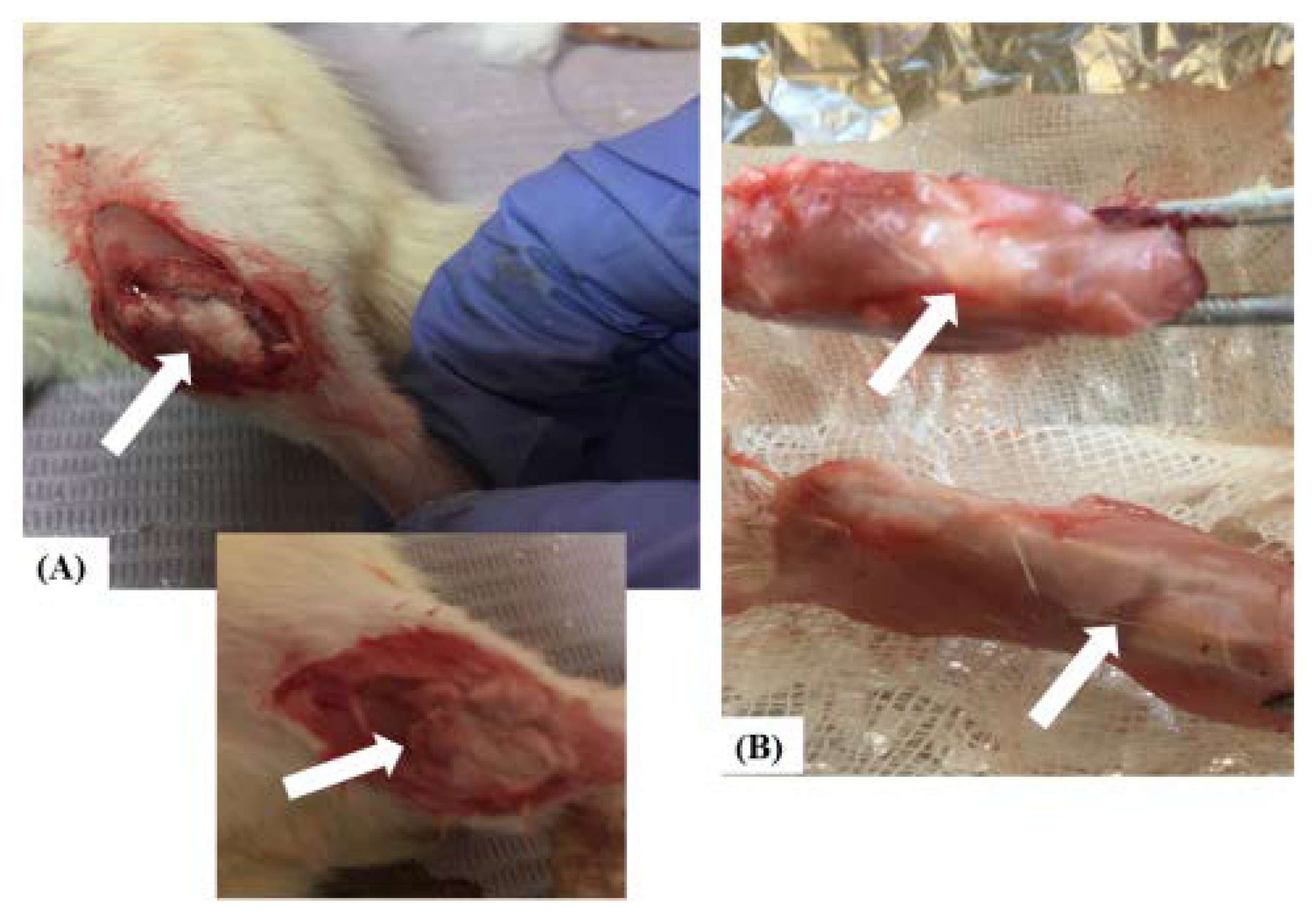
2.5.2. Surgical Operations and Experimental Design
- Group (1): Sham control group—bone fracture was left empty and untreated.
- Group (2): Pure MgS group—bone fracture was stuffed with MgS (50mg weight per animal).
- Group (3): MgS Ba-doped (3% wt) group—bone fracture was stuffed with MgS nanopowders doped with 3% wt percentage of BaO.
- Group (4): MgS Ba-doped (5% wt) group—bone fracture was stuffed with MgS nanopowders doped with 5% BaO.
2.5.3. Clinical Observation
2.5.4. Blood and Tissue Preparation
2.6. Biochemical Assays
2.6.1. Determination of Serum Transaminases and Alkaline Phosphatase
2.6.2. Evaluation of Serum Levels of Oxidative Stress Markers
2.7. Histological Examination of Tibial and Liver Samples
2.8. Statistical Analyses
3. Results
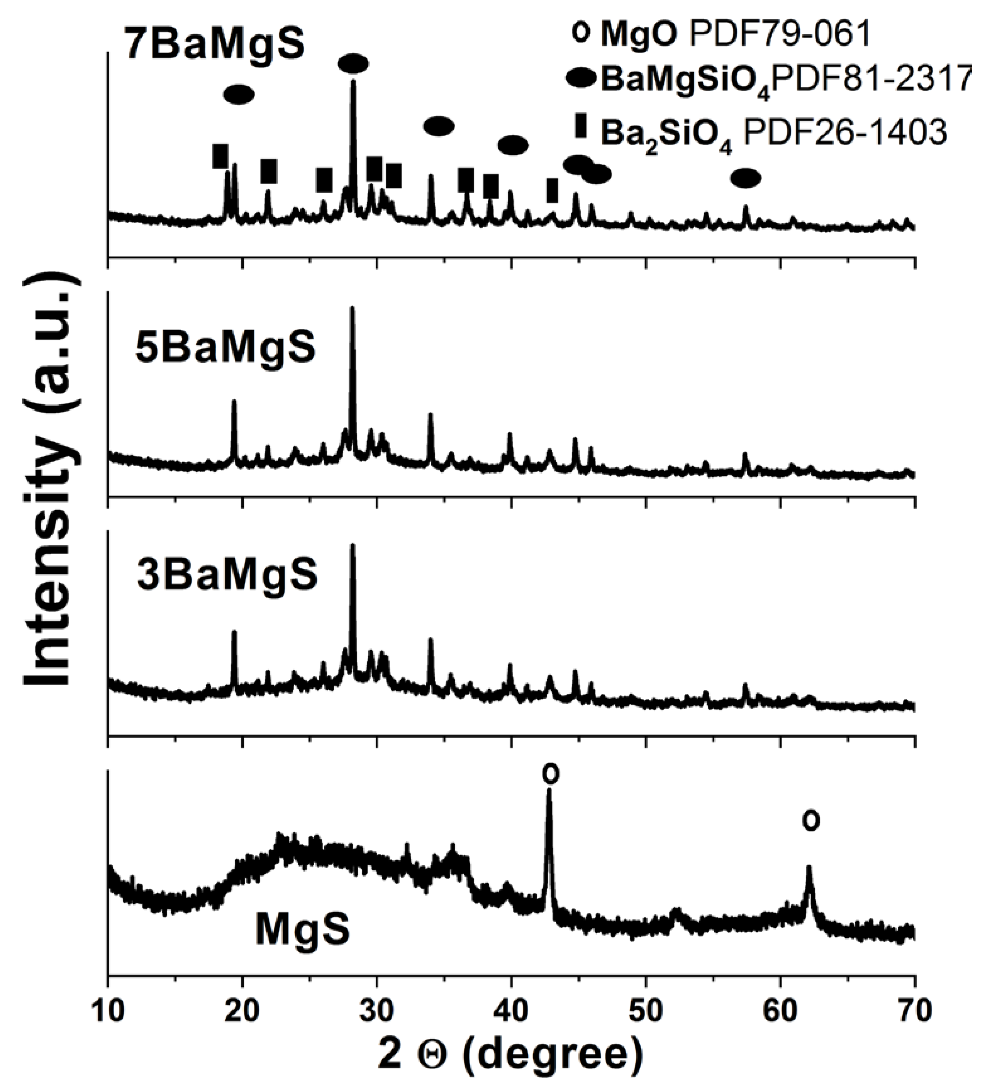
3.1. XRD Analysis
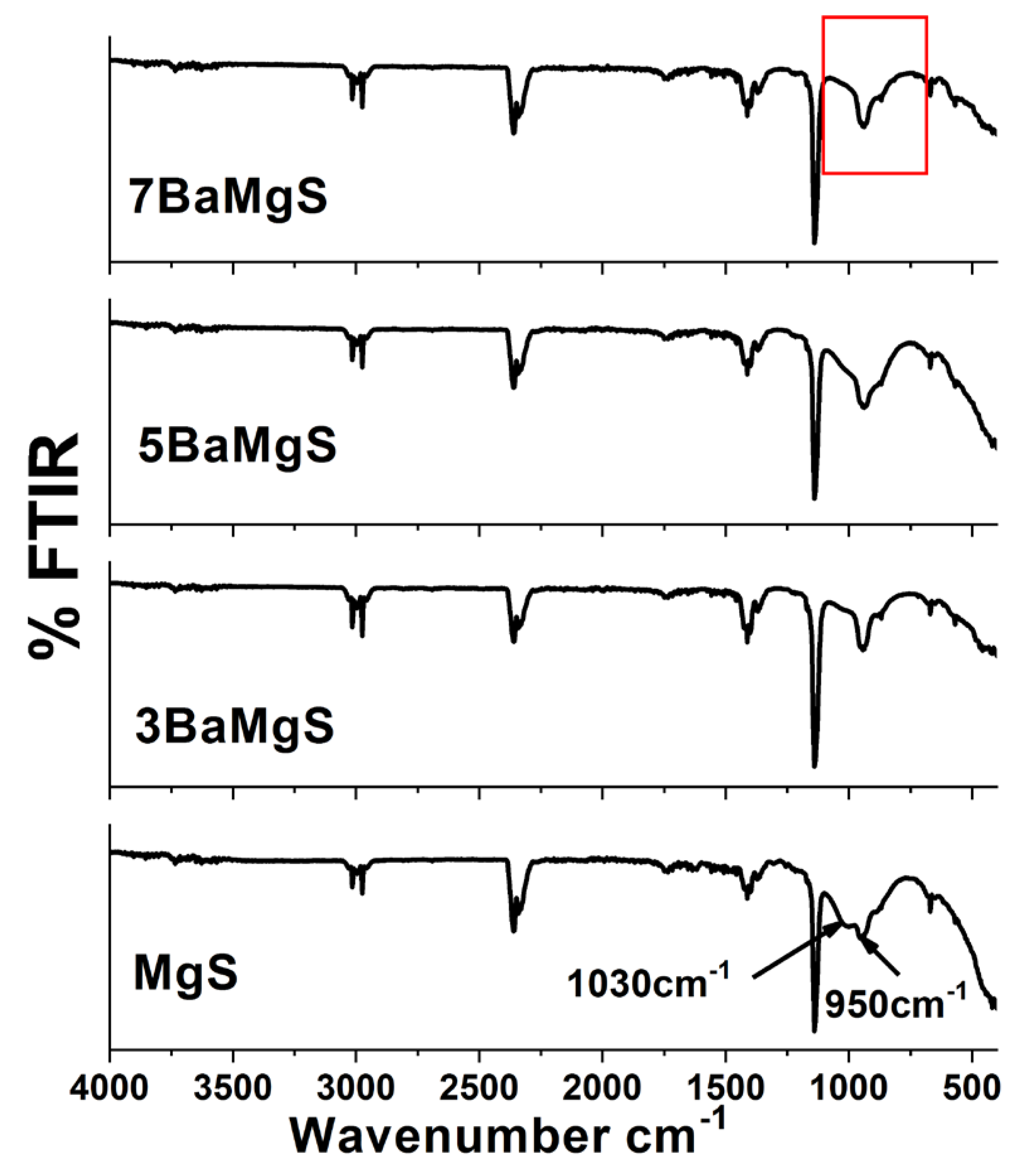
3.2. FTIR Characterization
3.3. Morphological and Elemental Analyses Results
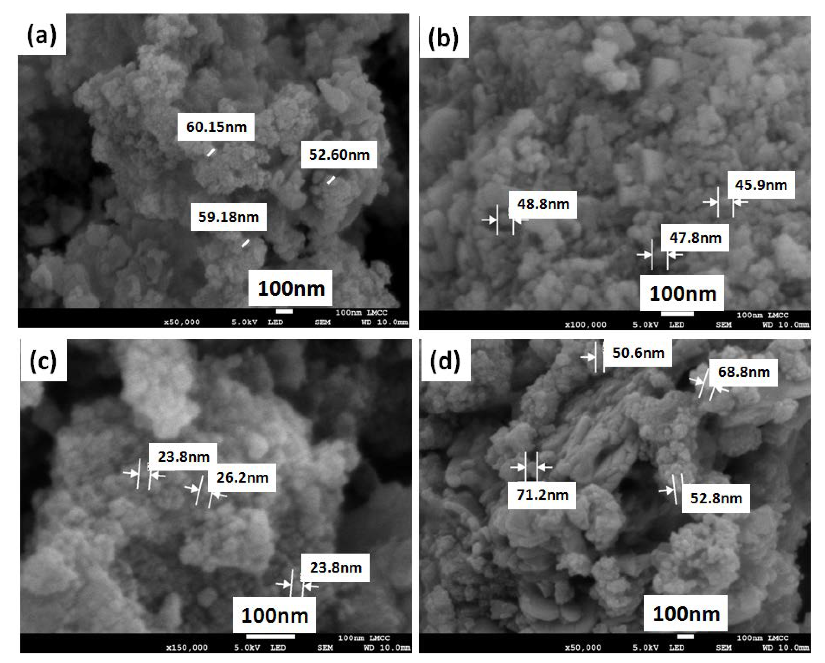
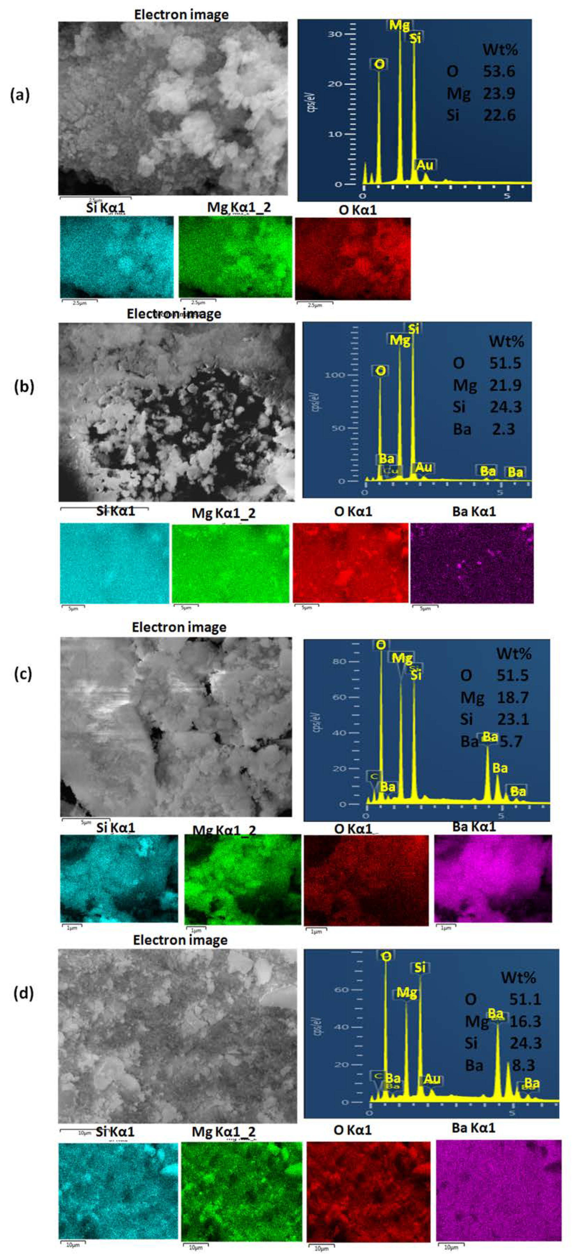
3.3.1. Scanning Electron Microscopy Examinations Results
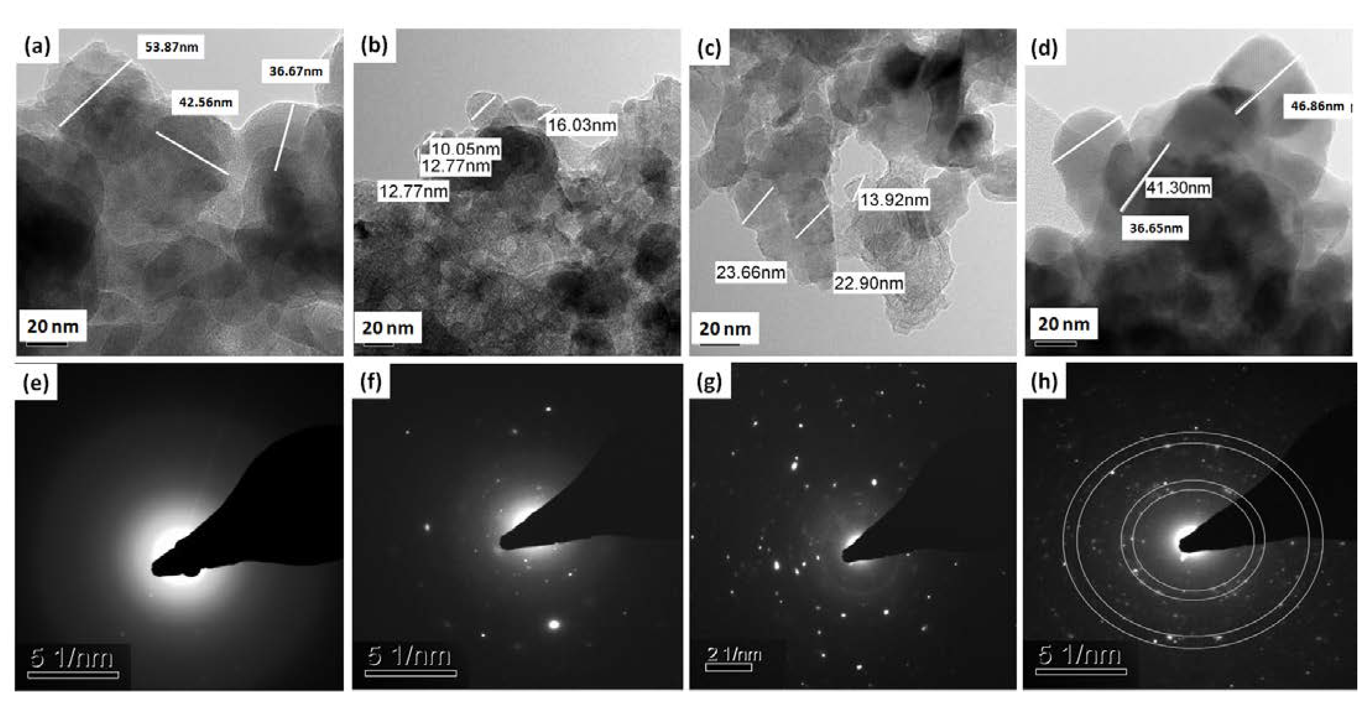
3.3.2. Transmission Electron Microscopy Examinations Results
3.4. Surface Area Measurements Results
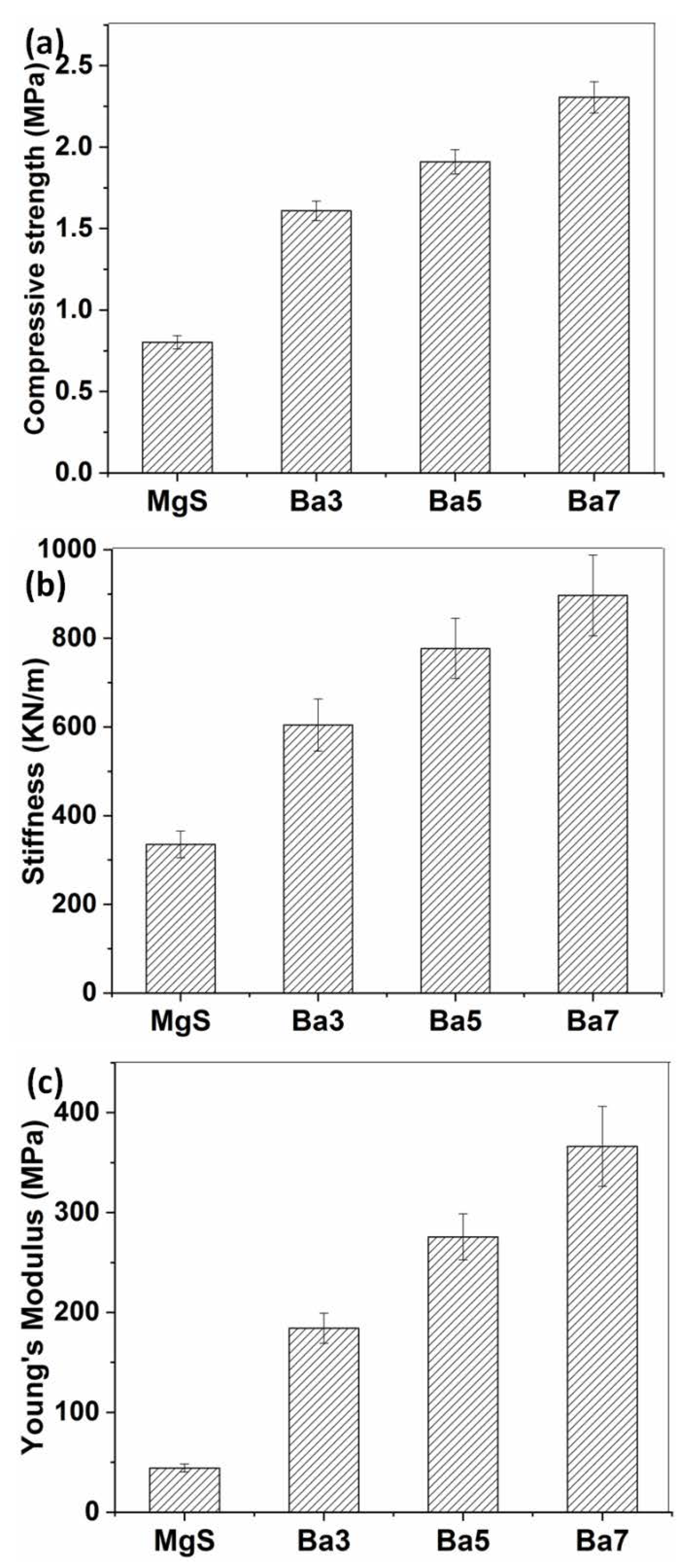
3.5. Mechanical Properties
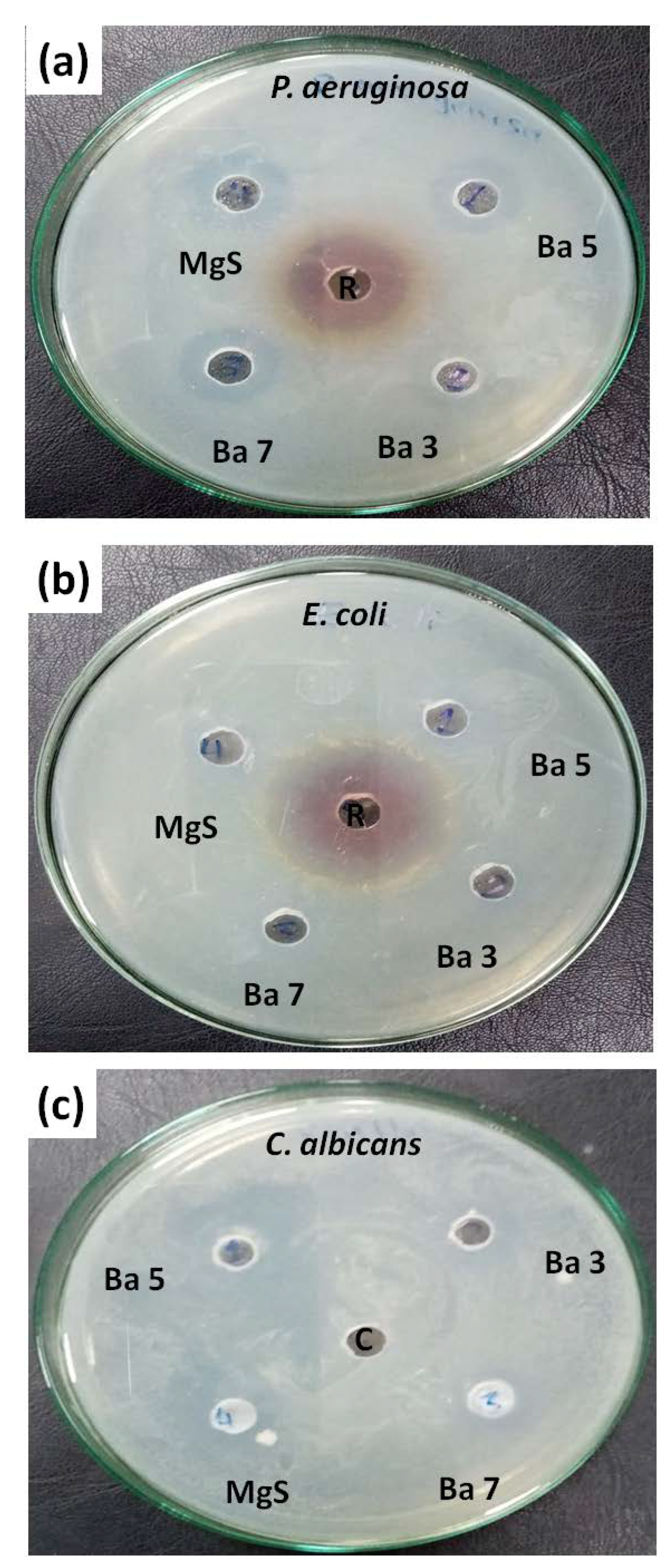
3.6. Antibacterial Investigations Results
3.7. In Vivo Animal Study
3.7.1. Clinical Evaluation
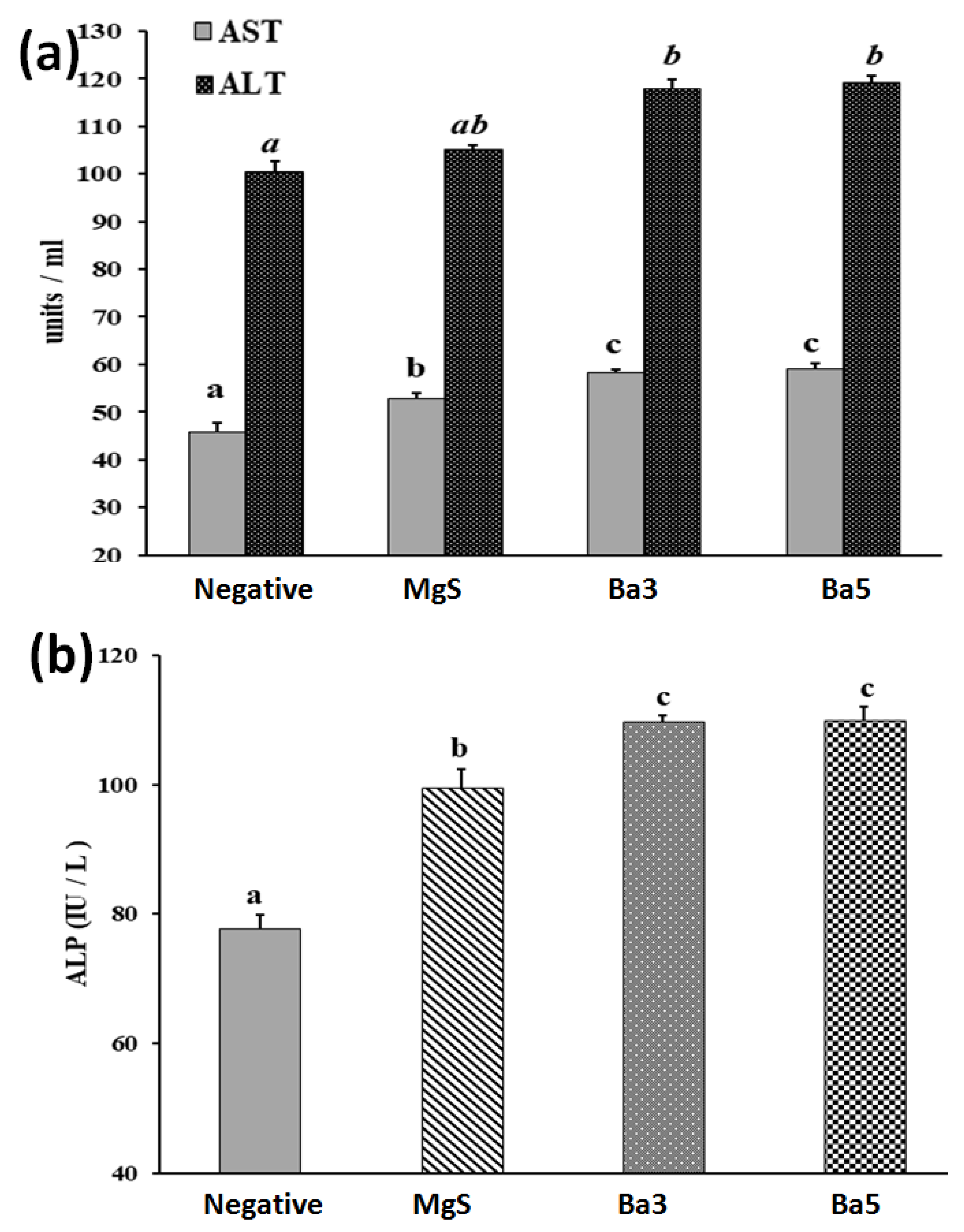
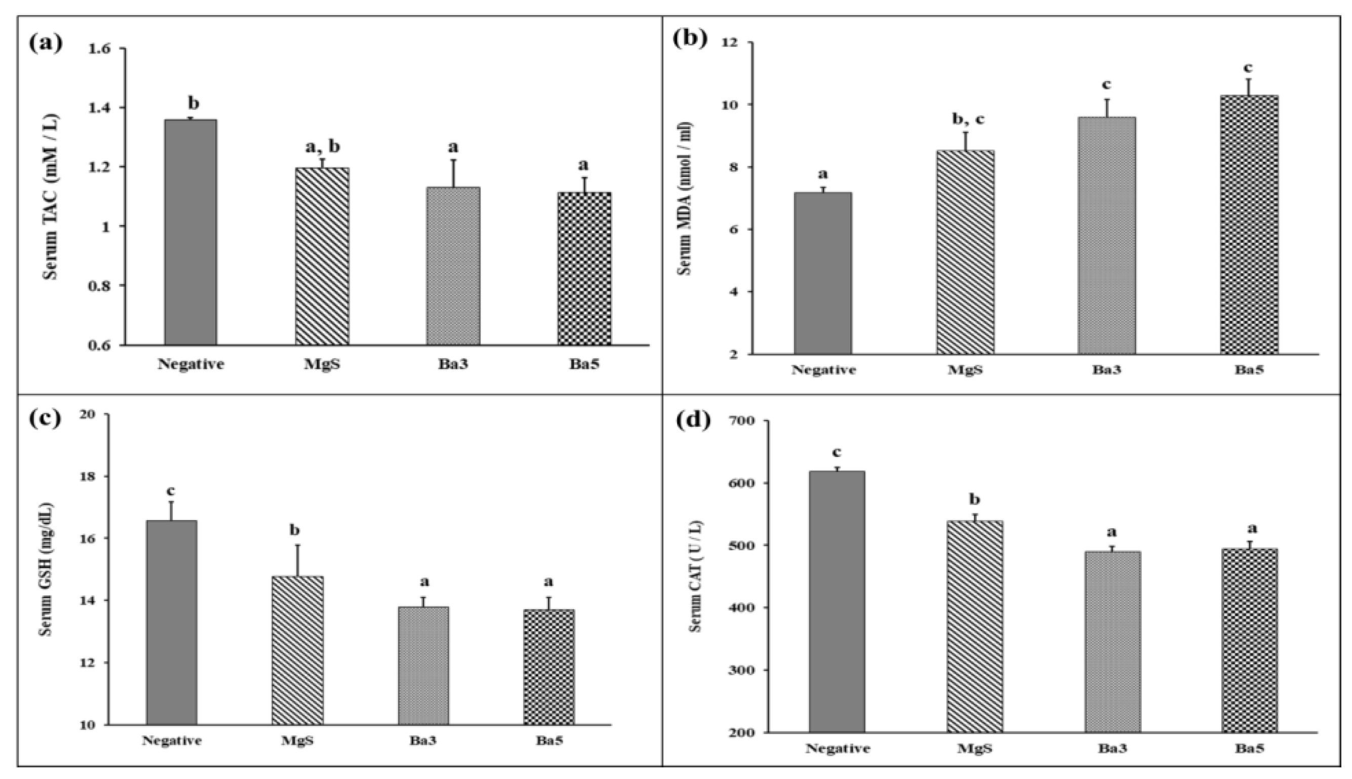
3.7.2. Hepatotoxicity Indices in Tibia-Fractured Rats
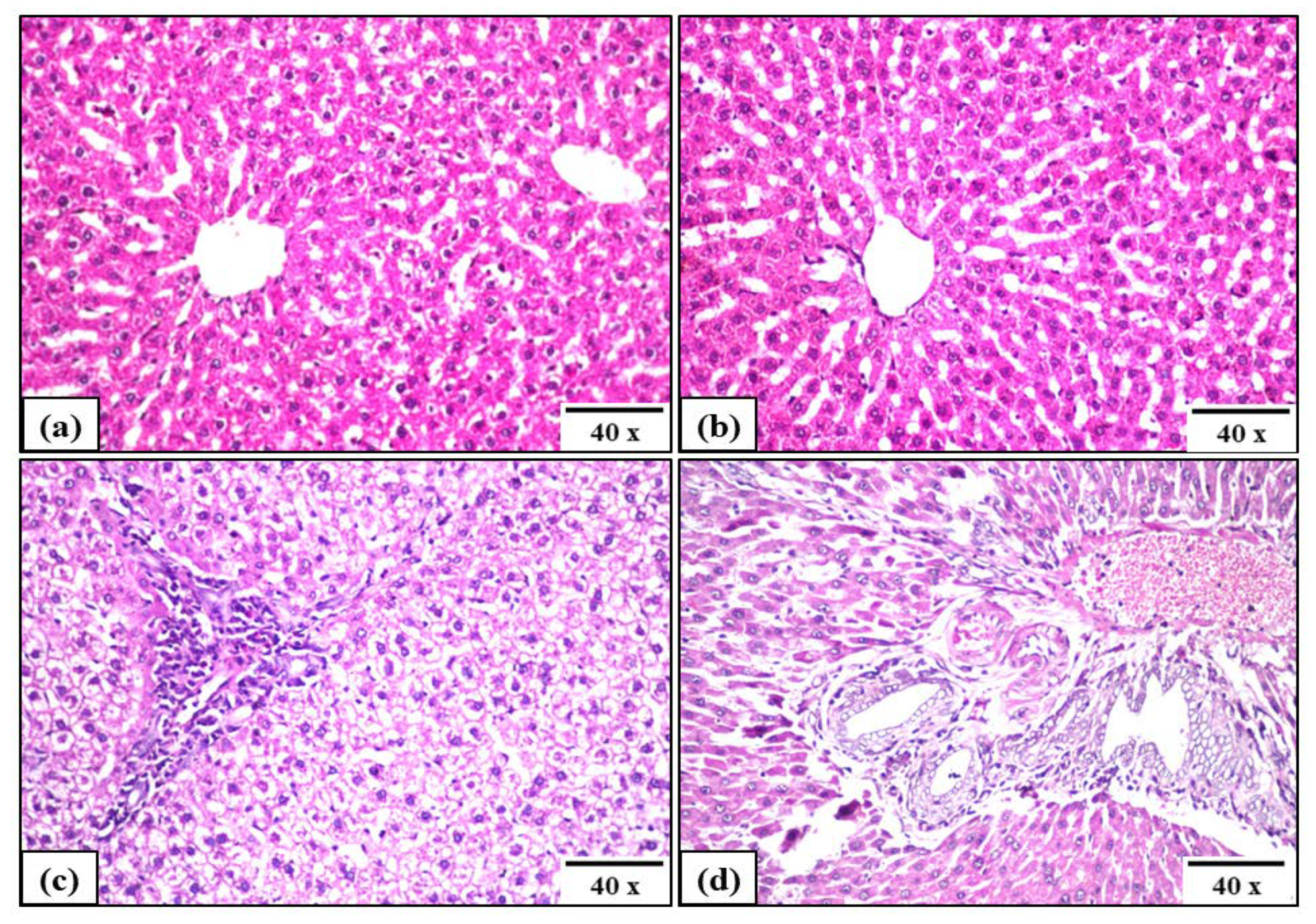
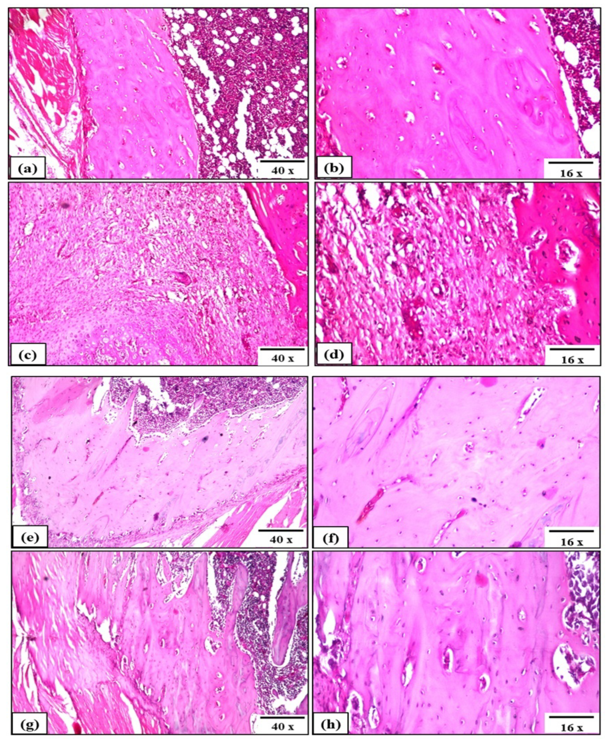
3.7.3. Histopathological Analysis of Tibia-Fractured Rats
4. Discussion
5. Conclusions
Author Contributions
Funding
Institutional Review Board Statement
Informed Consent Statement
Data Availability Statement
Conflicts of Interest
References
- Sabet, F.A.; Najafi, A.R.; Hamed, E.; Jasiuk, I. Modelling of bone fracture and strength at different length scales: A review. Interface Focus 2016, 6, 20150055. [Google Scholar] [CrossRef] [PubMed] [Green Version]
- Javaid, M.K.; Kyer, C.; Mitchell, P.J.; Chana, J.; Moss, C.; Edwards, M.H.; McLellan, A.R.; Stenmark, J.; Pierroz, D.D.; Schneider, M.C.; et al. Exco, effective secondary fracture prevention: Implementation of a global benchmarking of clinical quality using the IOF Capture the Fracture® Best Practice Framework tool. Osteoporos. Int. 2015, 26, 2573–2578. [Google Scholar] [CrossRef] [PubMed]
- Marmor, M.; Alt, V.; Latta, L.; Lane, J.; Rebolledo, B.; Egol, K.A.; Miclau, T. Osteoporotic fracture care: Are we closer to gold standards? J. Orthop. Trauma 2015, 29 (Suppl. S12), S53–S56. [Google Scholar] [CrossRef] [PubMed]
- Marsell, R.; Einhorn, T.A. The biology of fracture healing. Inj. Int. J. Care Inj. 2011, 42, 551–555. [Google Scholar] [CrossRef] [Green Version]
- Egermann, M.; Lill, C.A.; Griesbeck, K.; Evans, C.H.; Robbins, P.D.; Schneider, E.; Baltzer, A.W. Effect of BMP-2 gene transfer on bone healing in sheep. Gene Ther. 2006, 13, 1290–1299. [Google Scholar] [CrossRef] [PubMed]
- Phillips, A.M. Overview of the fracture healing cascade. Injury 2005, 36, S5–S7. [Google Scholar] [CrossRef]
- Tautzenberger, A.; Kovtun, A.; Ignatius, A. Nanoparticles and their potential for application in bone. Int. J. Nanomed. 2012, 7, 4545–4557. [Google Scholar] [CrossRef] [Green Version]
- Dimitriou, R.; Jones, E.; McGonagle, D.; Giannoudis, P.V. Bone regeneration: Current concepts and future directions. BMC Med. 2011, 9, 66. [Google Scholar] [CrossRef] [Green Version]
- Aydin, A.; Memisoglu, K.; Cengiz, A.; Atmaca, H.; Muezzinoglu, B.; Muezzinoglu, U.S. Effects of botulinum toxin a on fracture healing in rats: An experimental study. J. Orthop. Sci. 2012, 17, 796–801. [Google Scholar] [CrossRef]
- LaStayo, P.C.; Winters, K.M.; Hardy, M. Fracture healing: Bone healing, fracture management and current concepts related to the hand. J. Hand Ther. 2003, 16, 81–93. [Google Scholar] [CrossRef]
- Wang, Q.; Yan, J.; Yang, J.; Li, B. Nanomaterials promise better bone repair. Mater. Today 2016, 19, 451–463. [Google Scholar] [CrossRef]
- Perez, R.A.; Won, J.-E.; Knowles, J.C.; Kim, H.-W. Naturally and synthetic smart composite biomaterials for tissue regeneration. Adv. Drug Deliv. Rev. 2013, 65, 471–496. [Google Scholar] [CrossRef] [PubMed]
- Balansundarum, G.; Storey, D.M.; Webster, T.J. Novel nano-rough polymers for cartilage tissue engineering. Int. J. Nanomed. 2014, 9, 1845–1853. [Google Scholar]
- Mabrouk, M.; Das, D.B.; Salem, Z.A.; Beherei, H.H. Nanomaterials for Biomedical Applications: Production, Characterisations, Recent Trends and Difficulties. Molecules 2021, 26, 1077. [Google Scholar] [CrossRef]
- Florence, N.T.; Huguette, S.T.S.; Hubert, D.J.; Raceline, G.K.; Desire, D.D.P.; Pierre, K.; Theophile, D. Aqueous extract of Peperomia pellucida (L.) HBK accelerates fracture healing in Wistar rats. BMC Complement. Altern. Med. 2017, 17, 188. [Google Scholar] [CrossRef] [Green Version]
- Chen, Z.; Kang, L.; Meng, Q.Y.; Liu, H.; Wang, Z.; Guo, Z.; Cui, F.Z. Degradability of injectable calcium sulfate/mineralized collagen-based bone repair material and its effect on bone tissue regeneration. Mater. Sci. Eng. C Mater. Biol. Appl. 2014, 45, 94–102. [Google Scholar] [CrossRef]
- Guo, Z.; Liu, X.-M.; Ma, L.; Li, J.; Zhang, H.; Gao, Y.-P.; Yuan, Y. Effects of particle morphology, pore size and surface coating of mesoporous silica on Naproxen dissolution rate enhancement. Colloids Surf. B Biointerfaces 2013, 101, 228–235. [Google Scholar] [CrossRef]
- Lin, K.; Liu, Y.; Huang, H.; Chen, L.; Wang, Z.; Chang, J. Degradation and silicon excretion of the calcium silicate bioactive ceramics during bone regeneration using rabbit femur defect model. J. Mater. Sci. Mater. Med. 2015, 26, 197. [Google Scholar] [CrossRef]
- Khan, A.F.; Saleem, M.; Afzal, A.; Ali, A.; Khan, A.; Khan, A.R. Bioactive behavior of silicon substituted calcium phosphate based bioceramics for bone regeneration. Mater. Sci. Eng. C Mater. Biol. Appl. 2014, 35, 245–252. [Google Scholar] [CrossRef]
- Zhou, H.; Wu, X.; Wei, J.; Lu, X.; Zhang, S.; Shi, J.; Liu, C. Stimulated osteoblastic proliferation by mesoporous silica xerogel with high specific surface area. J. Mater. Sci. Mater. Med. 2011, 22, 731–739. [Google Scholar] [CrossRef] [PubMed]
- Bose, S.; Fielding, G.; Tarafder, S.; Bandyopadhyay, A. Understanding of dopant-induced osteogenesis and angiogenesis in calcium phosphate ceramics. Trends Biotechnol. 2013, 31, 594–605. [Google Scholar] [CrossRef] [PubMed] [Green Version]
- Alshemary, A.Z.; Goh, Y.-F.; Akram, M.; Kadir, M.R.A.; Hussain, R. Barium and fluorine doped synthetic hydroxyapatite: Characterization and in-vitro bioactivity analysis. Sci. Adv. Mater. 2015, 7, 249–257. [Google Scholar] [CrossRef]
- Zamora, L.L.; Perez-Gracia, M.T. Using digital photography to implement the McFarland method. J. R. Soc. Interface 2012, 9, 1892–1897. [Google Scholar] [CrossRef] [PubMed]
- Anitha, S.; Muthukumaran, S. Structural, optical and antibacterial investigation of La, Cu dual doped ZnO nanoparticles prepared by co-precipitation method. Mater. Sci. Eng. C Mater. Biol. Appl. 2020, 108, 110387. [Google Scholar] [CrossRef]
- Bigham-Sadeg, A.; Karimi, I.; Hoseini, F.; Oryan, A.; Sharifi, S.; Pakzad, A. Effects of honey and hydroxyapatite on bone healing in rats. Trauma Mon. 2018, 23, e56119. [Google Scholar]
- Reitman, S.; Frankel, S. A colorimetric method for the determination of serum glutamic oxalacetic and glutamic pyruvic transaminases. Am. J. Clin. Pathol. 1957, 28, 56–63. [Google Scholar] [CrossRef]
- Belfield, A.; Goldberg, D.M. Normal ranges and diagnostic value of serum 5′nucleotidase and alkaline phosphatase activities in infancy. Arch. Dis. Child. 1971, 46, 842–846. [Google Scholar] [CrossRef] [Green Version]
- Koracevic, D.; Koracevic, G.; Djordjevic, V.; Andrejevic, S.; Cosic, V. Method for the measurement of antioxidant activity in human fluids. J. Clin. Pathol. 2001, 54, 356–361. [Google Scholar] [CrossRef] [Green Version]
- Ohkawa, H.; Ohishi, N.; Yagi, K. Assay for lipid peroxides in animal tissues by thiobarbituric acid reaction. Anal. Biochem. 1979, 95, 351–358. [Google Scholar] [CrossRef]
- Tekin, S.; Seven, E. Assessment of serum catalase, reduced glutathione, and superoxide dismutase activities and malondialdehyde levels in keratoconus patients. Eye 2021, 1–5. [Google Scholar] [CrossRef]
- Krishna, H.; Avinash, K.; Shivakumar, A.; Al-Tayar, N.G.S.; Shrestha, A.K. A quantitative method for the detection and validation of catalase activity at physiological concentration in human serum, plasma and erythrocytes. Spectrochim. Acta A Mol. Biomol. Spectrosc. 2021, 251, 119358. [Google Scholar] [CrossRef] [PubMed]
- Bancroft, J.D.; Stevens, A.; Turner, D.R. Theory and Practice of Histological Techniques, 4th ed.; Churchill Livingstone: New York, NY, USA, 1996. [Google Scholar]
- Myat, M.-H.; Noor, A.-F.M.; Kawashita, M.; Ismail, Y.M.B. Enhanced sinterability and in vitro bioactivity of barium-doped akermanite Ceramic. Ceram. Int. 2020, 46, 19062–19068. [Google Scholar] [CrossRef]
- Sun, Z.; Xinhui, D.; Srinivasakannan, C.; Liang, J. Preparation of magnesium silicate/carbon composite for adsorption of rhodamine B. RSC Adv. 2018, 8, 7873–7882. [Google Scholar] [CrossRef] [Green Version]
- Peric, M.; Dumic-Cule, I.; Grcevic, D.; Matijasic, M.; Verbanac, D.; Paul, R.; Grgurevic, L.; Trkulja, V.; Bagi, C.M.; Vukicevic, S. The rational use of animal models in the evaluation of novel bone regenerative therapies. Bone 2015, 70, 73–86. [Google Scholar] [CrossRef] [Green Version]
- Kanasan, N.; Adzila, S.; Koh, C.T.; Rahman, H.A.; Panerselvan, G. Effects of magnesium doping on the properties of hydroxyapatite/sodium alginate biocomposite. Adv. Appl. Ceram. 2019, 118, 381–386. [Google Scholar] [CrossRef]
- Schatkoski, V.M.; doMontanheiro, A.; de Menezes, T.L.; Pereira, B.R.C.; Rodrigues, R.M.; Rodrigues, K.F.; Ribas, R.G.; da Silva, D.M.; Thim, G.P. Current advances concerning the most cited metal ions doped bioceramics and silicate-based bioactive glasses for bone tissue engineering. Ceram. Int. 2021, 47, 2999–3012. [Google Scholar] [CrossRef]
- Munirathinam, B.; Jaladurgam, N.R.; Magesh, J.; Narayanan, R.; Mol, J.M.; Neelakantan, L. Improved corrosion protection of titanium implant material by crystallographic texturing of Sr doped calcium phosphate electrodeposits. Thin Solid Films 2019, 675, 115–121. [Google Scholar] [CrossRef]
- Tabia, Z.; El Mabrouk, K.M.; Nouneh, B.K. Mesoporous bioactive glass nanoparticles doped with magnesium: Drug delivery and acellular in vitro bioactivity. RSC Adv. 2019, 9, 12232–12246. [Google Scholar] [CrossRef] [Green Version]
- Ni, S.; Chou, L.; Chang, J. Preparation and characterization of forsterite (Mg2SiO4) bioceramics. Ceram. Int. 2007, 33, 83–88. [Google Scholar] [CrossRef]
- Oudadesse, H.; Martin, S.; Derrien, A.; Lucas-Girot, A.; Cathelineau, G.; Blondiaux, G. Determination of Ca, P, Sr and Mg in the synthetic biomaterial aragonite by NAA. J. Radioanal. Nucl. Chem. 2004, 262, 479–483. [Google Scholar] [CrossRef]
- Diba, M.; Goudouri, O.M.; Tapia, F.; Boccaccini, A.R. Magnesium-containing bioactive polycrystalline silicate-based ceramics and glass-ceramics for biomedical applications. Curr. Opin. Solid State Mater. Sci. 2014, 18, 147–167. [Google Scholar] [CrossRef]
- Zhai, W.; Lu, H.; Chen, L.; Lin, X.; Huang, Y.; Dai, K.; Naoki, K.; Chen, G.; Chang, J. Silicate bioceramicsinduce angiogenesis during bone regeneration. Acta Biomater. 2012, 8, 341–349. [Google Scholar] [CrossRef] [PubMed]
- Nielsen, F.H. Update on the possible nutritional importance of silicon. J. Trace Elem. Med. Biol. 2014, 28, 379–382. [Google Scholar] [CrossRef] [PubMed]
- Jugdaohsingh, R.; Pedro, L.D.; Watson, A.; Powell, J.J. Silicon and boron differ in their localization and loading of bone. Bone Rep. 2014, 4, 9–15. [Google Scholar] [CrossRef] [Green Version]
- Shie, M.-Y.; Ding, S.-J.; Chang, H.-C. The role of silicon in osteoblast-like cell proliferation and apoptosis. Acta Biomater. 2011, 7, 2604–2614. [Google Scholar] [CrossRef]
- Reffitt, D.M.; Ogston, N.; Jugdaohsingh, R.; Cehung, H.F.J.; Evans, R.P.H.; Thompson, R.P.H.; Powell, J.J.; Hampson, G.N. Orthosilic acid stimulates collagen type I synthesis and osteoblastic differentiation in human osteoblast-like cells in vitro. Bone 2003, 32, 127–135. [Google Scholar] [CrossRef]
- European Food Society Authority (EFSA). Opinion of the Scientific Panel on Dietetic Products, Nutrition and Allergies on a request from the Commission related to the tolerable upper intake level of silicon. EFSA J. 2004, 60, 1–11. [Google Scholar]
- Götz, W.; Tobiasch, E.; Witzleben, S.; Schulze, M. Effects of silicon compounds on biomineralization, osteogenesis, and hard tissue formation. Pharmaceutics 2019, 11, 117. [Google Scholar] [CrossRef] [PubMed] [Green Version]
- Yonesaki, Y.; Takei, T.; Kumada, N.; Kinomura, N. Crystal structure of Eu2+-doped M3MgSi2O8 (M: Ba, Sr, Ca) compounds and their emission properties. J. Solid State Chem. 2009, 182, 547–554. [Google Scholar] [CrossRef]
- Joseph, T.; Sebastian, M.T. Microwave Dielectric Properties of (Sr1−xAx)2(Zn1−xBx)Si2O7 Ceramics (A = Ca, Ba and B = Co, Mg, Mn, Ni). J. Am. Ceram. Soc. 2010, 93, 147–154. [Google Scholar] [CrossRef]
- Singh, R.K.; Kannan, S. Synthesis, Structural analysis, Mechanical, antibacterial and Hemolytic activity of Mg2+ and Cu2+ co-substitutions in β-Ca3(PO4)2. Mater. Sci. Eng. C 2014, 45, 530–538. [Google Scholar] [CrossRef] [PubMed]
- Guo, Z.; Zhang, Z.; Zhang, N.; Gao, W.; Li, J.; Pu, Y.; He, B.; Xie, J. A Mg2+/polydopamine composite hydrogel for the acceleration of infected wound healing. Bioact. Mater. 2022, 15, 203–213. [Google Scholar] [CrossRef] [PubMed]
- Luque-Agudo, V.; Fernández-Calderón, M.C.; Pacha-Olivenza, M.A.; Pérez-Giraldo, C.; Gallardo-Moreno, A.M.; González-Martín, M.L. The role of magnesium in biomaterials related infections. Colloids Surf. B Biointerfaces 2020, 191, 110996. [Google Scholar] [CrossRef] [PubMed]
- Hickey, D.J.; Muthusamy, D.; Webster, T.J. Electrophoretic deposition of MgO nanoparticles imparts antibacterial properties to poly-L-lactic acid for orthopedic applications. J. Biomed. Mater. Res. Part A 2017, 105, 3136–3147. [Google Scholar] [CrossRef] [PubMed]
- Rodríguez-Sánchez, J.; Pacha-Olivenza, M.; González-Martín, M. Bactericidal effect of magnesium ions over planktonic and sessile Staphylococcus epidermidis and Escherichia coli. Mater. Chem. Phys. 2019, 221, 342–348. [Google Scholar] [CrossRef]
- Tong, G.; Du, F.; Wu, W.; Wu, R.; Liu, F.; Liang, Y. Enhanced reactive oxygen species (ROS) yields and antibacterial activity of spongy ZnO/ZnFe2O4 hybrid micro-hexahedra selectively synthesized through a versatile glucose-engineered co-precipitation/annealing process. J. Mater. Chem. B 2013, 1, 2647–2657. [Google Scholar] [CrossRef]
- Mabrouk, M.; Fouad, G.I.; El-Sayed, S.A.M.; Rizk, M.Z.; Beherei, H.H. Hepatotoxic and Neurotoxic Potential of Iron Oxide Nanoparticles in Wistar Rats: A Biochemical and Ultrastructural Study. Biol. Trace Elem. Res. 2021, 200, 3638–3665. [Google Scholar] [CrossRef]
- Mahdy, E.A.; Sahbal, K.M.; Mabrouk, M.; Beherei, H.H.; Abdel-Monem, Y.K. Enhancement of glass-ceramic performance by TiO2 doping: In Vitro cell viability, proliferation, and differentiation. Ceram. Int. 2021, 47, 6251–6261. [Google Scholar] [CrossRef]
- Ball, J.P.; Mound, B.A.; Nino, J.C.; Allen, J.B. Biocompatible evaluation of barium titanate foamed ceramic structures for orthopedic applications. J. Biomed. Mater. Res. Part A 2014, 102, 2089–2095. [Google Scholar] [CrossRef]
- Wani, M.Y.; Hashim, M.A.; Nabi, F.; Malik, M.A. Nanotoxicity: Dimensional and morphological concerns. Adv. Phys. Chem. 2011, 2011, 450912. [Google Scholar] [CrossRef] [Green Version]
- Kamitakahara, M.; Ohtsuki, C.; Miyazaki, T. Review paper: Behavior of ceramic biomaterials derived from tricalcium phosphate in physiological condition. J. Biomater. Appl. 2008, 23, 197–212. [Google Scholar] [CrossRef] [PubMed]
- Misch, C.E.; Qu, Z.; Bidez, M.W. Mechanical properties of trabecular bone in the human mandible: Implications for dental implant treatment planning and surgical placement. J. Oral Maxillofac. Surg. 1999, 57, 700–706. [Google Scholar] [CrossRef]
- Kravchenko, J.; Darrah, T.H.; Miller, R.K.; Lyerly, H.K.; Vengosh, A. A review of the health impacts of barium from natural and anthropogenic exposure. Environ. Geochem. Health 2014, 36, 797–814. [Google Scholar] [CrossRef]
- Emsley, J. The Elements; Clarendon Press: Oxford, UK; Oxford University Press: New York, NY, USA, 1998. [Google Scholar]
- Poddalgoda, D.; Macey, K.; Assad, H.; Krishnan, K. Development of biomonitoring equivalents for barium in urine and plasma for interpreting human biomonitoring data. Regul. Toxicol. Pharmacol. 2017, 86, 303–311. [Google Scholar] [CrossRef] [PubMed]
- Peana, M.; Medici, S.; Dadar, M.; Zoroddu, M.A.; Pelucelli, A.; Chasapis, C.T.; Bjørklund, G. Environmental barium: Potential exposure and health-hazards. Arch. Toxicol. 2021, 95, 2605–2612. [Google Scholar] [CrossRef]
- Makita, M.; Yamakado, K.; Nakatsuka, A.; Takaki, H.; Inaba, T.; Oshima, F.; Katayama, H.; Takeda, K. Effects of barium concentration on the radiopacity and biomechanics of bone cement: Experimental study. Radiat. Med. 2008, 26, 533–538. [Google Scholar] [CrossRef]


| Sample | SiO2 | MgO | BaO |
|---|---|---|---|
| MgS | 50 | 50 | -- |
| Ba3 | 50 | 47 | 3 |
| Ba5 | 50 | 45 | 5 |
| Ba7 | 50 | 43 | 7 |
| Sample | BET Surface Area (m²/g) | Pore Volume (cm³/g) | Pore Diameter (nm) |
|---|---|---|---|
| MgS | 122.63 ± 0.54 | 0.41± 0.04 | 10.78 ± 0.54 |
| Ba3 | 75.20 ± 0.41 | 0.22 ± 0.03 | 11.08 ± 0.62 |
| Ba5 | 64.66 ± 0.37 | 0.19 ± 0.05 | 11.10 ± 0.47 |
| Ba7 | 37.95 ± 0.30 | 0.13 ± 0.04 | 12.81 ± 0.83 |
Publisher’s Note: MDPI stays neutral with regard to jurisdictional claims in published maps and institutional affiliations. |
© 2022 by the authors. Licensee MDPI, Basel, Switzerland. This article is an open access article distributed under the terms and conditions of the Creative Commons Attribution (CC BY) license (https://creativecommons.org/licenses/by/4.0/).
Share and Cite
Mabrouk, M.; Ibrahim Fouad, G.; Beherei, H.H.; Das, D.B. Barium Oxide Doped Magnesium Silicate Nanopowders for Bone Fracture Healing: Preparation, Characterization, Antibacterial and In Vivo Animal Studies. Pharmaceutics 2022, 14, 1582. https://doi.org/10.3390/pharmaceutics14081582
Mabrouk M, Ibrahim Fouad G, Beherei HH, Das DB. Barium Oxide Doped Magnesium Silicate Nanopowders for Bone Fracture Healing: Preparation, Characterization, Antibacterial and In Vivo Animal Studies. Pharmaceutics. 2022; 14(8):1582. https://doi.org/10.3390/pharmaceutics14081582
Chicago/Turabian StyleMabrouk, Mostafa, Ghadha Ibrahim Fouad, Hanan H. Beherei, and Diganta Bhusan Das. 2022. "Barium Oxide Doped Magnesium Silicate Nanopowders for Bone Fracture Healing: Preparation, Characterization, Antibacterial and In Vivo Animal Studies" Pharmaceutics 14, no. 8: 1582. https://doi.org/10.3390/pharmaceutics14081582