
The pH-Responsive Liposomes—The Effect of PEGylation on Release Kinetics and Cellular Uptake in Glioblastoma Cells

 , and
, and 
Abstract
:1. Introduction
2. Materials and Methods
2.1. Materials
2.2. Liposome Preparation
2.3. Characterization of Liposomes
2.4. In Vitro pH-Responsive Calcein Release
2.5. Cell Culture
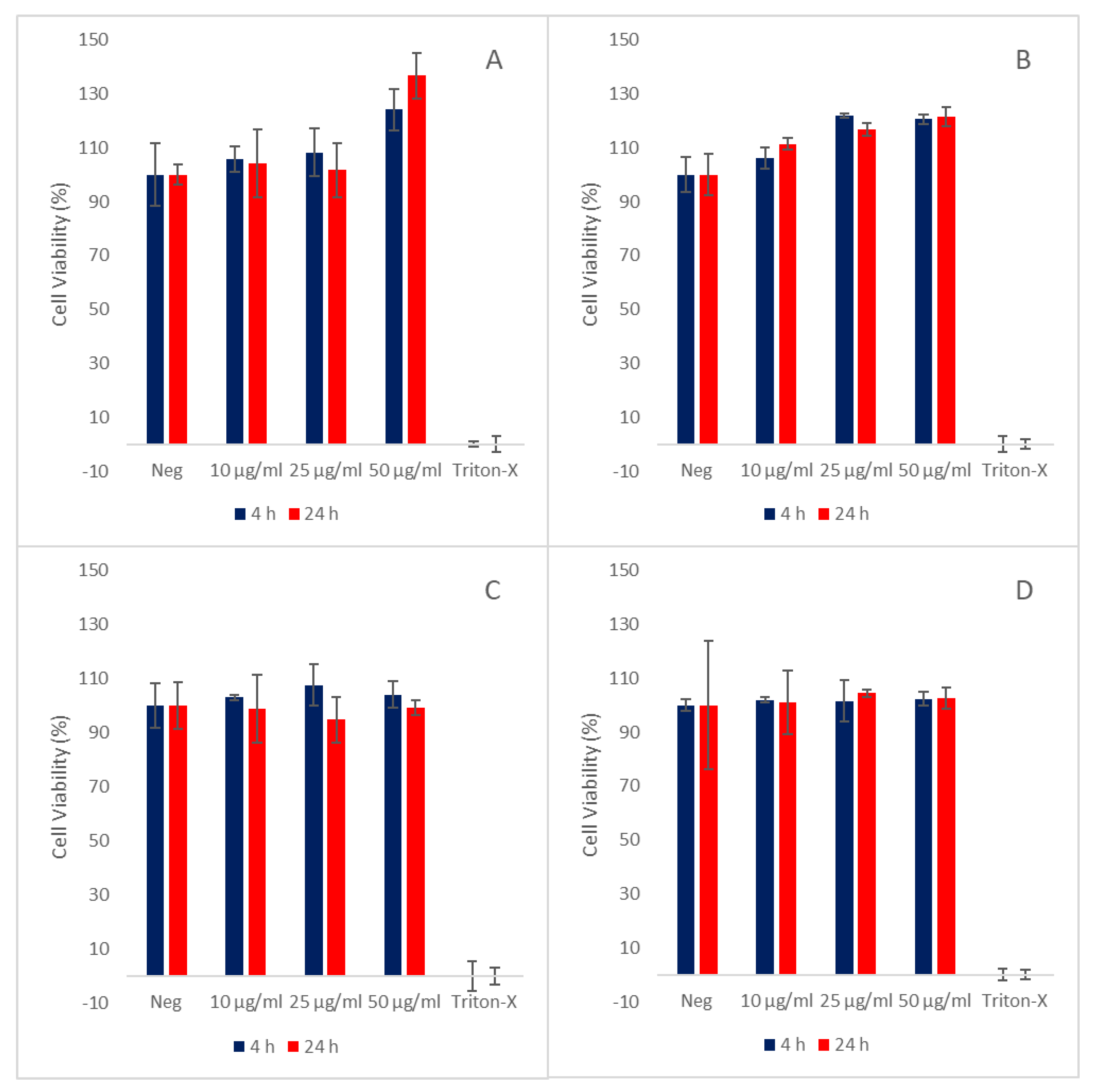
2.6. Cell Viability
2.7. Cellular Uptake
2.8. Statistical Analysis
3. Results
3.1. Liposomal Charcateristics
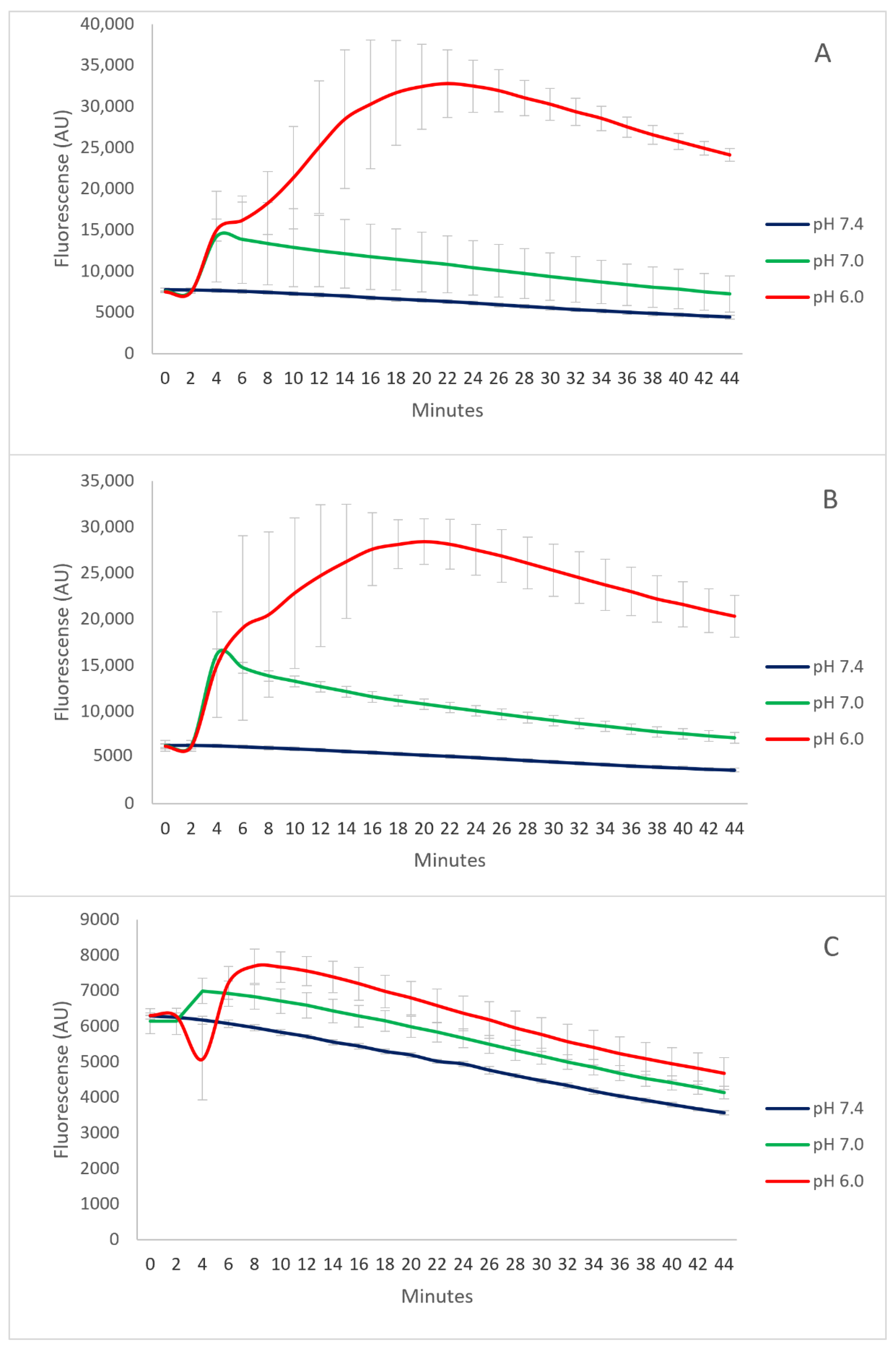
3.2. The pH-Responsive Calcein Release
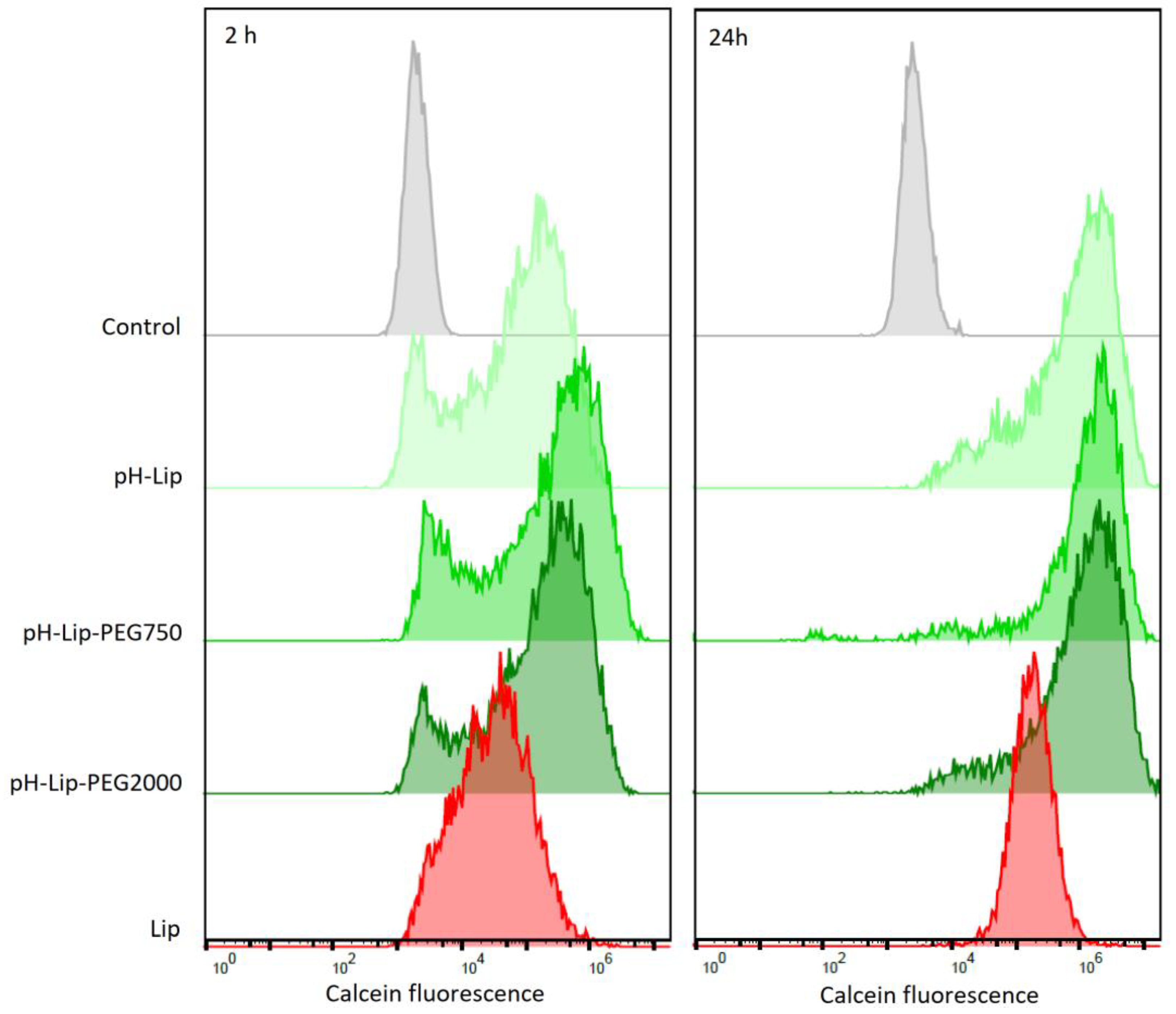
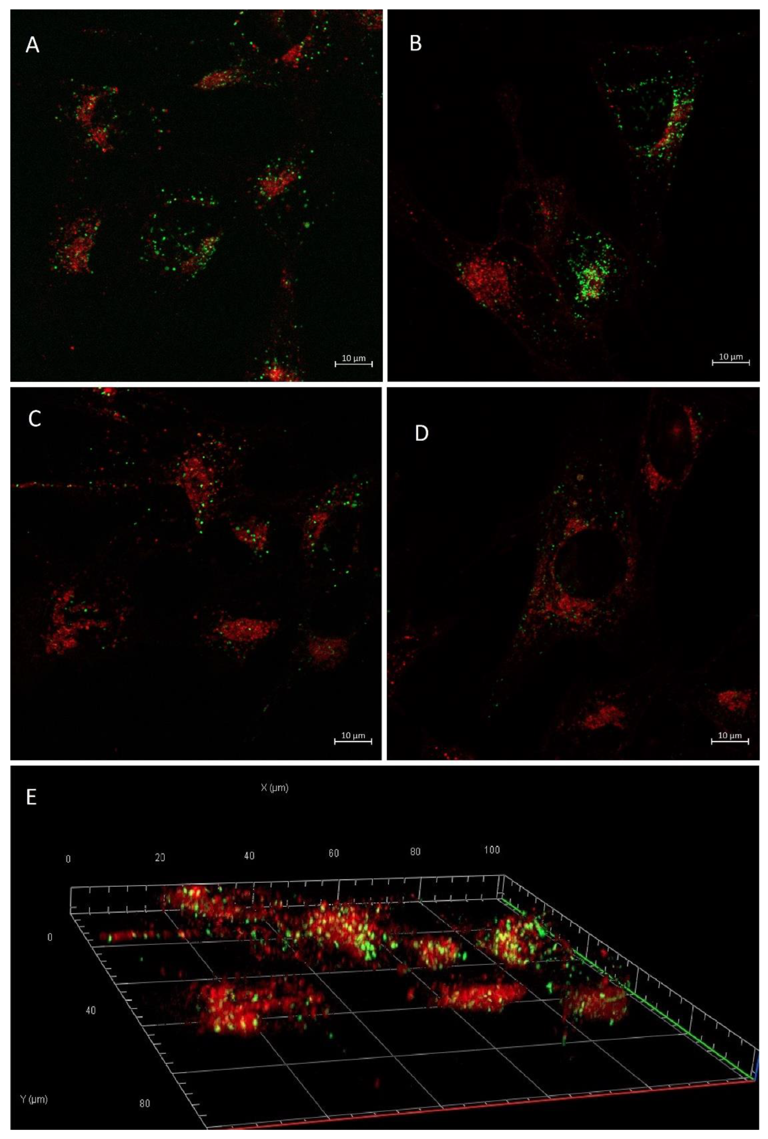
3.3. Cellular Uptake
4. Discussion
5. Conclusions
Supplementary Materials
Author Contributions
Funding
Institutional Review Board Statement
Informed Consent Statement
Data Availability Statement
Acknowledgments
Conflicts of Interest
References
- Hsu, J.F.; Chu, S.M.; Liao, C.C.; Wang, C.J.; Wang, Y.S.; Lai, M.Y.; Wang, H.C.; Huang, H.R.; Tsai, M.H. Nanotechnology and Nanocarrier-Based Drug Delivery as the Potential Therapeutic Strategy for Glioblastoma Multiforme: An Update. Cancers 2021, 13, 195. [Google Scholar] [CrossRef] [PubMed]
- Janjua, T.I.; Rewatkar, P.; Ahmed-Cox, A.; Saeed, I.; Mansfeld, F.M.; Kulshreshtha, R.; Kumeria, T.; Ziegler, D.S.; Kavallaris, M.; Mazzieri, R.; et al. Frontiers in the Treatment of Glioblastoma: Past, Present and Emerging. Adv. Drug Deliv. Rev. 2021, 171, 108–138. [Google Scholar] [CrossRef] [PubMed]
- Beier, C.P.; Schmid, C.; Gorlia, T.; Kleinletzenberger, C.; Beier, D.; Grauer, O.; Steinbrecher, A.; Hirschmann, B.; Brawanski, A.; Dietmaier, C.; et al. RNOP-09: Pegylated Liposomal Doxorubicine and Prolonged Temozolomide in Addition to Radiotherapy in Newly Diagnosed Glioblastoma—A Phase II Study. BMC Cancer 2009, 9, 308. [Google Scholar] [CrossRef] [Green Version]
- Ananda, S.; Nowak, A.K.; Cher, L.; Dowling, A.; Brown, C.; Simes, J.; Rosenthal, M.A. Phase 2 Trial of Temozolomide and Pegylated Liposomal Doxorubicin in the Treatment of Patients with Glioblastoma Multiforme Following Concurrent Radiotherapy and Chemotherapy. J. Clin. Neurosci. 2011, 18, 1444–1448. [Google Scholar] [CrossRef] [PubMed]
- Li, J.; Tan, T.; Zhao, L.; Liu, M.; You, Y.; Zeng, Y.; Chen, D.; Xie, T.; Zhang, L.; Fu, C.; et al. Recent Advancements in Liposome-Targeting Strategies for the Treatment of Gliomas: A Systematic Review. ACS Appl. Bio Mater. 2020, 3, 5500–5528. [Google Scholar] [CrossRef] [PubMed]
- Patel, A.P.; Fisher, J.L.; Nichols, E.; Abd-Allah, F.; Abdela, J.; Abdelalim, A.; Abraha, H.N.; Agius, D.; Alahdab, F.; Alam, T.; et al. Global, Regional, and National Burden of Brain and Other CNS Cancer, 1990–2016: A Systematic Analysis for the Global Burden of Disease Study 2016. Lancet Neurol. 2019, 18, 376–393. [Google Scholar] [CrossRef] [Green Version]
- Amarandi, R.M.; Ibanescu, A.; Carasevici, E.; Marin, L.; Dragoi, B. Liposomal-Based Formulations: A Path from Basic Research to Temozolomide Delivery Inside Glioblastoma Tissue. Pharmaceutics 2022, 14, 308. [Google Scholar] [CrossRef]
- Minniti, G.; De Sanctis, V.; Muni, R.; Filippone, F.; Bozzao, A.; Valeriani, M.; Osti, M.F.; De Paula, U.; Lanzetta, G.; Tombolini, V.; et al. Radiotherapy plus Concomitant and Adjuvant Temozolomide for Glioblastoma in Elderly Patients. J. Neurooncol. 2008, 88, 97–103. [Google Scholar] [CrossRef]
- Tan, A.C.; Ashley, D.M.; López, G.Y.; Malinzak, M.; Friedman, H.S.; Khasraw, M. Management of Glioblastoma: State of the Art and Future Directions. CA. Cancer J. Clin. 2020, 70, 299–312. [Google Scholar] [CrossRef]
- Ayub, A.; Wettig, S. An Overview of Nanotechnologies for Drug Delivery to the Brain. Pharmaceutics 2022, 14, 224. [Google Scholar] [CrossRef]
- Pandit, R.; Chen, L.; Götz, J. The Blood-Brain Barrier: Physiology and Strategies for Drug Delivery. Adv. Drug Deliv. Rev. 2020, 165–166, 1–14. [Google Scholar] [CrossRef] [PubMed]
- Chan, M.; Huang, W.; Satpathy, A.; Su, T.; Hsiao, M.; Liu, R. Progress and Viewpoints of Multifunctional Composite Nanomaterials for Glioblastoma Theranostics. Pharmaceutics 2022, 14, 456. [Google Scholar] [CrossRef] [PubMed]
- Bukhari, S.I.; Imam, S.S.; Ahmad, M.Z.; Vuddanda, P.R.; Alshehri, S.; Mahdi, W.A.; Ahmad, J. Recent Progress in Lipid Nanoparticles for Cancer Theranostics: Opportunity and Challenges. Pharmaceutics 2021, 13, 840. [Google Scholar] [CrossRef]
- Brown, T.D.; Habibi, N.; Wu, D.; Lahann, J.; Mitragotri, S. Effect of Nanoparticle Composition, Size, Shape, and Stiffness on Penetration across the Blood-Brain Barrier. ACS Biomater. Sci. Eng. 2020, 6, 4916–4928. [Google Scholar] [CrossRef] [PubMed]
- Nunes, S.S.; de Oliveira Silva, J.; Fernandes, R.S.; Miranda, S.E.M.; Leite, E.A.; de Farias, M.A.; Portugal, R.V.; Cassali, G.D.; Townsend, D.M.; Oliveira, M.C.; et al. PEGylated versus Non-PEGylated PH-Sensitive Liposomes: New Insights from a Comparative Antitumor Activity Study. Pharmaceutics 2022, 14, 272. [Google Scholar] [CrossRef]
- Germain, M.; Caputo, F.; Metcalfe, S.; Tosi, G.; Spring, K.; Åslund, A.K.O.; Pottier, A.; Schiffelers, R.; Ceccaldi, A.; Schmid, R. Delivering the Power of Nanomedicine to Patients Today. J. Control. Release 2020, 326, 164–171. [Google Scholar] [CrossRef]
- Kanamala, M.; Palmer, B.D.; Wilson, W.R.; Wu, Z. Characterization of a Smart PH-Cleavable PEG Polymer towards the Development of Dual PH-Sensitive Liposomes. Int. J. Pharm. 2018, 548, 288–296. [Google Scholar] [CrossRef]
- Halcrow, P.; Datta, G.; Ohm, J.E.; Soliman, M.L.; Chen, X.; Geiger, J.D. Role of Endolysosomes and PH in the Pathogenesis and Treatment of Glioblastoma. Cancer Rep. 2019, 2, e1177. [Google Scholar] [CrossRef]
- Webb, B.A.; Chimenti, M.; Jacobson, M.P.; Barber, D.L. Dysregulated PH: A Perfect Storm for Cancer Progression. Nat. Rev. Cancer 2011, 11, 671–677. [Google Scholar] [CrossRef]
- Shi, D.; Beasock, D.; Fessler, A.; Szebeni, J.; Ljubimova, J.Y.; Afonin, K.A.; Dobrovolskaia, M.A. To PEGylate or Not to PEGylate: Immunological Properties of Nanomedicine’s Most Popular Component, Polyethylene Glycol and Its Alternatives. Adv. Drug Deliv. Rev. 2022, 180, 114079. [Google Scholar] [CrossRef]
- Szatmári, T.; Lumniczky, K.; Désaknai, S.; Trajcevski, S.; Hídvégi, E.J.; Hamada, H.; Sáfrány, G. Detailed Characterization of the Mouse Glioma 261 Tumor Model for Experimental Glioblastoma Therapy. Cancer Sci. 2006, 97, 546–553. [Google Scholar] [CrossRef]
- Cauzzo, J.; Nystad, M.; Holsæter, A.M.; Basnet, P.; Škalko-Basnet, N. Following the Fate of Dye-Containing Liposomes in Vitro. Int. J. Mol. Sci. 2020, 21, 4847. [Google Scholar] [CrossRef] [PubMed]
- Lukyanov, A.N.; Elbayoumi, T.A.; Chakilam, A.R.; Torchilin, V.P. Tumor-Targeted Liposomes: Doxorubicin-Loaded Long-Circulating Liposomes Modified with Anti-Cancer Antibody. J. Control. Release 2004, 100, 135–144. [Google Scholar] [CrossRef]
- Mojarad-Jabali, S.; Farshbaf, M.; Hemmati, S.; Sarfraz, M.; Motasadizadeh, H.; Shahbazi Mojarrad, J.; Atyabi, F.; Zakeri-Milani, P.; Valizadeh, H. Comparison of Three Synthetic Transferrin Mimetic Small Peptides to Promote the Blood–Brain Barrier Penetration of Vincristine Liposomes for Improved Glioma Targeted Therapy. Int. J. Pharm. 2022, 613, 121395. [Google Scholar] [CrossRef] [PubMed]
- Smith, M.C.; Crist, R.M.; Clogston, J.D.; McNeil, S.E. Zeta Potential: A Case Study of Cationic, Anionic, and Neutral Liposomes. Anal. Bioanal. Chem. 2017, 409, 5779–5787. [Google Scholar] [CrossRef]
- Stupp, R.; Mason, W.P.; Van Den Bent, M.J.; Weller, M.; Fisher, B.; Taphoorn, M.J.B.; Belanger, K.; Brandes, A.A.; Marosi, C.; Bogdahn, U.; et al. Radiotherapy plus Concomitant and Adjuvant Temozolomide for Glioblastoma. N. Engl. J. Med. 2005, 352, 987–996. [Google Scholar] [CrossRef] [PubMed]
- Jain, K.K. A Critical Overview of Targeted Therapies for Glioblastoma. Front. Oncol. 2018, 8, 419. [Google Scholar] [CrossRef]
- Nowak, M.; Helgeson, M.E.; Mitragotri, S. Delivery of Nanoparticles and Macromolecules across the Blood–Brain Barrier. Adv. Ther. 2020, 3, 1900073. [Google Scholar] [CrossRef]
- Zhou, Y.; Peng, Z.; Seven, E.S.; Leblanc, R.M. Crossing the Blood-Brain Barrier with Nanoparticles. J. Control. Release 2018, 270, 290–303. [Google Scholar] [CrossRef]
- Gabizon, A.A.; de Rosales, R.T.M.; La-Beck, N.M. Translational Considerations in Nanomedicine: The Oncology Perspective. Adv. Drug Deliv. Rev. 2020, 158, 140–157. [Google Scholar] [CrossRef]
- Chauhan, V.P.; Stylianopoulos, T.; Boucher, Y.; Jain, R.K. Delivery of Molecular and Nanoscale Medicine to Tumors: Transport Barriers and Strategies. Annu. Rev. Chem. Biomol. Eng. 2011, 2, 281–298. [Google Scholar] [CrossRef] [PubMed]
- Simões, S.; Slepushkin, V.; Düzgünes, N.; Pedroso de Lima, M.C. On the Mechanisms of Internalization and Intracellular Delivery Mediated by PH-Sensitive Liposomes. Biochim. Biophys. Acta-Biomembr. 2001, 1515, 23–37. [Google Scholar] [CrossRef] [Green Version]
- Slepushkin, V.A.; Simões, S.; Dazin, P.; Newman, M.S.; Guo, L.S.; De Lima, M.C.P.; Düzgüneş, N. Sterically Stabilized PH-Sensitive Liposomes. Intracellular Delivery of Aqueous Contents and Prolonged Circulation in Vivo. J. Biol. Chem. 1997, 272, 2382–2388. [Google Scholar] [CrossRef] [PubMed] [Green Version]
- Nunes, S.S.; Fernandes, R.S.; Cavalcante, C.H.; da Costa César, I.; Leite, E.A.; Lopes, S.C.A.; Ferretti, A.; Rubello, D.; Townsend, D.M.; de Oliveira, M.C.; et al. Influence of PEG Coating on the Biodistribution and Tumor Accumulation of PH-Sensitive Liposomes. Drug Deliv. Transl. Res. 2019, 9, 123–130. [Google Scholar] [CrossRef]
- Kozma, G.T.; Shimizu, T.; Ishida, T.; Szebeni, J. Anti-PEG Antibodies: Properties, Formation, Testing and Role in Adverse Immune Reactions to PEGylated Nano-Biopharmaceuticals. Adv. Drug Deliv. Rev. 2020, 154–155, 163–175. [Google Scholar] [CrossRef]
- Yuan, F.; Dellian, M.; Fukumura, D.; Leunig, M.; Berk, D.A.; Jain, R.K.; Torchilin, V.P. Vascular Permeability in a Human Tumor Xenograft: Molecular Size Dependence and Cutoff Size. Cancer Res. 1995, 55, 3752–3756. [Google Scholar]
- Huth, U.S.; Schubert, R.; Peschka-Süss, R. Investigating the Uptake and Intracellular Fate of PH-Sensitive Liposomes by Flow Cytometry and Spectral Bio-Imaging. J. Control. Release 2006, 110, 490–504. [Google Scholar] [CrossRef]
- Alshehri, A.; Grabowska, A.; Stolnik, S. Pathways of Cellular Internalisation of Liposomes Delivered SiRNA and Effects on SiRNA Engagement with Target MRNA and Silencing in Cancer Cells. Sci. Rep. 2018, 8, 3748. [Google Scholar] [CrossRef]
- Zhao, Y.; Ren, W.; Zhong, T.; Zhang, S.; Huang, D.; Guo, Y.; Yao, X.; Wang, C.; Zhang, W.Q.; Zhang, X.; et al. Tumor-Specific PH-Responsive Peptide-Modified PH-Sensitive Liposomes Containing Doxorubicin for Enhancing Glioma Targeting and Anti-Tumor Activity. J. Control. Release 2016, 222, 56–66. [Google Scholar] [CrossRef]
- Poustforoosh, A.; Nematollahi, M.H.; Hashemipour, H.; Pardakhty, A. Recent Advances in Bio-Conjugated Nanocarriers for Crossing the Blood-Brain Barrier in (Pre-)Clinical Studies with an Emphasis on Vesicles. J. Control. Release 2022, 343, 777–797. [Google Scholar] [CrossRef]
- Farshbaf, M.; Mojarad-Jabali, S.; Hemmati, S.; Khosroushahi, A.Y.; Motasadizadeh, H.; Zarebkohan, A.; Valizadeh, H. Enhanced BBB and BBTB Penetration and Improved Anti-Glioma Behavior of Bortezomib through Dual-Targeting Nanostructured Lipid Carriers. J. Control. Release 2022, 345, 371–384. [Google Scholar] [CrossRef] [PubMed]


| Type of Liposome | Vesicle Size (nm) | PDI | ζ Potential (mV) |
|---|---|---|---|
| pH-Lip | 145.0 (±0.5) | 0.07 (±0.00) | −44.8 (±0.6) |
| pH-Lip_C | 163.9 (±0.9) | 0.11 (±0.01) | −37.2 (±1.6) |
| pH-Lip–PEG750 | 177.9 (±1.1) | 0.16 (±0.01) | −21.0 (±1.7) |
| pH-Lip–PEG750_C | 166.5 (±1.5) | 0.16 (±0.01) | −31.9 (±1.8) |
| pH-Lip–PEG2000 | 160.6 (±1.5) | 0.14 (±0.01) | −12.9 (±0.8) |
| pH-Lip–PEG2000_C | 161.7 (±0.8) | 0.12 (±0.01) | −16.1 (±0.8) |
| Lip | 156.4 (±2.0) | 0.14 (±0.01) | 0.08 (±0.7) |
| Lip_C | 160.1 (±0.6) | 0.09 (±0.01) | 4.29 (±0.9) |
Publisher’s Note: MDPI stays neutral with regard to jurisdictional claims in published maps and institutional affiliations. |
© 2022 by the authors. Licensee MDPI, Basel, Switzerland. This article is an open access article distributed under the terms and conditions of the Creative Commons Attribution (CC BY) license (https://creativecommons.org/licenses/by/4.0/).
Share and Cite
Rustad, E.A.L.; von Hofsten, S.; Kumar, R.; Lænsman, E.A.; Berge, G.; Škalko-Basnet, N. The pH-Responsive Liposomes—The Effect of PEGylation on Release Kinetics and Cellular Uptake in Glioblastoma Cells. Pharmaceutics 2022, 14, 1125. https://doi.org/10.3390/pharmaceutics14061125
Rustad EAL, von Hofsten S, Kumar R, Lænsman EA, Berge G, Škalko-Basnet N. The pH-Responsive Liposomes—The Effect of PEGylation on Release Kinetics and Cellular Uptake in Glioblastoma Cells. Pharmaceutics. 2022; 14(6):1125. https://doi.org/10.3390/pharmaceutics14061125
Chicago/Turabian StyleRustad, Eirik A. L., Susannah von Hofsten, Robin Kumar, Eirik A. Lænsman, Gerd Berge, and Nataša Škalko-Basnet. 2022. "The pH-Responsive Liposomes—The Effect of PEGylation on Release Kinetics and Cellular Uptake in Glioblastoma Cells" Pharmaceutics 14, no. 6: 1125. https://doi.org/10.3390/pharmaceutics14061125