
Mucoadhesive PLGA Nanospheres and Nanocapsules for Lactoferrin Controlled Ocular Delivery


 , , and
Abstract
:1. Introduction
2. Materials
3. Methods
3.1. Preparation of PLGA Nanoparticles
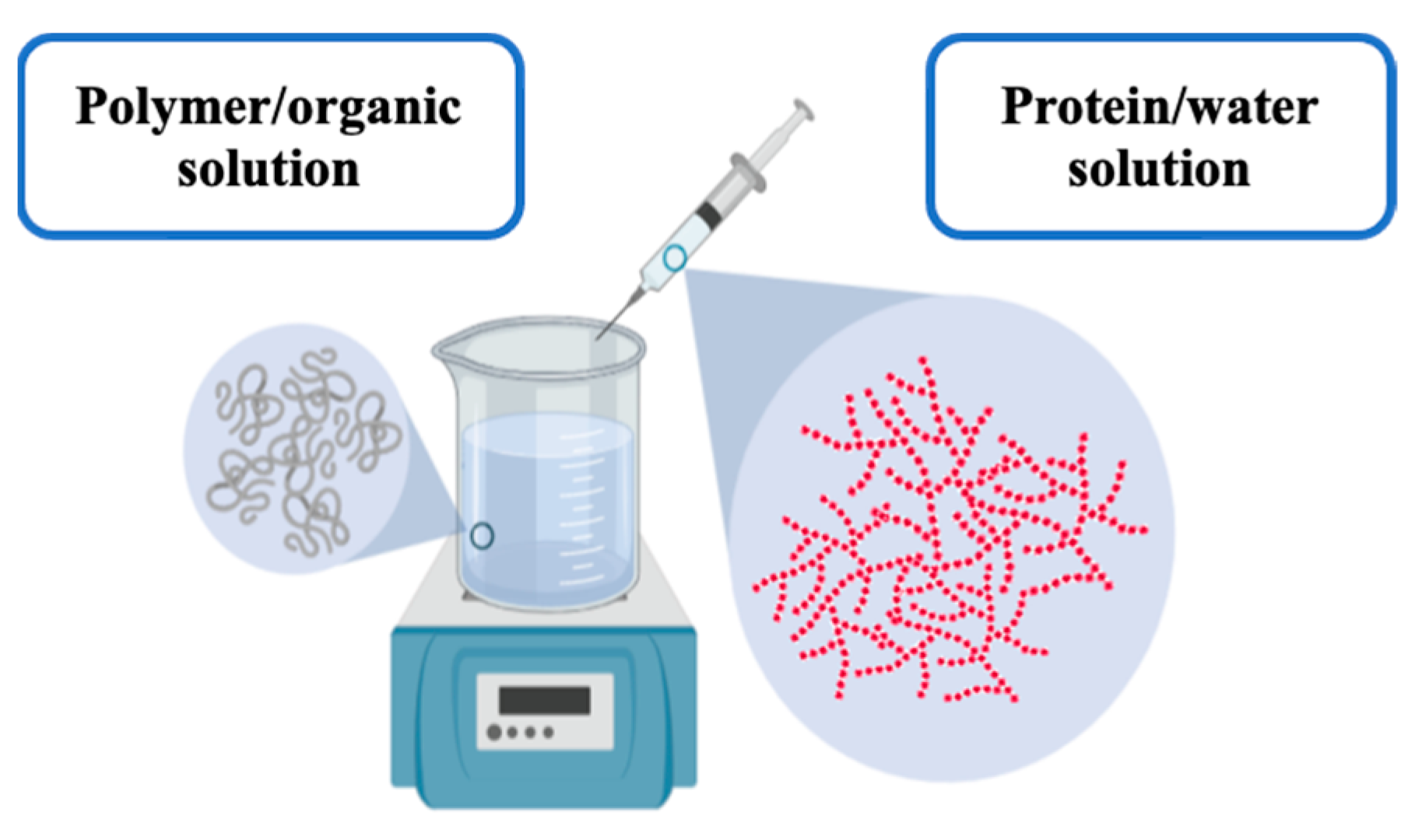
3.1.1. One-Step Nanoprecipitation Method: Nanospheres
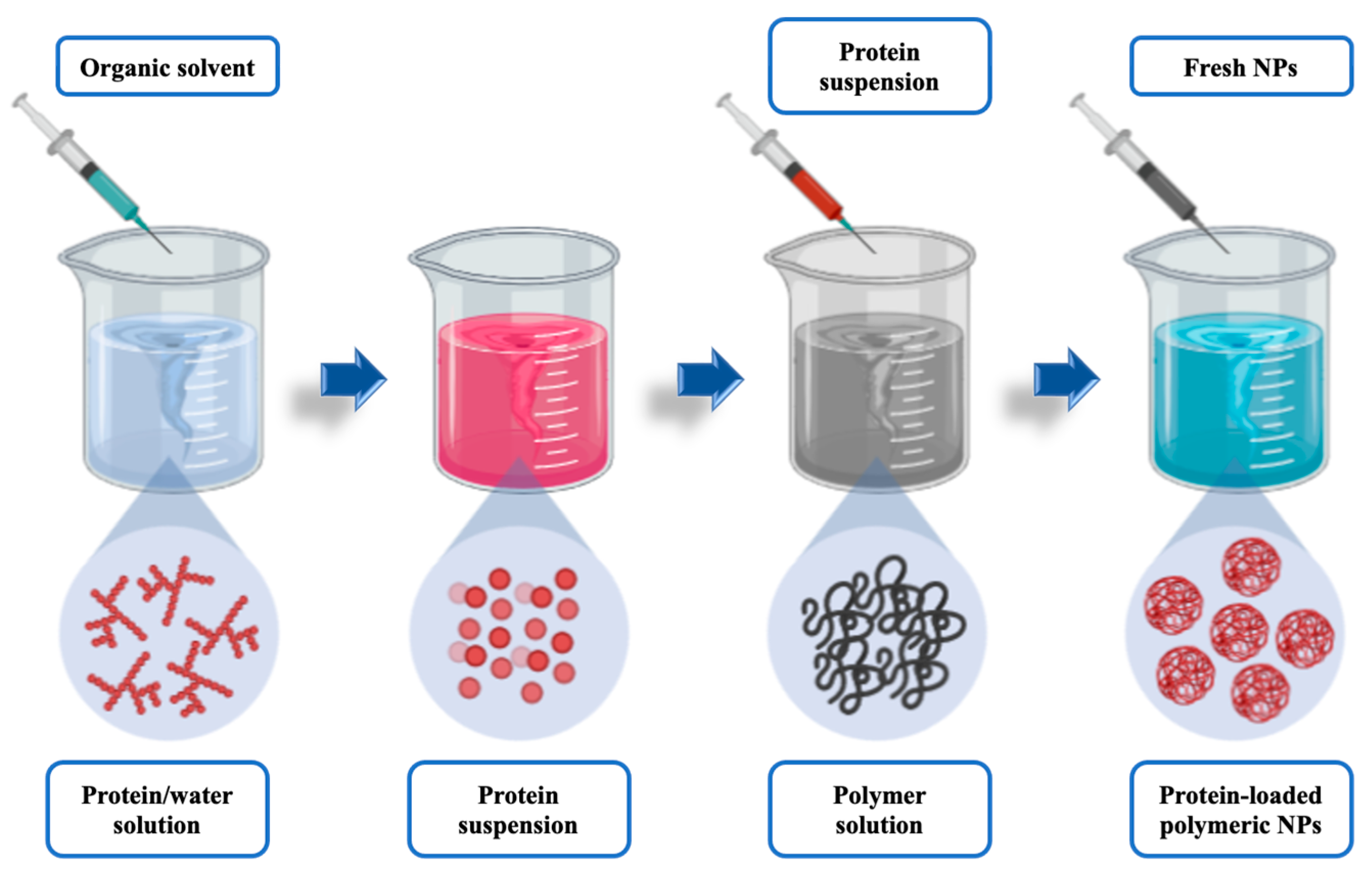
3.1.2. Two-Step Nanoprecipitation Method: Nanocapsules
3.2. Physicochemical Characterization of the Nanoparticles
3.2.1. Particle Size, Polydispersity, and Surface Charge
3.2.2. Morphological Evaluation
3.2.3. Production Yield (PY)
3.2.4. Encapsulation Efficiency (EE) and Loading Capacity (LC)
3.3. Stability Studies
3.3.1. Stability to Storage
3.3.2. Stability to pH
3.3.3. Stability to Ionic Strength
3.4. In Vitro Release Study
3.5. Citotoxicity Analysis
3.5.1. Bovine Corneal Opacity and Permeability Test (BCOP)
3.5.2. Hen’s Egg Test on the Chorioallantoic Membrane (HET-CAM)
3.6. Ocular Surface Retention Study
3.6.1. Ex Vivo Corneal Surface Retention
3.6.2. In Vivo Corneal Surface Retention Study
Evaluation of the Radiolabeling Stability and Efficiency of PLGA-Based Nanoparticles
Experimental In Vivo Evaluation of the Ocular Biopermanence of PLGA-Based Nanoparticles
3.7. Data Analysis
4. Results and Discussion
4.1. Preparation of Lactoferrin-Loaded PLGA Nanoparticles
4.1.1. One-Step Nanoprecipitation Method: Lactoferrin-Loaded PLGA Nanospheres
4.1.2. Two-Step Nanoprecipitation Method: Lactoferrin-Loaded PLGA Nanocapsules
4.2. Physicochemical Characterization of the Nanoparticles
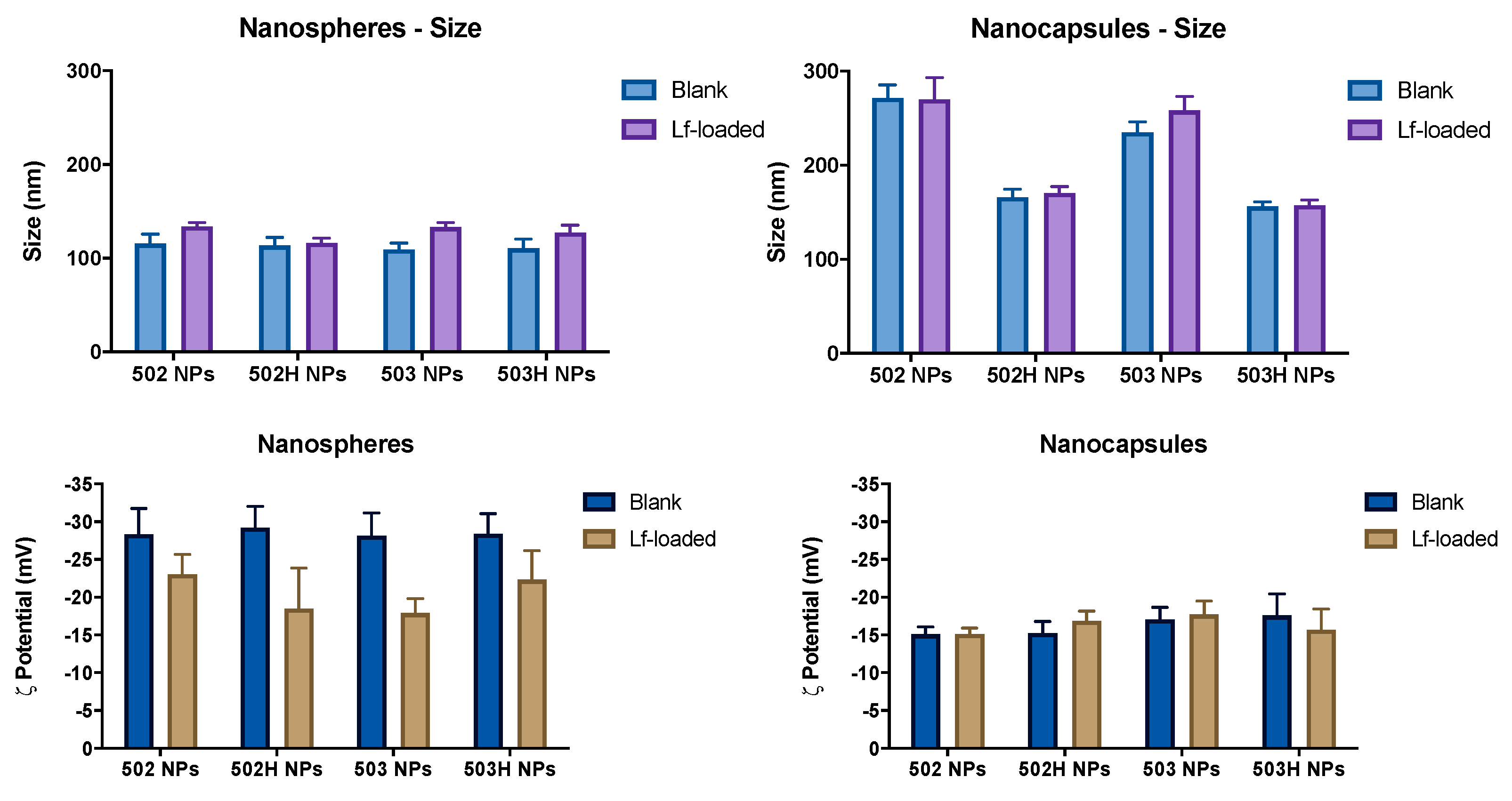
4.2.1. Particle Size Distribution and ζ Potential
4.2.2. Effect of Protein Loading on Particle Size and ζ Potential
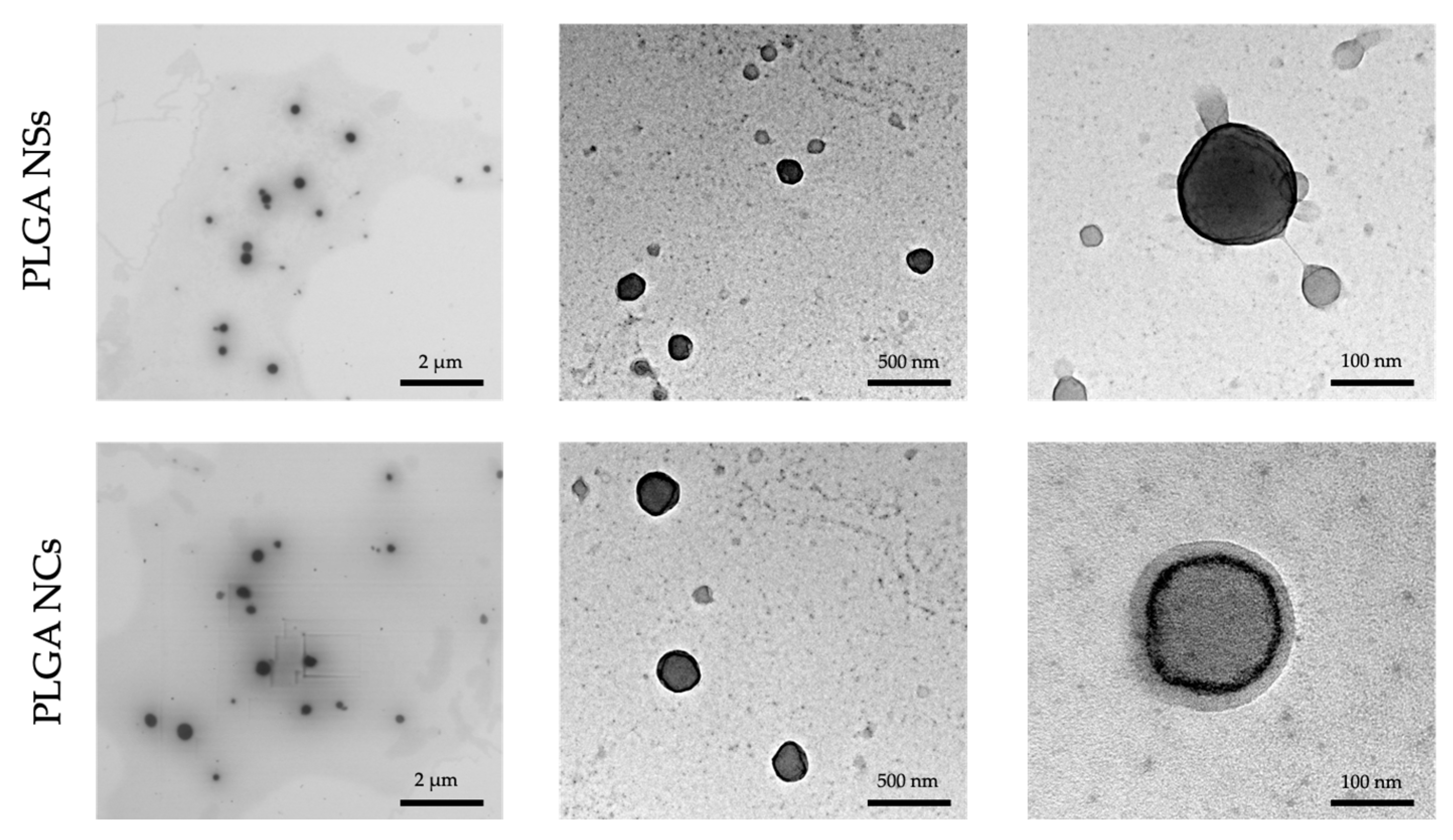
4.2.3. Morphological Evaluation
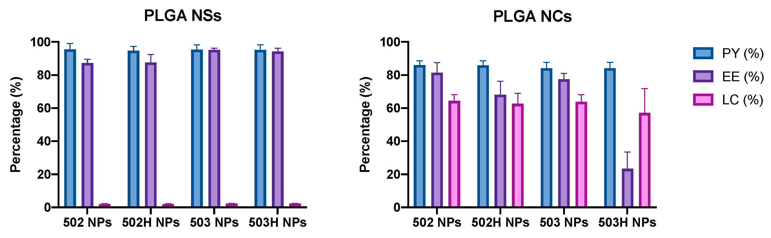
4.2.4. Production Yield (PY), Encapsulation Efficiency (EE) and Loading Capacity (LC) of Nanoparticles
4.3. Stability Studies
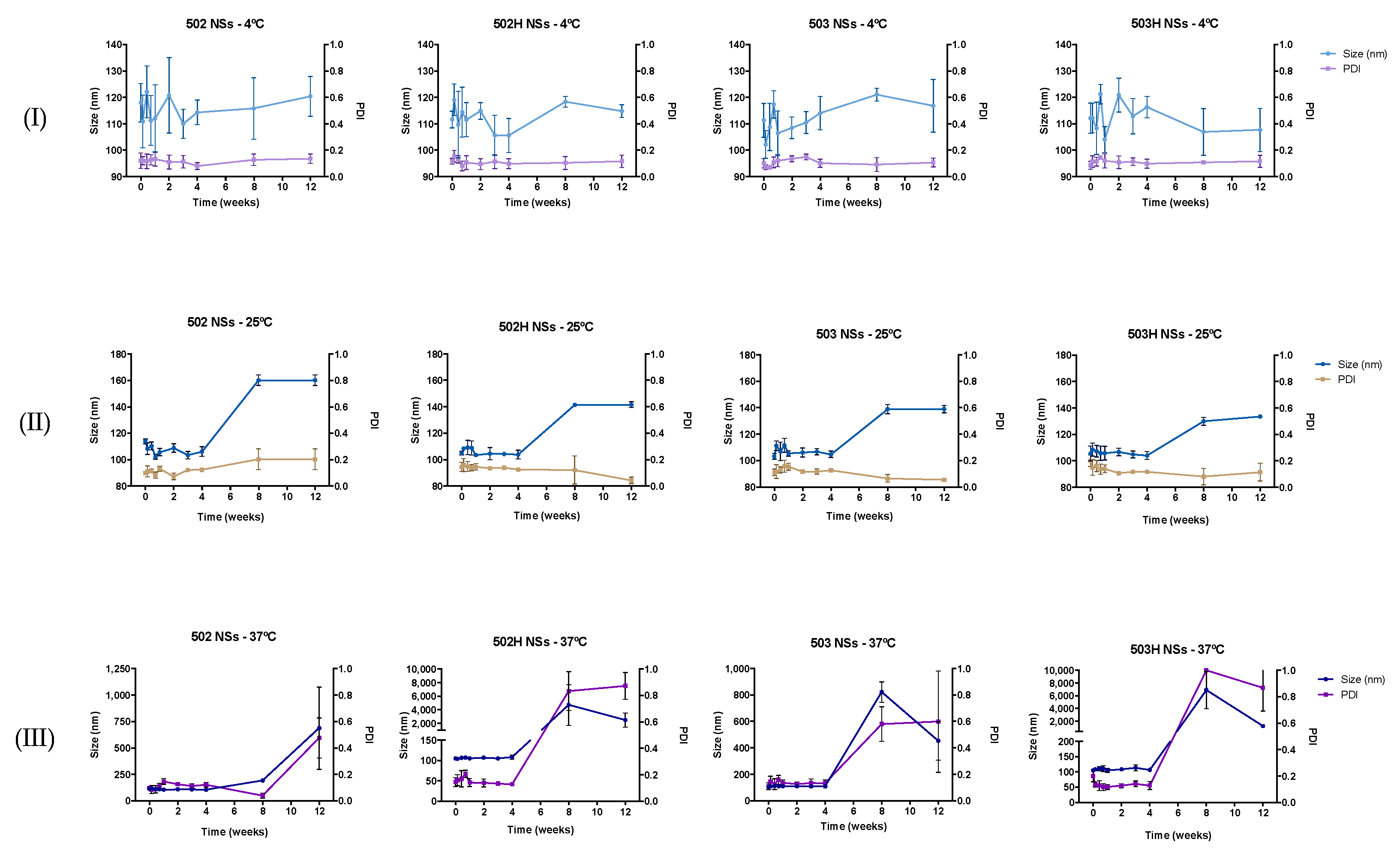
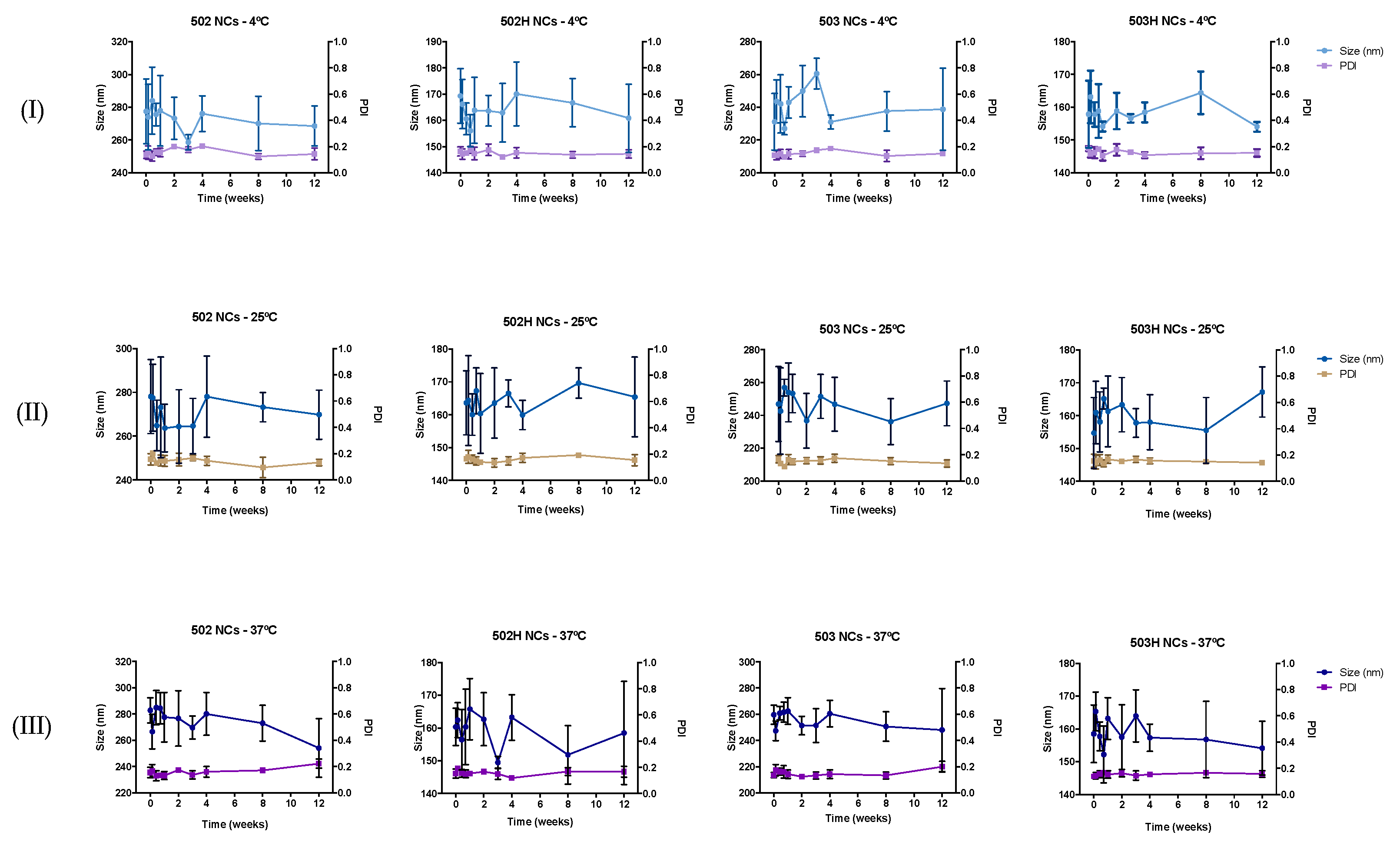
4.3.1. Stability to Storage
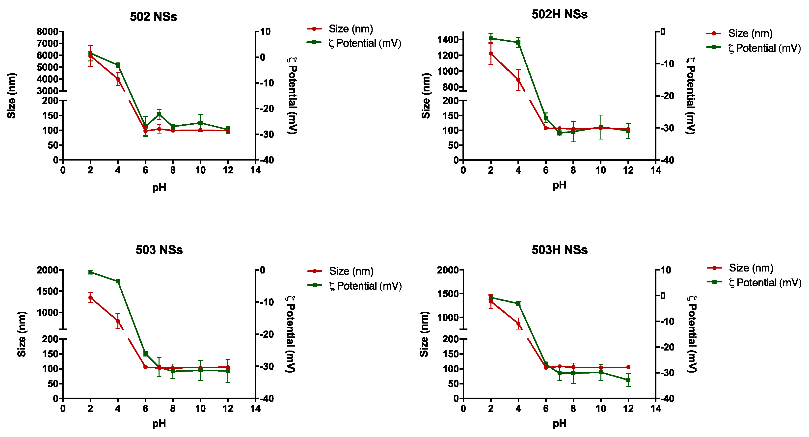
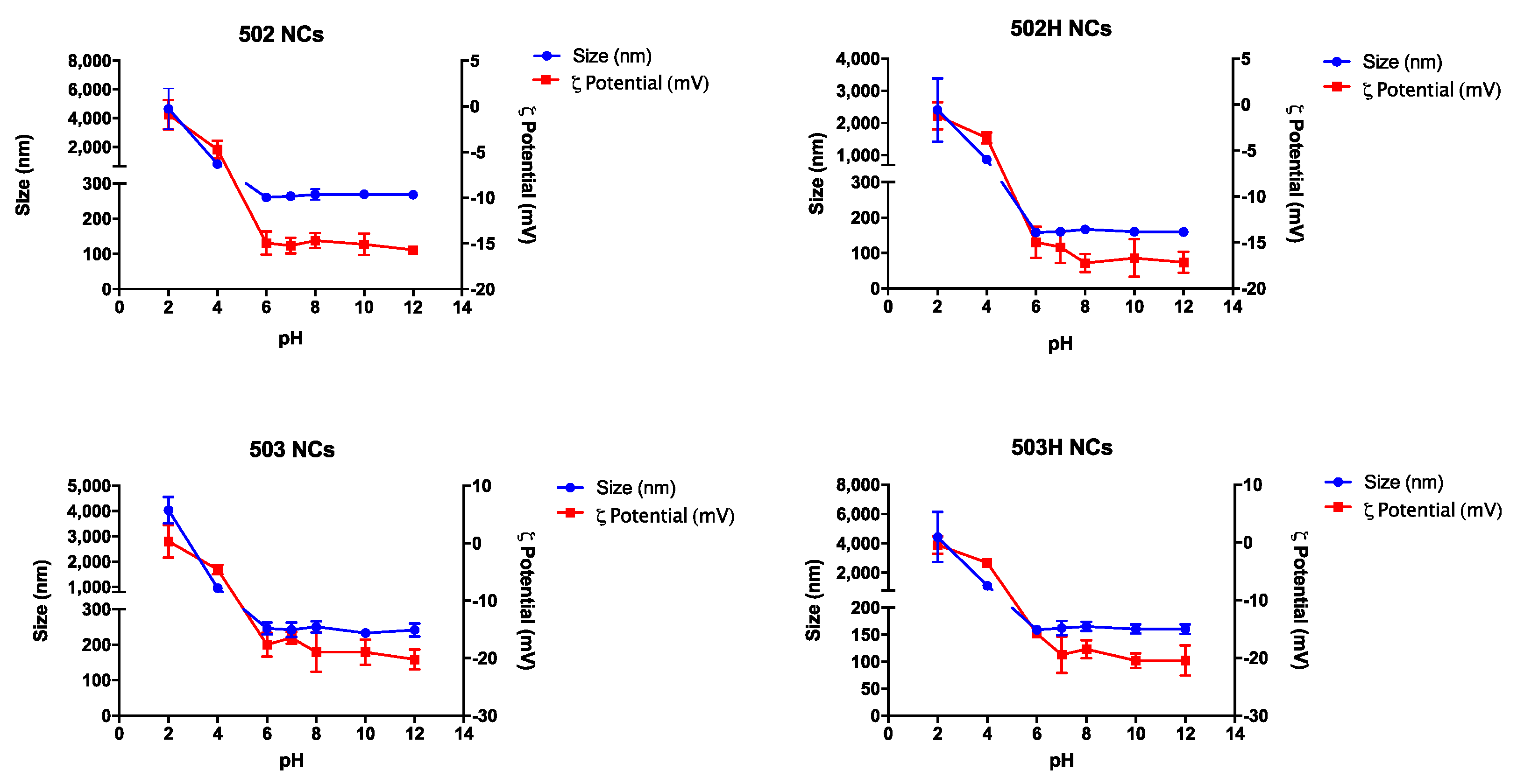
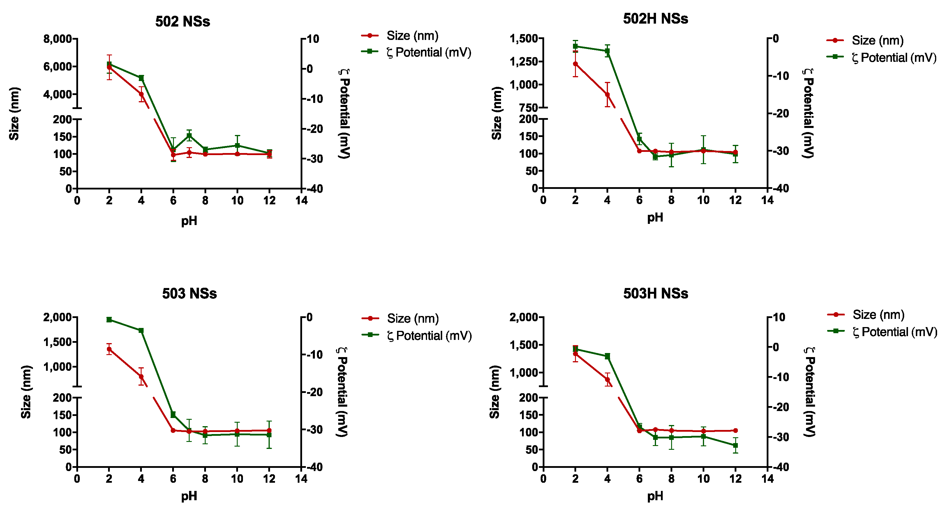
4.3.2. Stability to pH
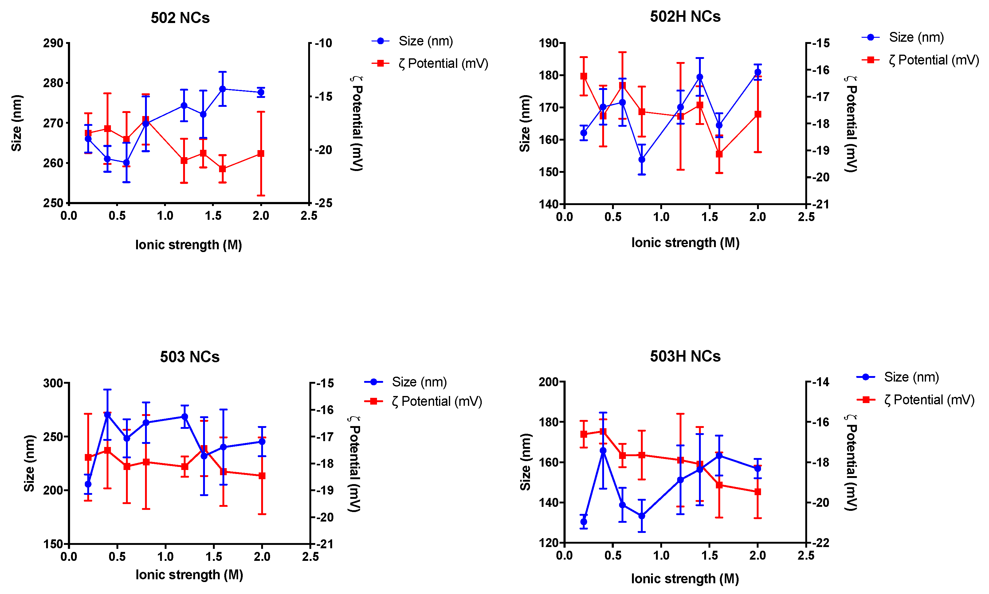
4.3.3. Stability to Ionic Strength
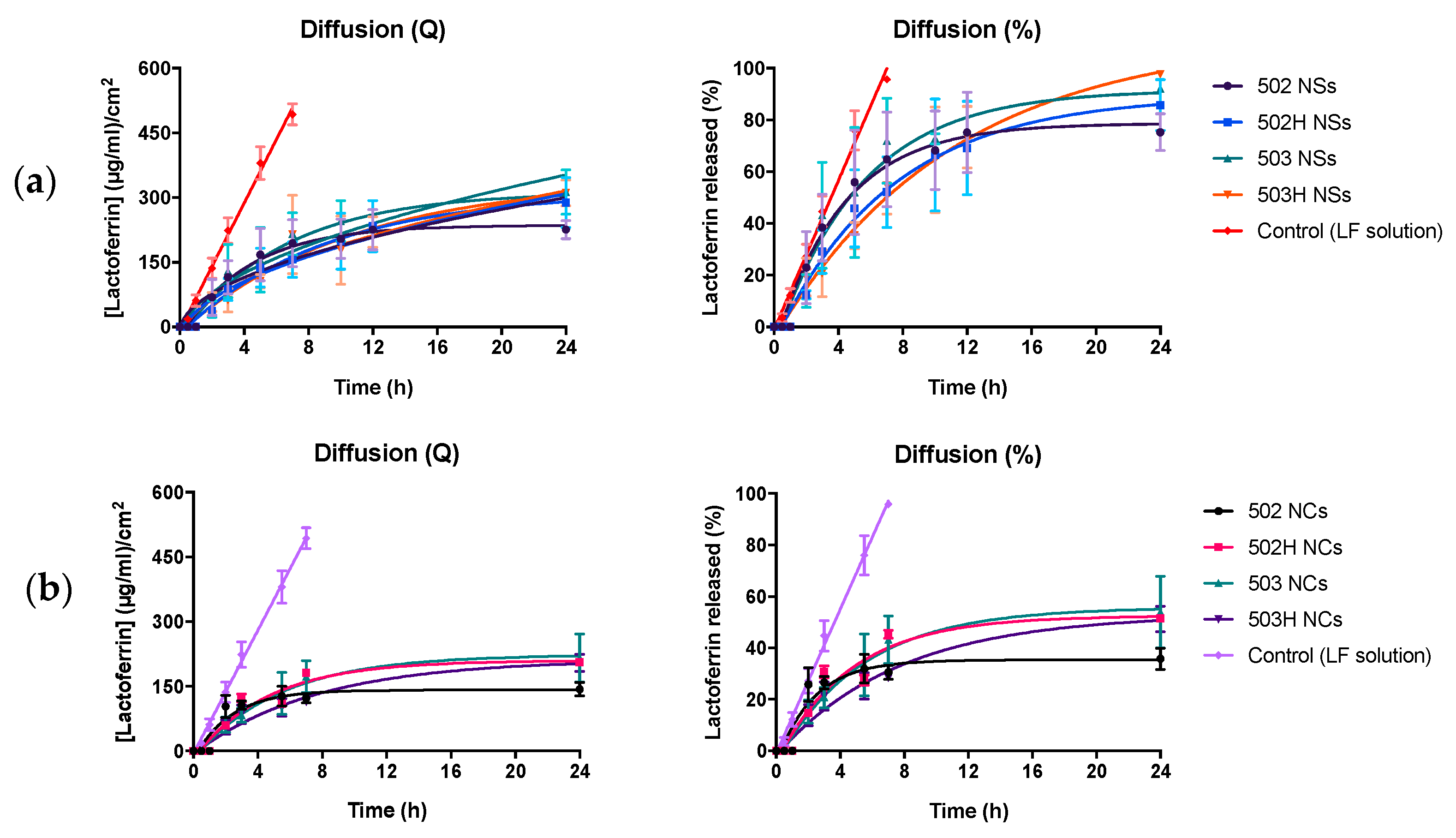
4.4. In Vitro Release Study
4.5. Citotoxicity Analysis
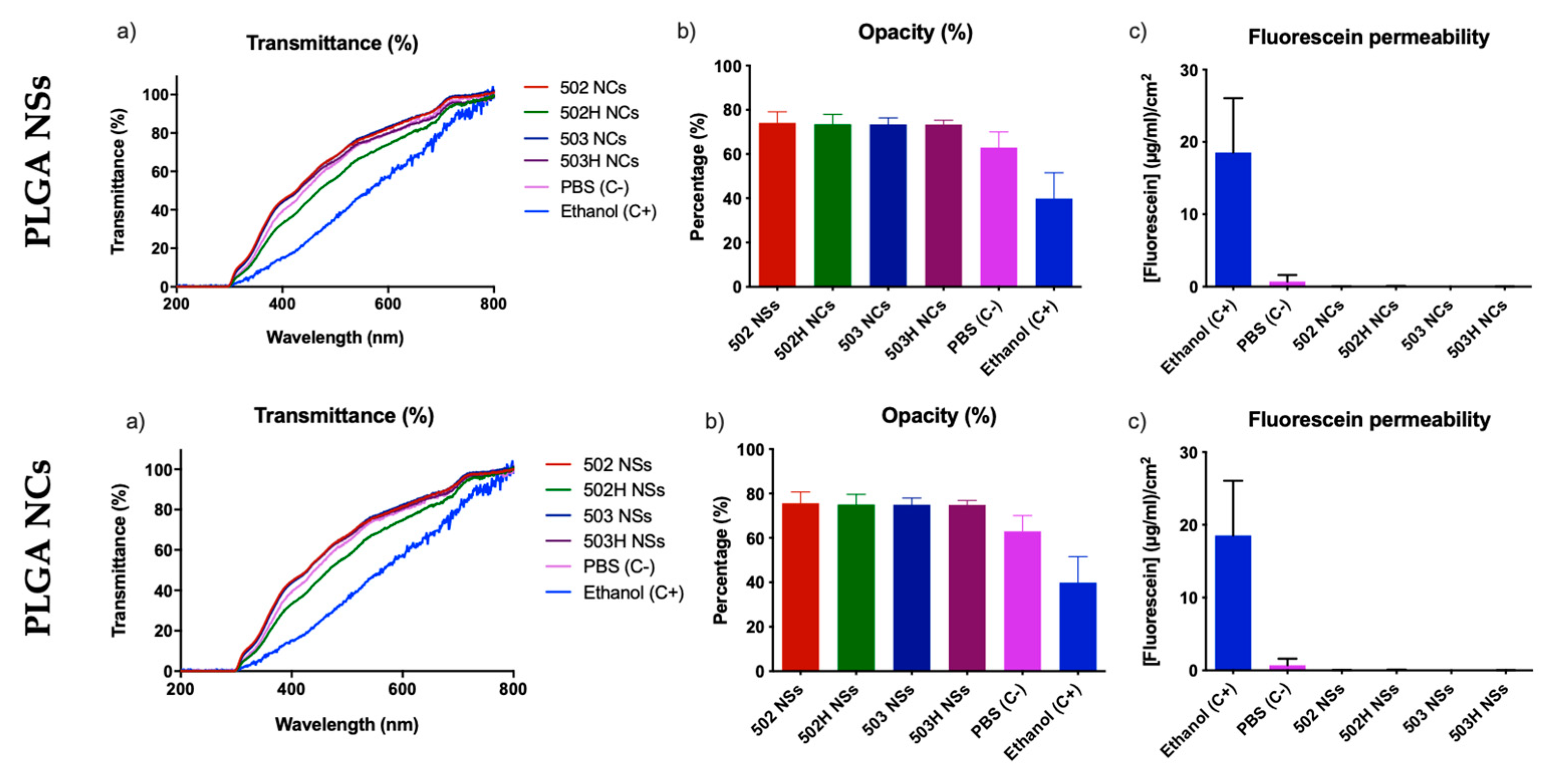
4.5.1. Bovine Corneal Opacity and Permeability Test (BCOP)
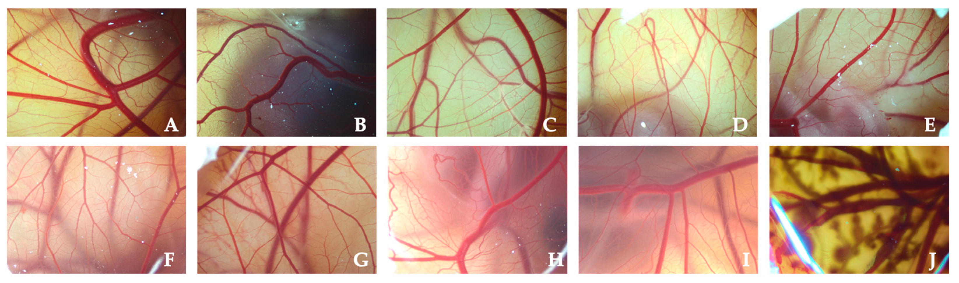
4.5.2. Hen’s Egg Test on the Chorioallantoic Membrane (HET-CAM)
4.6. Ocular Surface Retention Study
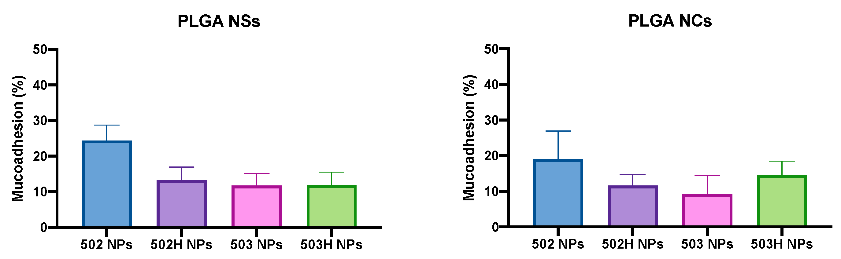
4.6.1. Ex Vivo Corneal Surface Model
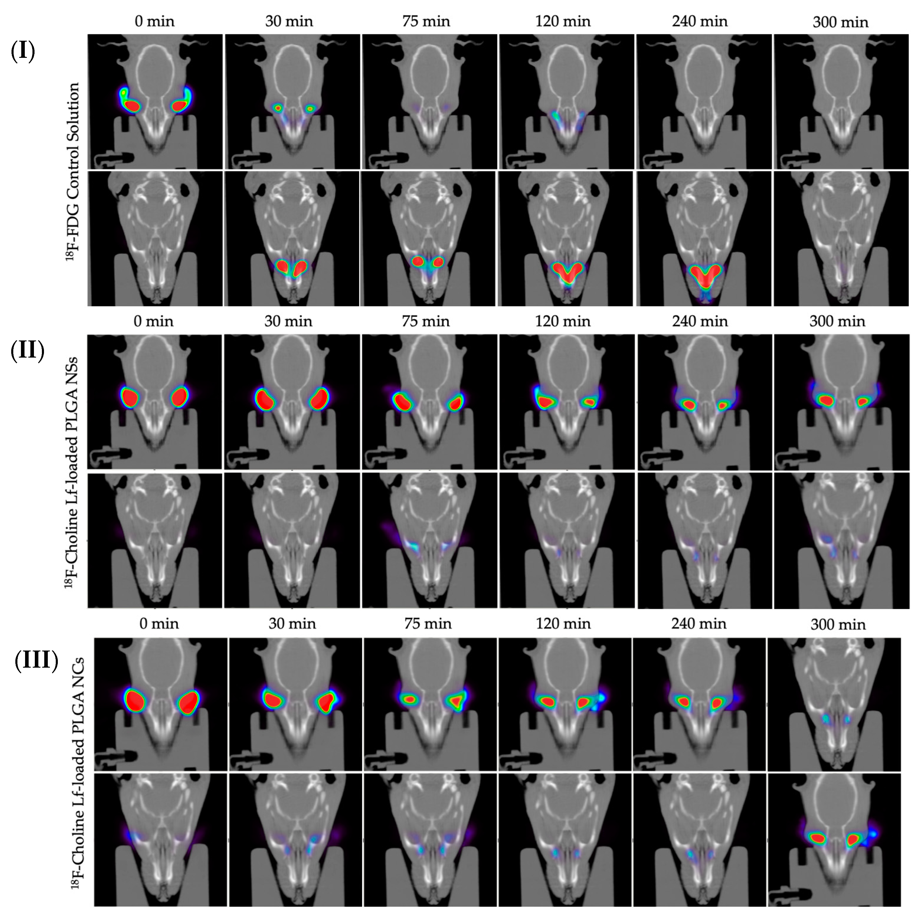
4.6.2. In Vivo Ocular Surface Permanence Study
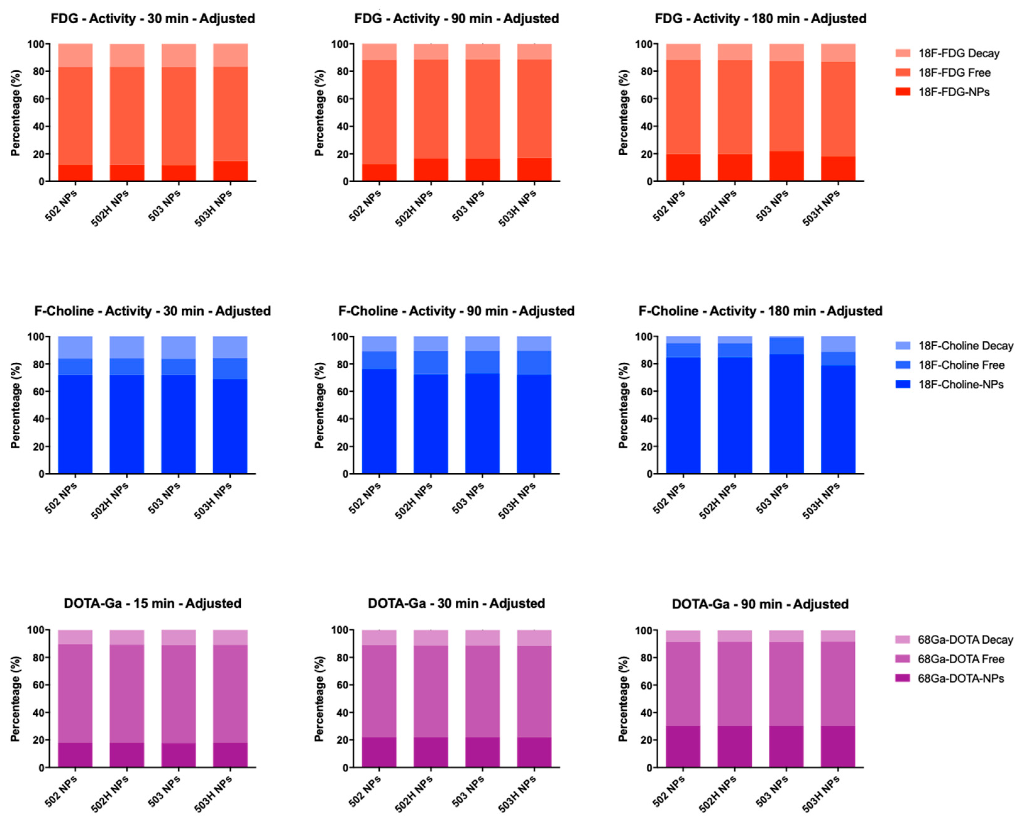
Evaluation of the Radiolabeling Stability and Efficiency of PLGA-Based Nanoparticles
Experimental In Vivo Evaluation of Ocular Surface Permanence
5. Conclusions
Author Contributions
Funding
Institutional Review Board Statement
Informed Consent Statement
Data Availability Statement
Acknowledgments
Conflicts of Interest
References
- Bucolo, C.; Drago, F.; Salomone, S. Ocular drug delivery: A clue from nanotechnology. Front. Pharmacol. 2012, 3, 188. [Google Scholar] [CrossRef] [PubMed] [Green Version]
- Sahoo, S.K.; Dilnawaz, F.; Krishnakumar, S. Nanotechnology in ocular drug delivery. Drug Discov. Today 2008, 13, 144–151. [Google Scholar] [CrossRef]
- Nagarwal, R.C.; Kant, S.; Singh, P.N.; Maiti, P.; Pandit, J.K. Polymeric nanoparticulate system: A potential approach for ocular drug delivery. J. Control. Release 2009, 136, 2–13. [Google Scholar] [CrossRef] [PubMed]
- Danhier, F.; Ansorena, E.; Silva, J.M.; Coco, R.; Le Breton, A.; Préat, V. PLGA-based nanoparticles: An overview of biomedical applications. J. Control. Release 2012, 161, 505–522. [Google Scholar] [CrossRef] [PubMed]
- Fessi, H.; Puisieux, F.; Devissaguet, J.P.; Ammoury, N.; Benita, S. Nanocapsule formation by interfacial polymer deposition following solvent displacement. Int. J. Pharm. 1989, 55, R1–R4. [Google Scholar] [CrossRef]
- Lepeltier, E.; Bourgaux, C.; Couvreur, P. Nanoprecipitation and the “Ouzo effect”: Application to drug delivery devices. Adv. Drug Deliv. Rev. 2014, 71, 86–97. [Google Scholar] [CrossRef] [PubMed]
- Beck-Broichsitter, M.; Nicolas, J.; Couvreur, P. Solvent selection causes remarkable shifts of the “Ouzo region” for poly(lactide-co-glycolide) nanoparticles prepared by nanoprecipitation. Nanoscale 2015, 7, 9215–9221. [Google Scholar] [CrossRef] [PubMed]
- Vitale, S.A.; Katz, J.L. Liquid Droplet Dispersions Formed by Homogeneous Liquid−Liquid Nucleation: “The Ouzo Effect”. Langmuir 2003, 19, 4105–4110. [Google Scholar] [CrossRef]
- Aubry, J.; Ganachaud, F.; Addad, J.-P.C.; Cabane, B. Nanoprecipitation of Polymethylmethacrylate by Solvent Shifting: 1. Boundaries. Langmuir 2009, 25, 1970–1979. [Google Scholar] [CrossRef]
- Legrand, P.; Lesieur, S.; Bochot, A.; Gref, R.; Raatjes, W.; Barratt, G.; Vauthier, C. Influence of polymer behaviour in organic solution on the production of polylactide nanoparticles by nanoprecipitation. Int. J. Pharm. 2007, 344, 33–43. [Google Scholar] [CrossRef] [PubMed]
- Yang, Z.; Foster, D.; Dhinojwala, A. Continuous production of polymer nanoparticles using a membrane-based flow cell. J. Colloid Interface Sci. 2017, 501, 150–155. [Google Scholar] [CrossRef] [Green Version]
- Hans, M.L.; Lowman, A.M. Biodegradable nanoparticles for drug delivery and targeting. Curr. Opin. Solid State Mater. Sci. 2002, 6, 319–327. [Google Scholar] [CrossRef]
- Soppimath, K.S.; Aminabhavi, T.M.; Kulkarni, A.R.; Rudzinski, W.E. Biodegradable polymeric nanoparticles as drug delivery devices. J. Control. Release 2001, 70, 1–20. [Google Scholar] [CrossRef]
- Corrigan, O.I.; Li, X. Quantifying drug release from PLGA nanoparticulates. Eur. J. Pharm. Sci. 2009, 37, 477–485. [Google Scholar] [CrossRef] [PubMed]
- Mu, L.; Feng, S.S. A novel controlled release formulation for the anticancer drug paclitaxel (Taxol®): PLGA nanoparticles containing vitamin E TPGS. J. Control. Release 2003, 86, 33–48. [Google Scholar] [CrossRef]
- Lemoine, D.; Préat, V. Polymeric nanoparticles as delivery system for influenza virus glycoproteins. J. Control. Release 1998, 54, 15–27. [Google Scholar] [CrossRef]
- Appelmelk, B.J.; An, Y.Q.; Geerts, M.; Thijs, B.G.; De Boer, H.A.; MacLaren, D.M.; De Graaff, J.; Nuijens, J.H. Lactoferrin is a lipid A-binding protein. Infect. Immun. 1994, 62, 2628–2632. [Google Scholar] [CrossRef] [PubMed] [Green Version]
- Hayashida, K.-I.; Kaneko, T.; Takeuchi, T.; Shimizu, H.; Ando, K.; Harada, E. Oral Administration of Lactoferrin Inhibits Inflammation and Nociception in Rat Adjuvant-Induced Arthritis. J. Veter. Med. Sci. 2004, 66, 149–154. [Google Scholar] [CrossRef] [PubMed] [Green Version]
- Schryvers, A.B.; Morris, L.J. Identification and characterization of the human lactoferrin-binding protein from Neisseria meningitidis. Infect. Immun. 1988, 56, 1144–1149. [Google Scholar] [CrossRef] [PubMed] [Green Version]
- Ellison, R.T.; Giehl, T.J. Killing of gram-negative bacteria by lactoferrin and lysozyme. J. Clin. Investig. 1991, 88, 1080–1091. [Google Scholar] [CrossRef] [PubMed]
- Takakura, N.; Wakabayashi, H.; Ishibashi, H.; Teraguchi, S.; Tamura, Y.; Yamaguchi, H.; Abe, S. Oral Lactoferrin Treatment of Experimental Oral Candidiasis in Mice. Antimicrob. Agents Chemother. 2003, 47, 2619–2623. [Google Scholar] [CrossRef] [PubMed] [Green Version]
- Marchetti, M.; Superti, F.; Ammendolia, M.G.; Rossi, P.; Valenti, P.; Seganti, L. Inhibition of poliovirus type 1 infection by iron-, manganese- and zinc-saturated lactoferrin. Med. Microbiol. Immunol. 1999, 187, 199–204. [Google Scholar] [CrossRef]
- Murphy, M.E.; Kariwa, H.; Mizutani, T.; Tanabe, H.; Yoshimatsu, K.; Arikawa, J.; Takashima, I. Characterization of in vitro and in vivo Antiviral Activity of Lactoferrin and Ribavirin upon Hantavirus. J. Veter. Med. Sci. 2001, 63, 637–645. [Google Scholar] [CrossRef] [PubMed] [Green Version]
- Berkhout, B.; Floris, R.; Recio, I.; Visser, S. The antiviral activity of the milk protein lactoferrin against the human immunodeficiency virus type 1. BioMetals 2004, 17, 291–294. [Google Scholar] [CrossRef]
- Eliassen, L.T.; Berge, G.; Sveinbjørnsson, B.; Svendsen, J.S.; Vorland, L.H.; Rekdal, Ø. Evidence for a Direct Antitumor Mechanism of Action of Bovine Lactoferricin. Anticancer Res. 2002, 22, 2703–2710. [Google Scholar]
- Tsuda, H.; Sekine, K.; Fujita, K.-I.; Iigo, M. Cancer prevention by bovine lactoferrin and underlying mechanisms—A review of experimental and clinical studies. Biochem. Cell Biol. 2002, 80, 131–136. [Google Scholar] [CrossRef]
- Pattamatta, U.; Willcox, M.; Stapleton, F.; Cole, N.; Garrett, Q. Bovine Lactoferrin Stimulates Human Corneal Epithelial Alkali Wound Healing In Vitro. Investig. Opthalmol. Vis. Sci. 2009, 50, 1636–1643. [Google Scholar] [CrossRef] [PubMed] [Green Version]
- Flanagan, J.L.; Willcox, M.D.P. Role of lactoferrin in the tear film. Biochimie 2009, 91, 35–43. [Google Scholar] [CrossRef] [PubMed]
- Vagge, A.; Senni, C.; Bernabei, F.; Pellegrini, M.; Scorcia, V.; Traverso, C.E.; Giannaccare, G. Therapeutic Effects of Lactoferrin in Ocular Diseases: From Dry Eye Disease to Infections. Int. J. Mol. Sci. 2020, 21, 6668. [Google Scholar] [CrossRef] [PubMed]
- Ibuki, M.; Shoda, C.; Miwa, Y.; Ishida, A.; Tsubota, K.; Kurihara, T. Lactoferrin Has a Therapeutic Effect via HIF Inhibition in a Murine Model of Choroidal Neovascularization. Front. Pharmacol. 2020, 11, 174. [Google Scholar] [CrossRef] [PubMed] [Green Version]
- Kymes, S.M.; Walline, J.; Zadnik, K.; Sterling, J.; Gordon, M.O.; Collaborative Longitudinal Evaluation of Keratoconus Study Group. Changes in the Quality-of-Life of People with Keratoconus. Am. J. Ophthalmol. 2008, 145, 611–617.e1. [Google Scholar] [CrossRef] [PubMed] [Green Version]
- Godefrooij, D.A.; de Wit, G.A.; Uiterwaal, C.S.; Imhof, S.M.; Wisse, R.P. Age-specific Incidence and Prevalence of Keratoconus: A Nationwide Registration Study. Am. J. Ophthalmol. 2017, 175, 169–172. [Google Scholar] [CrossRef] [PubMed]
- Sharif, R.; Bak-Nielsen, S.; Hjortdal, J.; Karamichos, D. Pathogenesis of Keratoconus: The intriguing therapeutic potential of Prolactin-inducible protein. Prog. Retin. Eye Res. 2018, 67, 150–167. [Google Scholar] [CrossRef] [Green Version]
- Tur, V.M.; MacGregor, C.; Jayaswal, R.; O’Brart, D.; Maycock, N. A review of keratoconus: Diagnosis, pathophysiology, and genetics. Surv. Ophthalmol. 2017, 62, 770–783. [Google Scholar] [CrossRef]
- Romero-Jiménez, M.; Santodomingo-Rubido, J.; Wolffsohn, J. Keratoconus: A review. Contact Lens Anterior Eye 2010, 33, 157–166. [Google Scholar] [CrossRef] [PubMed]
- Meiri, Z.; Keren, S.; Rosenblatt, A.; Sarig, T.; Shenhav, L.; Varssano, D. Efficacy of Corneal Collagen Cross-Linking for the Treatment of Keratoconus: A Systematic Review and Meta-Analysis. Cornea 2016, 35, 417–428. [Google Scholar] [CrossRef]
- Buddi, R.; Lin, B.; Atilano, S.; Zorapapel, N.C.; Kenney, M.C.; Brown, D.J. Evidence of Oxidative Stress in Human Corneal Diseases. J. Histochem. Cytochem. 2002, 50, 341–351. [Google Scholar] [CrossRef]
- Gondhowiardjo, T.D.; Van Haeringen, N.J.; Völker-Dieben, H.J.; Beekhuis, H.W.; Kok, J.H.; Van Rij, G.; Pels, L.; Kijlstra, A. Analysis of Corneal Aldehyde Dehydrogenase Patterns in Pathologic Corneas. Cornea 1993, 12, 146–154. [Google Scholar] [CrossRef]
- Atilano, S.R.; Coskun, E.P.; Chwa, M.; Jordan, N.; Reddy, V.; Le, K.; Wallace, D.C.; Kenney, M.C. Accumulation of Mitochondrial DNA Damage in Keratoconus Corneas. Investig. Opthalmol. Vis. Sci. 2005, 46, 1256–1263. [Google Scholar] [CrossRef]
- Lubrano, S.B.V.; Balzan, S. Enzymatic antioxidant system in vascular inflammation and coronary artery disease. World J. Exp. Med. 2015, 5, 218–224. [Google Scholar] [CrossRef]
- Cao, J.Y.; Dixon, S.J. Mechanisms of ferroptosis. Cell. Mol. Life Sci. 2016, 73, 2195–2209. [Google Scholar] [CrossRef] [Green Version]
- Zilka, O.; Shah, R.; Li, B.; Angeli, J.P.F.; Griesser, M.; Conrad, M.; Pratt, D.A. On the Mechanism of Cytoprotection by Ferrostatin-1 and Liproxstatin-1 and the Role of Lipid Peroxidation in Ferroptotic Cell Death. ACS Cent. Sci. 2017, 3, 232–243. [Google Scholar] [CrossRef] [PubMed]
- Dixon, S.J.; Lemberg, K.M.; Lamprecht, M.R.; Skouta, R.; Zaitsev, E.M.; Gleason, C.E.; Patel, D.N.; Bauer, A.J.; Cantley, A.M.; Yang, W.S.; et al. Ferroptosis: An Iron-Dependent Form of Nonapoptotic Cell Death. Cell 2012, 149, 1060–1072. [Google Scholar] [CrossRef] [PubMed] [Green Version]
- Varela-Fernández, R.; García-Otero, X.; Díaz-Tomé, V.; Regueiro, U.; López-López, M.; González-Barcia, M.; Lema, M.I.; Otero-Espinar, F.J. Design, Optimization, and Characterization of Lactoferrin-Loaded Chitosan/TPP and Chitosan/Sulfobutylether-β-cyclodextrin Nanoparticles as a Pharmacological Alternative for Keratoconus Treatment. ACS Appl. Mater. Interfaces 2021, 13, 3559–3575. [Google Scholar] [CrossRef] [PubMed]
- Bilati, U.; Allémann, E.; Doelker, E. Nanoprecipitation versus emulsion-based techniques for the encapsulation of proteins into biodegradable nanoparticles and process-related stability issues. AAPS PharmSciTech 2005, 6, E594–E604. [Google Scholar] [CrossRef] [PubMed] [Green Version]
- Weber, C.; Coester, C.; Kreuter, J.; Langer, K. Desolvation process and surface characterisation of protein nanoparticles. Int. J. Pharm. 2000, 194, 91–102. [Google Scholar] [CrossRef]
- Morales-Cruz, M.; Flores-Fernández, G.M.; Morales-Cruz, M.; Orellano, E.A.; Rodriguez-Martinez, J.A.; Ruiz, M.; Griebenow, K. Two-step nanoprecipitation for the production of protein-loaded PLGA nanospheres. Results Pharma Sci. 2012, 2, 79–85. [Google Scholar] [CrossRef] [PubMed] [Green Version]
- Varela-Fernández, R.; García-Otero, X.; Díaz-Tomé, V.; Regueiro, U.; López-López, M.; González-Barcia, M.; Lema, M.I.; Otero-Espinar, F.J. Lactoferrin-loaded nanostructured lipid carriers (NLCs) as a new formulation for optimized ocular drug delivery. Eur. J. Pharm. Biopharm. 2022, 172, 144–156. [Google Scholar] [CrossRef]
- Jithan, A.; Madhavi, K.; Madhavi, M.; Prabhakar, K. Preparation and characterization of albumin nanoparticles encapsulating curcumin intended for the treatment of breast cancer. Int. J. Pharm. Investig. 2011, 1, 119–125. [Google Scholar] [CrossRef] [Green Version]
- IHT Guideline. Stability Testing of New Drug Substances and Products. Q1A (R2) Current Step 4. 2003. pp. 1–24. Available online: file:///C:/Users/MDPI/Desktop/Q1A(R2)%20Guideline.pdf (accessed on 20 February 2022).
- Orthner, M.P.; Lin, G.; Avula, M.; Buetefisch, S.; Magda, J.; Rieth, L.W.; Solzbacher, F. Hydrogel Based Sensor Arrays (2 × 2) with Perforated Piezoresistive Diaphragms for Metabolic Monitoring (In Vitro). Sens. Actuators B Chem. 2010, 145, 807–816. [Google Scholar] [CrossRef] [PubMed] [Green Version]
- Salem, H.; Katz, S.A. Alternative Toxicological Methods; CRC Press: Boca Raton, FL, USA, 2003; ISBN 978-0-203-00879-9. [Google Scholar]
- Eskes, C.; Bessou, S.; Bruner, L.; Curren, R.; Jones, P.; Kreiling, R.; Liebsch, M.; McNamee, P.; Pape, W.; Prinsen, M.K.; et al. Subgroup 3. Eye Irritation. 76. Altern. Lab. Anim. ATLA 2005, 33, 47–81. [Google Scholar] [CrossRef]
- Sina, J.F.; Galer, D.M.; Sussman, R.G.; Gautheron, P.D.; Sargent, E.V.; Leong, B.; Shah, P.V.; Curren, R.D.; Miller, K. A Collaborative Evaluation of Seven Alternatives to the Draize Eye Irritation Test Using Pharmaceutical Intermediates. Fundam. Appl. Toxicol. 1995, 26, 20–31. [Google Scholar] [CrossRef] [PubMed]
- Wallig, M.A.; Haschek, W.M.; Rous-Seaux, C.G.; Bolon, B. (Eds.) Chapter 22-Special Senses. In Fundamentals of Toxicologic Pathology, 3rd ed.; Academic Press: Cambridge, MA, USA, 2018; pp. 673–747. ISBN 978-0-12-809841-7. [Google Scholar]
- Luepke, N. Hen’s egg chorioallantoic membrane test for irritation potential. Food Chem. Toxicol. 1985, 23, 287–291. [Google Scholar] [CrossRef]
- Kalweit, S.; Besoke, R.; Gerner, I.; Spielmann, H. A national validation project of alternative methods to the Draize rabbit eye test. Toxicol. Vitr. 1990, 4, 702–706. [Google Scholar] [CrossRef]
- Belgamwar, V.; Shah, V.; Surana, S.J. Formulation and evaluation of oral mucoadhesive multiparticulate system containing metoprolol tartarate: An in vitro-ex vivo characterization. Curr. Drug Deliv. 2009, 6, 113–121. [Google Scholar] [CrossRef]
- Prassl, R.; Gradauer, K.; Vonach, C.; Leitinger, G.; Kolb, D.; Fröhlich, E.; Roblegg, E.; Bernkop-Schnürch, A.; Prassl, R. Chemical coupling of thiolated chitosan to preformed liposomes improves mucoadhesive properties. Int. J. Nanomed. 2012, 7, 2523–2534. [Google Scholar] [CrossRef] [PubMed] [Green Version]
- Rojas, S.; Gispert, J.D.; Abad, S.; Buaki-Sogo, M.; Victor, V.M.; Garcia, H.; Herance, J.R. In Vivo Biodistribution of Amino-Functionalized Ceria Nanoparticles in Rats Using Positron Emission Tomography. Mol. Pharm. 2012, 9, 3543–3550. [Google Scholar] [CrossRef] [PubMed]
- Pérez-Campaña, C.; Gómez-Vallejo, V.; Martin, A.; Sebastián, E.S.; Moya, S.E.; Reese, T.; Ziolo, R.F.; Llop, J. Tracing nanoparticles in vivo: A new general synthesis of positron emitting metal oxide nanoparticles by proton beam activation. Analyst 2012, 137, 4902–4906. [Google Scholar] [CrossRef] [PubMed]
- Allmeroth, M.; Moderegger, D.; Biesalski, B.; Koynov, K.; Rösch, F.; Thews, O.; Zentel, R. Modifying the Body Distribution of HPMA-Based Copolymers by Molecular Weight and Aggregate Formation. Biomacromolecules 2011, 12, 2841–2849. [Google Scholar] [CrossRef] [PubMed]
- Allmeroth, M.; Moderegger, D.; Gündel, D.; Buchholz, H.-G.; Mohr, N.; Koynov, K.; Rösch, F.; Thews, O.; Zentel, R. PEGylation of HPMA-based block copolymers enhances tumor accumulation in vivo: A quantitative study using radiolabeling and positron emission tomography. J. Control. Release 2013, 172, 77–85. [Google Scholar] [CrossRef] [PubMed]
- Fernández-Ferreiro, A.; Silva-Rodríguez, J.; Otero-Espinar, F.J.; González-Barcia, M.; Lamas, M.J.; Ruibal, A.; Luaces-Rodriguez, A.; Vieites-Prado, A.; Sobrino, T.; Herranz, M.; et al. Positron Emission Tomography for the Development and Characterization of Corneal Permanence of Ophthalmic Pharmaceutical Formulations. Investig. Opthalmol. Vis. Sci. 2017, 58, 772–780. [Google Scholar] [CrossRef]
- The Association for Research in Vision and Ophthalmology-Statement for the Use of Animals in Ophthalmic and Vision Re-search. Available online: https://www.arvo.org/About/policies/statement-for-the-use-of-animals-in-ophthalmic-and-vision-research/ (accessed on 22 July 2020).
- Burden, N.; Aschberger, K.; Chaudhry, Q.; Clift, M.J.D.; Doak, S.H.; Fowler, P.; Johnston, H.; Landsiedel, R.; Rowland, J.; Stone, V. The 3Rs as a framework to support a 21st century approach for nanosafety assessment. Nanotoday 2017, 12, 10–13. [Google Scholar] [CrossRef]
- Zhang, Y.; Huo, M.; Zhou, J.; Xie, S. PKSolver: An add-in program for pharmacokinetic and pharmacodynamic data analysis in Microsoft Excel. Comput. Methods Programs Biomed. 2010, 99, 306–314. [Google Scholar] [CrossRef] [PubMed]
- Capretto, L.; Cheng, W.; Carugo, D.; Katsamenis, O.L.; Hill, M.; Zhang, X. Mechanism of co-nanoprecipitation of organic actives and block copolymers in a microfluidic environment. Nanotechnology 2012, 23, 375602. [Google Scholar] [CrossRef] [PubMed]
- Othman, R.; Vladisavljević, G.T.; Shahmohamadi, H.; Nagy, Z.K.; Holdich, R.G. Formation of size-tuneable biodegradable polymeric nanoparticles by solvent displacement method using micro-engineered membranes fabricated by laser drilling and electroforming. Chem. Eng. J. 2016, 304, 703–713. [Google Scholar] [CrossRef] [Green Version]
- Giteau, A.; Venier-Julienne, M.-C.; Marchal, S.; Courthaudon, J.-L.; Sergent, M.; Montero-Menei, C.; Verdier, J.-M.; Benoit, J.-P. Reversible protein precipitation to ensure stability during encapsulation within PLGA microspheres. Eur. J. Pharm. Biopharm. 2008, 70, 127–136. [Google Scholar] [CrossRef] [PubMed]
- Csaba, N.; Garcia-Fuentes, M.; Alonso, M.J. The performance of nanocarriers for transmucosal drug delivery. Expert Opin. Drug Deliv. 2006, 3, 463–478. [Google Scholar] [CrossRef] [PubMed]
- Varela-Fernández, R.; Díaz-Tomé, V.; Luaces-Rodríguez, A.; Conde-Penedo, A.; García-Otero, X.; Luzardo-Álvarez, A.; Fernández-Ferreiro, A.; Otero-Espinar, F.J. Drug Delivery to the Posterior Segment of the Eye: Biopharmaceutic and Pharmacokinetic Considerations. Pharmaceutics 2020, 12, 269. [Google Scholar] [CrossRef] [PubMed] [Green Version]
- Kumari, A.; Yadav, S.K.; Pakade, Y.B.; Singh, B.; Yadav, S.C. Development of biodegradable nanoparticles for delivery of quercetin. Colloids Surf. B Biointerfaces 2010, 80, 184–192. [Google Scholar] [CrossRef] [PubMed]
- Mohanraj, V.J.; Chen, Y. Nanoparticles—A review. Trop. J. Pharm. Res. 2006, 5, 561–573. [Google Scholar] [CrossRef] [Green Version]
- Oyarzun-Ampuero, F.; Brea, J.; Loza, M.; Torres, D.; Alonso, M. Chitosan–hyaluronic acid nanoparticles loaded with heparin for the treatment of asthma. Int. J. Pharm. 2009, 381, 122–129. [Google Scholar] [CrossRef]
- Schiffelers, R.M.; Woodle, M.C.; Scaria, P. Pharmaceutical Prospects for RNA Interference. Pharm. Res. 2004, 21, 1–7. [Google Scholar] [CrossRef] [PubMed] [Green Version]
- Tantra, R.; Tompkins, J.; Quincey, P. Characterisation of the de-agglomeration effects of bovine serum albumin on nanoparticles in aqueous suspension. Colloids Surf. B Biointerfaces 2010, 75, 275–281. [Google Scholar] [CrossRef]
- Parveen, S.; Sahoo, S.K. Long circulating chitosan/PEG blended PLGA nanoparticle for tumor drug delivery. Eur. J. Pharmacol. 2011, 670, 372–383. [Google Scholar] [CrossRef] [PubMed]
- Zahr, A.S.; Davis, C.A.; Pishko, M.V. Macrophage Uptake of Core−Shell Nanoparticles Surface Modified with Poly(ethylene glycol). Langmuir 2006, 22, 8178–8185. [Google Scholar] [CrossRef]
- Prego, C.; Torres, D.; Fernandez-Megia, E.; Novoa-Carballal, R.; Quiñoá, E.; Alonso, M.J. Chitosan–PEG nanocapsules as new carriers for oral peptide delivery: Effect of chitosan pegylation degree. J. Control. Release 2006, 111, 299–308. [Google Scholar] [CrossRef] [PubMed]
- Park, P.I.P.; Jonnalagadda, S. Predictors of glass transition in the biodegradable poly-lactide and poly-lactide-co-glycolide polymers. J. Appl. Polym. Sci. 2006, 100, 1983–1987. [Google Scholar] [CrossRef]
- Abdelkader, H.; Fathalla, Z.; Moharram, H.; Ali, T.; Pierscionek, B. Cyclodextrin Enhances Corneal Tolerability and Reduces Ocular Toxicity Caused by Diclofenac. Oxidative Med. Cell. Longev. 2018, 2018, 5260976. [Google Scholar] [CrossRef] [PubMed] [Green Version]
- Sreekumar, S.; Goycoolea, F.M.; Moerschbacher, B.M.; Rivera-Rodriguez, G.R. Parameters influencing the size of chitosan-TPP nano- and microparticles. Sci. Rep. 2018, 8, 4695. [Google Scholar] [CrossRef] [PubMed] [Green Version]
- Fredenberg, S.; Wahlgren, M.; Reslow, M.; Axelsson, A. The mechanisms of drug release in poly(lactic-co-glycolic acid)-based drug delivery systems—A review. Int. J. Pharm. 2011, 415, 34–52. [Google Scholar] [CrossRef] [PubMed]
- Budhian, A.; Siegel, S.J.; Winey, K.I. Controlling the in vitro release profiles for a system of haloperidol-loaded PLGA nanoparticles. Int. J. Pharm. 2008, 346, 151–159. [Google Scholar] [CrossRef]
- Gaspar, M.M.; Blanco, D.; Cruz, M.E.; Alonso, M.J. Formulation of l-asparaginase-loaded poly(lactide-co-glycolide) nanoparticles: Influence of polymer properties on enzyme loading, activity and in vitro release. J. Control. Release 1998, 52, 53–62. [Google Scholar] [CrossRef]
- Holgado, M.; Cózar-Bernal, M.; Salas, S.; Arias, J.L.; Alvarez-Fuentes, J.; Fernández-Arévalo, M.; Villafuerte, M.D.L.A.H. Protein-loaded PLGA microparticles engineered by flow focusing: Physicochemical characterization and protein detection by reversed-phase HPLC. Int. J. Pharm. 2009, 380, 147–154. [Google Scholar] [CrossRef] [PubMed]
- Mircioiu, C.; Voicu, V.; Anuta, V.; Tudose, A.; Celia, C.; Paolino, D.; Fresta, M.; Sandulovici, R.; Mircioiu, I. Mathematical Modeling of Release Kinetics from Supramolecular Drug Delivery Systems. Pharmaceutics 2019, 11, 140. [Google Scholar] [CrossRef] [PubMed] [Green Version]
- Lee, P.I. Modeling of drug release from matrix systems involving moving boundaries: Approximate analytical solutions. Int. J. Pharm. 2011, 418, 18–27. [Google Scholar] [CrossRef] [PubMed]
- Martins, S.; Sarmento, B.; Ferreira, D.C.; Souto, E.B. Lipid-based colloidal carriers for peptide and protein delivery-liposomes versus lipid nanoparticles. Int. J. Nanomed. 2007, 2, 595–607. [Google Scholar]
- Saw, C.L.L.; Heng, P.W.S.; Liew, C.V. Chick Chorioallantoic Membrane as an In Situ Biological Membrane for Pharmaceutical Formulation Development: A Review. Drug Dev. Ind. Pharm. 2008, 34, 1168–1177. [Google Scholar] [CrossRef] [PubMed]
- Vargas, A.; Zeisser-Labouèbe, M.; Lange, N.; Gurny, R.; Delie, F. The chick embryo and its chorioallantoic membrane (CAM) for the in vivo evaluation of drug delivery systems. Adv. Drug Deliv. Rev. 2007, 59, 1162–1176. [Google Scholar] [CrossRef] [PubMed]
- Schoubben, A.; Blasi, P.; Marenzoni, M.L.; Barberini, L.; Giovagnoli, S.; Cirotto, C.; Ricci, M. Capreomycin supergenerics for pulmonary tuberculosis treatment: Preparation, in vitro, and in vivo characterization. Eur. J. Pharm. Biopharm. 2013, 83, 388–395. [Google Scholar] [CrossRef] [PubMed]
- Kean, T.; Thanou, M. Biodegradation, biodistribution and toxicity of chitosan. Adv. Drug Deliv. Rev. 2010, 62, 3–11. [Google Scholar] [CrossRef] [PubMed]
- Luaces-Rodríguez, A.; Touriño-Peralba, R.; Alonso-Rodríguez, I.; García-Otero, X.; González-Barcia, M.; Rodríguez-Ares, M.T.; Martínez-Pérez, L.; Aguiar, P.; Gómez-Lado, N.; Silva-Rodríguez, J.; et al. Preclinical Characterization and Clinical Evaluation of Tacrolimus Eye Drops. Eur. J. Pharm. Sci. 2018, 120, 152–161. [Google Scholar] [CrossRef]


| Radiotracer | t1/2 (min) | Eβ+,max (KeV) | β+ Intensity (%) |
|---|---|---|---|
| 18F | 109.7 | 635 | 97 |
| 68Ga | 67.71 | 1899 | 89 |
| Hopfenberg | Higuchi | Peppas and Korsmeyer | ||||||
|---|---|---|---|---|---|---|---|---|
| Formulation | k | n | R | k | R | k | n | R |
| 502 NSs | 0.0377 | 3 | 0.9418 | 26.04 | 0.9265 | 50.57 | 0.58 | 0.9326 |
| 502H NSs | 0.0302 | 3 | 0.9679 | 23.99 | 0.9363 | 14.21 | 0.70 | 0.9610 |
| 503 NSs | 0.0394 | 3 | 0.9340 | 27.42 | 0.9061 | 20.09 | 0.92 | 0.9163 |
| 503H NSs | 0.0293 | 3 | 0.9789 | 24.00 | 0.9364 | 11.12 | 0.80 | 0.9787 |
| 502 NCs | 0.0215 | 3 | 0.7839 | 14.85 | 0.7924 | 0.57 | 0.57 | 0.7965 |
| 502H NCs | 0.0243 | 3 | 0.8798 | 17.85 | 0.8336 | 0.81 | 0.81 | 0.8826 |
| 503 NCs | 0.0237 | 3 | 0.9639 | 17.82 | 0.8909 | 0.93 | 0.93 | 0.9722 |
| 503H NCs | 0.0170 | 3 | 0.8914 | 12.76 | 0.8756 | 0.70 | 0.70 | 0.9043 |
| Formulation | K (min−1) | t1/2 (min) | % Dose 30 min | |||
|---|---|---|---|---|---|---|
| Mean | SD | Mean | SD | Mean | SD | |
| PLGA NSs | 0.008 | 0.0092 | 93.31 | 37.71 | 77.73 | 18.78 |
| PLGA NCs | 0.014 | 0.0107 | 51.32 | 17.45 | 49.72 | 8.19 |
| 18F-FDG control | 0.044 | 0.012 | 16.27 | 3.81 | 23.31 | 5.89 |
| 18F-Choline control | 0.013 | 0.012 | 53.19 | 11.28 | 42.90 | 5.33 |
Publisher’s Note: MDPI stays neutral with regard to jurisdictional claims in published maps and institutional affiliations. |
© 2022 by the authors. Licensee MDPI, Basel, Switzerland. This article is an open access article distributed under the terms and conditions of the Creative Commons Attribution (CC BY) license (https://creativecommons.org/licenses/by/4.0/).
Share and Cite
Varela-Fernández, R.; García-Otero, X.; Díaz-Tomé, V.; Regueiro, U.; López-López, M.; González-Barcia, M.; Isabel Lema, M.; Otero-Espinar, F.J. Mucoadhesive PLGA Nanospheres and Nanocapsules for Lactoferrin Controlled Ocular Delivery. Pharmaceutics 2022, 14, 799. https://doi.org/10.3390/pharmaceutics14040799
Varela-Fernández R, García-Otero X, Díaz-Tomé V, Regueiro U, López-López M, González-Barcia M, Isabel Lema M, Otero-Espinar FJ. Mucoadhesive PLGA Nanospheres and Nanocapsules for Lactoferrin Controlled Ocular Delivery. Pharmaceutics. 2022; 14(4):799. https://doi.org/10.3390/pharmaceutics14040799
Chicago/Turabian StyleVarela-Fernández, Rubén, Xurxo García-Otero, Victoria Díaz-Tomé, Uxía Regueiro, Maite López-López, Miguel González-Barcia, María Isabel Lema, and Francisco Javier Otero-Espinar. 2022. "Mucoadhesive PLGA Nanospheres and Nanocapsules for Lactoferrin Controlled Ocular Delivery" Pharmaceutics 14, no. 4: 799. https://doi.org/10.3390/pharmaceutics14040799