
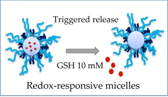
Redox-Responsive Crosslinked Mixed Micelles for Controllable Release of Caffeic Acid Phenethyl Ester


Abstract
:
1. Introduction
2. Materials and Methods
2.1. Materials
2.2. Analysis
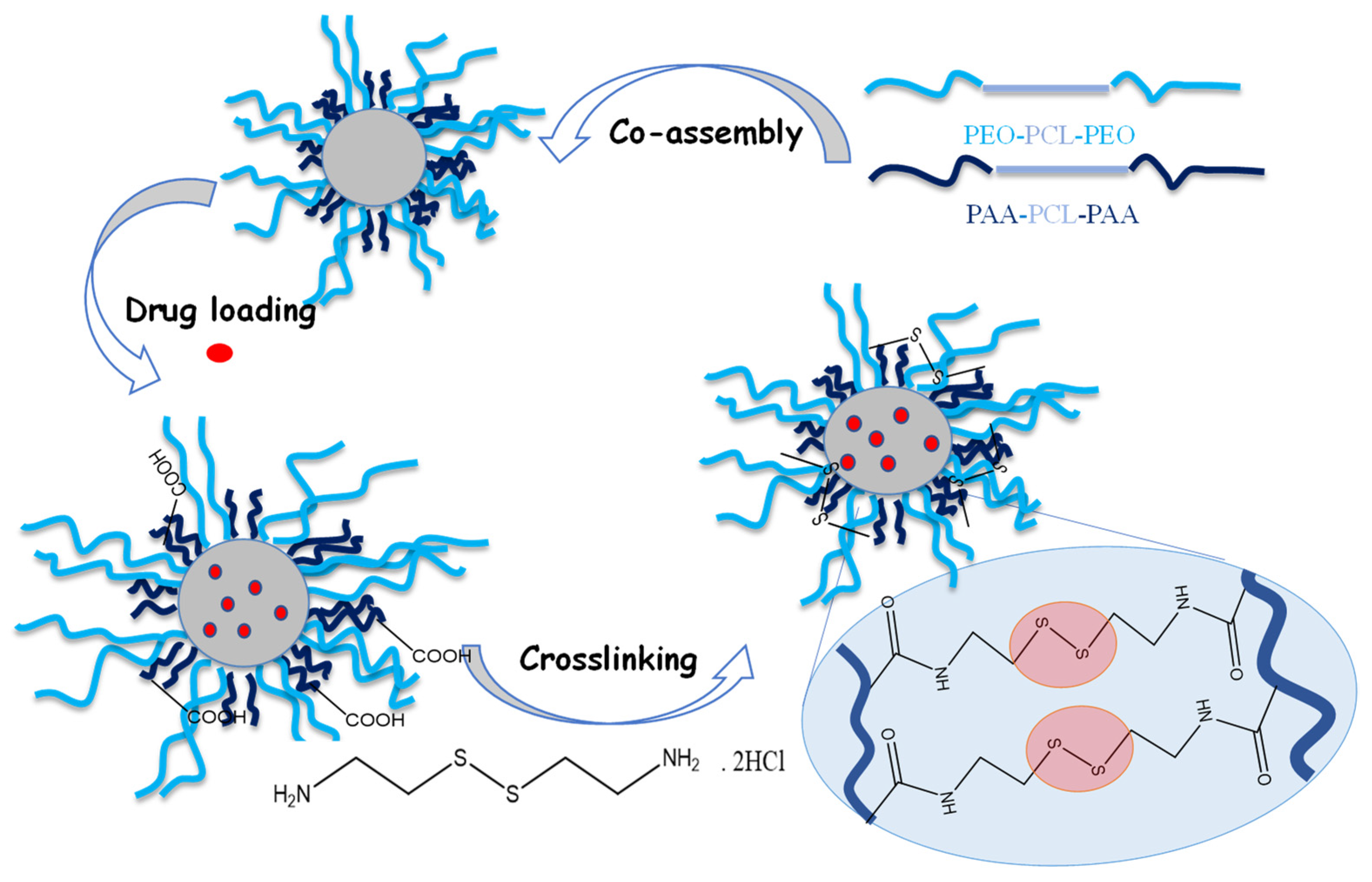
2.3. Synthesis of Block Copolymers
2.4. Preparation of Micelles
2.5. Preparation of Crosslinked Micelles
2.6. Drug Loading of Micellar Carriers
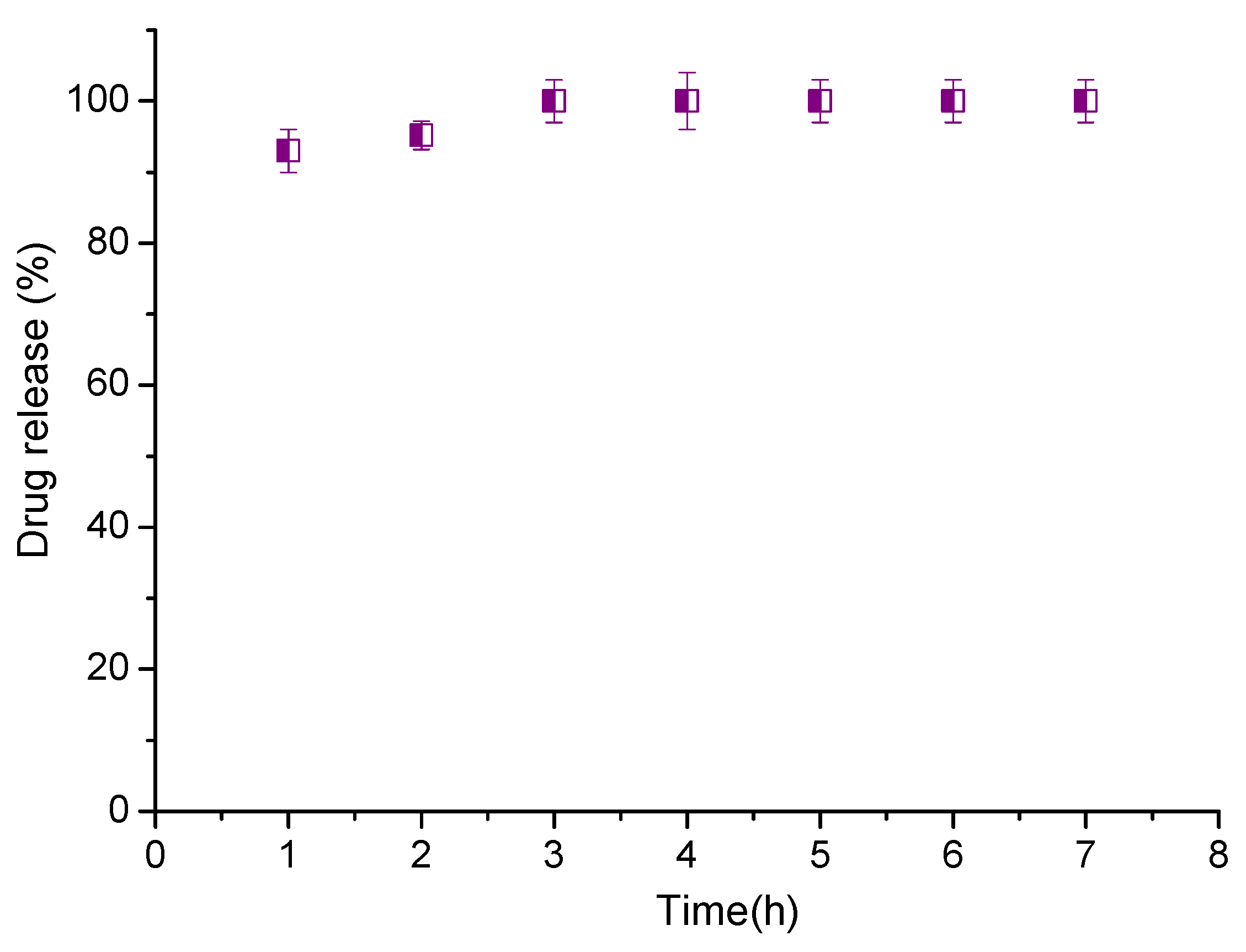
2.7. In Vitro Drug Release
3. Results
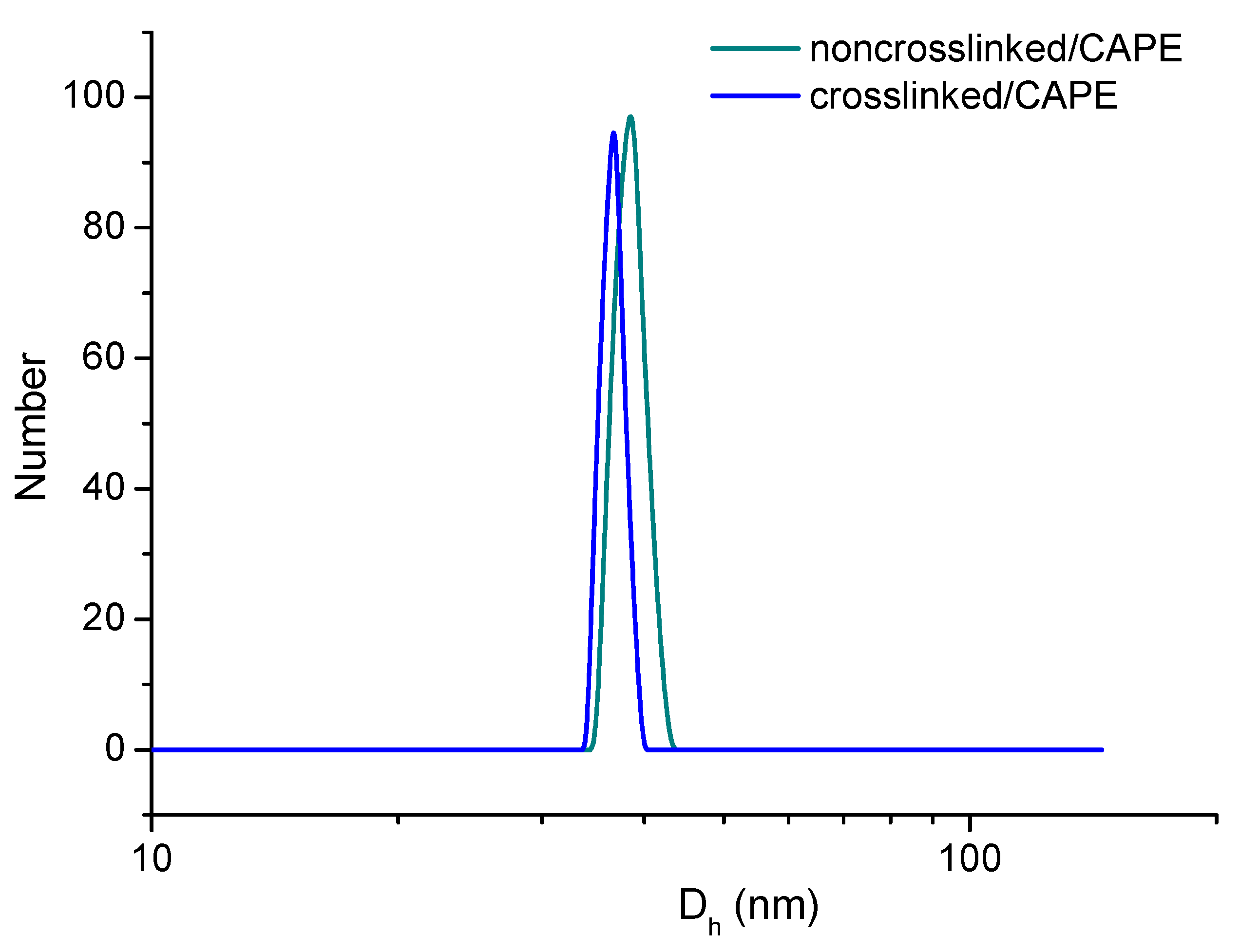
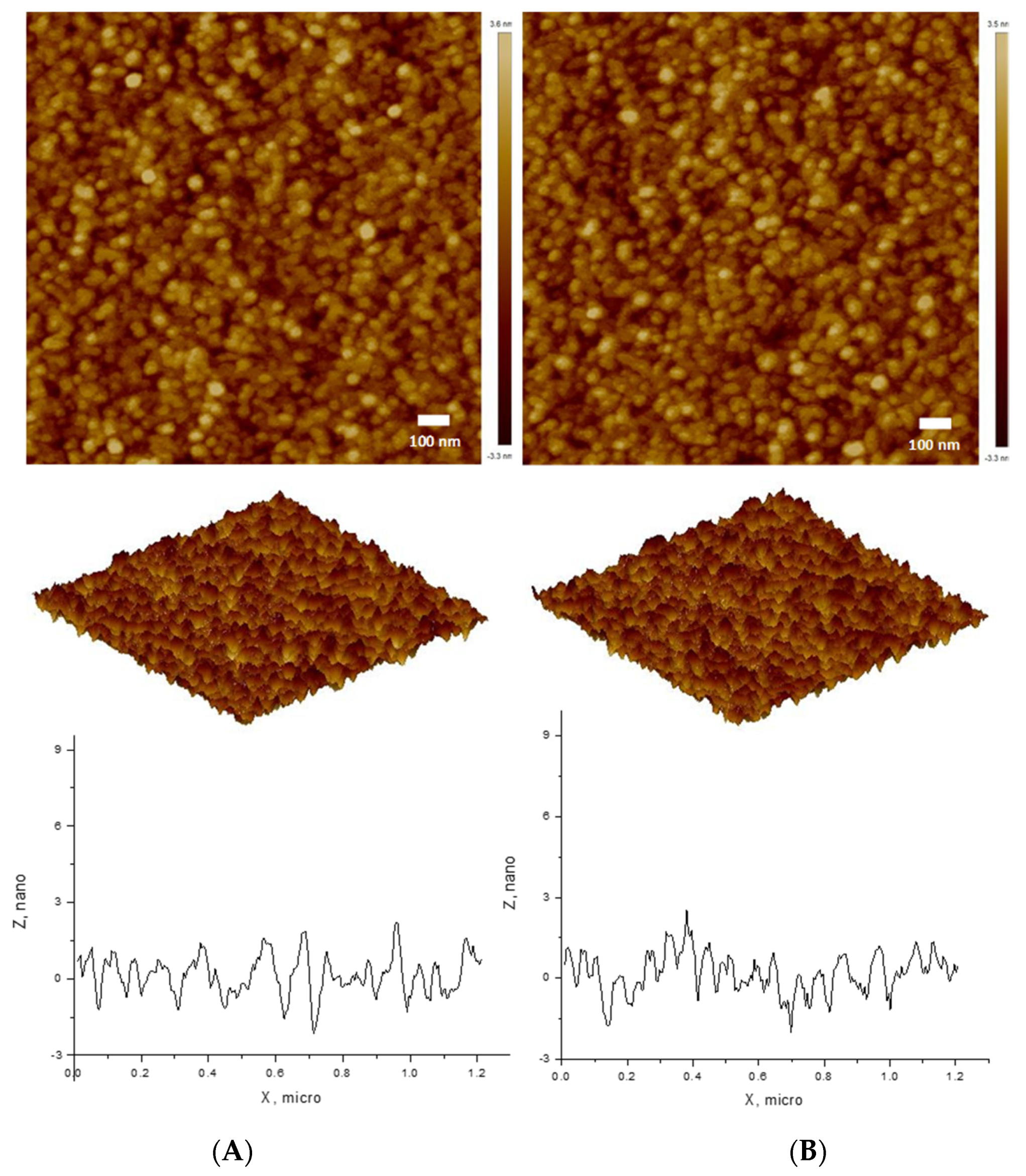
3.1. Polymeric Micelles
3.2. Drug Loading
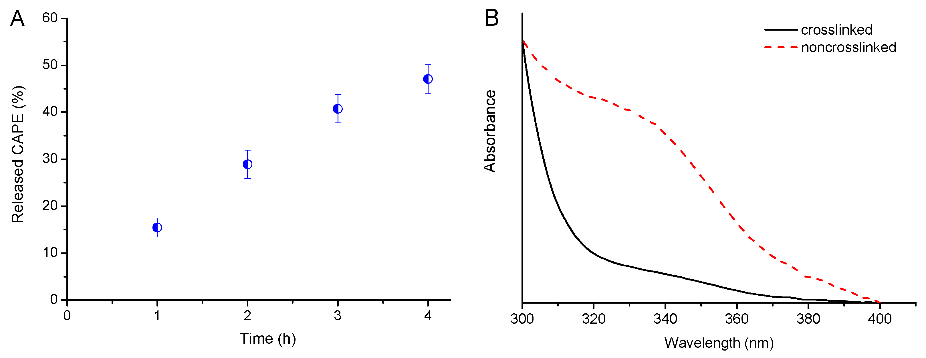
3.3. Drug Release
4. Discussion
5. Conclusions
Supplementary Materials
Author Contributions
Funding
Institutional Review Board Statement
Informed Consent Statement
Data Availability Statement
Acknowledgments
Conflicts of Interest
References
- Nichols, J.W.; Bae, Y.H. Odyssey of a cancer nanoparticle: From injection site to site of action. Nano Today 2012, 7, 606–618. [Google Scholar] [CrossRef] [PubMed] [Green Version]
- Ghosh, B.; Biswas, S. Polymeric micelles in cancer therapy: State of the art. J. Control. Release 2021, 332, 127–147. [Google Scholar] [CrossRef] [PubMed]
- Cabral, H.; Miyata, K.; Osada, K.; Kataoka, K. Block Copolymer Micelles in Nanomedicine Applications. Chem. Rev. 2018, 118, 6844–6892. [Google Scholar] [CrossRef] [PubMed] [Green Version]
- Van Vlerken, L.E.; Vyas, T.K.; Amiji, M.M. Poly(ethylene glycol)-modified Nanocarriers for Tumor-targeted and Intracellular Delivery. Pharm. Res. 2007, 24, 1405–1414. [Google Scholar] [CrossRef] [PubMed]
- Davoodi, P.; Lee, L.Y.; Sunil, V.; Sun, Y.; Soh, S.; Wang, C.-H. Drug delivery systems for programmed and on-demand release. Adv. Drug Deliv. Rev. 2018, 132, 104–138. [Google Scholar] [CrossRef] [PubMed]
- Dai, Y.; Chen, X.; Zhang, X. Recent advances in stimuli-responsive polymeric micelles via click chemistry. Polym. Chem. 2019, 10, 34–44. [Google Scholar] [CrossRef]
- Blanco, E.; Kessinger, C.W.; Sumer, B.D.; Gao, J. Multifunctional micellar nanomedicine for cancer therapy. Exp. Biol. Med. 2009, 234, 123–131. [Google Scholar] [CrossRef] [PubMed] [Green Version]
- Jhaveri, A.M.; Torchilin, V.P. Multifunctional polymeric micelles for delivery of drugs and siRNA. Front. Pharmacol. 2014, 5, 77. [Google Scholar] [CrossRef] [PubMed] [Green Version]
- Persi, E.; Duran-Frigola, M.; Damaghi, M.; Roush, W.R.; Aloy, P.; Cleveland, J.L.; Gillies, R.J.; Ruppin, E. Systems analysis of intracellular pH vulnerabilities for cancer therapy. Nat. Commun. 2018, 9, 2997. [Google Scholar] [CrossRef] [PubMed]
- Kinoh, H.; Miura, Y.; Chida, T.; Liu, X.; Mizuno, K.; Fukushima, S.; Morodomi, Y.; Nishiyama, N.; Cabral, H.; Kataoka, K. Nanomedicines eradicating cancer stem-like cells in vivo by pH-triggered intracellular cooperative action of loaded drugs. ACS Nano 2016, 10, 5643–5655. [Google Scholar] [CrossRef]
- Chan, N.; An, S.Y.; Oh, J.K. Dual location disulfide degradable interlayer-crosslinked micelles with extended sheddable coronas exhibiting enhanced colloidal stability and rapid release. Polym. Chem. 2014, 5, 1637–1649. [Google Scholar] [CrossRef]
- Lin, W.; Ma, G.; Kampf, N.; Yuan, Z.; Chen, S. Development of long-circulating zwitterionic cross-linked micelles for active-targeted drug delivery. Biomacromolecules 2016, 17, 2010–2018. [Google Scholar] [CrossRef] [PubMed]
- Li, Y.; Xiao, K.; Luo, J.; Xiao, W.; Lee, J.S.; Gonik, A.M.; Kato, J.; Dong, T.A.; Lam, K.S. Well-defined, reversible disulfide cross-linked micelles for on-demand paclitaxel delivery. Biomaterials 2011, 32, 6633e6645. [Google Scholar] [CrossRef] [PubMed] [Green Version]
- Yang, Z.; Fu, K.; Yu, J.; Zhou, P.; Cheng, Z. “pH-triggered” drug release using shell cross-linked micelles from aqueous RAFT-synthesized PAPMA-b-PNIPAM copolymers. J. Polym. Res. 2018, 25, 164. [Google Scholar] [CrossRef]
- Koo, A.N.; Lee, H.J.; Kim, S.E.; Chang, J.H.; Park, C.; Kim, C.; Park, J.H.; Lee, S.C. Disulfide-cross-linked PEG-poly(amino acid)s copolymer micelles for glutathione-mediated intracellular drug delivery. Chem. Commun. 2008, 6570–6572. [Google Scholar] [CrossRef] [PubMed]
- Wang, Y.-C.; Li, Y.; Sun, T.-M.; Xiong, M.-H.; Wu, J.; Yang, Y.-Y.; Wang, J. Core–Shell–Corona Micelle Stabilized by Reversible Cross-Linkage for Intracellular Drug Delivery. Macromol. Rapid Commun. 2010, 31, 1201–1206. [Google Scholar] [CrossRef] [PubMed]
- Yue, J.; Wang, R.; Liu, S.; Wu, S.; Xie, Z.; Huang, Y.; Jing, X. Reduction-responsive shell-crosslinked micelles prepared from Y-shaped amphiphilic block copolymers as a drug carrier. Soft Matter 2012, 8, 7426. [Google Scholar] [CrossRef]
- Li, W.; Fan, X.; Lv, X.; Du, J.; Liu, Q.; Lin, J.; Hu, Z.; Li, Z. Reduction-responsive shell cross-linked micelles derived from amphiphilic triblock copolymer as anticancer drug delivery carrier. Mater. Sci. Eng. C 2019, 96, 383–390. [Google Scholar] [CrossRef] [PubMed]
- Xiong, D.; Yao, N.; Gu, H.; Wang, J.; Zhang, L. Stimuli-responsive shell cross-linked micelles from amphiphilic four-arm star copolymers as potential nanocarriers for “pH/redox-triggered” anticancer drug release. Polymer 2017, 114, 161–172. [Google Scholar] [CrossRef]
- Liu, Y.; Chen, F.; Zhang, K.; Wang, Q.; Chena, Y.; Luo, X. pH-Responsive reversibly cross-linked micelles by phenol–yne click via curcumin as a drug delivery system in cancer chemotherapy. J. Mater. Chem. B 2019, 7, 3884–3893. [Google Scholar] [CrossRef]
- Petrov, P.D.; Yoncheva, K.; Gancheva, V.; Konstantinov, S.; Trzebicka, B. Multifunctional block copolymer nanocarriers for co-delivery of silver nanoparticles and curcumin: Synthesis and enhanced efficacy against tumor cells. Eur. Polym. J. 2016, 81, 24–33. [Google Scholar] [CrossRef]
- Akyol, S.; Ozturk, G.; Ginis, Z.; Armutcu, F.; Yigitoglu, M.R.; Akyol, O. In vivo and in vitro antineoplastic actions of caffeic acid phenethyl ester (CAPE): Therapeutic perspectives. Nutr. Cancer 2013, 65, 515–526. [Google Scholar] [CrossRef] [PubMed]
- Lee, H.Y.; Jeong, Y.I.; Kim, E.J.; Lee, K.D.; Choi, S.H.; Kim, Y.J.; Kim, D.H.; Choi, K.C. Preparation of caffeic acid phenethyl ester-incorporated nanoparticles and their biological activity. J. Pharm. Sci. 2015, 104, 144–154. [Google Scholar] [CrossRef] [PubMed]
- Yoncheva, K.; Tzankova, V.; Yordanov, Y.; Tzankov, B.; Grancharov, G.; Aluani, D.; Bankova, V.; Popova, M.; Trusheva, B.; Kondeva-Burdina, M.; et al. Evaluation of antioxidant activity of caffeic acid phenethyl ester loaded block copolymer micelles. Biotechnol. Biotechnol. Equip. 2019, 33, 64–74. [Google Scholar] [CrossRef] [Green Version]
- Grancharov, G.; Atanasova, M.-D.; Aluani, D.; Yoncheva, K.; Tzankova, V.; Trusheva, B.; Forys, A.; Trzebicka, B.; Petrov, P.D. Functional block copolymers bearing pendant cinnamyl groups for enhanced solubilization of caffeic acid phenethyl ester. Polym. J. 2020, 52, 435–447. [Google Scholar] [CrossRef]
- Atanasova, M.-D.; Grancharov, G.; Petrov, P.D. Poly(ethylene oxide)-block-poly(α-cinnamyl-ε-caprolactone-co-ε-caprolactone) diblock copolymer nanocarriers for enhanced solubilization of caffeic acid phenethyl ester. J. Polym. Sci. 2021, 59, 251–260. [Google Scholar] [CrossRef]
- Kamenova, K.; Grancharov, G.; Tzankov, B.; Aluani, D.; Tzankova, V.; Tzankov, S.; Yoncheva, K.; Petrov, P.D. Mixed micellar system for codelivery of doxorubicin and caffeic acid phenethyl ester: Design and enhanced antitumor activity. Polym. J. 2021, 53, 471–479. [Google Scholar] [CrossRef]
- Kamenova, K.; Haladjova, E.; Grancharov, G.; Kyulavska, M.; Tzankova, V.; Aluani, D.; Yoncheva, K.; Pispas, S.; Petrov, P.D. Co-assembly of block copolymers as a tool for developing novel micellar carriers of insulin for controlled drug delivery. Eur. Polym. J. 2018, 104, 1–9. [Google Scholar] [CrossRef]


| Sample | Dh (nm) | ζ Potential (mV) |
|---|---|---|
| Non-crosslinked | 32 ± 2 | −22 ± 2 |
| Crosslinked | 29 ± 2 | −9 ± 1 |
| Non-crosslinked/CAPE | 38 ± 3 | −22 ± 2 |
| Crosslinked/CAPE | 36 ± 3 | −9 ± 1 |
Publisher’s Note: MDPI stays neutral with regard to jurisdictional claims in published maps and institutional affiliations. |
© 2022 by the authors. Licensee MDPI, Basel, Switzerland. This article is an open access article distributed under the terms and conditions of the Creative Commons Attribution (CC BY) license (https://creativecommons.org/licenses/by/4.0/).
Share and Cite
Kamenova, K.; Grancharov, G.; Kortenova, V.; Petrov, P.D. Redox-Responsive Crosslinked Mixed Micelles for Controllable Release of Caffeic Acid Phenethyl Ester. Pharmaceutics 2022, 14, 679. https://doi.org/10.3390/pharmaceutics14030679
Kamenova K, Grancharov G, Kortenova V, Petrov PD. Redox-Responsive Crosslinked Mixed Micelles for Controllable Release of Caffeic Acid Phenethyl Ester. Pharmaceutics. 2022; 14(3):679. https://doi.org/10.3390/pharmaceutics14030679
Chicago/Turabian StyleKamenova, Katya, Georgy Grancharov, Vasilena Kortenova, and Petar D. Petrov. 2022. "Redox-Responsive Crosslinked Mixed Micelles for Controllable Release of Caffeic Acid Phenethyl Ester" Pharmaceutics 14, no. 3: 679. https://doi.org/10.3390/pharmaceutics14030679