Review

Role of Anti-Cancer Peptides as Immunomodulatory Agents: Potential and Design Strategy

by
Amit Kumar Tripathi
* and
Jamboor K. Vishwanatha
*
Department of Microbiology, Immunology and Genetics, University of North Texas Health Science Center, Fort Worth, TX 76107, USA
*
Authors to whom correspondence should be addressed.
Pharmaceutics 2022, 14(12), 2686; https://doi.org/10.3390/pharmaceutics14122686
Submission received: 11 November 2022 / Revised: 27 November 2022 / Accepted: 30 November 2022 / Published: 1 December 2022
(This article belongs to the Special Issue Peptide-Based Drugs for Cancer Therapies)

Abstract

:
The usage of peptide-based drugs to combat cancer is gaining significance in the pharmaceutical industry. The collateral damage caused to normal cells due to the use of chemotherapy, radiotherapy, etc. has given an impetus to the search for alternative methods of cancer treatment. For a long time, antimicrobial peptides (AMPs) have been shown to display anticancer activity. However, the immunomodulatory activity of anti-cancer peptides has not been researched very extensively. The interconnection of cancer and immune responses is well-known. Hence, a search and design of molecules that can show anti-cancer and immunomodulatory activity can be lead molecules in this field. A large number of anti-cancer peptides show good immunomodulatory activity by inhibiting the pro-inflammatory responses that assist cancer progression. Here, we thoroughly review both the naturally occurring and synthetic anti-cancer peptides that are reported to possess both anti-cancer and immunomodulatory activity. We also assess the structural and biophysical parameters that can be utilized to improve the activity. Both activities are mostly reported by different groups, however, we discuss them together to highlight their interconnection, which can be used in the future to design peptide drugs in the field of cancer therapeutics.

1. Introduction

Rudolf Virchow first observed that persistent inflammation transcends into cancer and tumor tissues show a large infiltration of inflammatory cells. Later on, Dvorak showed that carcinogenesis and inflammation have commonalities in terms of proliferation, migration, cytokine secretion and angiogenesis. He described cancer as a “wound that does not heal” [1]. In modern times, the three main methods of cancer treatment so far have been Chemotherapy, radiotherapy and immunotherapy. Chemotherapy is the most established method of treatment that kill fast-dividing cancer cells. However, most cancer drugs have very poor cell selectivity and kill normal cells along with cancer cells indiscriminately [2,3]. Moreover, continuous use of this therapy increases the possibility of drug resistance in the body along with the chances of recurrence. Radiotherapy is the second type of cancer therapy that uses high-energy beams to kill cancer cells. X-rays are the most commonly used energy beams but, protons or other types of energy can also be used. Unfortunately, radiotherapy causes collateral damage as, despite the advancements in modern types of equipment, the radiation kills normal cells along with the targeted cancer cells. Immunotherapy is the third kind of therapy that improves the patient’s immune system to exert an anti-tumor effect. This treatment method has fewer side effects than chemotherapy and radiotherapy, and the therapeutic effects are long-lasting [4]. As a standard job, the immune system detects cancerous cells due to the presence of abnormal cell surface markers. Biopsies of patients show various immune cells in and around tumors [5]. These cells, called tumor-infiltrating lymphocytes or TILs, are a sign that the immune system is responding to the tumor. Cancer patients whose tumors contain TILs often have a lesser level of cancer severity than those whose tumors do not contain them. The interrelationship between inflammation, innate immunity and cancer is well known [6]. Persistent inflammation triggers cancer initiation that is characterized by infiltration of mononuclear immune cells (including macrophages, lymphocytes, and plasma cells), tissue destruction, fibrosis, and increased angiogenesis [7].

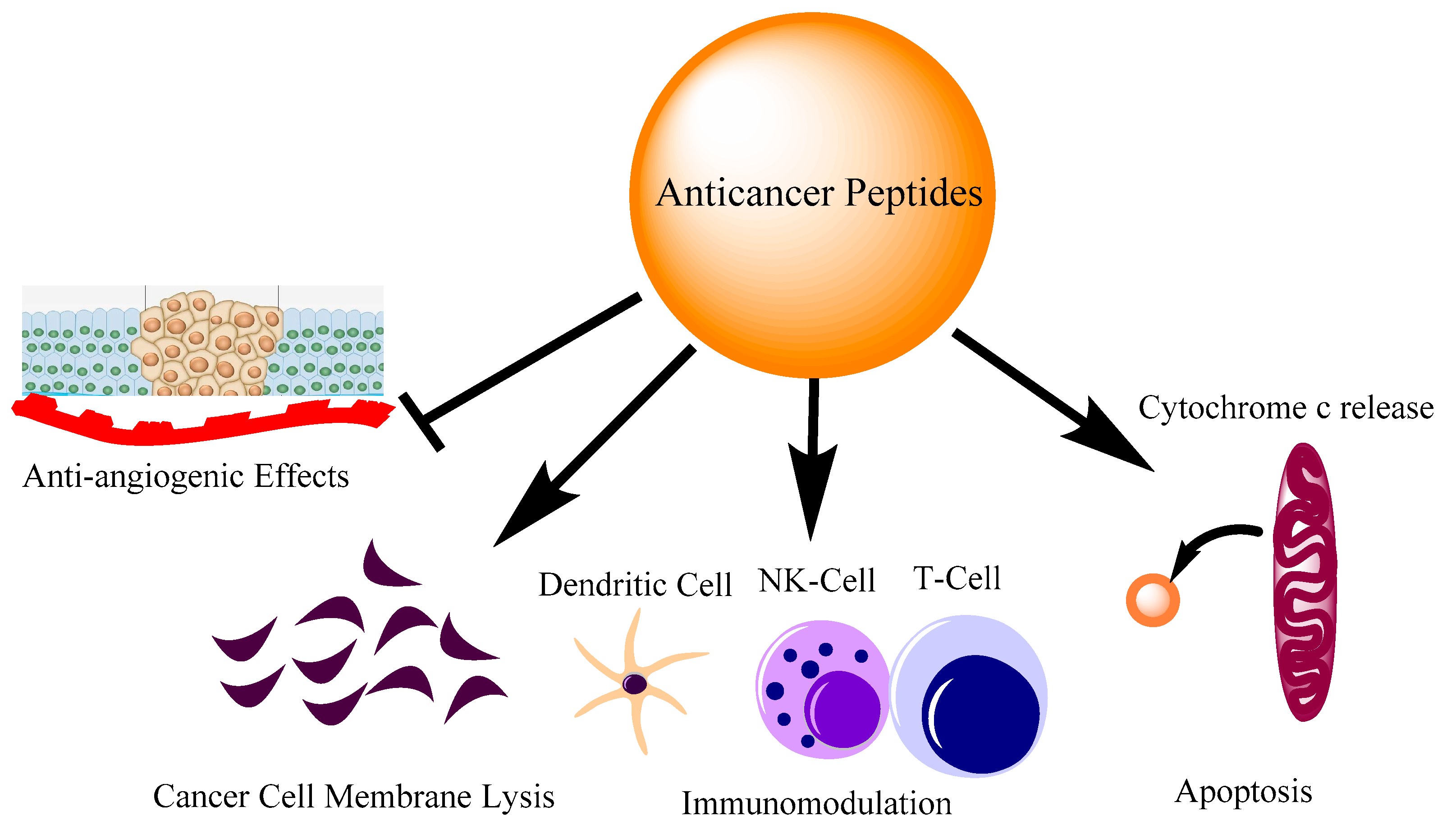
2. Where Anti-Cancer Peptides Stand

The discovery of peptide hormone insulin gave an impetus to peptide therapeutics. During the last two decades, peptides have grown as encouraging healing mediators in many areas such as diabetes [8], cardiovascular diseases [9] and cancer treatment [10] (Figure 1). Improvements in peptide design have increased its applications in other fields as well [11,12]. The beginning of the 21st century witnessed rapid advancements in various interdisciplinary fields like analytics, structural biology Computer-assisted drug discovery, and bioinformatics tools which have made peptide design much easier and also minimized the chances of drug failure. This has led to the opening of new areas of drug discovery where peptide synthesis, chemical modifications and the evaluation of biological activities can be done simultaneously that speed up the process of lead molecule identifications. Insights in the global market for peptide therapeutics is projected to record a value of USD 44.43 billion in 2026, progressing at a CAGR of 6.95%, over the period 2022–2026. Currently, more than 80 peptide-based drugs are present in the market for the treatment of a wide range of diseases including cancer, osteoporosis, diabetes, etc. [13]. It is estimated that up to 400–600 peptide drugs are in preclinical trials. After 2017, USFDA has already approved more than 10 peptide-based drugs. Of these, LupkynisTM and Zegalogue were recently approved in 2021, while ImcivreeTM, Victoza, LUPRON DEPOT, Zoladex, Sandostatin and Somatuline received approval in 2020 [14]. The three drugs which have touched global sales of over $1 billion are goserelin, leuprolide and octreotide [15]. Goserelin is a synthetic decapeptide analog of luteinizing hormone-releasing hormone (LHRH). It has anti-cancer activity and is used in treatments of both breast and prostate cancer [16] Leuprolide is a peptide analog of gonadotropin-releasing hormone (GnRH) which is used as a palliative treatment of prostate cancer and many other conditions [17]. Studies have shown that it also possesses an immunomodulatory effect. The investigations showed that Leuprolide Acetate (LA) administration to Experimental autoimmune encephalomyelitis (EAE) rats considerably inhibited the activation of NF-κB which is central to both inflammation and cancer progression. It was shown that treatment with LA reduced the production of TNF-α, IL-1β and other inflammatory cytokines which play an important role in both inflammation and cancer initiation [18]. The third successful peptide drug Octreotide is an analog of somatostatin. It helps to temporarily reduce the tumor size and diminish cancer development. Due to its large therapeutic abilities, octreotide has evolved as a backbone of clinical cancer therapeutics [19,20]. With the advent of bioinformatics tools, various short peptides have been identified from the naturally occurring proteins that have shown a high affinity for cancer cells. The structure-function relationships of anti-cancer peptides showed that certain biophysical parameters are present in these peptides that attract them to the cancer cells. Based on these parameters, several naturally occurring and synthetic peptides are being identified that show anti-cancerous properties.

3. Naturally Occurring AMPs/ACPs with Immunomodulatory Activities

The helicity is strongly correlated with antimicrobial and anti-cancer activity [21]. Naturally occurring anti-microbial peptides have shown potent anti-cancer activities in in vitro experiments in several independent studies (Table 1). Besides, naturally occurring proteins that are associated with cancer progression pathways have been used to design immunomodulatory peptides that can inhibit LPS-mediated inflammatory responses [22,23]. LL-37, Magainin-II, Melittin and other naturally occurring anti-microbial peptides have shown appreciable anti-cancer activity [24,25,26]. Additionally, they contain good anti-inflammatory activity which is an important immunomodulatory activity to tackle the dual problem of cancer and inflammation [27]. Melittin (GIGAVLKVLTTGLPALISWIKRKRQQ), a peptide first isolated from bee venom has shown to be a promising candidate for cancer therapy. It is an alpha-helical anticancer peptide that plays a key role in immunomodulation and inhibits proinflammatory agents. The anti-cancer properties exerted by melittin are very similar to anti-cancer drugs and involve cell cycle arrest, anti-proliferative activity on cancer cells and activation of caspases [28]. Besides these, melittin is also shown to induce apoptosis in cancer cells through ROS generation and the diffusion of mitochondrial membrane potential [29].
Unfortunately, melittin is a non-cell selective peptide and displays cytotoxicity to normal cells as well [30]. Therefore, to achieve its true therapeutic potential, suitable analogs of it are to be designed that show reduced cytotoxicity to normal cells but retain the anti-cancer activity. Asthana et al. have identified a leucine zipper motif in melittin which can be used as a switch to design bioactive analogs [31]. These analogs can be used for their anti-cancer and immunomodulatory activities but with reduced cytotoxicity to normal cells. Srivastava et al. showed that melittin can neutralize the lipopolysaccharide-induced proinflammatory pathways in RAW 264.7 and primary macrophage cells and the leucine zipper motif present in the peptide played an important role in its immunomodulatory activity [32]. Liu et al. created a bifunctional fusion protein melittin-MIL-2, which was a recombinant of melittin and a mutant IL-2 [33]. The melittin-MIL-2 displayed potent anti-cancer activities in comparison to Melittin and rIL-2 alone. The MIL-2 displayed anti-proliferative activity against cancer cells derived from different tissues. In the in vivo experiments, the MIL-2 was able to inhibit the tumor growth in liver, lung and ovary cancer cells. The investigators also showed that exposure of MIL-2 was able to reduce the ability of breast cancer cells to metastasize to the lungs. Another promising antimicrobial peptide that can be used for the dual role of anticancer and immunomodulatory activities is Magainin II (KWKLFKKIKFLHSAKKF). It was first isolated from the skin of Xenopus laevis frogs. Studies have shown that magainin II inhibited the cell proliferation of bladder cancer cells while did not cause any toxicity to normal fibroblast 3T3 cells [34]. Although Magainin II did not show effective anti-cancer activity on human breast cancer cells MDA-MB-231, it displays good anti-cancer activity against lung cancer cell line A549 [25]. It is not toxic to human immortalized epidermal cells under similar conditions. While Magainin II itself does not cause any immunomodulatory activity, hybrid peptides designed using cecropin A17 and Magainin II showed potent anti-cancer and anti-inflammatory activity [35,36]. A cecropin A–magainin II hybrid peptide called P18 (KWKLFKKIPKFLHLAKKF) was effective against human leukemia K562 cells [37]. Tang et al. showed that P18 induced necrosis in these cells instead of activating the apoptotic pathway. The mechanism of action of the peptide involved the diffusing of the plasma membrane potential in the cells after peptide exposure [37]. Nan et al. while studying the immunomodulatory activity of P18 showed that when mouse macrophage cell line RAW264.7 was challenged with LPS from E. coli in the absence or presence of P18, it inhibited the LPS-mediated production of pro-inflammatory mediators and cytokines viz. nitrite (NO), TNF-α and IL-1β [38]. This was a classic example of using two naturally occurring peptide sequences to design a hybrid peptide with immunomodulatory activity, which was not present in the parent peptide. The judicious substitution of key residues at either terminus of an anti-cancer peptide often improves its biological activity [39]. However, it is essential to make a conservative replacement so that the biophysical parameters like charge, hydrophobicity, etc. remain similar to the parent peptide. Along similar lines, Arias et al. designed improved analogs of tritrpticin (VRRFPWWWPFLRR) for potent anti-cancer activity against the Jurkat leukemia cell line [40]. By designing a series of analogs of tritrpticin, they found that if the arginines at both the termini are replaced with lysines or lysine-derivatives, it improves the cell-selectivity of the peptides towards Jurkat leukemia cells as opposed to normal peripheral blood mononuclear cells (PBMCs). Interestingly, arginine to lysine substitution also enhanced the biological activity in other sequentially similar peptides including indolicidin (ILPWKWPWWPWRR) and puroindoline A (PuroA FPVTWKWWKWWKG-NH2) [41]. Ghiselli et al. checked the anti-inflammatory activity of Indolicidin, a closely related peptide to tritrpticin in two rat models of polymicrobial peritonitis. The investigators used two different models to induce sepsis. One by intraperitoneal injection of LPS and the other by using cecal ligation and puncture (CLP model) of inflammation. The results showed that indolicidin treated group decreased the bacterial burden in visceral organs like peritoneum, spleen and liver and plasma levels of LPS-mediated production of TNF-α and IL-6 were also inhibited. This signifies the dual role of the peptide as both an immunomodulatory and anticancer peptide in addition to being a well-established antimicrobial peptide [42].
Anti-cancer peptides with immunomodulatory activities have also been reported from marine sources [43]. Marine fishes are rich sources of anti-cancer peptides possessing immunomodulatory activities [44]. Marine animals possess poor immune systems and live in ecological niches where they are exposed to diverse pathogens. Iijima et al. purified Chrysophsins in marine fish Chrysophsis major. There are three different forms of chrysophsins [45]. The majority of the anti-cancer and immunomodulatory studies are done on two isoforms, Chrysophsin-I (FFGWLIKGAIHAGKAIHGLIHRRRH) and Chrysophsin-II (FFGWLIRGAIHAGKAIHGLIHRRRH). Hsu et al. checked the anti-cancer activity of Chrysophsin-1. Their results showed that the peptide followed a lytic mechanism to kill cancer cells mostly through pore formation. The inhibition ratio was less for normal cell lines viz. NIH-3T3 and WS-1 [46]. Tripathi et al. identified the GXXXXG motif in Chrysophsin I and designed various proline-substituted analogs of the Chrysophsin-1 and showed that the peptides in addition to the anti-cancer activity as described by other workers also possess potent immunomodulatory activity. The authors were able to show that one of the proline-substituted analogs rescued the mice from the lethal dose of LPS [47]. Temporin L (FVQWFSKFLGRIL), another highly studied alpha-helical anti-microbial peptide has significant anti-cancer and immunomodulatory activities [48]. Swithenbank et al., while studying the activity of Temporins and Bombinin H2 (LIGPVLGLVGSALGGLLKKI) on lung cancer cell lines reported that Temporin L exposure to cancer cell lines viz. A-549 and Calu-3 caused significant cytotoxicity in a dose-dependent manner [49]. Srivastava et al. investigated the immunomodulatory activity of Temporin L. They identified a phenylalanine zipper in Temporin L that can be used to design Temporin L analogs that are less toxic to normal cells and exhibit anti-endotoxin activities [50]. Studies have shown that Temporin L directly binds to LPS and can be a therapeutic agent in septic shock [51].
Interestingly, phenylalanine heptad repeats have also been used to design synthetic peptides that contain potent anti-cancer and immunomodulatory activities. Tripathi et al. showed that if the phenylalanine residues are replaced with proline in a synthetic peptide designed on the basis of phenylalanine heptad repeats, the resultant peptide exhibit potent anti-cancer and immunomodulatory activities. The proline substituted analogs of parent peptide designed on phenylalanine heptad repeats also inhibit migration in MDA-MB-231 breast cancer cells and induce programmed cell death by activating the intrinsic pathway of apoptosis. The same peptides contained anti-endotoxin activities as they inhibit the LPS-mediated NF-kB nuclear translocation and inhibit the production of pro-inflammatory cytokines [52]. Hepcidin (ICIFCCGCCHRSKCGMCCKT) is also a good example of an anti-cancer peptide containing immunomodulatory activity [53]. Cytotoxicity data of hepcidin on myeloma cells indicate that it causes plasma membrane damage and DNA fragmentation in these cancer cells to exhibit its anti-cancerous activities [54]. An independent study about the immunomodulatory activity of hepcidin showed that it can up-regulate the expression of both pro- and anti-inflammatory cytokines like TNF-α, IL-1β, and IL-10 in teleost leukocytes. [53]. The mRNA expression was also found to be high in the organs like the spleen and head kidney. LL-37(LLGDFFRKSKEKIGKEFKRIVQRIKDFLRNLVPRTES) is probably the most widely studied Cathelicidin. Originally investigated for its antimicrobial activities, LL-37 soon was reported to inhibit a wide range of cancers and is very context-specific [55]. The role of LL-37 in colon cancer seems to be most interesting. A differential expression of LL-37 has been observed in normal colon and cancer colon mucosa. It had been reported that LL-37 gets downregulated as colon cancer progresses [56]. This has led to the idea of using LL-37 as a colon cancer biomarker [57]. It can also induce apoptosis by upregulating the levels of Bax/Bak and downregulating the BCL-2 levels [58]. LL-37 has also been shown to increase the PUMA (p53 upregulated modulator of apoptosis) expression which is a modulator of apoptosis in colon cancer cells [24]. Besides this, LL-37 also increases the nuclear translocation of apoptosis-inducing-factor (AIF) and endonuclease G (EndoG) in colon cancer cells to induce apoptosis [56]. FK-16, a derivate of full-length LL-37 containing the same amino acid residues from 17 to 32 followed a similar mechanism as the parental LL-37 to cause apoptosis by increasing the nuclear levels of apoptosis-inducing-factor (AIF) and endonuclease G (EndoG) in colon cancer cells in a caspase-independent manner [59]. LL-37 also has a very potent immunomodulatory activity on different types of immune cells. It is shown to have antisepsis properties and has been proven to neutralize the inflammatory responses activated by bacterial components like LPS and LTA. Culturing the bone marrow-derived macrophages with LPS with or without LL-37 showed that the LL-37 was able to almost completely cancel out the LPS-mediated TNF-α and brought it to an almost basal level [60].

3.1. Amino Acid Arrangement and Their Biophysical Parameters Determine Anti-Cancer and Immunomodulatory Properties

Amino acid residues in an anti-cancer peptide can dictate its cell permeability. Leucine, lysine, histidine and glycine are abundant in peptides having anti-cancer and immunomodulatory activity [61]. Studies have reported that electrostatic interaction between cationic amino acid residues and negatively charged components of cancer cell membrane mostly phosphatidylserine is the first interaction to begin the anti-cancer activity [62]. Similarly, anti-cancer peptides eg. Piscidin-1 exhibiting anti-endotoxin activity interact with lipopolysaccharide (LPS) on similar lines [63,64]. The LPS molecules also contain anionic groups making them ideal candidates for electrostatic interactions by cationic ACPs [65]. Glutamic and aspartic acid are shown to inhibit cell proliferation in hepatoma cells and inhibit the AKT phosphorylation, a key signal transduction protein in cancer biology [66]. Proline in anti-cancer peptides impart proteolytic stability, aids in membrane interaction and brings conformational changes to the secondary structure of the peptides [67]. Phenylalanine imparts hydrophobicity to the peptides, which is an important biophysical parameter for both anti-cancer and immunomodulatory activity. It has been observed that the incorporation of phenylalanine amino acid sometimes improves the anti-cancer activity of the peptides like Galaxamide [68]. Tyrosine and tryptophan are hydrophobic amino acids, which is an important biophysical attribute for anti-cancer activity. Tryptophan plays an important role in the anti-cancer activity of peptides such as indolicidin and trans-activator of transportation (TAT)-Ras GTPase-activating protein-326 peptides [61,69]. Tryptophan contributes to the cell-penetrating ability of the ACP to facilitate its entry to the cell involving an endocytic pathway and DNA-binding [70,71]. Thus, it has been shown that cationic and hydrophobic amino acid residues are critical to both anti-cancer and immunomodulatory activity of the peptides and play an important role in the preliminary interaction of the peptide and cell membrane interactions [72,73,74,75,76].

3.2. Knowledge of Structural Determinants of AMPs/ACPs Are Same as for Immunomodulatory Peptides

There are several bio-physical parameters in AMPs, which are usually present in anti-cancer peptides, and their proper knowledge can be used to design ACPs without using any computer or software applications. By judiciously choosing the amino acids at specific positions and utilizing the structural determinants like charge, hydrophobicity, etc. potent antimicrobial peptides could be designed which possess the important attribute of cell selectivity.

3.2.1. Size

ACPs despite similar biological behaviors can vary in terms of length. The number of residues in the amino acid sequence of ACPs ranges from 5 to lesser than 100, however the most ACPs fall in the range of 15–50 residues [77]. They can be as small as KTH-222, having a length of 8 amino acids (NH2-LKGQLRCI-C02H), or as long as LL-37 and PR-39 [78,79,80].

3.2.2. Amino Acid Prevalence

Biological behavior of an ACP is a mere manifestation of its structural components, and these parameters depend on the amino acid sequence and prevalence of specific amino acids in the peptide sequence [81]. It is observed that many anti-cancer and immunomodulatory peptides more often contain basic residues like lysine or arginine than acidic residues like aspartate and glutamate [82,83]. On the other side, hydrophobic residues such as alanine, leucine, isoleucine, phenylalanine and tryptophan are well represented in ACPs and contribute to acquiring a stable conformation in a membrane environment [84]. L-K6 and K4R2Nal2-S1 are examples of such peptides [85,86].

3.2.3. Charge

The presence of positive charge is one of the most important parameters to initiate electrostatic interactions with negatively charged cancer cell membranes, and encourage self-promoted uptakes of ACPs [86]. Hence, it is not surprising that most of the cationic ACPs target the anionic membranes of cancer cells. Apart from many natural anti-cancer peptides, synthetic peptides like IK-13 and LK-13 reported by Hadianamrei et al. were designed chiefly based on positive charge [87].

3.2.4. Conformation

Based on the amino acid composition and their positions, ACPs acquire different secondary structures including α- helices, β-sheet, loops and extended helical conformations [87]. Amphipathic α-helical peptides are the most prevalent, followed by β-sheet peptides [88,89,90]. However, a large majority of ACPs can assume intermediate structures or random coiled but still display good anti-cancer activity such as proline-arginine-rich and tryptophan-rich peptides. Melittin, Bovine lactoferrin (LfcinB) and Alloferon are examples of alpha-helical, β-sheet and random-coiled anticancer peptides, respectively [91,92].

3.2.5. Hydrophobicity, Amphipathicity and Hydrophobic Moment

The presence of a threshold percentage of hydrophobicity is necessary to perform the biological activities in ACPs [93]. Nearly all ACPs, e.g., Temporin A exhibit moderate hydrophobic moments and amphipathic conformations upon interaction with target membranes [49]. The amphipathicity in β-sheet ACPs can be created by well-organized polar and nonpolar surfaces [94]. It is observed that an increase in hydrophobicity at certain positions in the sequence of a peptide may promote peptide amphipathicity. Generally, this phenomenon results in the enhancement of mean hydrophobicity of peptides, but the site-specific hydrophobicity is proportional to its hydrophobic moment and subsequently, the amphipathic characters of peptides.

3.2.6. Polar Angle

Polar angle is an estimation of the relative distribution of polar and nonpolar residues on two opposite faces of a peptide in an amphipathic helix. Increased segregation between hydrophilic and hydrophobic domains of the peptide would increase the polar angle as in VmCT1 analogs [95]. The polar angles are correlated with the net ability of the peptide to induce lethal pores in membranes, and peptides with smaller polar angles stimulate unstable pore formation than peptides with larger polar angles [96].

3.2.7. Peptide Self Assembly

Self-assembling peptides (SAPs) are small peptide sequences alternating in hydrophilic and hydrophobic amino acid residues [97]. Peptide self-assembly is a process in which peptides spontaneously form ordered aggregates when they encounter different microenvironments [98]. Many physical and chemical interactions stabilize this state [99]. Self-assembling peptides such as RADA16 and E3PA (AAAAGGGEEE) have great potential in cancer therapy as they can be utilized for cancer cell targeting, forming nanostructures, drug delivery, etc. [100,101,102].

3.2.8. Chemical Modifications

Modifications, both at the side chain and the main chain have been reported to improve anti-cancer activity [103,104]. The replacement of natural amino acids with non-natural amino acids in the main chain has been done by investigators to improve biological activity. Similarly, many chemical modifications in the side chain such as PEGylation, phosphorylation, adding fatty acids and glycosylation have been reported to facilitate the entry of the peptide into the target cancer cell that thereby reduces the IC50 values of the concerned peptide [105,106]. Lee et al. have found that PEGylation of a cationic antimicrobial decapeptide KSL-W (KKVVFWVKFK) improves the survival of mice in a sepsis model. It was also shown that the chemically modified peptide nullified the lipopolysaccharide (LPS)-induced inflammatory responses in human umbilical vein endothelial cells compared with unmodified KSLW [107].

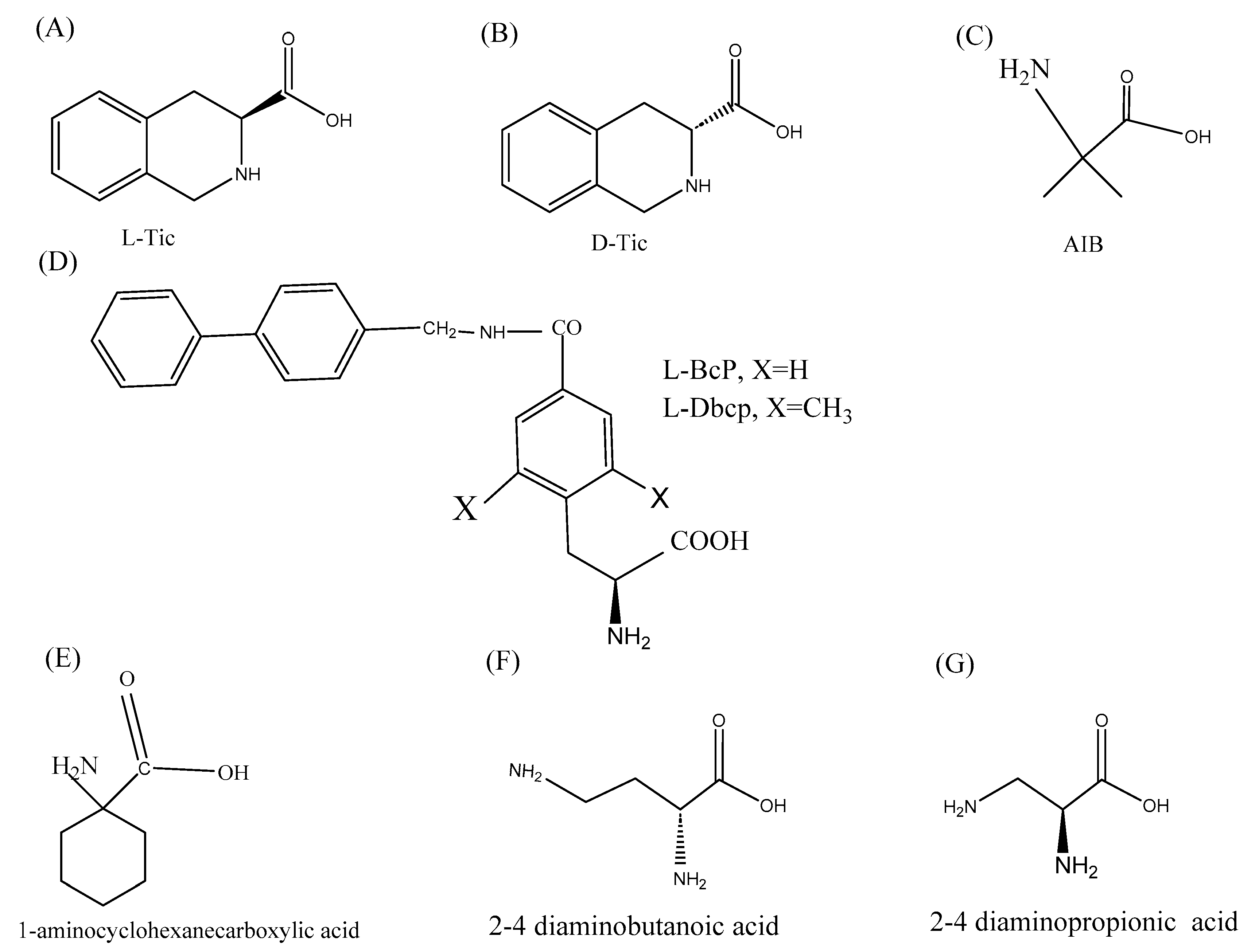
3.3. Role of Non-Natural Amino Acids in Improving the Anti-Cancer Activity
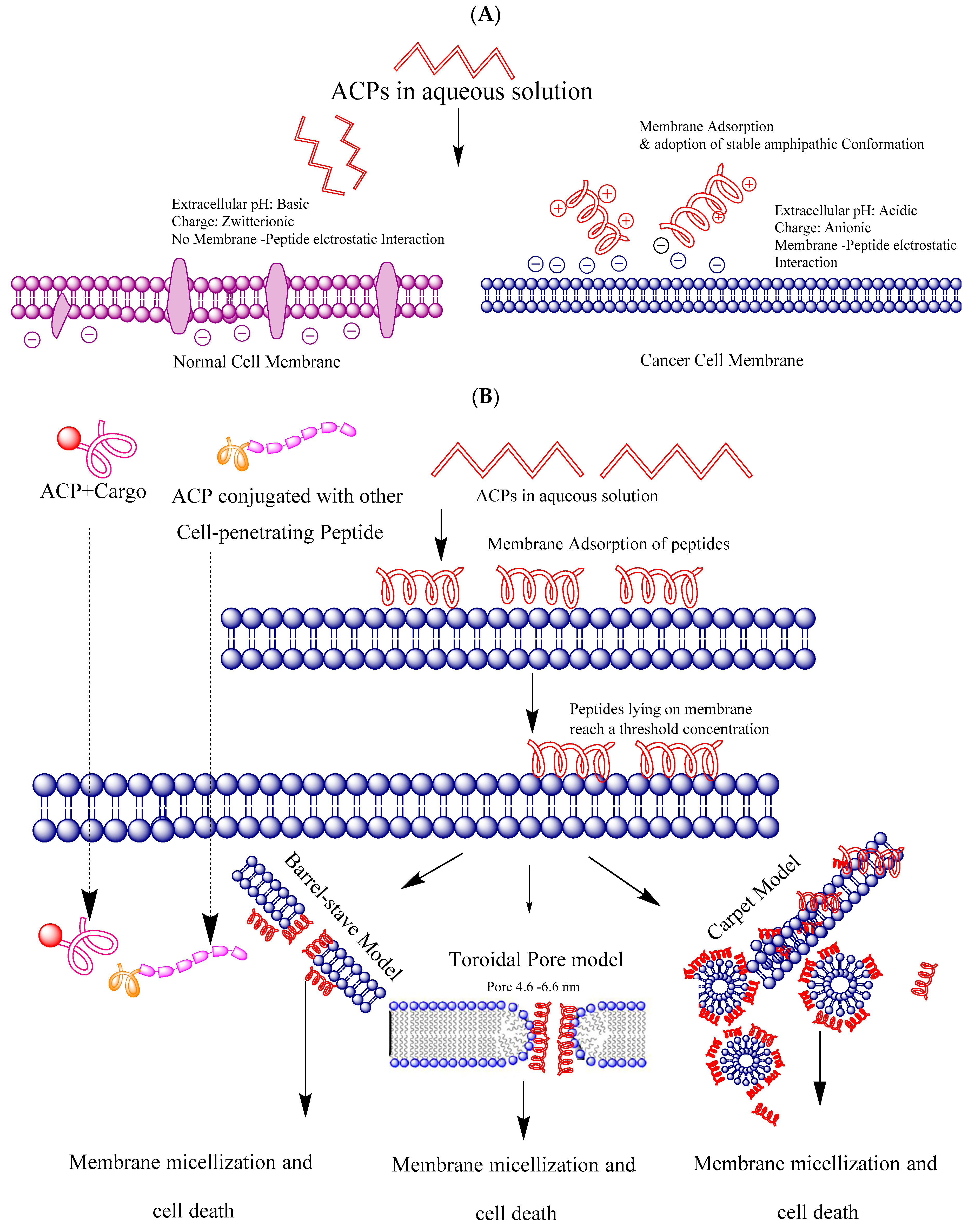
Non-natural amino acids contain structural and biochemical properties that are unique to them and are not present in naturally occurring amino acids. The introduction of non-natural amino acids and other chemical groups impart conformational flexibility to the peptide which in turn improves their selectivity to cancer cells. The unnatural amino acids also evoke immune responses against the cancer cells. The substitutions of the natural amino acids Ala and Leu with their unnatural analogs β-alanine and nor-Leu make them unrecognizable by the proteolytic enzymes in the body which increases their half-life and bioavailability [108]. Tørfoss et al. showed that the cyclization of short peptides improves the anti-cancer activity of peptides [109]. They showed that the IC50 values of cyclic peptides reduce drastically against the Ramos cancer cells as opposed to their linear derivatives. Another simple change to design anti-cancer peptides with high therapeutic indexes is the introduction of d-amino acids. Substituting the l-amino acids with the corresponding d-amino acids results in structural alterations which make the peptide less hemolytic and cytotoxic to normal cells and also significantly improve the proteolytic stability of the peptide [110]. 1,2,3,4-tetrahydroisoquinoline-3-carboxylic acid, also known as Tic falls within the category of non-natural chiral α–amino acids (Figure 2). Tic can be used to design peptides that can be used for their dual role of Anti-cancer and immunomodulatory properties. Owing to the distinctive geometrical conformation, it can be used as a substitute for aromatic amino acids like tyrosine and phenylalanine and the imino acid proline to design conformationally constrained analogs [111]. Besides that, the percent hydrophobicity of Tic is more than that of proline hence it can be used to enhance the percent hydrophobicity of the peptide, which is an important biophysical parameter for biological activity. Octahydroindole-2-carboxylic acid (Oic) is another α-amino acid having a bicyclic structure and can be used instead of proline to impart rigidity to the peptide backbone. Oic is more lipophilic than proline and hence its introduction in the peptide design may improve the absorption and distribution through the cell membranes [112]. Amnoisobutyric acid, (Aib) is known to induce helical structures in peptides [113]. Since helicity is an important property of many anticancer and immunomodulatory peptides, it can be used at strategic positions in place of classical amino acids to design anti-cancer and immunomodulatory peptides. Alanine and leucine residues are very common in ACPs. However, to improve the activities, both amino acids can be substituted with 1-aminocyclohexane carboxylic acid (A6c)/A5c. Similarly, 2,4-diaminobutanoic acid/2,3-diaminopropionic acid can be used for lysine to design less cytotoxic analogs with improved biological activities [94,114]. It is worth mentioning that the design of amino acid analogs is as important as designing the biologically active anti-cancer peptides [115,116]. The introduction of non-natural amino acids readily improves the anti-cancer activity, which can help to speed up the translation of ACPs to clinical settings.

4. Cell Selectivity

The difference in the membrane composition of a cancer cell versus a normal cell plays a very crucial role to dictate the membrane binding and consequent anti-cancer activity of cationic amphiphilic peptides. In particular, membranes of different cancer cells contain phosphatidylserine, sialic acid-containing lipids and proteins, and heparan sulfate, which impart an overall anionic nature to it. Contrary to it, the non-cancerous cells are zwitterionic in nature due to the presence of phosphatidylcholine and sphingomyelin [117] (Figure 3A). The cell surface of almost all the cancer cell membranes displays phosphatidylserine. The latter is present in the inner leaflet of the lipid bilayer in normal cells in sharp contrast to the cancer cells. This has led to the development of the idea that phosphatidylserine could be used as a diagnostic cancer marker [118]. Apart from membrane composition differences, microvilli surface area is also seen to be high in cancer cells which could also be potential targets of cationic amphiphilic peptides [119,120]. Leon et al. while working on a synthetic ACP viz. HB43 also called FLAK50 (FAKLLAKLAKKLL) showed that phosphatidylserine (PS) plays an important role in the cell selectivity of the peptide in distinguishing a cancerous cell from a non-cancer cell [121]. Using various biophysical and in silico approaches the authors show that lysine side chains of HB43 and the carboxylate group of phosphatidylserine catalyze the alpha-helical conformation that facilitates its internalization in cancer cells. Membrane permeabilization assays demonstrated that the peptide-membrane interaction may lead to the destabilization of PS-containing vesicles with respect to PC-containing ones, which were used as non-cancerous membrane mimetic vesicles. What did the authors not investigate could be the immunomodulatory activity of the peptide.HB43 is a leucine-lysine rich peptide. Rosenfeld et al. and Azmi et al. have shown that such peptides can have a strong ability to inhibit the LPS-mediated inflammation by directly binding to the peptide [122,123]. Electrostatic interaction between the anionic cancer cell membrane and the anti-cancer peptides is a well-established and widely studied mechanism of APCs. This is the first interaction that occurs between the two. Koo et al. while studying the biophysical characterization of LTX-315 (K-K-W-W-K-K-W-Dip-K-NH2) Anticancer Peptide found that electrostatic interactions were the main mechanism for the peptide’s anti-cancer activity. Their results showed that the cationic LTX-315 peptide selectively disrupted negatively charged phospholipid membranes to a greater extent than zwitterionic or positively charged phospholipid membranes [124]. Camilio et al. harnessed the immunomodulatory activity of the LTX-315 peptide by using it in combination with doxorubicin [125]. LTX-315 displayed a strong additive antitumoral effect in combination with doxorubicin and induced immune-mediated changes in the tumor microenvironment. Their results displayed a complete regression of breast tumors grown from 4T1 cells in the majority of animals treated. Furthermore, imaging techniques and histological examination showed that the combination induced strong local necrosis, followed by an increase in the infiltration of CD4+ and CD8+ immune cells into the tumor parenchymal tissue. In an independent study, Sveinbjornsson et al. showed that LTX-315 induces ICD through its membranolytic mode of action, leading to the release of potent immunostimulants in addition to a wide spectrum of tumor antigens, thus creating an essential premise for tumor-specific immune responses [126]. Intratumoral treatment with LTX-315 resulted in complete regression of orthotopic B16 melanomas in 80% of animals [126].

5. Mechanism of Membrane Targeting and Entry to the Cell

All classes of Anti-cancer peptides interact differently with the cancer cell membrane. However, in all the models suggested, either the peptides form a pore through which the cytoplasmic content eludes out, or they can directly penetrate and enter the cell, i.e., the cell-penetrating peptides (Figure 3B). The following are four different mechanisms that facilitate the entry of ACPs.

5.1. Barrel-Stave Model

Ehrenstein & Lecar proposed the ‘barrel-stave’ model in 1977 [127]. ACPs reach the cancer cell membrane and accumulate as monomers on the surface, which then oligomerizes because it is energetically unfavorable for a single amphipathic α-helix or β-sheet to transverse the membrane as a monomer and after that, it forms pores followed by the formation a ring-like pattern on membrane exteriors. Later they align perpendicularly to the membrane. In their perpendicular arrangement, the peptides begin to insert into the lipid core of the cell membrane resulting in a shape of a barrel whose staves are the α-helix or β-sheet of peptides. In this model, the membrane is neither deformed nor bent during the precise drilling like the insertion process by ACPs. Alamethicin is known to respond to this model.

5.2. The Carpet Model

Pouny et al. proposed the carpet model in which the peptides aggregate onto the bilayer surface in a parallel manner by keeping their hydrophobic surfaces aligned towards the target cell membranes and they maintain their parallel alignment to the membrane throughout the process [128]. ACPs then intercalate to the membrane in a detergent-like manner and leading the membrane to break into small pockets and micelles. In contrast to the barrel stave and toroidal pore model, no particular pore-forming stage is evidenced in the carpet mechanism, and peptides practically never insert into membranes. Dermaseptin and LL-37 peptides are known to follow this model [129,130].

5.3. Toroidal Pore Model

This model has been studied in some antimicrobial peptides viz. magainin II, protegrin-1 and cecropin A [131]. A primary difference between the toroidal pore and barrel-stave models is that in the toroid pore, lipids are intercalated with peptides in the transmembrane channel. In this model, peptides in the extracellular environment take an α-helical structure as they interact with the anionic and hydrophobic cancer cell membrane. The bound peptides create a breach in the membrane and induce a continuous bend resulting in the formation of toroidal pores.

5.4. Cell-Penetrating Mechanism

These are the class of anti-cancer peptides that do not operate at the membrane level. Instead, they are internalized through the plasma membrane and affect the enzymatic activity and intracellular targets such as DNA and RNA [132]. In addition to being rich in histidine, lysine and arginine, the cell-penetrating peptides are commonly known to have a lipophilic and hydrophilic tail that facilitates their translocation across the membrane [133]. Many researchers believe that the cell-penetrating peptides can be used to transport cargo into the cells and can be harnessed for their target delivery. There are web servers to predict the cell-penetrating peptides [134].

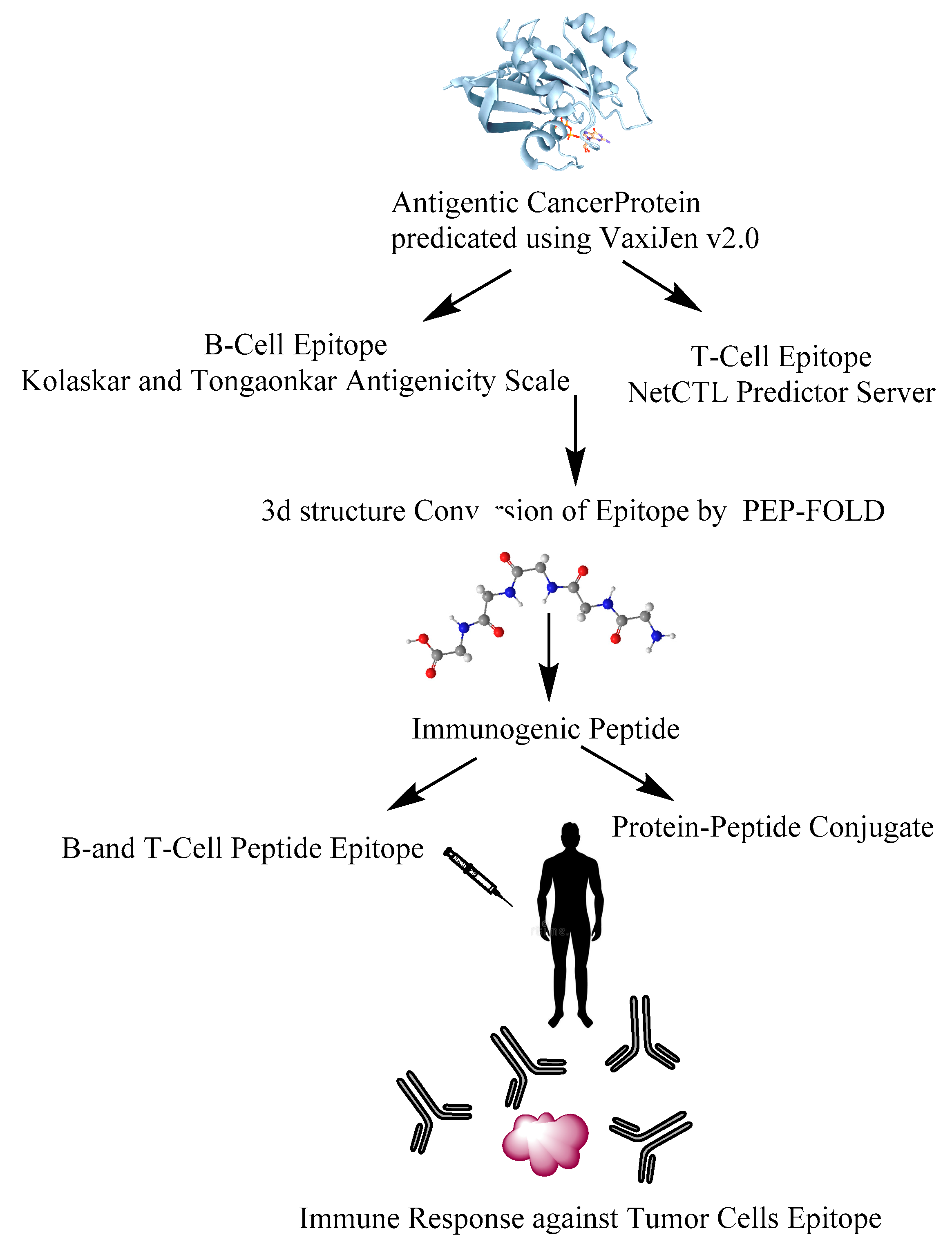
6. Design of Anti-Cancer Peptides as Vaccines to Influence the Immune System

Due to the complex pathophysiology of cancer, the development of peptide vaccines has always been a challenge [135]. However, various in silico approaches have been made to design anti-cancer peptide vaccines. To design the anti-cancer peptide vaccines, the initial step is to identify the antigenicity of the target protein. This can be done by utilizing softwares like ANTIGENpro or VaxiJen [136,137]. It has been observed that the former is more specific and precise than VaxiJen and the resultant probability indicated high antigenicity of the protein [136]. The B-cell epitopes on protein antigens can be determined by using Kolaskar and Tongaonkar antigenicity scale [138]. T-cell epitopes can be selected by NetCTL prediction server [139]. The three-dimensional structure of the epitope can be predicted by PEP-FOLD [140]. The immunogenic peptide thus created might be used to mount an immune response against the tumor cell epitopes. (Figure 4). The use of combinatorial technologies such as using page display libraries can also be utilized to identify peptide molecules that can bind to receptors on the cancer cell surface for cell internalization [141,142,143]. The molecules selected in this way can be subsequently synthesized and modified to obtain peptide drugs with high affinity for the target molecule. In one approach, targets associated with inflammation can be used to screen out therapeutic peptides from a random phage library (usually Ph. D. -7 library) without making a phage library. Another method involves constructing the phage that displays candidate peptides on the surface. After that, an affinity selection technique termed biopanning can be used to select peptides that bind to a given target of interest.

7. Limitations of Anti-Cancer Peptides with Immunomodulatory Activity and Plausible Resolution

There has been promising development in the field of cancer to push anti-cancer peptides as the lead molecules in cancer therapeutics. Most of the anti-cancer peptides preferably bind to the cancer cells more than normal cells due to their cationic nature. However, the observed anti-cancer effects of the peptides depend mostly on compositional differences between cancer and non-cancer cells, where the former frequently display higher content of negatively charged lipids and other membrane components, such as phosphatidylserine and gangliosides, which result in higher peptide binding and membrane insertion, in turn destabilizing such membranes. The short plasma life of peptides is another drawback that hinders the clinical translation of anti-cancer peptides. Like cancer cell membranes, there are many anionic compounds present in blood that can suppress the anti-cancer activity of the peptides. Naturally occurring peptides like melittin, chrysophsin, etc. are equally cytotoxic to both normal and cancer cells [47,144]. In addition to this, many anticancer peptides work at higher concentration ranges and hence chemical modifications are needed to lower their activity concentration, which increases their cost of production.
Hence, more peptide analogs need to be made that could retain the anti-cancer activity and display low cytotoxicity to normal cells. An emphasis should be given to the synthesis and incorporation of non-natural amino acids to increase the plasma life of the designed peptides. More emphasis needs to be given to synthesizing peptides using conjugations. A combined effort to create more open peptide databases should also be made to know the amino acid compositions that make bioactive peptides. It will also reduce the “trial and error effort” to a large extent. This also underlines the need to perform more in vivo experiments on more complex animal models to accurately evaluate the true potential of anti-cancer peptides.

8. Summary and Concluding Remarks

During COVID-19 pandemic, antiviral peptides became a center of attraction due to the development of peptide vaccines targeted against SARS-CoV-2 [145,146]. Although the pandemic is mostly over, however it emphasized the need for harnessing the potential of therapeutic peptides. Diseases like cancer are always a challenge and its deep connection to inflammation provides an opportunity to harness peptides as both anti-cancer and immunomodulatory agents. A combined and collaborative approach of the latest technologies such as immunoinformatic characterization, computer-assisted drug design (CADD) and epitope-based design along with the PK/PD experiments and animal studies can help in the development of new peptide molecules. Such rationally designed peptides can target both the inflammatory and the cancer nodes of the disease and translate to clinical settings.

Author Contributions

A.K.T. wrote the original draft. J.K.V. supervision and scientific edits. All authors have read and agreed to the published version of the manuscript.

Funding

This research received no external funding.

Institutional Review Board Statement

Not applicable.

Informed Consent Statement

Not applicable.

Data Availability Statement

The data supporting the findings of this study are available from the corresponding authors upon reasonable request.

Acknowledgments

Amit Kumar Tripathi acknowledge his Ph.D. supervisor Jimut Kanti Ghosh for his guidance.

Conflicts of Interest

The authors declare no conflict of interest.

References

  1. Byun, J.S.; Gardner, K. Wounds that will not heal: Pervasive cellular reprogramming in cancer. Am. J. Pathol. 2013, 182, 1055–1064. [Google Scholar] [CrossRef] [PubMed] [Green Version]
  2. Mitchison, T.J. The proliferation rate paradox in antimitotic chemotherapy. Mol. Biol. Cell 2012, 23, 1–6. [Google Scholar] [CrossRef]
  3. Liu, B.; Ezeogu, L.; Zellmer, L.; Yu, B.; Xu, N.; Joshua Liao, D. Protecting the normal in order to better kill the cancer. Cancer Med. 2015, 4, 1394–1403. [Google Scholar] [CrossRef] [PubMed]
  4. Wirsdorfer, F.; de Leve, S.; Jendrossek, V. Combining Radiotherapy and Immunotherapy in Lung Cancer: Can We Expect Limitations Due to Altered Normal Tissue Toxicity? Int. J. Mol. Sci. 2018, 20, 24. [Google Scholar] [CrossRef] [Green Version]
  5. Lara, O.D.; Krishnan, S.; Wang, Z.; Corvigno, S.; Zhong, Y.; Lyons, Y.; Dood, R.; Hu, W.; Qi, L.; Liu, J.; et al. Tumor core biopsies adequately represent immune microenvironment of high-grade serous carcinoma. Sci. Rep. 2019, 9, 17589. [Google Scholar] [CrossRef] [Green Version]
  6. Coussens, L.M.; Werb, Z. Inflammation and cancer. Nature 2002, 420, 860–867. [Google Scholar] [CrossRef] [PubMed]
  7. Gonzalez, H.; Hagerling, C.; Werb, Z. Roles of the immune system in cancer: From tumor initiation to metastatic progression. Genes Dev. 2018, 32, 1267–1284. [Google Scholar] [CrossRef] [Green Version]
  8. Greenwood, H.C.; Bloom, S.R.; Murphy, K.G. Peptides and their potential role in the treatment of diabetes and obesity. Rev. Diabet. Stud. 2011, 8, 355–368. [Google Scholar] [CrossRef] [PubMed] [Green Version]
  9. Grieco, P.; Gomez-Monterrey, I. Natural and synthetic peptides in the cardiovascular diseases: An update on diagnostic and therapeutic potentials. Arch. Biochem. Biophys. 2019, 662, 15–32. [Google Scholar] [CrossRef] [PubMed]
  10. Naeimi, R.; Bahmani, A.; Afshar, S. Investigating the role of peptides in effective therapies against cancer. Cancer Cell Int. 2022, 22, 139. [Google Scholar] [CrossRef]
  11. Wang, L.; Wang, N.; Zhang, W.; Cheng, X.; Yan, Z.; Shao, G.; Wang, X.; Wang, R.; Fu, C. Therapeutic peptides: Current applications and future directions. Signal Transduct. Target Ther. 2022, 7, 48. [Google Scholar] [CrossRef] [PubMed]
  12. Recio, C.; Maione, F.; Iqbal, A.J.; Mascolo, N.; De Feo, V. The Potential Therapeutic Application of Peptides and Peptidomimetics in Cardiovascular Disease. Front. Pharm. 2016, 7, 526. [Google Scholar] [CrossRef] [PubMed] [Green Version]
  13. Lau, J.L.; Dunn, M.K. Therapeutic peptides: Historical perspectives, current development trends, and future directions. Bioorg. Med. Chem. 2018, 26, 2700–2707. [Google Scholar] [CrossRef] [PubMed]
  14. Al Shaer, D.; Al Musaimi, O.; Albericio, F.; de la Torre, B.G. 2021 FDA TIDES (Peptides and Oligonucleotides) Harvest. Pharm 2022, 15, 222. [Google Scholar] [CrossRef] [PubMed]
  15. Cheng, T.F.; Wang, J.D.; Uen, W.C. Cost-utility analysis of adjuvant goserelin (Zoladex) and adjuvant chemotherapy in premenopausal women with breast cancer. BMC Cancer 2012, 12, 33. [Google Scholar] [CrossRef] [PubMed] [Green Version]
  16. Cockshott, I.D. Clinical pharmacokinetics of goserelin. Clin. Pharm. 2000, 39, 27–48. [Google Scholar] [CrossRef]
  17. Swayzer, D.V.; Gerriets, V. Leuprolide; StatPearls: Treasure Island, FL, USA, 2022. [Google Scholar]
  18. Guzman-Soto, I.; Salinas, E.; Quintanar, J.L. Leuprolide Acetate Inhibits Spinal Cord Inflammatory Response in Experimental Autoimmune Encephalomyelitis by Suppressing NF-kappaB Activation. Neuroimmunomodulation 2016, 23, 33–40. [Google Scholar] [CrossRef]
  19. Katai, M.; Sakurai, A.; Inaba, H.; Ikeo, Y.; Yamauchi, K.; Hashizume, K. Octreotide as a rapid and effective painkiller for metastatic carcinoid tumor. Endocr. J. 2005, 52, 277–280. [Google Scholar] [CrossRef] [Green Version]
  20. Theodoropoulou, M.; Zhang, J.; Laupheimer, S.; Paez-Pereda, M.; Erneux, C.; Florio, T.; Pagotto, U.; Stalla, G.K. Octreotide, a somatostatin analogue, mediates its antiproliferative action in pituitary tumor cells by altering phosphatidylinositol 3-kinase signaling and inducing Zac1 expression. Cancer Res. 2006, 66, 1576–1582. [Google Scholar] [CrossRef] [Green Version]
  21. Huang, Y.B.; He, L.Y.; Jiang, H.Y.; Chen, Y.X. Role of helicity on the anticancer mechanism of action of cationic-helical peptides. Int. J. Mol. Sci. 2012, 13, 6849–6862. [Google Scholar] [CrossRef]
  22. Kumari, T.; Verma, D.P.; Kuldeep, J.; Dhanabal, V.B.; Verma, N.K.; Sahai, R.; Tripathi, A.K.; Saroj, J.; Ali, M.; Mitra, K.; et al. 10-Residue MyD88-Peptide Adopts beta-Sheet Structure, Self-Assembles, Binds to Lipopolysaccharides, and Rescues Mice from Endotoxin-Mediated Lung-Infection and Death. ACS Chem. Biol. 2022, 75, 2431–2446. [Google Scholar] [CrossRef]
  23. Tandon, A.; Harioudh, M.K.; Ishrat, N.; Tripathi, A.K.; Srivastava, S.; Ghosh, J.K. An MD2-derived peptide promotes LPS aggregation, facilitates its internalization in THP-1 cells, and inhibits LPS-induced pro-inflammatory responses. Cell. Mol. Life Sci. 2018, 75, 2431–2446. [Google Scholar] [CrossRef] [PubMed]
  24. Chen, X.; Zou, X.; Qi, G.; Tang, Y.; Guo, Y.; Si, J.; Liang, L. Roles and Mechanisms of Human Cathelicidin LL-37 in Cancer. Cell Physiol. Biochem. 2018, 47, 1060–1073. [Google Scholar] [CrossRef] [PubMed]
  25. Liu, S.; Yang, H.; Wan, L.; Cai, H.W.; Li, S.F.; Li, Y.P.; Cheng, J.Q.; Lu, X.F. Enhancement of cytotoxicity of antimicrobial peptide magainin II in tumor cells by bombesin-targeted delivery. Acta Pharm. Sin. 2011, 32, 79–88. [Google Scholar] [CrossRef]
  26. Ceremuga, M.; Stela, M.; Janik, E.; Gorniak, L.; Synowiec, E.; Sliwinski, T.; Sitarek, P.; Saluk-Bijak, J.; Bijak, M. Melittin-A Natural Peptide from Bee Venom Which Induces Apoptosis in Human Leukaemia Cells. Biomolecules 2020, 10, 247. [Google Scholar] [CrossRef] [PubMed] [Green Version]
  27. Lee, G.; Bae, H. Anti-Inflammatory Applications of Melittin, a Major Component of Bee Venom: Detailed Mechanism of Action and Adverse Effects. Molecules 2016, 21, 616. [Google Scholar] [CrossRef] [Green Version]
  28. Tipgomut, C.; Wongprommoon, A.; Takeo, E.; Ittiudomrak, T.; Puthong, S.; Chanchao, C. Melittin Induced G1 Cell Cycle Arrest and Apoptosis in Chago-K1 Human Bronchogenic Carcinoma Cells and Inhibited the Differentiation of THP-1 Cells into Tumour- Associated Macrophages. Asian Pac. J. Cancer Prev. 2018, 19, 3427–3434. [Google Scholar] [CrossRef] [Green Version]
  29. Kong, G.M.; Tao, W.H.; Diao, Y.L.; Fang, P.H.; Wang, J.J.; Bo, P.; Qian, F. Melittin induces human gastric cancer cell apoptosis via activation of mitochondrial pathway. World J. Gastroenterol. 2016, 22, 3186–3195. [Google Scholar] [CrossRef] [Green Version]
  30. Gajski, G.; Domijan, A.M.; Zegura, B.; Stern, A.; Geric, M.; Novak Jovanovic, I.; Vrhovac, I.; Madunic, J.; Breljak, D.; Filipic, M.; et al. Melittin induced cytogenetic damage, oxidative stress and changes in gene expression in human peripheral blood lymphocytes. Toxicon 2016, 110, 56–67. [Google Scholar] [CrossRef]
  31. Asthana, N.; Yadav, S.P.; Ghosh, J.K. Dissection of antibacterial and toxic activity of melittin: A leucine zipper motif plays a crucial role in determining its hemolytic activity but not antibacterial activity. J. Biol. Chem. 2004, 279, 55042–55050. [Google Scholar] [CrossRef]
  32. Srivastava, R.M.; Srivastava, S.; Singh, M.; Bajpai, V.K.; Ghosh, J.K. Consequences of alteration in leucine zipper sequence of melittin in its neutralization of lipopolysaccharide-induced proinflammatory response in macrophage cells and interaction with lipopolysaccharide. J. Biol. Chem. 2012, 287, 1980–1995. [Google Scholar] [CrossRef] [Green Version]
  33. Liu, M.; Wang, H.; Liu, L.; Wang, B.; Sun, G. Melittin-MIL-2 fusion protein as a candidate for cancer immunotherapy. J. Transl. Med. 2016, 14, 155. [Google Scholar] [CrossRef] [Green Version]
  34. Zasloff, M. Magainins, a class of antimicrobial peptides from Xenopus skin: Isolation, characterization of two active forms, and partial cDNA sequence of a precursor. Proc. Natl. Acad. Sci. USA 1987, 84, 5449–5453. [Google Scholar] [CrossRef] [PubMed] [Green Version]
  35. Shin, S.Y.; Lee, M.K.; Kim, K.L.; Hahm, K.S. Structure-antitumor and hemolytic activity relationships of synthetic peptides derived from cecropin A-magainin 2 and cecropin A-melittin hybrid peptides. J. Pept. Res. 1997, 50, 279–285. [Google Scholar] [CrossRef]
  36. Ryu, S.; Choi, S.Y.; Acharya, S.; Chun, Y.J.; Gurley, C.; Park, Y.; Armstrong, C.A.; Song, P.I.; Kim, B.J. Antimicrobial and anti-inflammatory effects of Cecropin A(1-8)-Magainin2(1-12) hybrid peptide analog p5 against Malassezia furfur infection in human keratinocytes. J. Investig. Derm. 2011, 131, 1677–1683. [Google Scholar] [CrossRef] [PubMed] [Green Version]
  37. Tang, C.; Shao, X.; Sun, B.; Huang, W.; Qiu, F.; Chen, Y.; Shi, Y.K.; Zhang, E.Y.; Wang, C.; Zhao, X. Anticancer mechanism of peptide P18 in human leukemia K562 cells. Org. Biomol. Chem. 2010, 8, 984–987. [Google Scholar] [CrossRef]
  38. Nan, Y.H.; Jeon, Y.J.; Park, I.S.; Shin, S.Y. Antimicrobial peptide P18 inhibits inflammatory responses by LPS- but not by IFN-gamma-stimulated macrophages. Biotechnol. Lett. 2008, 30, 1183–1187. [Google Scholar] [CrossRef]
  39. Almeida, J.R.; Mendes, B.; Lancellotti, M.; Franchi, G.C., Jr.; Passos, O.; Ramos, M.J.; Fernandes, P.A.; Alves, C.; Vale, N.; Gomes, P.; et al. Lessons from a Single Amino Acid Substitution: Anticancer and Antibacterial Properties of Two Phospholipase A2-Derived Peptides. Curr. Issues Mol. Biol. 2021, 44, 46–62. [Google Scholar] [CrossRef] [PubMed]
  40. Arias, M.; Haney, E.F.; Hilchie, A.L.; Corcoran, J.A.; Hyndman, M.E.; Hancock, R.E.W.; Vogel, H.J. Selective anticancer activity of synthetic peptides derived from the host defence peptide tritrpticin. Biochim. Biophys. Acta Biomembr. 2020, 1862, 183228. [Google Scholar] [CrossRef]
  41. Arias, M.; Piga, K.B.; Hyndman, M.E.; Vogel, H.J. Improving the Activity of Trp-Rich Antimicrobial Peptides by Arg/Lys Substitutions and Changing the Length of Cationic Residues. Biomolecules 2018, 8, 19. [Google Scholar] [CrossRef]
  42. Ghiselli, R.; Giacometti, A.; Cirioni, O.; Mocchegiani, F.; Orlando, F.; Silvestri, C.; Di Matteo, F.; Abbruzzetti, A.; Scalise, G.; Saba, V. Efficacy of the bovine antimicrobial peptide indolicidin combined with piperacillin/tazobactam in experimental rat models of polymicrobial peritonitis. Crit. Care Med. 2008, 36, 240–245. [Google Scholar] [CrossRef] [PubMed]
  43. Zhang, Q.T.; Liu, Z.D.; Wang, Z.; Wang, T.; Wang, N.; Wang, N.; Zhang, B.; Zhao, Y.F. Recent Advances in Small Peptides of Marine Origin in Cancer Therapy. Mar. Drugs 2021, 19, 115. [Google Scholar] [CrossRef] [PubMed]
  44. Kang, H.K.; Choi, M.C.; Seo, C.H.; Park, Y. Therapeutic Properties and Biological Benefits of Marine-Derived Anticancer Peptides. Int. J. Mol. Sci. 2018, 19, 919. [Google Scholar] [CrossRef] [Green Version]
  45. Iijima, N.; Tanimoto, N.; Emoto, Y.; Morita, Y.; Uematsu, K.; Murakami, T.; Nakai, T. Purification and characterization of three isoforms of chrysophsin, a novel antimicrobial peptide in the gills of the red sea bream, Chrysophrys major. Eur. J. Biochem. 2003, 270, 675–686. [Google Scholar] [CrossRef]
  46. Hsu, J.C.; Lin, L.C.; Tzen, J.T.; Chen, J.Y. Characteristics of the antitumor activities in tumor cells and modulation of the inflammatory response in RAW264.7 cells of a novel antimicrobial peptide, chrysophsin-1, from the red sea bream (Chrysophrys major). Peptides 2011, 32, 900–910. [Google Scholar] [CrossRef] [PubMed]
  47. Tripathi, A.K.; Kumari, T.; Harioudh, M.K.; Yadav, P.K.; Kathuria, M.; Shukla, P.K.; Mitra, K.; Ghosh, J.K. Identification of GXXXXG motif in Chrysophsin-1 and its implication in the design of analogs with cell-selective antimicrobial and anti-endotoxin activities. Sci. Rep. 2017, 7, 3384. [Google Scholar] [CrossRef] [PubMed] [Green Version]
  48. Rinaldi, A.C.; Mangoni, M.L.; Rufo, A.; Luzi, C.; Barra, D.; Zhao, H.; Kinnunen, P.K.; Bozzi, A.; Di Giulio, A.; Simmaco, M. Temporin L: Antimicrobial, haemolytic and cytotoxic activities, and effects on membrane permeabilization in lipid vesicles. Biochem. J. 2002, 368, 91–100. [Google Scholar] [CrossRef] [Green Version]
  49. Swithenbank, L.; Cox, P.; Harris, L.G.; Dudley, E.; Sinclair, K.; Lewis, P.; Cappiello, F.; Morgan, C. Temporin A and Bombinin H2 Antimicrobial Peptides Exhibit Selective Cytotoxicity to Lung Cancer Cells. Science 2020, 2020, 3526286. [Google Scholar] [CrossRef]
  50. Srivastava, S.; Kumar, A.; Tripathi, A.K.; Tandon, A.; Ghosh, J.K. Modulation of anti-endotoxin property of Temporin L by minor amino acid substitution in identified phenylalanine zipper sequence. Biochem. J. 2016, 473, 4045–4062. [Google Scholar] [CrossRef]
  51. Giacometti, A.; Cirioni, O.; Ghiselli, R.; Mocchegiani, F.; Orlando, F.; Silvestri, C.; Bozzi, A.; Di Giulio, A.; Luzi, C.; Mangoni, M.L.; et al. Interaction of antimicrobial peptide temporin L with lipopolysaccharide in vitro and in experimental rat models of septic shock caused by gram-negative bacteria. Antimicrob. Agents Chemother. 2006, 50, 2478–2486. [Google Scholar] [CrossRef]
  52. Tripathi, A.K.; Kumari, T.; Tandon, A.; Sayeed, M.; Afshan, T.; Kathuria, M.; Shukla, P.K.; Mitra, K.; Ghosh, J.K. Selective phenylalanine to proline substitution for improved antimicrobial and anticancer activities of peptides designed on phenylalanine heptad repeat. Acta. Biomater. 2017, 57, 170–186. [Google Scholar] [CrossRef]
  53. Alvarez, C.A.; Santana, P.A.; Salinas-Parra, N.; Beltran, D.; Guzman, F.; Vega, B.; Acosta, F.; Mercado, L. Immune Modulation Ability of Hepcidin from Teleost Fish. Animals 2022, 12, 1586. [Google Scholar] [CrossRef] [PubMed]
  54. Conrad, D.M.; Hilchie, A.L.; McMillan, K.A.M.; Liwski, R.S.; Hoskin, D.W.; Power Coombs, M.R. The Acute Phase Protein Hepcidin Is Cytotoxic to Human and Mouse Myeloma Cells. Anticancer Res. 2021, 41, 601–608. [Google Scholar] [CrossRef] [PubMed]
  55. Piktel, E.; Niemirowicz, K.; Wnorowska, U.; Watek, M.; Wollny, T.; Gluszek, K.; Gozdz, S.; Levental, I.; Bucki, R. The Role of Cathelicidin LL-37 in Cancer Development. Arch. Immunol. Ther. Exp. (Warsz) 2016, 64, 33–46. [Google Scholar] [CrossRef] [PubMed] [Green Version]
  56. Ren, S.X.; Cheng, A.S.; To, K.F.; Tong, J.H.; Li, M.S.; Shen, J.; Wong, C.C.; Zhang, L.; Chan, R.L.; Wang, X.J.; et al. Host immune defense peptide LL-37 activates caspase-independent apoptosis and suppresses colon cancer. Cancer Res. 2012, 72, 6512–6523. [Google Scholar] [CrossRef] [Green Version]
  57. Porter, R.J.; Murray, G.I.; Alnabulsi, A.; Humphries, M.P.; James, J.A.; Salto-Tellez, M.; Craig, S.G.; Wang, J.M.; Yoshimura, T.; McLean, M.H. Colonic epithelial cathelicidin (LL-37) expression intensity is associated with progression of colorectal cancer and presence of CD8(+) T cell infiltrate. J. Pathol. Clin. Res. 2021, 7, 495–506. [Google Scholar] [CrossRef]
  58. Tuomela, J.M.; Sandholm, J.A.; Kaakinen, M.; Hayden, K.L.; Haapasaari, K.M.; Jukkola-Vuorinen, A.; Kauppila, J.H.; Lehenkari, P.P.; Harris, K.W.; Graves, D.E.; et al. Telomeric G-quadruplex-forming DNA fragments induce TLR9-mediated and LL-37-regulated invasion in breast cancer cells in vitro. Breast. Cancer Res. Treat. 2016, 155, 261–271. [Google Scholar] [CrossRef]
  59. Ren, S.X.; Shen, J.; Cheng, A.S.; Lu, L.; Chan, R.L.; Li, Z.J.; Wang, X.J.; Wong, C.C.; Zhang, L.; Ng, S.S.; et al. FK-16 derived from the anticancer peptide LL-37 induces caspase-independent apoptosis and autophagic cell death in colon cancer cells. PLoS ONE 2013, 8, e63641. [Google Scholar] [CrossRef] [Green Version]
  60. Scott, M.G.; Davidson, D.J.; Gold, M.R.; Bowdish, D.; Hancock, R.E. The human antimicrobial peptide LL-37 is a multifunctional modulator of innate immune responses. J. Immunol. 2002, 169, 3883–3891. [Google Scholar] [CrossRef] [Green Version]
  61. Chiangjong, W.; Chutipongtanate, S.; Hongeng, S. Anticancer peptide: Physicochemical property, functional aspect and trend in clinical application (Review). Int. J. Oncol. 2020, 57, 678–696. [Google Scholar] [CrossRef]
  62. Wodlej, C.; Riedl, S.; Rinner, B.; Leber, R.; Drechsler, C.; Voelker, D.R.; Choi, J.Y.; Lohner, K.; Zweytick, D. Interaction of two antitumor peptides with membrane lipids—Influence of phosphatidylserine and cholesterol on specificity for melanoma cells. PLoS ONE 2019, 14, e0211187. [Google Scholar] [CrossRef] [Green Version]
  63. Cheng, M.H.; Pan, C.Y.; Chen, N.F.; Yang, S.N.; Hsieh, S.; Wen, Z.H.; Chen, W.F.; Wang, J.W.; Lu, W.H.; Kuo, H.M. Piscidin-1 Induces Apoptosis via Mitochondrial Reactive Oxygen Species-Regulated Mitochondrial Dysfunction in Human Osteosarcoma Cells. Sci. Rep. 2020, 10, 5045. [Google Scholar] [CrossRef] [PubMed] [Green Version]
  64. Kumar, A.; Tripathi, A.K.; Kathuria, M.; Shree, S.; Tripathi, J.K.; Purshottam, R.K.; Ramachandran, R.; Mitra, K.; Ghosh, J.K. Single Amino Acid Substitutions at Specific Positions of the Heptad Repeat Sequence of Piscidin-1 Yielded Novel Analogs That Show Low Cytotoxicity and In Vitro and In Vivo Antiendotoxin Activity. Antimicrob. Agents Chemother. 2016, 60, 3687–3699. [Google Scholar] [CrossRef] [PubMed] [Green Version]
  65. Adams, P.G.; Lamoureux, L.; Swingle, K.L.; Mukundan, H.; Montano, G.A. Lipopolysaccharide-induced dynamic lipid membrane reorganization: Tubules, perforations, and stacks. Biophys. J. 2014, 106, 2395–2407. [Google Scholar] [CrossRef] [PubMed] [Green Version]
  66. Yamaguchi, Y.; Yamamoto, K.; Sato, Y.; Inoue, S.; Morinaga, T.; Hirano, E. Combination of aspartic acid and glutamic acid inhibits tumor cell proliferation. Biomed. Res. 2016, 37, 153–159. [Google Scholar] [CrossRef] [Green Version]
  67. Liscano, Y.; Onate-Garzon, J.; Delgado, J.P. Peptides with Dual Antimicrobial-Anticancer Activity: Strategies to Overcome Peptide Limitations and Rational Design of Anticancer Peptides. Molecules 2020, 25, 4245. [Google Scholar] [CrossRef]
  68. Bai, D.; Yu, S.; Zhong, S.; Zhao, B.; Qiu, S.; Chen, J.; Lunagariya, J.; Liao, X.; Xu, S. d-Amino Acid Position Influences the Anticancer Activity of Galaxamide Analogs: An Apoptotic Mechanism Study. Int. J. Mol. Sci. 2017, 18, 544. [Google Scholar] [CrossRef] [Green Version]
  69. Barras, D.; Chevalier, N.; Zoete, V.; Dempsey, R.; Lapouge, K.; Olayioye, M.A.; Michielin, O.; Widmann, C. A WXW motif is required for the anticancer activity of the TAT-RasGAP317-326 peptide. J. Biol. Chem. 2014, 289, 23701–23711. [Google Scholar] [CrossRef] [Green Version]
  70. Walrant, A.; Bauza, A.; Girardet, C.; Alves, I.D.; Lecomte, S.; Illien, F.; Cardon, S.; Chaianantakul, N.; Pallerla, M.; Burlina, F.; et al. Ionpair-pi interactions favor cell penetration of arginine/tryptophan-rich cell-penetrating peptides. Biochim. Biophys. Acta Biomembr. 2020, 1862, 183098. [Google Scholar] [CrossRef]
  71. Jobin, M.L.; Blanchet, M.; Henry, S.; Chaignepain, S.; Manigand, C.; Castano, S.; Lecomte, S.; Burlina, F.; Sagan, S.; Alves, I.D. The role of tryptophans on the cellular uptake and membrane interaction of arginine-rich cell penetrating peptides. Biochim. Biophys. Acta 2015, 1848, 593–602. [Google Scholar] [CrossRef]
  72. Tyagi, A.; Kapoor, P.; Kumar, R.; Chaudhary, K.; Gautam, A.; Raghava, G.P. In silico models for designing and discovering novel anticancer peptides. Sci. Rep. 2013, 3, 2984. [Google Scholar] [CrossRef] [PubMed] [Green Version]
  73. Huang, Y.B.; Wang, X.F.; Wang, H.Y.; Liu, Y.; Chen, Y. Studies on mechanism of action of anticancer peptides by modulation of hydrophobicity within a defined structural framework. Mol. Cancer Ther. 2011, 10, 416–426. [Google Scholar] [CrossRef] [PubMed] [Green Version]
  74. Yang, Q.Z.; Wang, C.; Lang, L.; Zhou, Y.; Wang, H.; Shang, D.J. Design of potent, non-toxic anticancer peptides based on the structure of the antimicrobial peptide, temporin-1CEa. Arch. Pharm. Res. 2013, 36, 1302–1310. [Google Scholar] [CrossRef] [PubMed]
  75. Piyadasa, H.; Hemshekhar, M.; Osawa, N.; Lloyd, D.; Altieri, A.; Basu, S.; Krokhin, O.V.; Halayko, A.J.; Mookherjee, N. Disrupting Tryptophan in the Central Hydrophobic Region Selectively Mitigates Immunomodulatory Activities of the Innate Defence Regulator Peptide IDR-1002. J. Med. Chem. 2021, 64, 6696–6705. [Google Scholar] [CrossRef] [PubMed]
  76. Hemshekhar, M.; Faiyaz, S.; Choi, K.G.; Krokhin, O.V.; Mookherjee, N. Immunomodulatory Functions of the Human Cathelicidin LL-37 (aa 13-31)-Derived Peptides are Associated with Predicted alpha-Helical Propensity and Hydrophobic Index. Biomolecules 2019, 9, 501. [Google Scholar] [CrossRef] [PubMed] [Green Version]
  77. Zhao, Y.; Wang, S.; Fei, W.; Feng, Y.; Shen, L.; Yang, X.; Wang, M.; Wu, M. Prediction of Anticancer Peptides with High Efficacy and Low Toxicity by Hybrid Model Based on 3D Structure of Peptides. Int. J. Mol. Sci. 2021, 22, 5630. [Google Scholar] [CrossRef]
  78. Kozlowski, M.R.; Kozlowski, R.E. A novel, small peptide with activity against human pancreatic cancer. Am. J. Cancer Res. 2020, 10, 1356–1365. [Google Scholar]
  79. Ohtake, T.; Fujimoto, Y.; Ikuta, K.; Saito, H.; Ohhira, M.; Ono, M.; Kohgo, Y. Proline-rich antimicrobial peptide, PR-39 gene transduction altered invasive activity and actin structure in human hepatocellular carcinoma cells. Br. J. Cancer 1999, 81, 393–403. [Google Scholar] [CrossRef] [Green Version]
  80. Ding, X.; Bian, D.; Li, W.; Xie, Y.; Li, X.; Lv, J.; Tang, R. Host defense peptide LL-37 is involved in the regulation of cell proliferation and production of pro-inflammatory cytokines in hepatocellular carcinoma cells. Amino. Acids. 2021, 53, 471–484. [Google Scholar] [CrossRef]
  81. Agrawal, P.; Bhagat, D.; Mahalwal, M.; Sharma, N.; Raghava, G.P.S. AntiCP 2.0: An updated model for predicting anticancer peptides. Brief. Bioinform. 2021, 22, bbaa153. [Google Scholar] [CrossRef] [PubMed]
  82. Maraming, P.; Klaynongsruang, S.; Boonsiri, P.; Peng, S.F.; Daduang, S.; Rungsa, P.; Tavichakorntrakool, R.; Chung, J.G.; Daduang, J. Anti-metastatic Effects of Cationic KT2 Peptide (a Lysine/Tryptophan-rich Peptide) on Human Melanoma A375.S2 Cells. In Vivo 2021, 35, 215–227. [Google Scholar] [CrossRef] [PubMed]
  83. Lind, D.S. Arginine and cancer. J. Nutr. 2004, 134, 2837S–2841S, discussion 2853S. [Google Scholar] [CrossRef] [PubMed] [Green Version]
  84. Burdukiewicz, M.; Sidorczuk, K.; Rafacz, D.; Pietluch, F.; Bakala, M.; Slowik, J.; Gagat, P. CancerGram: An Effective Classifier for Differentiating Anticancer from Antimicrobial Peptides. Pharmaceutics 2020, 12, 1045. [Google Scholar] [CrossRef] [PubMed]
  85. Wang, C.; Dong, S.; Zhang, L.; Zhao, Y.; Huang, L.; Gong, X.; Wang, H.; Shang, D. Cell surface binding, uptaking and anticancer activity of L-K6, a lysine/leucine-rich peptide, on human breast cancer MCF-7 cells. Sci. Rep. 2017, 7, 8293. [Google Scholar] [CrossRef] [Green Version]
  86. Chu, H.L.; Yip, B.S.; Chen, K.H.; Yu, H.Y.; Chih, Y.H.; Cheng, H.T.; Chou, Y.T.; Cheng, J.W. Novel antimicrobial peptides with high anticancer activity and selectivity. PLoS ONE 2015, 10, e0126390. [Google Scholar] [CrossRef] [Green Version]
  87. Hadianamrei, R.; Tomeh, M.A.; Brown, S.; Wang, J.; Zhao, X. Rationally designed short cationic alpha-helical peptides with selective anticancer activity. J. Colloid. Interface Sci. 2022, 607, 488–501. [Google Scholar] [CrossRef]
  88. Hadianamrei, R.; Tomeh, M.A.; Brown, S.; Wang, J.; Zhao, X. Correlation between the secondary structure and surface activity of beta-sheet forming cationic amphiphilic peptides and their anticancer activity. Colloids Surf. B Biointerfaces 2022, 209, 112165. [Google Scholar] [CrossRef]
  89. Pan, F.; Li, Y.; Ding, Y.; Lv, S.; You, R.; Hadianamrei, R.; Tomeh, M.A.; Zhao, X. Anticancer effect of rationally designed alpha-helical amphiphilic peptides. Colloids Surf. B Biointerfaces 2022, 220, 112841. [Google Scholar] [CrossRef]
  90. Huang, Y.; Feng, Q.; Yan, Q.; Hao, X.; Chen, Y. Alpha-helical cationic anticancer peptides: A promising candidate for novel anticancer drugs. Mini Rev. Med. Chem. 2015, 15, 73–81. [Google Scholar] [CrossRef]
  91. Bae, S.; Oh, K.; Kim, H.; Kim, Y.; Kim, H.R.; Hwang, Y.I.; Lee, D.S.; Kang, J.S.; Lee, W.J. The effect of alloferon on the enhancement of NK cell cytotoxicity against cancer via the up-regulation of perforin/granzyme B secretion. Immunobiology 2013, 218, 1026–1033. [Google Scholar] [CrossRef]
  92. Bellamy, W.; Takase, M.; Yamauchi, K.; Wakabayashi, H.; Kawase, K.; Tomita, M. Identification of the bactericidal domain of lactoferrin. Biochim. Biophys. Acta 1992, 1121, 130–136. [Google Scholar] [CrossRef]
  93. Guo, F.; Zhang, Y.; Dong, W.; Guan, Y.; Shang, D. Effect of hydrophobicity on distinct anticancer mechanism of antimicrobial peptide chensinin-1b and its lipoanalog PA-C1b in breast cancer cells. Int. J. Biochem. Cell. Biol. 2022, 143, 106156. [Google Scholar] [CrossRef]
  94. Xie, M.; Liu, D.; Yang, Y. Anti-cancer peptides: Classification, mechanism of action, reconstruction and modification. Open. Biol. 2020, 10, 200004. [Google Scholar] [CrossRef]
  95. Pedron, C.N.; Andrade, G.P.; Sato, R.H.; Torres, M.T.; Cerchiaro, G.; Ribeiro, A.O.; Oliveira, V.X., Jr. Anticancer activity of VmCT1 analogs against MCF-7 cells. Chem. Biol. Drug Des. 2018, 91, 588–596. [Google Scholar] [CrossRef]
  96. Uematsu, N.; Matsuzaki, K. Polar angle as a determinant of amphipathic alpha-helix-lipid interactions: A model peptide study. Biophys. J. 2000, 79, 2075–2083. [Google Scholar] [CrossRef] [PubMed] [Green Version]
  97. Delfi, M.; Sartorius, R.; Ashrafizadeh, M.; Sharifi, E.; Zhang, Y.; De Berardinis, P.; Zarrabi, A.; Varma, R.S.; Tay, F.R.; Smith, B.R.; et al. Self-assembled peptide and protein nanostructures for anti-cancer therapy: Targeted delivery, stimuli-responsive devices and immunotherapy. Nano Today 2021, 38, 101119. [Google Scholar] [CrossRef]
  98. Lee, S.; Trinh, T.H.T.; Yoo, M.; Shin, J.; Lee, H.; Kim, J.; Hwang, E.; Lim, Y.B.; Ryou, C. Self-Assembling Peptides and Their Application in the Treatment of Diseases. Int. J. Mol. Sci. 2019, 20, 5850. [Google Scholar] [CrossRef] [Green Version]
  99. Edwards-Gayle, C.J.C.; Hamley, I.W. Self-assembly of bioactive peptides, peptide conjugates, and peptide mimetic materials. Org. Biomol. Chem. 2017, 15, 5867–5876. [Google Scholar] [CrossRef] [PubMed] [Green Version]
  100. Cui, H.; Webber, M.J.; Stupp, S.I. Self-assembly of peptide amphiphiles: From molecules to nanostructures to biomaterials. Biopolymers 2010, 94, 1–18. [Google Scholar] [CrossRef] [Green Version]
  101. Pentlavalli, S.; Coulter, S.; Laverty, G. Peptide Nanomaterials for Drug Delivery Applications. Curr. Protein Pept. Sci. 2020, 21, 401–412. [Google Scholar] [CrossRef] [PubMed]
  102. Liu, J.; Zhang, L.; Yang, Z.; Zhao, X. Controlled release of paclitaxel from a self-assembling peptide hydrogel formed in situ and antitumor study in vitro. Int. J. Nanomed. 2011, 6, 2143–2153. [Google Scholar] [CrossRef]
  103. Han, Y.Y.; Liu, H.Y.; Han, D.J.; Zong, X.C.; Zhang, S.Q.; Chen, Y.Q. Role of glycosylation in the anticancer activity of antibacterial peptides against breast cancer cells. Biochem. Pharm. 2013, 86, 1254–1262. [Google Scholar] [CrossRef]
  104. Apostolopoulos, V.; Bojarska, J.; Chai, T.T.; Elnagdy, S.; Kaczmarek, K.; Matsoukas, J.; New, R.; Parang, K.; Lopez, O.P.; Parhiz, H.; et al. A Global Review on Short Peptides: Frontiers and Perspectives. Molecules 2021, 26, 430. [Google Scholar] [CrossRef]
  105. Morita, K.; Nishimura, K.; Yamamoto, S.; Shimizu, N.; Yashiro, T.; Kawabata, R.; Aoi, T.; Tamura, A.; Maruyama, T. In Situ Synthesis of an Anticancer Peptide Amphiphile Using Tyrosine Kinase Overexpressed in Cancer Cells. JACS Au 2022, 2, 2023–2028. [Google Scholar] [CrossRef]
  106. Moradi, S.V.; Hussein, W.M.; Varamini, P.; Simerska, P.; Toth, I. Glycosylation, an effective synthetic strategy to improve the bioavailability of therapeutic peptides. Chem. Sci. 2016, 7, 2492–2500. [Google Scholar] [CrossRef] [Green Version]
  107. Lee, W.; Park, E.J.; Min, G.; Choi, J.; Na, D.H.; Bae, J.S. Dual Functioned Pegylated Phospholipid Micelles Containing Cationic Antimicrobial Decapeptide for Treating Sepsis. Theranostics 2017, 7, 3759–3767. [Google Scholar] [CrossRef]
  108. Jaber, S.; Iliev, I.; Angelova, T.; Nemska, V.; Sulikovska, I.; Naydenova, E.; Georgieva, N.; Givechev, I.; Grabchev, I.; Danalev, D. Synthesis, Antitumor and Antibacterial Studies of New Shortened Analogues of (KLAKLAK)2-NH2 and Their Conjugates Containing Unnatural Amino Acids. Molecules 2021, 26, 898. [Google Scholar] [CrossRef]
  109. Torfoss, V.; Isaksson, J.; Ausbacher, D.; Brandsdal, B.O.; Flaten, G.E.; Anderssen, T.; Cavalcanti-Jacobsen Cde, A.; Havelkova, M.; Nguyen, L.T.; Vogel, H.J.; et al. Improved anticancer potency by head-to-tail cyclization of short cationic anticancer peptides containing a lipophilic beta(2,2) -amino acid. J. Pept. Sci. 2012, 18, 609–619. [Google Scholar] [CrossRef]
  110. Jia, F.; Wang, J.; Peng, J.; Zhao, P.; Kong, Z.; Wang, K.; Yan, W.; Wang, R. D-amino acid substitution enhances the stability of antimicrobial peptide polybia-CP. Acta. Biochim. Biophys. Sin. (Shanghai) 2017, 49, 916–925. [Google Scholar] [CrossRef] [Green Version]
  111. Zhang, Y.; Fang, H.; Xu, W. Applications and modifications of 1,2,3,4-tetrahydroisoquinoline-3-carboxylic acid (Tic) in peptides and peptidomimetics design and discovery. Curr. Protein Pept. Sci. 2010, 11, 752–758. [Google Scholar] [CrossRef]
  112. Sayago, F.J.; Calaza, M.I.; Jimenez, A.I.; Cativiela, C. Versatile methodology for the synthesis and alpha-functionalization of (2R,3aS,7aS)-octahydroindole-2-carboxylic acid. Tetrahedron 2008, 64, 84–91. [Google Scholar] [CrossRef] [PubMed]
  113. Tsuji, G.; Misawa, T.; Doi, M.; Demizu, Y. Extent of Helical Induction Caused by Introducing alpha-Aminoisobutyric Acid into an Oligovaline Sequence. ACS Omega 2018, 3, 6395–6399. [Google Scholar] [CrossRef] [PubMed]
  114. Hicks, R.P. Antibacterial and anticancer activity of a series of novel peptides incorporating cyclic tetra-substituted C(alpha) amino acids. Bioorg. Med. Chem. 2016, 24, 4056–4065. [Google Scholar] [CrossRef]
  115. Burgess, A.W. Designing amino acids to determine the local conformations of peptides. Proc. Natl. Acad. Sci. USA 1994, 91, 2649–2653. [Google Scholar] [CrossRef] [Green Version]
  116. Garton, M.; Nim, S.; Stone, T.A.; Wang, K.E.; Deber, C.M.; Kim, P.M. Method to generate highly stable D-amino acid analogs of bioactive helical peptides using a mirror image of the entire PDB. Proc. Natl. Acad. Sci. USA 2018, 115, 1505–1510. [Google Scholar] [CrossRef] [Green Version]
  117. Desai, T.J.; Toombs, J.E.; Minna, J.D.; Brekken, R.A.; Udugamasooriya, D.G. Identification of lipid-phosphatidylserine (PS) as the target of unbiasedly selected cancer specific peptide-peptoid hybrid PPS1. Oncotarget 2016, 7, 30678–30690. [Google Scholar] [CrossRef] [Green Version]
  118. Sharma, B.; Kanwar, S.S. Phosphatidylserine: A cancer cell targeting biomarker. Semin. Cancer Biol. 2018, 52, 17–25. [Google Scholar] [CrossRef]
  119. Memmel, S.; Sukhorukov, V.L.; Horing, M.; Westerling, K.; Fiedler, V.; Katzer, A.; Krohne, G.; Flentje, M.; Djuzenova, C.S. Cell surface area and membrane folding in glioblastoma cell lines differing in PTEN and p53 status. PLoS ONE 2014, 9, e87052. [Google Scholar] [CrossRef] [Green Version]
  120. Ma, L.; Han, X.; Gu, J.; Li, J.; Lou, W.; Jin, C.; Saiyin, H. The physiological characteristics of the basal microvilli microvessels in pancreatic cancers. Cancer Med. 2020, 9, 5535–5545. [Google Scholar] [CrossRef]
  121. Herrera-Leon, C.; Ramos-Martin, F.; Antonietti, V.; Sonnet, P.; D’Amelio, N. The impact of phosphatidylserine exposure on cancer cell membranes on the activity of the anticancer peptide HB43. FEBS J. 2022, 289, 1984–2003. [Google Scholar] [CrossRef]
  122. Rosenfeld, Y.; Papo, N.; Shai, Y. Endotoxin (lipopolysaccharide) neutralization by innate immunity host-defense peptides. Peptide properties and plausible modes of action. J. Biol. Chem. 2006, 281, 1636–1643. [Google Scholar] [CrossRef] [PubMed]
  123. Azmi, S.; Srivastava, S.; Mishra, N.N.; Tripathi, J.K.; Shukla, P.K.; Ghosh, J.K. Characterization of antimicrobial, cytotoxic, and antiendotoxin properties of short peptides with different hydrophobic amino acids at “a” and “d” positions of a heptad repeat sequence. J. Med. Chem. 2013, 56, 924–939. [Google Scholar] [CrossRef] [PubMed]
  124. Koo, D.J.; Sut, T.N.; Tan, S.W.; Yoon, B.K.; Jackman, J.A. Biophysical Characterization of LTX-315 Anticancer Peptide Interactions with Model Membrane Platforms: Effect of Membrane Surface Charge. Int. J. Mol. Sci. 2022, 23, 558. [Google Scholar] [CrossRef] [PubMed]
  125. Camilio, K.A.; Wang, M.Y.; Mauseth, B.; Waagene, S.; Kvalheim, G.; Rekdal, O.; Sveinbjornsson, B.; Maelandsmo, G.M. Combining the oncolytic peptide LTX-315 with doxorubicin demonstrates therapeutic potential in a triple-negative breast cancer model. Breast Cancer Res. 2019, 21, 9. [Google Scholar] [CrossRef] [PubMed]
  126. Sveinbjornsson, B.; Camilio, K.A.; Haug, B.E.; Rekdal, O. LTX-315: A first-in-class oncolytic peptide that reprograms the tumor microenvironment. Future Med. Chem. 2017, 9, 1339–1344. [Google Scholar] [CrossRef] [Green Version]
  127. Ehrenstein, G.; Lecar, H. Electrically gated ionic channels in lipid bilayers. Q. Rev. Biophys. 1977, 10, 1–34. [Google Scholar] [CrossRef]
  128. Pouny, Y.; Rapaport, D.; Mor, A.; Nicolas, P.; Shai, Y. Interaction of antimicrobial dermaseptin and its fluorescently labeled analogues with phospholipid membranes. Biochemistry 1992, 31, 12416–12423. [Google Scholar] [CrossRef]
  129. Majewska, M.; Zamlynny, V.; Pieta, I.S.; Nowakowski, R.; Pieta, P. Interaction of LL-37 human cathelicidin peptide with a model microbial-like lipid membrane. Bioelectrochemistry 2021, 141, 107842. [Google Scholar] [CrossRef]
  130. Li, M.; Xi, X.; Ma, C.; Chen, X.; Zhou, M.; Burrows, J.F.; Chen, T.; Wang, L. A Novel Dermaseptin Isolated from the Skin Secretion of Phyllomedusa tarsius and Its Cationicity-Enhanced Analogue Exhibiting Effective Antimicrobial and Anti-Proliferative Activities. Biomolecules 2019, 9, 628. [Google Scholar] [CrossRef] [Green Version]
  131. Sengupta, D.; Leontiadou, H.; Mark, A.E.; Marrink, S.J. Toroidal pores formed by antimicrobial peptides show significant disorder. Biochim. Biophys. Acta 2008, 1778, 2308–2317. [Google Scholar] [CrossRef] [Green Version]
  132. Raucher, D.; Ryu, J.S. Cell-penetrating peptides: Strategies for anticancer treatment. Trends. Mol. Med. 2015, 21, 560–570. [Google Scholar] [CrossRef] [PubMed]
  133. Bitler, B.G.; Schroeder, J.A. Anti-cancer therapies that utilize cell penetrating peptides. Recent Pat. Anticancer Drug Discov. 2010, 5, 99–108. [Google Scholar] [CrossRef]
  134. Nasiri, F.; Atanaki, F.F.; Behrouzi, S.; Kavousi, K.; Bagheri, M. CpACpP: In Silico Cell-Penetrating Anticancer Peptide Prediction Using a Novel Bioinformatics Framework. ACS Omega 2021, 6, 19846–19859. [Google Scholar] [CrossRef]
  135. Buteau, C.; Markovic, S.N.; Celis, E. Challenges in the development of effective peptide vaccines for cancer. Mayo. Clin. Proc. 2002, 77, 339–349. [Google Scholar] [CrossRef] [Green Version]
  136. Magnan, C.N.; Zeller, M.; Kayala, M.A.; Vigil, A.; Randall, A.; Felgner, P.L.; Baldi, P. High-throughput prediction of protein antigenicity using protein microarray data. Bioinformatics 2010, 26, 2936–2943. [Google Scholar] [CrossRef] [Green Version]
  137. Doytchinova, I.A.; Flower, D.R. VaxiJen: A server for prediction of protective antigens, tumour antigens and subunit vaccines. BMC Bioinform. 2007, 8, 4. [Google Scholar] [CrossRef] [PubMed] [Green Version]
  138. Kolaskar, A.S.; Tongaonkar, P.C. A semi-empirical method for prediction of antigenic determinants on protein antigens. FEBS Lett. 1990, 276, 172–174. [Google Scholar] [CrossRef] [Green Version]
  139. Larsen, M.V.; Lundegaard, C.; Lamberth, K.; Buus, S.; Lund, O.; Nielsen, M. Large-scale validation of methods for cytotoxic T-lymphocyte epitope prediction. BMC Bioinform. 2007, 8, 424. [Google Scholar] [CrossRef] [PubMed] [Green Version]
  140. Thevenet, P.; Shen, Y.; Maupetit, J.; Guyon, F.; Derreumaux, P.; Tuffery, P. PEP-FOLD: An updated de novo structure prediction server for both linear and disulfide bonded cyclic peptides. Nucleic Acids Res. 2012, 40, W288–W293. [Google Scholar] [CrossRef] [Green Version]
  141. Zhang, K.; Tang, Y.; Chen, Q.; Liu, Y. The Screening of Therapeutic Peptides for Anti-Inflammation through Phage Display Technology. Int. J. Mol. Sci. 2022, 23, 8554. [Google Scholar] [CrossRef]
  142. Karami Fath, M.; Babakhaniyan, K.; Zokaei, M.; Yaghoubian, A.; Akbari, S.; Khorsandi, M.; Soofi, A.; Nabi-Afjadi, M.; Zalpoor, H.; Jalalifar, F.; et al. Anti-cancer peptide-based therapeutic strategies in solid tumors. Cell Mol. Biol. Lett. 2022, 27, 33. [Google Scholar] [CrossRef] [PubMed]
  143. Aloisio, A.; Nistico, N.; Mimmi, S.; Maisano, D.; Vecchio, E.; Fiume, G.; Iaccino, E.; Quinto, I. Phage-Displayed Peptides for Targeting Tyrosine Kinase Membrane Receptors in Cancer Therapy. Viruses 2021, 13, 649. [Google Scholar] [CrossRef] [PubMed]
  144. Askari, P.; Namaei, M.H.; Ghazvini, K.; Hosseini, M. In vitro and in vivo toxicity and antibacterial efficacy of melittin against clinical extensively drug-resistant bacteria. BMC Pharm. Toxicol. 2021, 22, 42. [Google Scholar] [CrossRef] [PubMed]
  145. Di Natale, C.; La Manna, S.; De Benedictis, I.; Brandi, P.; Marasco, D. Perspectives in Peptide-Based Vaccination Strategies for Syndrome Coronavirus 2 Pandemic. Front. Pharm. 2020, 11, 578382. [Google Scholar] [CrossRef]
  146. Yang, H.; Cao, J.; Lin, X.; Yue, J.; Zieneldien, T.; Kim, J.; Wang, L.; Fang, J.; Huang, R.P.; Bai, Y.; et al. Developing an Effective Peptide-Based Vaccine for COVID-19: Preliminary Studies in Mice Models. Viruses 2022, 14, 449. [Google Scholar] [CrossRef]
Figure 1. Different modes of action of anti-cancer peptides. The action of peptides involves inhibiting angiogenesis, direct cell membrane lysis, immune cell regulation and apoptosis by cytochrome c release from mitochondria.
Pharmaceutics 14 02686 g001
Figure 2. (AG), Non-natural amino acid analogs and chemical compounds that can be used to alter the secondary structure and improve the anti-cancer activity of the peptides.
Pharmaceutics 14 02686 g002
Figure 3. Cell Selectivity and Models of cancer cell membrane permeation by ACPs. (A) ACPs fail to attain the stable secondary structure in a normal cell membrane microenvironment due to zwitterionic charge and basic pH. The ACPs interact with anionic lipids of the cancer cell membrane to initiate anti-cancer activity. (B) The peptides directly penetrating the membrane can be used to deliver cargo into the cell. The non-permeable peptides can be conjugated with Cell-penetrating peptides (CPP) to facilitate their entry into the cell. Other peptides can breach the membrane integrity by Barrel-stove, Toroidal pore, or carpet model after interacting electrostatically with the cancer cell membrane.
Pharmaceutics 14 02686 g003
Figure 4. A simplified schematic diagram to design peptide vaccines against tumor cells.
Pharmaceutics 14 02686 g004
Table 1. Peptide and the amino acid sequences discussed in the Review.
Sl NoPeptide NameSequence
1LL-37LLGDFFRKSKEKIGKEFKRIVQRIKDFLRNLVPRTES
2Magainin IIGIGKFLHSAKKFGKAFVGEIMNS
3MelittinGIGAVLKVLTTGLPALISWIKRKRQQ
4P18KWKLFKKIPKFLHLAKKF
5TritrpticinVRRFPWWWPFLRR
6IndolicidinILPWKWPWWPWRR
7PuroAFPVTWKWWKWWKG
8Chrysophsin-1FFGWLIKGAIHAGKAIHGLIHRRRH
9Chrysophsin-2FFGWLIRGAIHAGKAIHGLIHRRRH
10Temporin LFVQWFSKFLGRIL
11Temporin AFLPLIGRVLSGIL
12Bombinin H2LIGPVLGLVGSALGGLLKKI
13HepcidinICIFCCGCCHRSKCGMCCKT
14KSL-WKKVVFWVKFK
15HB43FAKLLAKLAKKLL
16LTX-315 *KKWWKKW-DipK
16KTH-222LKGQLRCI
17K4R2-Nal2-S1 **KKKKRR-Nal-Nal-KKWRKWLAKK
18PR-39RRRPRPPYLPRPRPPPFFPPRLPPRIPPGFPPRFPPRFP
19L-K6IKKILSKIKKLLK
20IK-13CIIKKIIKKIIKK
21LK-13CLLKKLLKKLLKK
22AlloferonHGVSGHGQHGVHG
23LactoferricinB (LfcinB)FKCRRWQWRMKKLGAPSITCVRRAF
24RADA16RADARADARADARADA
25E3PAAAAAGGGEEE
26FLAK50FAKLLAKLAKKLL
27VmCT1FLGALWNVAKSVF
* Dip is β-diphenylalanine. ** Nal is β-naphthylalanine.
Publisher’s Note: MDPI stays neutral with regard to jurisdictional claims in published maps and institutional affiliations.

