Review

Mitochondrial Dysfunction: Pathophysiology and Mitochondria-Targeted Drug Delivery Approaches

1
Centre for Interdisciplinary Research in Basic Sciences, Jamia Millia Islamia, New Delhi 110025, India
2
Department of Biosciences, Jamia Millia Islamia, New Delhi 110025, India
*
Author to whom correspondence should be addressed.
These authors contributed equally to this work.
Pharmaceutics 2022, 14(12), 2657; https://doi.org/10.3390/pharmaceutics14122657
Submission received: 9 November 2022 / Revised: 25 November 2022 / Accepted: 27 November 2022 / Published: 30 November 2022
(This article belongs to the Special Issue Advances in Mitochondria-Targeted Drug Delivery)

Abstract

:
Mitochondria are implicated in a wide range of functions apart from ATP generation, and, therefore, constitute one of the most important organelles of cell. Since healthy mitochondria are essential for proper cellular functioning and survival, mitochondrial dysfunction may lead to various pathologies. Mitochondria are considered a novel and promising therapeutic target for the diagnosis, treatment, and prevention of various human diseases including metabolic disorders, cancer, and neurodegenerative diseases. For mitochondria-targeted therapy, there is a need to develop an effective drug delivery approach, owing to the mitochondrial special bilayer structure through which therapeutic molecules undergo multiple difficulties in reaching the core. In recent years, various nanoformulations have been designed such as polymeric nanoparticles, liposomes, inorganic nanoparticles conjugate with mitochondriotropic moieties such as mitochondria-penetrating peptides (MPPs), triphenylphosphonium (TPP), dequalinium (DQA), and mitochondrial protein import machinery for overcoming barriers involved in targeting mitochondria. The current approaches used for mitochondria-targeted drug delivery have provided promising ways to overcome the challenges associated with targeted-drug delivery. Herein, we review the research from past years to the current scenario that has identified mitochondrial dysfunction as a major contributor to the pathophysiology of various diseases. Furthermore, we discuss the recent advancements in mitochondria-targeted drug delivery strategies for the pathologies associated with mitochondrial dysfunction.

1. Introduction

Mitochondria are among the most important organelles in eukaryotic cells and have a distinctive structure composed of lipid-bilayer membranes [1]. A mitochondrion has a unique structure comprising four parts: the outer mitochondrial membrane (OMM), the inter-membranous space (IMS), the inner mitochondrial membrane (IMM), and the matrix, with each part performing a specific role. The permeability of mitochondrial lipid membranes differs; the outer membrane is permeable to a broad range of small molecules, but the inner membrane is selective [2]. The passage of molecules through the IMM is controlled by a variety of specialized channel proteins [3]. Therefore, compared to the cytoplasm, the mitochondrial matrix has a remarkably different electrochemical potential and composition. Moreover, mitochondria are the only organelles that have their own genomes, i.e., a circular form of DNA with 16,500 circular base pairs and 37 genes. These mitochondrial DNAs (mtDNAs) encode 2 ribosomal RNAs (rRNAs), 13 messenger RNAs (mRNAs), and 22 transfer RNAs (tRNAs), which are all required for the synthesis of 13 proteins that are components of the electron transport chain (ETC) for performing oxidative phosphorylation [4]. Mutations in either mtDNA or nuclear DNA genes coding for mitochondrial proteins may lead to the onset of mitochondrial diseases [5,6]. The identification of mitochondria as an emerging pharmaceutical target has led to the development of several mitochondria-targeting strategies for the effective treatment of diseases associated with mitochondrial dysfunction. Some of the current drugs’ limitations include low solubility, non-selective biodistribution, and poor bioavailability. Nanopreparations have the potential to overcome the present barriers by providing a sustained and targeted medication delivery system to mitochondria. Recently, NPs and traditional chemotherapeutic drugs have been conjugated to create biocompatible, multifunctional mitochondria-targeted nanoplatforms. Furthermore, nanopreparations may also improve therapeutic compounds’ pharmacokinetic characteristics and bio-distribution patterns. This technique is also being utilized to create targeted medicine delivery systems and hybrid nanostructures that can be activated by light (also known as photodynamic and/or photothermal therapy). In this review, we have discussed the role of mitochondrial dysfunction in the pathophysiology of various diseases. Further, we have also focused on mitochondria-based therapy involving different targeting mechanisms and the current approaches for mitochondria-targeted drug delivery.

2. Physiological Importance of Mitochondria

2.1. Mitochondria and Oxidative Phosphorylation

Mitochondria are implicated in various critical processes in animal cells, such as oxidative phosphorylation (OXPHOS), the tricarboxylic acid (TCA) cycle, fatty-acid oxidation, calcium ion homeostasis in association with the endoplasmic reticulum (ER) [7], amino acid metabolism [8], and the regulation of apoptosis [9]. The production of ATP for energy is the primary function of mitochondria. There are two ways by which cells produce ATP; in the cytosol through glycolysis and in mitochondria by oxidative phosphorylation. Substrates such as pyruvate and fatty acids are oxidized through TCA and β-oxidation pathways, respectively. The by-products of both the processes, flavin adenine dinucleotide (FADH2) and nicotinamide adenine dinucleotide (NADH) are used by the electron transport chain (ETC) of mitochondria to generate ATP. The ETC comprises protein complexes that lie within the inner mitochondrial membrane [10]. The electrons transported by NADH and FADH2 are transferred to complex I (NADH dehydrogenase) and complex II (succinate dehydrogenase) of protein complexes. After that, these electrons are transported by coenzyme Q to complex III (cytochrome bc1) and finally through complex IV (cytochrome c oxidase) to oxygen molecules. This sequential passage of electrons along these protein complexes is accompanied by the generation of a proton gradient across the IMM, which is further utilized by FO F1 ATP synthase in the formation of ATP. Therefore, it is clear that any damage that impairs the mitochondrial capacity to carry out these critical functions will have a significant impact on ATP synthesis that would be detrimental to cellular functioning [11].

2.2. Mitochondria and Reactive Oxygen Species (ROS)

ROS are produced as a consequence of oxygen metabolism, which includes hydrogen peroxide (H2O2), superoxide anions (O2), and hydroxyl radicals (OH) [12]. These ROS are mainly generated from oxidative phosphorylation. The primary member of ROS is a superoxide anion (O2) produced by both complex I and complex III of the ETC [13]. These overproduced superoxide anions are endogenously controlled by superoxide dismutase (SOD) via their conversion into hydrogen peroxide, which in turn is converted into water by catalase or peroxidase enzymes. However, in various diseases such as neurological diseases, cardiovascular disorders, and autoimmune diseases, a disturbance of this redox balance occurs in mitochondria, which activates inflammasomes, RIG-I-like receptors (RLRs), and mitogen-activated protein kinases (MAPK), leading to the activation of innate immune and inflammatory responses [14]. Numerous anti-oxidants such as coenzyme Q10 [15], vitamin E [16], apocynin [17], and SOD mimetic [18] in conjugation with small cationic molecules such as triphenylphosphonium (TPP+) have been used in controlling imbalanced redox species in mitochondria.

2.3. Mitochondria and Calcium Homeostasis

The endoplasmic reticulum in cells is primarily responsible for storing calcium cations; however, mitochondria can also temporarily store calcium [19]. In different kinds of healthy cells, mitochondria can withstand intracellular calcium concentrations between 50 and 500 nM. This buffering capacity is maintained via the calcium uniporter located in the IMM [20]. Ca2+ ions can permeate through the outer mitochondrial membrane; when the Ca2+ ion concentration surpasses a 1 mM concentration in extreme conditions, the calcium uniporter channel opens and transfers Ca2+ ions from the cytosol to the matrix of mitochondria [21]. Calcium homeostasis is important for various metabolic functions. Calcium is intricately involved in synaptic plasticity, organelle movement, and neurotransmitter vesicle release in brain [22]. In cellular signaling pathways, Ca2+ ions are critically involved and balance cellular signaling among cells [23].

2.4. Mitochondria and Apoptosis

A highly controlled type of cell death called apoptosis is managed by mitochondria. It is a crucial process in the development (e.g., in the modeling of limbs and neurodevelopment) and lifelong maintenance of tissue homeostasis. In terms of morphology, cells undergoing apoptosis show membrane blebbing and chromatin condensation. Therefore, apoptosis can also be easily characterized. The mitochondrial pathway (extrinsic) and the death receptor pathway are the two pathways through which apoptosis manifests itself in mammalian cells [24]. The mitochondrial apoptosis pathway can respond to both intracellular and extracellular cues, as exemplified by DNA damage. Cytochrome c, which typically transports electrons between complexes III and IV of the ETC, is the most effective signaling molecule in the apoptotic pathway. In apoptosis, however, cytochrome c release leads to the loss of mitochondrial membrane potential, resulting in the permeabilization of the OMM. This release of cytochrome c from the mitochondrial intermembrane space to the cytosol activates various caspase enzymes that cause apoptosis [25].

2.5. Mitochondria and Fe/S Clusters

The biosynthesis of various protein cofactors, including Moco, heme, lipoic acid, biotin, and iron–sulfur (Fe/S) clusters, is another important function of mitochondria [26]. Among these, Fe/S clusters are of particular importance due to their involvement in electron transfer reactions as well as in catalytic and regulatory processes. Moreover, they also serve as sulfur donors during the synthesis of lipoic acid and biotin. There are many types of Fe/S clusters, but [2Fe-2S] and [4Fe-4S] are the most prevalent and simplest clusters [27]. Mitochondrial Fe–S biosynthesis is initiated by the iron–sulfur cluster (ISC) assembly, which consists of more than 15 components [28]. Apart from mitochondrial Fe/S, this iron–sulfur cluster (ISC) assembly machinery in mitochondria is also required for the biosynthesis of cytosolic Fe/S clusters [29]. In mitochondria, these Fe/S proteins are specifically involved in the TCA cycle (aconitase), fatty acid oxidation (ETF-ubiquinone oxidoreductase), the electron transfer chain (respiratory complexes I–III), and in biotin and lipoate biosynthesis (lipoate and biotin synthases) [29]. Dysfunction in assembly with respect to the formation of Fe/S proteins is linked with severe and frequently fatal neurodegenerative, metabolic, or hematological diseases [30,31].

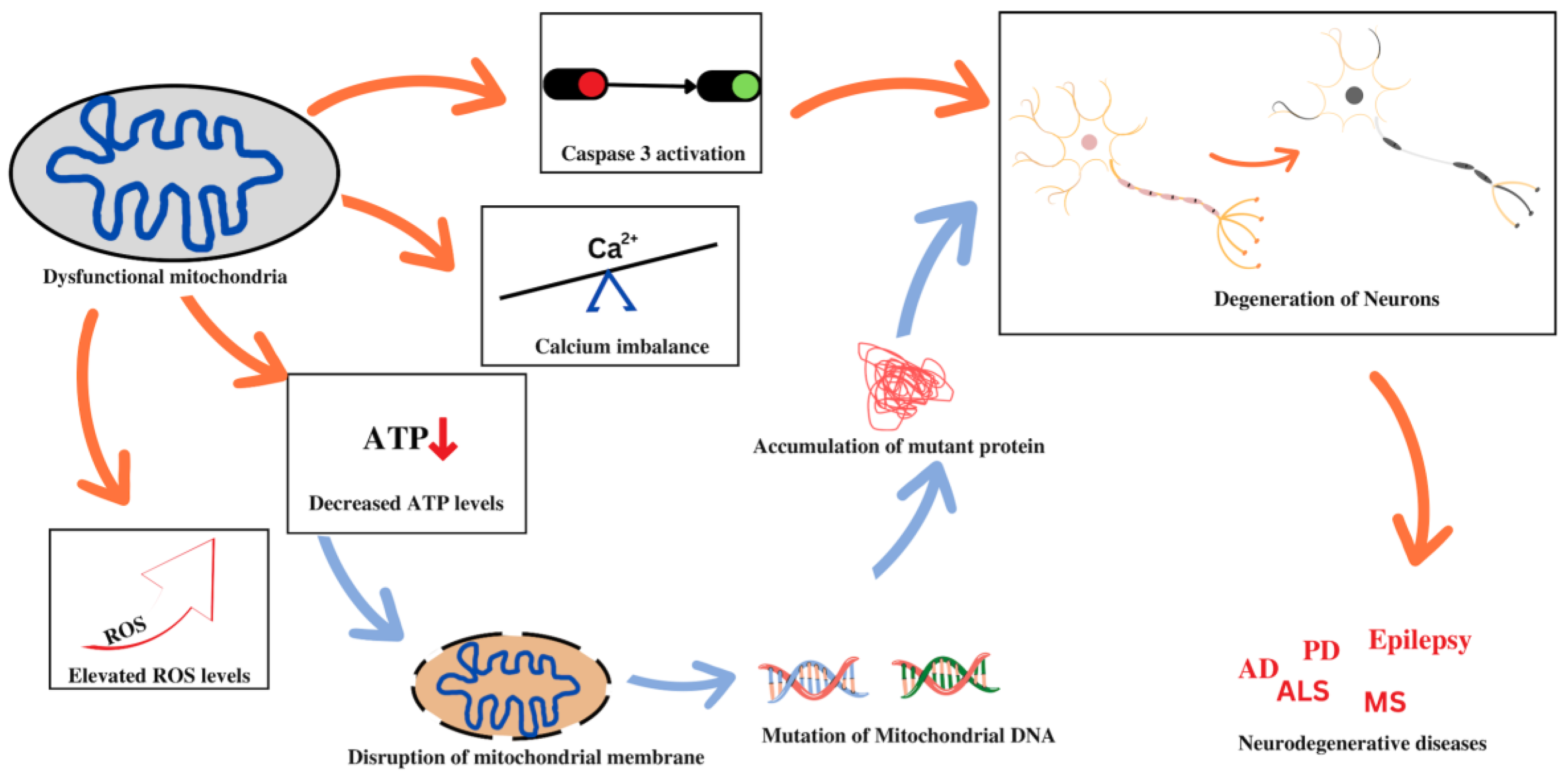
3. Role of Mitochondrial Dysfunction in Pathophysiology

The functioning of every enucleated cell in the human body depends on healthy mitochondria. A disturbance of the basic mitochondrial functions such as bioenergetic functions, the regulation of oxidative stress, and the homeostasis of ions is typically characterized as mitochondrial dysfunction. In the process of an ETC, the release of ROS from the mitochondria causes many detrimental effects, such as mtDNA/RNA damage, lipid oxidation, protein oxidation, the activation of the Ca2+-dependent mitochondrial permeability transition pore, and the release of cytochrome c, which causes the formation of apoptosomes and eventually leads to apoptosis. Both the oxidative stress-related and hereditary mitochondrial DNA abnormalities result in mitochondrial dysfunction, which triggers the cascade of cell death signals leading to organ failure and diseases. Numerous diseases, including diabetes [32], heart failure and ischemia-reperfusion damage [33], cancers [34], Alzheimer’s, and Parkinson’s disease [35], have recently been linked to mitochondrial dysfunction. Therefore, numerous studies are being undertaken to clarify the way in which mitochondrial dysfunction relates to the pathophysiology of different diseases. Herein, we have discussed the pathophysiology of mitochondrial dysfunction in various diseases.

3.1. Mitochondrial Dysfunction and Cancer

Several past investigations have demonstrated that mitochondria play a key role in the etiology of several ailments, including cancer. There are several reports suggesting the involvement of mitochondrial dysfunction in tumors or cancers [34,36]. The commonly found pathologies include reduced oxidative phosphorylation/ATP synthesis, reactive oxygen species over-production, altered calcium homeostasis, and a surge in inflammation [37]. When compared to normal cells/tissues, cancer cells are more prone to fluctuations in energy metabolism. Faster cell proliferation, longevity, and the reoccurrence of tumor cells are also linked to mitochondrial dysfunction. Furthermore, it has been repeatedly observed that mitochondrial failure results in physiological conditions such as hypoxia and acidosis in cancer patients [38]. In addition, there are studies reporting that cancer cells usually have a higher membrane potential of 220 mV than the 140 mVof normal cells [39,40], which may lead to enhanced anabolism, irresponsiveness towards anti-growth factors, and, most importantly, unregulated apoptosis and futile autophagy [41]. Several notable differences in the structure and function of mitochondria between normal and cancer cells have been revealed. A few of the key differences are listed below.

3.1.1. Metabolic Alterations Associated with Cancer

In cancer, various alterations in the functions of mitochondria have been reported, viz., lactic acid over-production accompanied by decreased pyruvate oxidation [42], enhanced glutaminolytic activity [43], gluconeogenesis increment [44], and lower fatty acid oxidation [45]. The bioenergetics guided by mitochondrial functions show a metabolic anomaly in the case of tumor cells in terms of respiratory substrate selection, calcium-buffering capability, or the reduced catalytic activity of mitochondrial enzymes (e.g., Cytochrome c oxidase) [46].

3.1.2. Structural Differences Associated with Cancer

The lipid distribution of inner membrane of a tumorous cell has a higher percentage of cholesterol, altered total phospholipid content, and/or variations in the number of individual phospholipids [47]. When compared to their non-malignant counterparts, cancer cells display variations in the structure and/or relative distribution of numerous proteins. A typical example includes the altered function of F1 ATPase in hepatic cells due to a significant decline in the levels of the b subunit of the F1 component of mitochondrial ATPase accompanied by the up-regulation of ATPase inhibitor protein (IF 1) [48,49]. Conversely, the expression of an pro-apoptotic IMM protein called BAX is decreased in some cancer cell lines [50,51].

3.1.3. OXPHOS Pathway Differences Associated with Cancer

OXPHOS is the process by which ATP is generated in normal cells. However, certain cancer-causing agents, such as radiation, carcinogens, and/or oncogenes, convert normal cells to cancer cells, and these cancer cells subsequently alter the ATP generation process from OXPHOS to Glycolysis [52]. This switch in the metabolic pathway is associated with variations in the normal values of several elements such as mitochondrial membrane potential (MMP; m), glutathione (GSH), pH, and reactive oxygen species (ROS) [53,54].

3.1.4. Physiological Differences Associated with Cancer

In normal cells, ATP production consumes the majority of mitochondrial oxygen, only leaving a small percentage for other processes such as ROS production, whereas cancer cells have significantly higher mitochondrial oxygen levels than normal cells due to the hypoxic extra-tumoral environment [53]. This is owning to the fact that there is reduced ATP production in the mitochondria of tumor cells, which, in turn, results in a 2.5-fold lower oxygen consumption rate (OCR) than that of the mitochondria of healthy cells [53].

3.1.5. Necrosis Associated with Cancer

The role of mitochondria in necrotic cell death has long been recognized [55]. A few situations, most notably oxidative stress and calcium build-up inside mitochondria, can cause a high-conductance leak resulting in the opening of IMM [56]. As a consequence, the electrochemical proton gradient falls, halting ATP synthesis followed by activating ROS generation. In addition, some physiological conditions of necrotic cell death like pH-dependent ischemia/reperfusion injury have also been reported to cause mitochondrial failure [57,58]. Tumor cells were able to strategically prevent hypoxia-mediated cell death by down-regulating p53, a cancer suppressor protein that controls cellular stress response [59].

3.2. Mitochondrial Dysfunction and Neurodegenerative Diseases

The aberrant folding and consequent accumulation of proteins within the cell body of neurons are indications of neurodegenerative disorders. This is plausible due to perturbations in mitochondrial function that can have profound repercussions on the structure and functioning of the neurons, thereby resulting in neurodegeneration. Some of these disorders will be discussed in this section in relation to mitochondrial dysfunction. A vast number of studies indicate that impaired brain metabolism or mitochondrial dysfunctions are some of the best-documented anomalies and early signs in brains affected by major neurodegenerative disorders. Mitochondrial dysfunction associated with non-maternal inheritance has been extensively reported in several neurological diseases, most notably amyotrophic lateral sclerosis (ALS) [60], Parkinson’s disease (PD) [61], Alzheimer’s disease (AD) [62], multiple sclerosis [63], schizophrenia [64], epilepsy [65], neuropathic pain, and Huntington’s disease (HD) [65]. Figure 1 depicts the association between mitochondrial dysfunction and various neurodegenerative diseases. Despite the varying mode of transmission and the unknown specific cause, the common and remarkable signature of mitochondrial dysfunction in such diseases is the malfunctioning of the respiratory chain, resulting in a variety of clinical manifestations. Other causes of mitochondrial malfunction and abnormal mitochondrial morphology include mtDNA mutations [66], oxidative damage, and mitochondrial protein aggregation [67]. The imbalance between ROS production and oxidation leads to oxidative stress, which disrupts the functioning of mitochondrial respiratory chain, affects calcium homeostasis, alters membrane permeability, increases heteroplasmic mt DNA levels, and weakens mitochondrial defense systems [68,69]. Oxidative stress may damage cellular components of mitochondria such as proteins, nucleic acids, and lipids, and contribute to the production of intracellular ROS, leading to mtDNA mutations in neurodegenerative disorders. These involve a variety of proteins that regulate oxidative phosphorylation (OXPHOS) and mitochondrial dynamics and are thus involved in regulating the integrity of the mitochondrial structure. Furthermore, mitochondrial failure largely affects mitochondrial biogenesis and dynamics, which are both associated with a variety of age-related neurodegenerative disorders [70]. In this section, we have briefly explored the role of mitochondrial dysfunction in various neurodegenerative diseases.

3.2.1. Alzheimer’s Disease

Alzheimer’s disease is the most common cause of dementia and is becoming one of the most costly, fatal, and burdensome diseases of the 21st century [71,72,73]. Its pathological hallmarks include nerve cell degeneration, the appearance of neurotic plaques, and neurofibrillary tangles [74,75,76]. Even though the precise mechanism of AD pathogenesis remains unknown, increasing evidence implies that mitochondrial dysfunction and oxidative stress play a significant role in the disease’s etiology [62]. Previous research has found that mtDNA and enzyme abnormalities in the brains of AD patients were followed by the altered morphology and mass of the organelle [77]. These patients were found to have altered mitochondrial enzymatic activity in their brains that led to impaired mechanisms of OXPHOS and the tricarboxylic acid (TCA) cycle [78,79], which further results in low ATP production and an increase in oxidative stress [80,81]. According to the most widely accepted explanation, tau (τ) and Aβ (amyloid β) damage neuronal cells in AD by interfering with the supply of energy and the antioxidative response, resulting in mitochondrial and synaptic dysfunction. The α-ketoglutarate dehydrogenase enzyme complex (α-KDHC) is a crucial mitochondrial enzyme for oxidative metabolism, and its activity diminishes with age. The mechanism of its inactivation is unknown; however, Q. Shi et al. have found that α-KDHC function can be restored, and can be a viable therapeutic method for Alzheimer’s disease [82]. Another study indicates that AD disrupts mitochondrial biogenesis as well as the dynamics of fission and fusion, resulting in an unequal distribution of mitochondria in the neurons [83]. Aβ is thought to be responsible for activating downstream cascades in microglia that cause mitochondrial dysfunction and exacerbate inflammation and cytotoxicity in AD patients [84]. Thus, investigations explaining the operative pathways of mitochondrial anomalies in AD may aid in a better understanding of the etiology of this neurodegenerative illness and may assist in the advancement of therapeutic options to safeguard synaptic activity and consequent cognitive function.

3.2.2. Parkinson’s Disease

The role of mitochondrial dysfunction as an activator, propagator, or bystander in Parkinson’s disease (PD) has been a mystery for decades. PD is the second most prevalent neurodegenerative disorder in the world, and it, like AD, has both hereditary and environmental risk factors [85]. The continuous loss of dopaminergic neurons as well as the aggregation of fibrous protein such as α-synuclein deposits in the cytoplasm of neurons (i.e., Lewy bodies) and nerve fibers (i.e., Lewy neurites) in the substantia nigra of the brain are pathological signatures of PD [61]. This disease manifests both motor and non-motor symptoms, wherein some motor symptoms include myotonia, rest tremors, hypokinesia, and aberrant posture [86], while non-motor symptoms include anxiety, sadness, constipation, frequent micturition, sleep behavior disorder (SBD), complications in rapid eye movement (REM), and cognitive difficulties [87]. In rare cases, PD is caused by mutations in the PINK1 (PTEN-induced kinase 1) or PRKN (parkin RBR E3 ubiquitin protein ligase) genes, which affect the selective autophagic clearance of damaged mitochondria (mitophagy) [88]. According to F. Wauters et al., the most prevalent monogenic cause of PD is a mutation in the gene encoding LRRK2 (leucine-rich repeat kinase 2) [89]. RAB10, a GTPase-activating protein (GAP) and a substrate of LRRK2, accumulates on the depolarized mitochondria via signals from activated PINK1 and PRKN genes. It further binds the autophagy receptor OPTN (optineurin), which enhances OPTN accumulation on depolarized mitochondria and aids mitophagy [90]. Moreover, several studies have also suggested that mtDNA mutations [91,92] and abnormalities in complex I of the mitochondrial ETC [93] play an essential role in the etiopathogenesis of PD. According to theories put forth by researchers [94], the substantia nigra—a group of dopaminergic neurons of the midbrain that undergoes age-related neurodegeneration in PD patients, has high oxidative capacities, and that seems to be particularly sensitive to mitochondrial dysfunction [95], loss in the respiratory chain and, subsequently, high oxidative stress—that is uniquely susceptible to the accumulation of somatic mtDNA mutations and deletions over time [96]. Post-mortem samples revealed that the levels of somatic mtDNA deletions in the substantia nigra of PD patients were marginally higher than those in controls of a comparable age [96] and other neuropathological disorders [97]. Consequently, deletions may result from a compromised mitochondrial replication system, specifically, from mitochondrial DNA polymerase γ (POLG) mutations in the mitochondrial polymerase [98]. In accordance with these findings, it is essential to explore drug development strategies to prevent mtDNA dysfunction as a potential way in which to slow the progression of PD.

3.2.3. Multiple Sclerosis

Multiple sclerosis (MS) is a metabolically dependent neurodegenerative disorder that is caused by persistent axonal loss of the brain and the spinal cord, which thereby initiates cognitive decline and physical disability among the affected. This chronic and progressive autoimmune disease has a complex pathophysiology that is characterized by flare-ups of inflammation and the breakdown of the myelin sheath (an insulating layer of fat that protects nerve fibers) in the central nervous system (CNS) [99]. A disrupted myelin sheath may result in the blocking or impedance of nerve signals and hamper the control of various physiological functions such as vision, sensation, muscle coordination, and strength. In the past several years, it has become obvious that malfunctioning mitochondria are key contributors to axonal and neuronal damage [100]. Animal and histological investigations indicate that invading leukocytes and activated microglia play a critical role in neuronal mitochondrial dysfunction [63]. Disruptions in mitochondrial function were accompanied by significant alterations in the morphology and density of the organelle, which cause inflammatory lesions in MS patients. However, at the onset of disease manifestation, the biochemical activity of mitochondrial complex I was specifically compromised in these spinal tissues [101]. So, it can be concluded that these mechanisms create an imbalance in energy and further contribute to irreversible impairment and neurodegeneration. Thus, extensive research and multiple mitochondria-targeted neuroprotective treatments must be developed as a part of the standard MS treatment regimen.

3.2.4. Amyotrophic Lateral Sclerosis

Amyotrophic lateral sclerosis (ALS) is a motor neuron disease (MND) characterized by the loss of upper motor neurons in the motor cortex and lower motor neurons in the brain stem and the spinal cord. Progressive limb degeneration results in muscular atrophy, paralysis, and, eventually, the death (respiratory failure) of the patient [102]. From a genetic standpoint, ALS can be of two types, namely, familial and sporadic, the latter one being most common (~90%) amongst affected individuals [103]. Further, the pathology of the aforementioned disease includes mitochondrial degeneration, oxidative stress, glutamate excitotoxicity, decreased axonal transport, glial cell disease, and defective RNA metabolism [102]. Thiol is oxidized during mitochondrial dysfunction, resulting in a cascade of multiple processes such as calcium imbalance and the breakdown of the mitochondrial membrane promoting cell death [104]. Analysis of ALS models revealed that the build-up of aberrant mitochondria in motor neuron axons leads to defective mitochondrial transport in ALS [104]. Furthermore, mitochondrial damage limits NAD+ and ATP production while increasing ROS production, leading to mtDNA mutations, structural deformity, aberrant protein aggregation, and, eventually, the loss of motor neurons [104]. Additionally, cell culture and animal investigations have revealed faulty calcium homeostasis and ROS overproduction in relation to abnormal oxidative metabolism in mitochondria [105]. Moreover, abnormalities in mitochondrial dynamics and the interruption of axonal transport were also identified in ALS models [106]. As a result, mitochondrial degradation is determined to be the cause of most familial or sporadic ALS cases and addressing ALS-associated mitochondrial deregulation pharmacologically may present a possibility for delaying disease progression.

3.3. Mitochondrial Dysfunction and Cardiovascular Diseases (CVD)

In 2019, Asia accounted for 58% of the 18.6 million CVD deaths globally [107]. Many cardiac illnesses, including atherosclerosis, ischemia-reperfusion injury, heart failure, and hypertension, are thought to be accompanied by mitochondrial dysfunction, most likely as a result of insufficient cellular energy production and unchecked ROS production. mtDNA damage is a significant contributor to mitochondrial dysfunction and is a key phenotypic feature [6]. In these cases, apoptosis, inflammation, fibrosis, and cardiac remodeling are stimulated, and sarcomere protein function is impaired. Other characteristics of mitochondrial dysfunction include decreased numbers of mitochondria in tissues, the stimulation of apoptosis and inflammation, the absence or dysfunction of mitochondrial enzymes, and impaired mitochondrial biogenesis [108,109]. In this section, we have briefly discussed the role of mitochondrial dysfunction in various cardiovascular diseases.

3.3.1. Atherosclerosis

Atherosclerosis, a chronic inflammatory disease, is characterized by the accumulation of lipids, primarily cholesterol, and other compounds such as fatty materials, cellular waste products, calcium, and fibrin within the artery wall [110]. The progression of atherosclerosis depends on numerous factors and one of its hallmarks is ROS overproduction, which is involved in various processes. Proteins, lipids, and nucleic acids can suffer oxidative damage from prolonged exposure to ROS or excessive ROS production [111]. Endothelial nitric oxide synthase (eNOS) converts L-arginine into L-citrulline to produce the majority of the nitric oxide (NO) produced in the endothelium. Intracellular arginine levels, which are, in turn, governed by mitochondrial arginase II, are the primary regulators of NO levels. As a result, NO needs a functioning mitochondrial respiratory chain (MRC) to maintain proper levels [112]. The major causes of decreased NO concentrations in the cell are eNOS breakdown brought on by elevated ROS levels and the loss of mitochondrial membrane potential. Lower NO secretion and production are the initial signs of endothelial change in early atherosclerosis [113]. Improper mitophagy also increases mitochondrial damage and the release of ROS, mtDNA, and K+ into the cytoplasm, which encourages NLRP3 inflammasome activation. Mitophagy is a biological mechanism involved in mitochondrial rejuvenation. In fact, the NRLP3 protein can directly interact with released mtDNA to start an inflammatory response [114].

3.3.2. Ischemic Stroke

Strokes continue to number among the major causes of death in developed nations and constitute the primary cause of physical and intellectual disability in adults. Stroke is still the most common cause of mortality in affluent countries and the main factor in adult physical and intellectual disability [115,116]. When the blood supply to the brain tissue provided by blocked arteries is reduced, an ischemia event takes place. Cell death ultimately results from altered cellular homeostasis caused by a scarcity of oxygen and nutrients [117,118,119]. Various enzymes have been shown to play a role in ischemia [120]. Numerous isoforms of nitric oxide synthase (NOS) activated post-ischemic stroke, such as neuronal, endothelial, and inducible NOSs, mediate excessive NO production [121,122,123,124]. The B-cell lymphoma (BCL-2) protein family is a significant regulator of the permeability of the OMM and is essential for the intrinsic apoptotic pathway [125]. BCL-2 may have a significant role in the regulation of neuronal death in cerebral ischemic strokes according to several reports [126,127,128,129,130].
Aside from the BCL-2 pathway, several other major apoptotic pathways involve the release of pro-apoptotic factors such as apoptosis-inducing factor (AIF) and the second mitochondrion-derived activator of caspase (SMAC) from the mitochondria. The pro-apoptotic factor SMAC releases from mitochondria and binds to the X-chromosome-linked inhibitor of apoptosis protein (XIAP), suppressing its anti-apoptotic activity and preventing serial procaspase activation and thus further inducing apoptosis following cerebral ischemia [131,132]. Another major pro-apoptotic factor, AIF, is a mitochondrial protein which was discovered to be a caspase-independent modulator of the degradation phase of apoptosis. AIF was proposed to serve as a mitochondrial effector of apoptotic cell death after its transfer from the mitochondria to the nucleus [133]. BH3-interacting domain death agonist (Bid) is a cytosolic protein that induces the release of cytochrome c, which leads to programmed cell death. The Bid protein has been demonstrated to maintain AIF in the nuclei, which speeds up and strengthens the apoptotic process [134]. Moreover, AIF has also been proven to block poly (ADP-ribose) polymerase. Experiments performed on animal models of ischemic stroke show the translocation of AIF with apoptotic DNA fragmentation, which happens prior to or concurrently with the release of cytochrome c from mitochondria [135]. Furthermore, the role of AIF is also seen to govern neuronal death brought about by oxygen-glucose deprivation, glutamate-induced toxicity, and experimental ischemic stroke in vivo [134].

4. Strategies for Mitochondria-Targeted Therapy

The identification of mitochondria as an emerging pharmaceutical target has led to the development of several mitochondrial targeting strategies for the effective treatment of diseases based on mitochondrial dysfunction. The primary challenge in exploiting mitochondria as a target is the delivery of therapeutic molecules. As we described above, permeability of both the membranes in mitochondria is different. The transition pore in the OMM is wider, and it is through this pore that therapeutic molecules can easily traverse. Whereas the highly folded hard IMM has narrower transition slits that separate the intermembrane space and the mitochondrial matrix, making it difficult for many therapeutic molecules to cross the mitochondrial matrix. Thus, research has been directed towards improving therapeutic delivery and reducing the unwanted effects of the delivered drugs. Numerous drug delivery approaches are being developed in light of the specific properties of mitochondria, including their membrane potential and lipophilicity [136], specialized protein import machinery [137], and distinctive phospholipid composition in the IMM [138], as shown in Figure 2. The first characteristic of a mitochondrion is that it contains a negative charge. The potential (ΔΨm) between the matrix and the intermembranous space is around 180 mV; mitochondria exploit this potential as a proton-motive force for ATP production [139]. However, this ΔΨm was exploited for mitochondrial targeting by using positively charged ions (cations) that are attracted to the mitochondria via electrostatic forces. The second distinguishing feature of mitochondria is the unique structure of their IMM and lipid composition, which mostly consists of cardiolipin and may be exploited for targeted drug delivery to the mitochondria. The phospholipid cardiolipin plays a key role in apoptosis and provides structural support for respiratory chain complexes [140].
Mitochondria generally lose most of their genetic material while forming endosymbiotic interactions with the host eukaryotic cell, forcing them to require proteins encoded in the nuclear genome. As a consequence, mitochondria possess protein import machinery that identifies proteins with a specific amino acid sequence [141]. Thus, drug delivery to mammalian mitochondria is accomplished using one of these approaches, or by a combination of both. Herein, we have discussed various molecules that are being used for drug delivery in mitochondria.

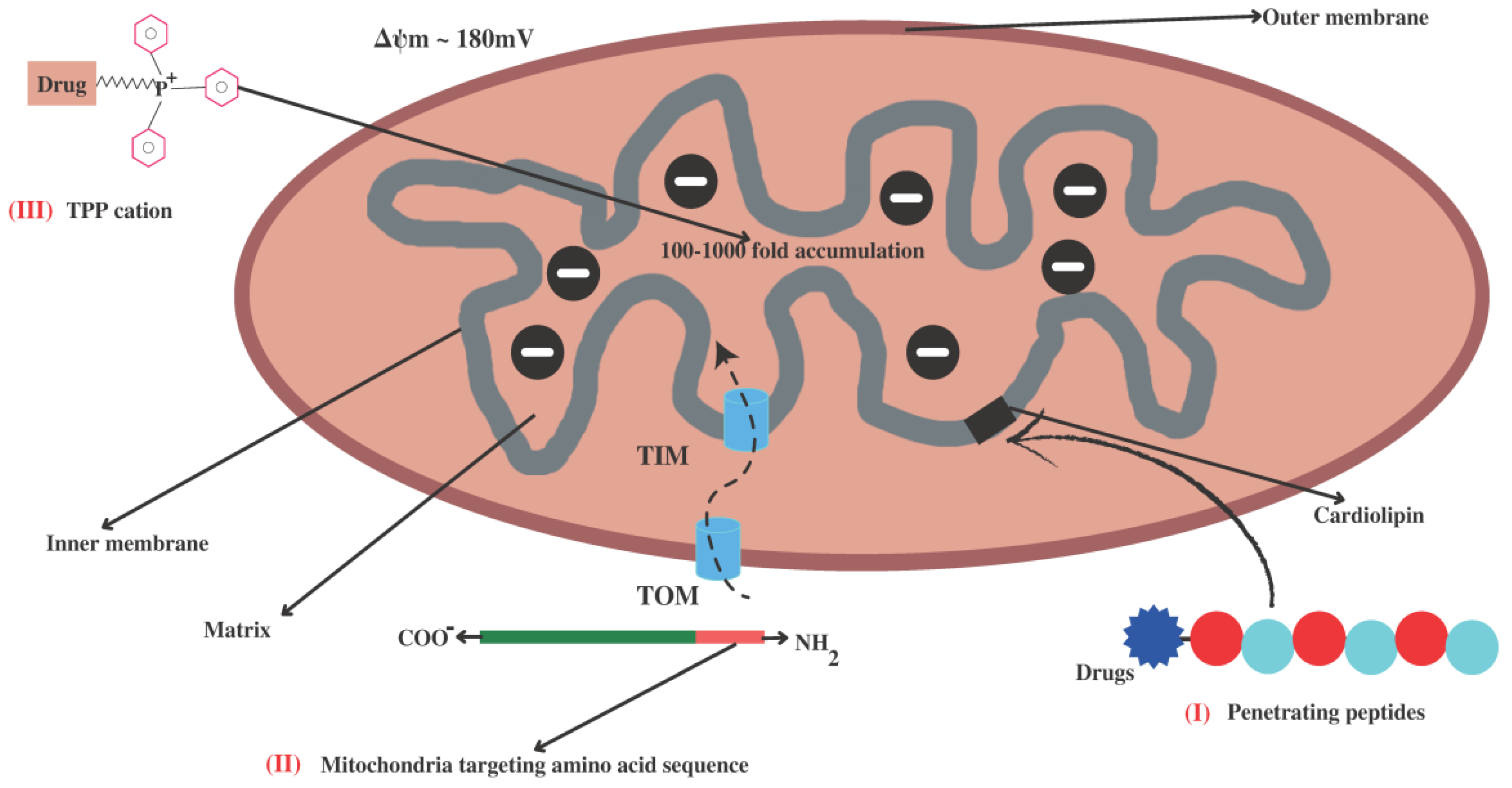
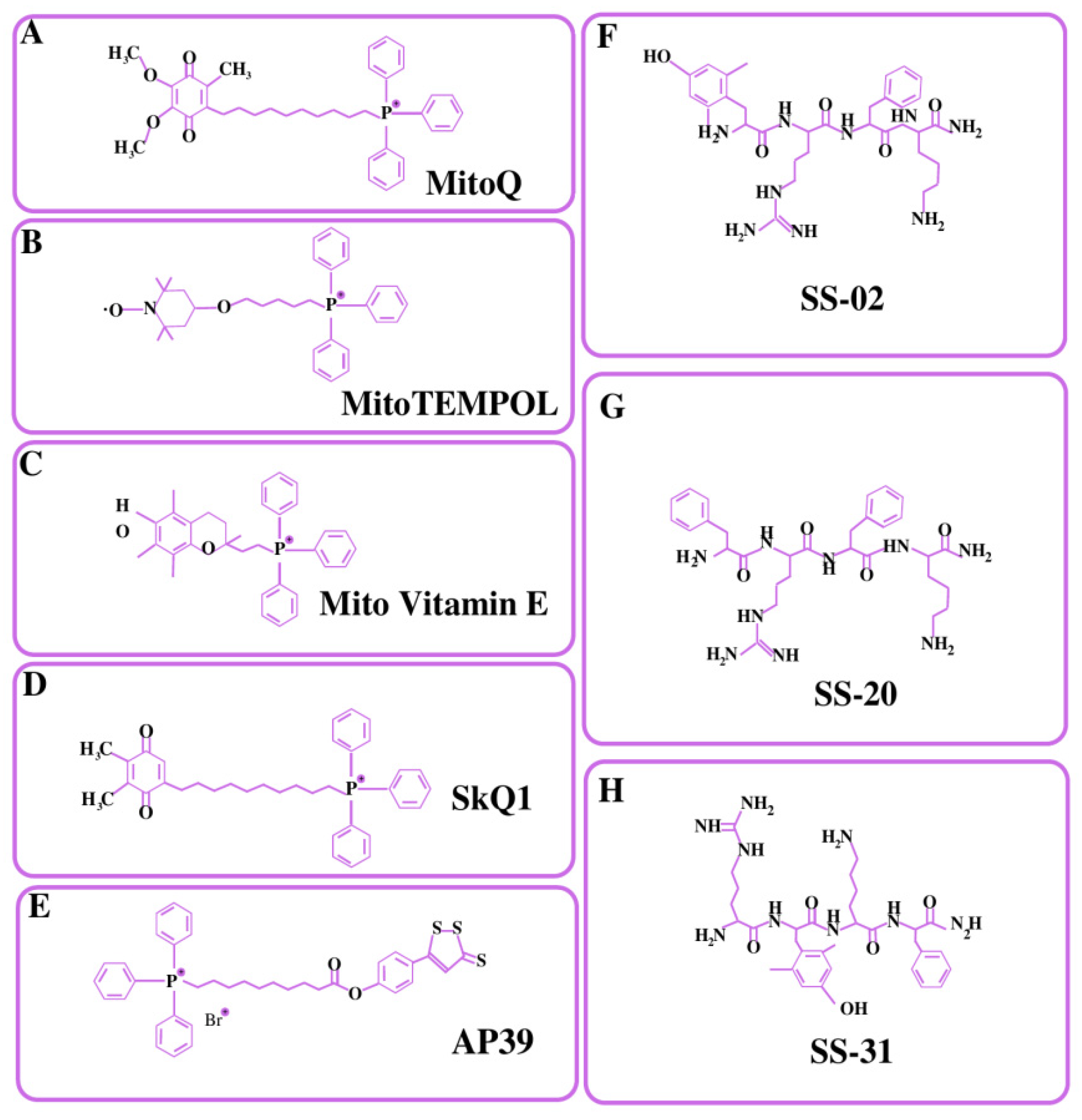
4.1. Small Lipophilic Cationic Molecules Targeting Mitochondria

4.1.1. Triphenylphosphonium Cation (TPP+)

In mitochondria, there is a significant transmembrane potential of 140–180 mV (negative inside) that may be used to transport positively charged molecules to mitochondria [142]. Murphy and colleagues pioneered the use of lipophilic cations as mitochondrial carriers, which are also known as mitochondriotropic ligands [143]. TPP+ is a lipophilic cation, and the potential gradient created by its charge dispersion over the surface area causes its accumulation inside the mitochondrial matrix [144]. The percentage of the TPP+ concentration inside the negatively charged membrane compartments rises by one order of magnitude for every 60 mV of negative membrane potential. The plasma membrane voltage typically varies between −30 and −60 mV, which is enough to cause an up to 10-fold buildup of TPP within the cell. Typical mitochondria, on the other hand, have a membrane potential of −180 mV, which facilitates an increased accumulation of TPP+ inside the mitochondria by 1000 times [144]. TPP+ aids in the delivery of mitochondrial drugs such as AP39 [145], Mito-Vit-E [136], Mito-Q [146], SkQ1 [147], and 2,2,6,6-tetramethyl-4-[5-(triphenylphosphonium) pentoxy] piperidine-1-oxybromide (Mito-TEMPOL) [148]. The structural formulas of all these drugs have been depicted in Figure 3. AP39 is mitochondria-targeting motif that comprises TPP+ attached to a H2S donor moiety (dithiol-77 ethione) through an aliphatic linker. There is emerging evidence regarding the roles of H2S in reducing the release of mitochondrial death signals and maintaining mitochondrial integrity [149]. Several studies suggested that AP39 are capable of preventing hyperglycemia-induced oxidative stress [150], reducing amyloid-β deposition in the brain, and ameliorating spatial memory deficits in APP/PS1 mice [151]. Mito-Vit-E was made by combining vitamin E with TPP+, with the latter driving the compound in mitochondria. Mito-Vit-E is an anti-oxidant that has been shown to revive mitochondrial activity and prevent cell death [136]. Mito-Q is produced through the conjugation of ubiquinone with TPP+ and is commonly utilized as an antioxidant to prevent cell death. SkQ1 are the derivatives of plastoquinone attached via the C10 hydrophobic linker to the TPP cation. SkQ1 has shown greater anti-oxidant activity and binding affinity with cardiolipin as compared to mito-Q, and has shown decreased protein oxidation, the prevention of cell apoptosis, decreased ROS levels, and the prevention of lipid peroxidation [147]. Several studies reported that SkQ1 showed high efficacy in a wide variety of eye diseases such as glaucoma, retinopathy, and dry eye syndrome [152,153]. Mito-TEMPOL is a well-known superoxide scavenger that targets mitochondria. Mito-TEMPOL therapy may reduce ATP depletion-induced necrosis and apoptosis by maintaining mitochondrial integrity and lowering BAX translocation to mitochondria. Mito-Q, Mito-TEMPOL, and Mito-Vit-E were shown to be more effective than untargeted antioxidants (vitamin E and ubiquinone10) at lower concentrations in protecting cells against peroxide-induced oxidative damage and death [146]. However, murphy et al. reported that a TPP+ concentration above 10 µM might have detrimental effects on mitochondrial damage control due to proton leakage and mitochondrial membrane depolarization [143].

4.1.2. Rhodamine

Rhodamine derivatives have a high binding affinity for mitochondrial membranes so they can be used as agents that specifically target the mitochondria and disrupt the electron transport chain [154]. Rhodamine’s lipophilic and cationic characteristics, which enable it to pass through double mitochondrial membranes and stay inside the negatively charged mitochondrial matrix, are thought to constitute the mechanism causing its accumulation in mitochondria [155]. When TPP+ was replaced with rhodamine 19, it was found to be a potential drug carrier to mitochondria. This was accomplished by the chemical alteration of a TPP–drug conjugate so as to produce rhodamine 19–drug conjugates [147]. It inhibited stroke-induced brain swelling and averted neurological impairment, providing a potential example of a moderate uncoupler that is effective in the treatment of brain pathologies related to oxidative stress [156].

4.1.3. Pyridinium Salts

Pyridinium salts are lipophilic delocalized cations that can be utilized as mitochondrial targeting groups. Large conjugated systems are created by modifying pyridinium salts with ethylenic bonds. The pyridine salts’ ability to withdraw electrons and their conjugated systems can induce or govern molecular luminescence, which is useful for the optical detection of mitochondria. However, pyridine salts’ lipophilicity hampers their ability to reach tissues in vivo.
A breakthrough in the use of pyridinium salts in vivo was made by Cheng’s team when they created a human serum albumin-facilitated pyridine salt complex that could easily access tumor tissue, had mitochondria-targeting ability, and caused tumor cell apoptosis [157]. The pyridine salt molecule’s cytotoxicity was a flaw in its mitochondrial detection, but it showed advantages in the treatment of tumors [157].

4.2. Mitochondria-Targeting Signal Peptides

The nuclear genome contains the genetic information for more than 1000 mitochondrial proteins. Cells usually utilize the mitochondrial protein import machinery to import nuclear-encoded mitochondrial proteins. These proteins typically have a 20–40-amino acid cleavable targeting region at their N-terminus. This sequence is generally positively charged, has amphiphilic helices, and is not conserved [158]. The movement of the imported proteins to the translocase of the inner membrane (TIM) complex at the IMM is carried out by the translocase of the outer membrane’s (TOM) importing complex, which is situated on the OMM. Following their passage through the IMM, mitochondrial peptidases break the targeting sequence, and mitochondrial chaperones fold the imported protein into a mature structure [159]. The majority of research efforts have focused on developing a mitochondrial targeting sequence for DNA or gene delivery into the mitochondria as a potential treatment to cure mitochondrial diseases [137,160]. This method is also utilized to investigate the precise involvement of mitochondria in the onset of some diseases, such as the modification of mitochondrial function and cell death caused by Aβ-peptide accumulation in mitochondria [161]. However, no prospective medicine including signal peptides for mitochondrial targeting has yet been evaluated, as this strategy has several practical limitations, i.e., effective targeting peptides are lengthy, large molecules are difficult to transport, the hydrophilicity and cellular permeability of these peptides are low, and the cost of chemical production is quite high. These limitations significantly restrict the application of mitochondria-targeted compounds (MTCs) based on mitochondrial targeting signal peptides.

4.3. Cardiolipin Targeting (Penetrating Peptides)

Peptide-based delivery is an additional method utilized to facilitate mitochondrial targeting. The advantages of peptide-based delivery scaffolds are their simplicity in synthesis, adaptability, biocompatibility, and high absorption in both cells and in vivo [162]. The properties of lipophilicity and a positive charge were used to design small peptides with which to target mitochondria. Recent studies have shown that many cell-penetrating peptides can penetrate the mitochondrial membrane [163]. Szeto–Schiller (SS peptides) is an illustration of a small peptide-targeting moiety that targets mitochondrial cardiolipin with specificity in order to enhance mitochondrial plasticity and re-establish optimal bioenergetics. Based on their lipophilicity and charges, alternate aromatic residues and basic amino acids are being used to design such types of peptides to target mitochondria [164]. The accumulation of these peptides occurs specifically in the IMM, where they scavenge ROS, prevent the opening of the mitochondrial permeability transition pores, and, ultimately, prevent the release of cytochrome c. Various peptides (SS-01 to SS-31) were prepared by using these amino acids with slight modifications, from which (SS-01, SS-02, and SS-31) have shown antioxidant efficacy. This effect was due to the presence of aromatic amino acids, such as Dmt (dimethyl tyrosine) and Tyrosine, which subsequently scavenged ROS because unreactive tyrosyl or dityrosine radicals can react with superoxide radicals to form tyrosine hydroperoxide [138]. Contrary to TPP+, which causes toxicity at 10 µM, Szeto–Schiller peptides did not show toxicity even at 1 mM. Preclinical studies of SS-31(D-Arg-Dmt-Lys-Phe-NH2) suggest beneficial effects with respect to muscle aging, atherosclerosis, ischemia, osteoarthritis, diabetes, and glaucoma. To determine the efficacy and safety of these peptides, several clinical trials are undergoing for different diseases such as cardiovascular diseases, kidney diseases, cerebral ischemia, etc. [138,164].
Many other peptides such as cationic amphipathic α-helical D-(KLAKLAK)2 have also been synthesized in an attempt to improve the potencies of anticancer peptides [165,166]. In their study, Li et al. developed a peptide (P11LRR) consisting of arginine-modified polyproline amphiphilic molecules that formed a helical structure [167]. It was observed that the accumulation of P11LRR in mitochondria was driven by the mitochondrial transmembrane potential, as the elimination of mitochondrial potential leads to the inhibition of peptide localization. P11LRR-conjugated dimethyl tyrosine (a molecule that is supposed to be antioxidant) was able to reduce the chemically induced reactive oxygen species within the mitochondria, thereby serving as an excellent mitochondrial drug delivery vector [167]. Similar to this, various cell-penetrating peptides have been designed [168] despite having some drawbacks, such as fast elimination from the body, intracellular/vesicular entrapment, and non-specific internalization [169].

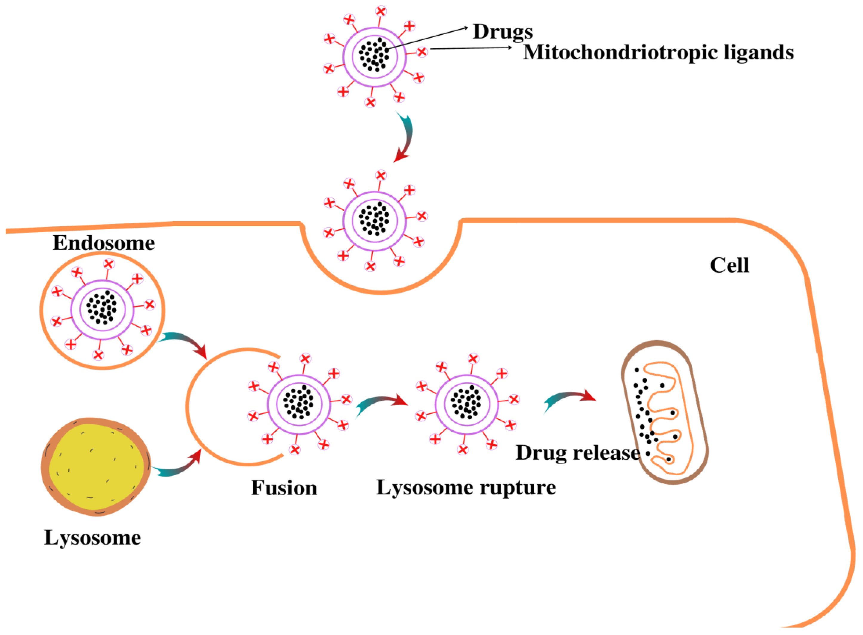
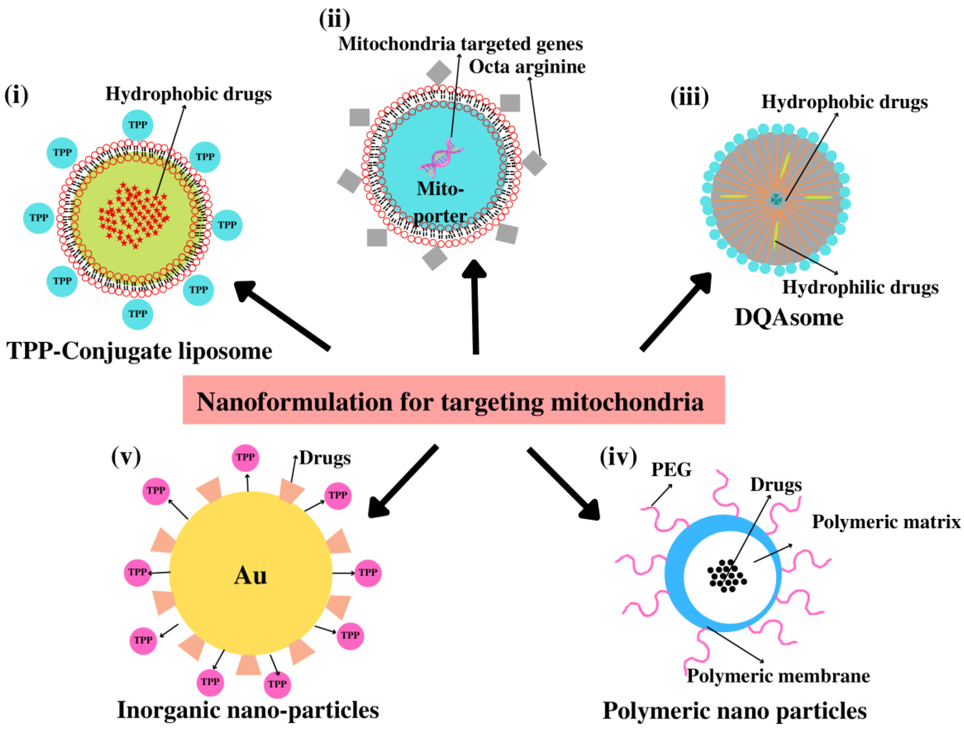
4.4. Nanoparticle (NPs)-Based Drug Delivery

4.4.1. Dequalinium (DQA)

Dequalinium, a single-chain amphiphilic compound with two delocalized cation centers that self-assemble into liposome-like cationic vesicles, has been shown to possess mitochondria-targeting characteristics [170]. It is capable of transporting DNA and drugs via nonspecific endocytic pathways to mitochondria, as shown in Figure 4. Various studies have suggested that DQA is capable of delivering DNA, and antisense RNA specifically, to the mitochondria [171,172]. Owing to this property, DQAsomes could also be utilized to encapsulate anti-cancer drugs such as curcumin and paclitaxel. These studies demonstrated that the free forms of curcumin and paclitaxel have less antioxidant and tumoricidal activity as compared to the encapsulated drug in DQAsomes [173,174]. DQAsomes are now considered to constitute a unique drug-delivery system due to the selective accumulation of the drug DQA in mitochondria and its anticarcinoma activity.

4.4.2. Liposomes

Liposomes are globular structures formed from phospholipid-based vesicles that contain one or more lipid bilayers and cholesterol [175]. They are suitable for the incorporation of both hydrophobic and hydrophilic drugs due to their aqueous center and the lipid bilayer surrounding them. Liposomes are used to deliver drugs since they are biologically compatible, biodegradable, can self-assemble, nontoxic, can carry large drugs, and have several properties that can be changed to control their biological characteristics [176]. Drugs can be loaded into liposomes in a variety of ways, such as by encasing them in the aqueous region of the liposomes or lipophilic bilayers or by electrostatically adsorbing them to the liposome’s surface [177]. Various liposome systems have been developed for mitochondria-targeted delivery. In a study, chlorine e6 (Ce6, a photosensitizer) and IR 780 iodide (photothermal and near infra-red agent) were encapsulated in biocompatible liposomes and the attachment of TPP+ on the surfaces of liposomes greatly facilitated their mitochondria-targeted delivery and showed higher toxicity in HeLa cells in vitro, leading to the enhanced efficacy of photodynamic therapy [178]. Further research has been directed in this field and involved the use of the liposystem as a vehicle for the controlled release of a drug [179]. In another study, a stearyl residue was conjugated to TPP+ and this conjugated form was incorporated into a liposome. The ceramide-loaded stearyl-conjugated TPP liposomes (STPP) showed significantly decreased tumor volumes in BALB/c mice with non-specific toxicity [180]. For specific targeting, a novel polyethylene glycol-phosphatidylethanolamine (PEG-PE) conjugate was synthesized by attaching a TPP+ group to the distal end of the PEG block (TPP-PEG-PE). Further, this conjugate was encapsulated into a liposome and its toxicity, mitochondrial targeting capacity, and efficacy in delivering paclitaxel (PTX) to cancer cells were investigated in vitro and in vivo as compared to STPP conjugates. They found that the TPP-PEG-PE-modified liposomes were less cytotoxic and showed enhanced mitochondrial targeting capacity compared to the STPP-liposomes [181]. In another study, STPP-liposome hybrid cerasomes (CER), which are based on the Si–O–Si framework and liposomes, were developed to combat the instability and aggregation of the liposome system. Wang et al. synthesized this nano-hybrid cerasome modified with triphosphonium (TPP) and it showed excellent biocompatibility, good stability, and sustainable drug release behavior in mitochondria [182].
Yamada et al. created the MITO-Porter liposome to transport genome-targeting nucleic acids to the mitochondria. It is a liposome-based transporter that effectively transports molecules to the cytoplasm [183,184], as well as to mitochondria through membrane fusion [185]. High-density octa-arginine (R8) was used by Yamada et al. to coat the MITO-Porter surface, which led to macropinocytosis rather than clathrin-mediated endocytosis and enabled particles to reach the cell without being harmed. Later, Yasuzaki et al. employed MITO-Porter to encapsulate propidium iodide, a fluorescent dye used for staining nucleic acids that could enable the visualization of mtDNA. Further investigation into this strategy is being conducted for photodynamic cancer therapy [186] and mitochondrial gene therapy [187,188].

4.4.3. Polymeric Nanoparticles

Polymeric NPs could be used to target mitochondria for drug delivery since they are biodegradable and biocompatible. Additionally, as they are simple to make, surface alterations can be performed easily and can be customized to drug-release characteristics. Various polymers such as poly (lactic-co-glycolic acid) (PLGA), poly (glycolic acid) (PGA), poly (lactic acid) (PLA), and polycaprolactone (PCL) can be formed into nanoparticles through emulsification-solvent evaporation or nanoprecipitation [189,190]. These NPs can encapsulate both hydrophilic and hydrophobic drugs with minor modification of FDA-approved hydrophilic blocks such as polyethylene glycol (PEG). These PEG molecules augment the residence time in vivo and are generally used to conjugate the targeting moieties [2]. Through various chemical reactions, numerous nanocarriers have been made for drug delivery. To assess the use of these nano-carriers, different drugs have been employed as their payloads, including 2, 4-dinitrophenol (an anti-obesity treatment), curcumin (AD), and α-tocopheryl succinate (a cancer medication) [191]. In comparison to non-targeted constructs or treatments in their free form, the drug therapeutic index for cancer, AD, and obesity is noticeably improved by targeted nanocarriers such as PLGA-b-PEG-TPP NP [191].

4.4.4. Inorganic Nanoparticles

Inorganic nanoparticles have a smaller and more homogeneous particle size than organic NPs. In contrast to liposomes, dendrimers, and micelles, metallic nanoparticles (Metal NPs) such as silver and gold have features including surface plasmon resonance (SPR), enhanced Rayleigh scattering, and abilities beneficial for imaging biological systems [192]. These metallic nanoparticles can be easily conjugated with peptides, antibodies, DNA, and RNA to target certain cells, and with biocompatible polymers (polyethylene glycol) to extend their circulation in vivo for medication and gene delivery applications. Therefore, metal NPs have been employed as core components because of their bio-inertness, simplicity in synthesis, and characterization.
The use of silver nanoparticles (AgNPs) as nanocarriers for the treatment of cancer has shown promising results [193]. They possess special antiviral, antibacterial, and antimicrobial activities. Additionally, AgNPs, with or without conjugates, have been recognized for their anticancer activities, which makes them effective drug carriers for the treatment of cancer [23]. AuNPs have been used to provide targeted medication delivery by conjugating with various mitochondrial moieties. We have summarized the different mitochondria-targeting nanoformulations in Table 1. In the human cancer cell lines, Caco-2, HeLa, and MCF-7, Oladimeji et al. investigated the mitochondria-targeted delivery of betulinic acid (BA) via mitochondriotropic TPP+-functionalized epigallocatechin gallate (EGCG)-capped gold NPs (AuNPs). The IC50 values of these nanocomplexes were 3.12–13.2 µM in vitro, compared to 9.74–36.31 µM for free BA, and they likewise demonstrated a considerable reduction in cancer cell proliferation [194]. The majority of traditional chemotherapeutics have harmful side effects that restrict the maximum tolerable dose and compromise their therapeutic efficiency by indiscriminately killing both healthy and malignant cells. To solve this issue, Sun et al. created the DNA-guided missile-integrated nanospacecraft (GM-NSC), a nanocomposite formed of gold nanoparticles (AuNPs) and a high-density multilayer DNA crown that is constructed from highly organized DNA tetrahedral units (DNA Tetra). Each DNA tetrahedral unit consists of three parts: an explosive bolt (E-bolt), a triphenylphosphonium (TPP) unit that targets mitochondria, and an aptamer that targets cancer cells [195]. Good biocompatibility, high cargo-loading capacity, adequate in vivo biodistribution, and therapeutic efficiency without side effects were demonstrated by GM-NSC, making it a viable alternative drug delivery system for targeted cancer treatment [195]. Figure 5 depicts the various nanoformulations currently in use for mitochondria-targeted drug delivery.

5. Conclusions

Mitochondria are among the most important cellular organelles, which, in addition to energy production, are involved in calcium signaling, cell growth and differentiation, cell cycle regulation, and apoptosis. Mitochondrial dysfunction is a potential target for diagnostic and therapeutic interventions for many diseases owing to its role concerning higher cell death, decreased ATP synthesis, and increased mtROS production in the pathogenesis of many diseases. The mitochondrial membranes and plasma membranes restrict therapeutic molecules’ ability to reach mitochondria. Although there is a bioactive additive with MitoQ and eye drops with SkQ1, there is still a scarcity of pharmacological formulations in the market that efficiently target mitochondria. The reason for which is the lack of an effective delivery system that can direct therapeutic molecules for selective accumulation inside mitochondria. The ability of NPs to localize within mitochondria and target particular cells facilitates treatments associated with mitochondrial dysfunction disorders. Nano-formulation approaches, such as those incorporating liposomes, inorganic NPs, and polymeric NPs conjugated with mitochondriotropic ligands, have been shown to carry a variety of payloads to mitochondria in in vitro models, although several clinical and pre-clinical studies still need to be conducted to understand the safety of these drug-delivery systems. The field of mitochondria-targeted nanomedicine is gaining considerable interest and efforts are also being made for the preparation of potent nano-formulations; however, a better understanding of the subject is still needed in order to use them as potential therapeutics for a wide range of diseases.

Author Contributions

Conceptualization, R.W. and A.I.; writing—original draft preparation, T.K., R.W., Z.Z., A.A., P.B. and J.A.; writing—review and editing, R.W., T.K. and A.I.; Supervision, M.I.H. and A.I.; project administration, R.W. and A.I. All authors have read and agreed to the published version of the manuscript.

Funding

This work was supported by the grants from the Indian Council of Medical Research ISRM/12/(127)/2020 and Science and Engineering Research Board (SERB) CRG/2018/004641.

Institutional Review Board Statement

Not applicable.

Informed Consent Statement

Not APPLICABLE.

Data Availability Statement

The information that supports the findings of this study is available in this article.

Acknowledgments

The authors are grateful to the FIST Program (SR/FST/LSI-541/2012) and Jamia Millia Islamia (a Central University) for providing the infrastructural support used to carry out this work. RW acknowledges the DST-INSPIRE for INSPIRE Fellowship (IF-180728). TK acknowledges the University Grant Commission for Junior Research Fellowship (191620050310). ZZ acknowledges the CSIR for Junior Research Fellowship (09/466(0239)/2020-EMR-I). AA acknowledges the PMRF for the fellowship (PMRF ID-3302520).

Conflicts of Interest

All authors declare that they have no conflict of interest.

Abbreviations

IMMInner mitochondrial membrane
OMMOuter mitochondrial membrane
IMSIntermembrane space
TPPTriphenylphosphonium
DmtDimethyltyrosine
DQADequalinium
MitoETPP-vitamin E
Mito-QTPP-ubiquinone
STPPStearyl conjugated TPP liposomes
PEGPolyethylene glycol
PTXPaclitaxel
PLGAPoly (lactic-co-glycolic acid)
BCL-2 B-cell lymphoma
ALSAmyotrophic lateral sclerosis
PDParkinson’s disease
ADAlzheimer’s disease
MSMultiple sclerosis

References

  1. Javadov, S.; Kuznetsov, A.V. Mitochondria: The Cell Powerhouse and Nexus of Stress; Frontiers Media SA: Lausanne, Switzerland, 2013; Volume 4, p. 207. [Google Scholar]
  2. Pathak, R.K.; Kolishetti, N.; Dhar, S. Targeted nanoparticles in mitochondrial medicine. Wiley Interdiscip. Rev. Nanomed. Nanobiotechnol. 2015, 7, 315–329. [Google Scholar] [CrossRef] [PubMed] [Green Version]
  3. Ross, M.F.; Kelso, G.; Blaikie, F.H.; James, A.M.; Cocheme, H.M.; Filipovska, A.; Da Ros, T.; Hurd, T.; Smith, R.A.; Murphy, M.P. Lipophilic triphenylphosphonium cations as tools in mitochondrial bioenergetics and free radical biology. Biochemistry 2005, 70, 222–230. [Google Scholar] [CrossRef] [PubMed]
  4. D’Souza, A.R.; Minczuk, M. Mitochondrial transcription and translation: Overview. Essays Biochem. 2018, 62, 309–320. [Google Scholar]
  5. Wallace, D.C.; Fan, W. Energetics, epigenetics, mitochondrial genetics. Mitochondrion 2010, 10, 12–31. [Google Scholar] [CrossRef] [PubMed] [Green Version]
  6. Waseem, R.; Shamsi, A.; Kazim, S.N.; Islam, A. An insight into mitochondrial dysfunction and its implications in neurological diseases. Curr. Drug Targets 2021, 22, 1585–1595. [Google Scholar] [CrossRef] [PubMed]
  7. Schapira, A.H. Mitochondrial disease. Lancet 2006, 368, 70–82. [Google Scholar] [CrossRef]
  8. McBride, H.M.; Neuspiel, M.; Wasiak, S. Mitochondria: More than just a powerhouse. Curr. Biol. 2006, 16, R551–R560. [Google Scholar] [CrossRef] [Green Version]
  9. Green, D.R. Apoptotic pathways: The roads to ruin. Cell 1998, 94, 695–698. [Google Scholar] [CrossRef] [Green Version]
  10. Kühlbrandt, W. Structure and function of mitochondrial membrane protein complexes. BMC Biol. 2015, 13, 89. [Google Scholar] [CrossRef] [Green Version]
  11. Bhatti, J.S.; Bhatti, G.K.; Reddy, P.H. Mitochondrial dysfunction and oxidative stress in metabolic disorders—A step towards mitochondria based therapeutic strategies. Biochim. Biophys. Acta (BBA)-Mol. Basis Dis. 2017, 1863, 1066–1077. [Google Scholar] [CrossRef]
  12. Turrens, J.F. Mitochondrial formation of reactive oxygen species. J. Physiol. 2003, 552, 335–344. [Google Scholar] [CrossRef] [PubMed]
  13. Fridovich, I. Superoxide radical and superoxide dismutases. Annu. Rev. Biochem. 1995, 64, 97–112. [Google Scholar] [CrossRef] [PubMed]
  14. Li, X.; Fang, P.; Mai, J.; Choi, E.T.; Wang, H.; Yang, X.-f. Targeting mitochondrial reactive oxygen species as novel therapy for inflammatory diseases and cancers. J. Hematol. Oncol. 2013, 6, 19. [Google Scholar] [CrossRef] [PubMed] [Green Version]
  15. Saini, R. Coenzyme Q10: The essential nutrient. J. Pharm. Bioallied Sci. 2011, 3, 466–467. [Google Scholar] [CrossRef]
  16. Cheng, G.; Zielonka, J.; McAllister, D.M.; Mackinnon, A.C.; Joseph, J.; Dwinell, M.B.; Kalyanaraman, B. Mitochondria-targeted vitamin E analogs inhibit breast cancer cell energy metabolism and promote cell death. BMC Cancer 2013, 13, 285. [Google Scholar] [CrossRef] [Green Version]
  17. Jin, H.; Kanthasamy, A.; Ghosh, A.; Anantharam, V.; Kalyanaraman, B.; Kanthasamy, A.G. Mitochondria-targeted antioxidants for treatment of Parkinson’s disease: Preclinical and clinical outcomes. Biochim. Biophys. Acta (BBA)-Mol. Basis Dis. 2014, 1842, 1282–1294. [Google Scholar] [CrossRef] [Green Version]
  18. Kelso, G.F.; Maroz, A.; Cochemé, H.M.; Logan, A.; Prime, T.A.; Peskin, A.V.; Winterbourn, C.C.; James, A.M.; Ross, M.F.; Brooker, S. A mitochondria-targeted macrocyclic Mn (II) superoxide dismutase mimetic. Chem. Biol. 2012, 19, 1237–1246. [Google Scholar] [CrossRef] [Green Version]
  19. Bagur, R.; Hajnóczky, G. Intracellular Ca2+ sensing: Its role in calcium homeostasis and signaling. Mol. Cell 2017, 66, 780–788. [Google Scholar] [CrossRef] [Green Version]
  20. Jouaville, L.S.; Pinton, P.; Bastianutto, C.; Rutter, G.A.; Rizzuto, R. Regulation of mitochondrial ATP synthesis by calcium: Evidence for a long-term metabolic priming. Proc. Natl. Acad. Sci. USA 1999, 96, 13807–13812. [Google Scholar] [CrossRef] [Green Version]
  21. Romero-Garcia, S.; Prado-Garcia, H. Mitochondrial calcium: Transport and modulation of cellular processes in homeostasis and cancer. Int. J. Oncol. 2019, 54, 1155–1167. [Google Scholar] [CrossRef] [Green Version]
  22. Ryan, K.C.; Ashkavand, Z.; Norman, K.R. The role of mitochondrial calcium homeostasis in Alzheimer’s and related diseases. Int. J. Mol. Sci. 2020, 21, 9153. [Google Scholar] [CrossRef] [PubMed]
  23. David, E.C. Calcium signaling. Cell 1995, 47, 423–431. [Google Scholar]
  24. Pradelli, L.A.; Bénéteau, M.; Ricci, J.-E. Mitochondrial control of caspase-dependent and-independent cell death. Cell. Mol. Life Sci. 2010, 67, 1589–1597. [Google Scholar] [CrossRef]
  25. Li, P.; Nijhawan, D.; Budihardjo, I.; Srinivasula, S.M.; Ahmad, M.; Alnemri, E.S.; Wang, X. Cytochrome c and dATP-dependent formation of Apaf-1/caspase-9 complex initiates an apoptotic protease cascade. Cell 1997, 91, 479–489. [Google Scholar] [CrossRef] [PubMed] [Green Version]
  26. Beinert, H.; Holm, R.H.; Munck, E. Iron-sulfur clusters: Nature’s modular, multipurpose structures. Science 1997, 277, 653–659. [Google Scholar] [CrossRef] [PubMed]
  27. Peters, J.W.; Broderick, J.B. Emerging paradigms for complex iron-sulfur cofactor assembly and insertion. Annu. Rev. Biochem. 2012, 81, 429–450. [Google Scholar] [CrossRef] [PubMed]
  28. Rouault, T.A. Biogenesis of iron-sulfur clusters in mammalian cells: New insights and relevance to human disease. Dis. Model. Mech. 2012, 5, 155–164. [Google Scholar] [CrossRef] [Green Version]
  29. Lill, R. Function and biogenesis of iron–sulphur proteins. Nature 2009, 460, 831–838. [Google Scholar] [CrossRef]
  30. Navarro-Sastre, A.; Tort, F.; Stehling, O.; Uzarska, M.A.; Arranz, J.A.; Del Toro, M.; Labayru, M.T.; Landa, J.; Font, A.; Garcia-Villoria, J. A fatal mitochondrial disease is associated with defective NFU1 function in the maturation of a subset of mitochondrial Fe-S proteins. Am. J. Hum. Genet. 2011, 89, 656–667. [Google Scholar] [CrossRef] [Green Version]
  31. Sheftel, A.; Stehling, O.; Lill, R. Iron–sulfur proteins in health and disease. Trends Endocrinol. Metab. 2010, 21, 302–314. [Google Scholar] [CrossRef]
  32. Sivitz, W.I.; Yorek, M.A. Mitochondrial dysfunction in diabetes: From molecular mechanisms to functional significance and therapeutic opportunities. Antioxid. Redox Signal. 2010, 12, 537–577. [Google Scholar] [CrossRef] [PubMed] [Green Version]
  33. Chistiakov, D.A.; Shkurat, T.P.; Melnichenko, A.A.; Grechko, A.V.; Orekhov, A.N. The role of mitochondrial dysfunction in cardiovascular disease: A brief review. Ann. Med. 2018, 50, 121–127. [Google Scholar] [CrossRef] [PubMed]
  34. Porporato, P.E.; Filigheddu, N.; Pedro, J.M.B.-S.; Kroemer, G.; Galluzzi, L. Mitochondrial metabolism and cancer. Cell Res. 2018, 28, 265–280. [Google Scholar] [CrossRef] [PubMed]
  35. Wang, Y.; Xu, E.; Musich, P.R.; Lin, F. Mitochondrial dysfunction in neurodegenerative diseases and the potential countermeasure. CNS Neurosci. Ther. 2019, 25, 816–824. [Google Scholar] [CrossRef] [PubMed] [Green Version]
  36. Hsu, C.-C.; Tseng, L.-M.; Lee, H.-C. Role of mitochondrial dysfunction in cancer progression. Exp. Biol. Med. 2016, 241, 1281–1295. [Google Scholar] [CrossRef] [Green Version]
  37. Zorov, D.B.; Juhaszova, M.; Sollott, S.J. Mitochondrial reactive oxygen species (ROS) and ROS-induced ROS release. Physiol. Rev. 2014, 94, 909–950. [Google Scholar] [CrossRef] [Green Version]
  38. Emami Nejad, A.; Najafgholian, S.; Rostami, A.; Sistani, A.; Shojaeifar, S.; Esparvarinha, M.; Nedaeinia, R.; Haghjooy Javanmard, S.; Taherian, M.; Ahmadlou, M. The role of hypoxia in the tumor microenvironment and development of cancer stem cell: A novel approach to developing treatment. Cancer Cell Int. 2021, 21, 62. [Google Scholar] [CrossRef]
  39. Fu, X.; Shi, Y.; Qi, T.; Qiu, S.; Huang, Y.; Zhao, X.; Sun, Q.; Lin, G. Precise design strategies of nanomedicine for improving cancer therapeutic efficacy using subcellular targeting. Signal Transduct. Target. Ther. 2020, 5, 262. [Google Scholar] [CrossRef]
  40. Forrest, M.D. Why cancer cells have a more hyperpolarised mitochondrial membrane potential and emergent prospects for therapy. BioRxiv 2015, 025197. [Google Scholar] [CrossRef] [Green Version]
  41. Galluzzi, L.; Morselli, E.; Kepp, O.; Vitale, I.; Rigoni, A.; Vacchelli, E.; Michaud, M.; Zischka, H.; Castedo, M.; Kroemer, G. Mitochondrial gateways to cancer. Mol. Asp. Med. 2010, 31, 1–20. [Google Scholar] [CrossRef]
  42. Mazurek, S.; Boschek, C.; Eigenbrodt, E. The role of phosphometabolites in cell proliferation, energy metabolism, and tumor therapy. J. Bioenerg. Biomembr. 1997, 29, 315–330. [Google Scholar] [CrossRef] [PubMed]
  43. Fisher, B.; Costantino, J.P.; Wickerham, D.L.; Redmond, C.K.; Kavanah, M.; Cronin, W.M.; Vogel, V.; Robidoux, A.; Dimitrov, N.; Atkins, J. Tamoxifen for prevention of breast cancer: Report of the National Surgical Adjuvant Breast and Bowel Project P-1 Study. JNCI J. Natl. Cancer Inst. 1998, 90, 1371–1388. [Google Scholar] [CrossRef] [PubMed] [Green Version]
  44. Lundholm, K.; Edström, S.; Karlberg, I.; Ekman, L.; Schersten, T. Glucose turnover, gluconeogenesis from glycerol, and estimation of net glucose cycling in cancer patients. Cancer 1982, 50, 1142–1150. [Google Scholar] [CrossRef]
  45. Ockner, R.K.; Kaikaus, R.M.; Bass, N.M. Fatty-acid metabolism and the pathogenesis of hepatocellular carcinoma: Review and hypothesis. Hepatology 1993, 18, 669–676. [Google Scholar] [CrossRef]
  46. Pedersen, P.L.; Greenawalt, J.W.; Reynafarje, B.; Hullihen, J.; Decker, G.L.; Soper, J.W.; Bustamente, E. Preparation and characterization of mitochondria and submitochondrial particles of rat liver and liver-derived tissues. Methods Cell Biol. 1978, 20, 411–481. [Google Scholar] [PubMed]
  47. Garcea, R.L.; Alberts, B. Comparative studies of histone acetylation in nucleosomes, nuclei, and intact cells. Evidence for special factors which modify acetylase action. J. Biol. Chem. 1980, 255, 11454–11463. [Google Scholar] [CrossRef] [PubMed]
  48. Capuano, F.; Guerrieri, F.; Papa, S. Oxidative phosphorylation enzymes in normal and neoplastic cell growth. J. Bioenerg. Biomembr. 1997, 29, 379–384. [Google Scholar] [CrossRef] [PubMed]
  49. Cuezva, J.M.; Ostronoff, L.K.; Ricart, J.; de Heredia, M.L.; Di Liegro, C.M.; Izquierdo, J.M. Mitochondrial biogenesis in the liver during development and oncogenesis. J. Bioenerg. Biomembr. 1997, 29, 365–377. [Google Scholar] [CrossRef]
  50. Rampino, N.; Yamamoto, H.; Ionov, Y.; Li, Y.; Sawai, H.; Reed, J.C.; Perucho, M. Somatic frameshift mutations in the BAX gene in colon cancers of the microsatellite mutator phenotype. Science 1997, 275, 967–969. [Google Scholar] [CrossRef]
  51. Brimmell, M.; Mendiola, R.; Mangion, J.; Packham, G. BAX frameshift mutations in cell lines derived from human haemopoietic malignancies are associated with resistance to apoptosis and microsatellite instability. Oncogene 1998, 16, 1803–1812. [Google Scholar] [CrossRef] [Green Version]
  52. Shirmanova, M.V.; Druzhkova, I.N.; Lukina, M.M.; Matlashov, M.E.; Belousov, V.V.; Snopova, L.B.; Prodanetz, N.N.; Dudenkova, V.V.; Lukyanov, S.A.; Zagaynova, E.V. Intracellular pH imaging in cancer cells in vitro and tumors in vivo using the new genetically encoded sensor SypHer2. Biochim. Biophys. Acta (BBA)-Gen. Subj. 2015, 1850, 1905–1911. [Google Scholar] [CrossRef] [PubMed]
  53. Cho, H.; Cho, Y.-Y.; Shim, M.S.; Lee, J.Y.; Lee, H.S.; Kang, H.C. Mitochondria-targeted drug delivery in cancers. Biochim. Biophys. Acta (BBA)-Mol. Basis Dis. 2020, 1866, 165808. [Google Scholar] [CrossRef] [PubMed]
  54. Zorova, L.D.; Popkov, V.A.; Plotnikov, E.Y.; Silachev, D.N.; Pevzner, I.B.; Jankauskas, S.S.; Babenko, V.A.; Zorov, S.D.; Balakireva, A.V.; Juhaszova, M. Mitochondrial membrane potential. Anal. Biochem. 2018, 552, 50–59. [Google Scholar] [CrossRef] [PubMed]
  55. Gogvadze, V. Targeting mitochondria in fighting cancer. Curr. Pharm. Des. 2011, 17, 4034–4046. [Google Scholar] [CrossRef] [Green Version]
  56. Chipuk, J.; Bouchier-Hayes, L.; Green, D. Mitochondrial outer membrane permeabilization during apoptosis: The innocent bystander scenario. Cell Death Differ. 2006, 13, 1396–1402. [Google Scholar] [CrossRef] [Green Version]
  57. Kristián, T.; Siesjö, B.K. Calcium in ischemic cell death. Stroke 1998, 29, 705–718. [Google Scholar] [CrossRef]
  58. Lemasters, J.J.; Qian, T.; Bradham, C.A.; Brenner, D.A.; Cascio, W.E.; Trost, L.C.; Nishimura, Y.; Nieminen, A.-L.; Herman, B. Mitochondrial dysfunction in the pathogenesis of necrotic and apoptotic cell death. J. Bioenerg. Biomembr. 1999, 31, 305–319. [Google Scholar] [CrossRef]
  59. Moll, U.; Schramm, L. p53--an acrobat in tumorigenesis. Crit. Rev. Oral Biol. Med. 1998, 9, 23–37. [Google Scholar] [CrossRef] [Green Version]
  60. Calió, M.L.; Henriques, E.; Siena, A.; Bertoncini, C.R.A.; Gil-Mohapel, J.; Rosenstock, T.R. Mitochondrial dysfunction, neurogenesis, and epigenetics: Putative implications for amyotrophic lateral sclerosis neurodegeneration and treatment. Front. Neurosci. 2020, 14, 679. [Google Scholar] [CrossRef]
  61. Malpartida, A.B.; Williamson, M.; Narendra, D.P.; Wade-Martins, R.; Ryan, B.J. Mitochondrial dysfunction and mitophagy in Parkinson’s disease: From mechanism to therapy. Trends Biochem. Sci. 2021, 46, 329–343. [Google Scholar] [CrossRef]
  62. Wang, W.; Zhao, F.; Ma, X.; Perry, G.; Zhu, X. Mitochondria dysfunction in the pathogenesis of Alzheimer’s disease: Recent advances. Mol. Neurodegener. 2020, 15, 30. [Google Scholar] [CrossRef] [PubMed]
  63. Witte, M.E.; Mahad, D.J.; Lassmann, H.; van Horssen, J. Mitochondrial dysfunction contributes to neurodegeneration in multiple sclerosis. Trends Mol. Med. 2014, 20, 179–187. [Google Scholar] [CrossRef] [PubMed]
  64. Muyderman, H.; Chen, T. Mitochondrial dysfunction in amyotrophic lateral sclerosis–a valid pharmacological target? Br. J. Pharmacol. 2014, 171, 2191–2205. [Google Scholar] [CrossRef] [PubMed] [Green Version]
  65. Folbergrová, J.; Kunz, W.S. Mitochondrial dysfunction in epilepsy. Mitochondrion 2012, 12, 35–40. [Google Scholar] [CrossRef]
  66. Sulaiman, S.A.; Mohd Rani, Z.; Mohd Radin, F.Z.; Abdul Murad, N.A. Advancement in the diagnosis of mitochondrial diseases. J. Transl. Genet. Genom. 2020, 4, 159–187. [Google Scholar] [CrossRef]
  67. Jurcau, A. Insights into the pathogenesis of neurodegenerative diseases: Focus on mitochondrial dysfunction and oxidative stress. Int. J. Mol. Sci. 2021, 22, 11847. [Google Scholar] [CrossRef]
  68. Larosa, V.; Remacle, C. Insights into the respiratory chain and oxidative stress. Biosci. Rep. 2018, 38, BSR20171492. [Google Scholar] [CrossRef] [Green Version]
  69. Guo, C.; Sun, L.; Chen, X.; Zhang, D. Oxidative stress, mitochondrial damage and neurodegenerative diseases. Neural Regen. Res. 2013, 8, 2003. [Google Scholar]
  70. Selvaraji, S.; Poh, L.; Natarajan, V.; Mallilankaraman, K.; Arumugam, T.V. Negative conditioning of mitochondrial dysfunction in age-related neurodegenerative diseases. Cond. Med. 2019, 2, 30. [Google Scholar]
  71. Alzheimer’s Association. 2019 Alzheimer’s disease facts and figures. Alzheimer’s Dement. 2019, 15, 321–387. [Google Scholar] [CrossRef]
  72. Alzheimer’s Association. 2018 Alzheimer’s disease facts and figures. Alzheimer’s Dement. 2018, 14, 367–429. [Google Scholar] [CrossRef]
  73. Waseem, R.; Shamsi, A.; Mohammad, T.; Alhumaydhi, F.A.; Kazim, S.N.; Hassan, M.I.; Ahmad, F.; Islam, A. Multispectroscopic and molecular docking insight into elucidating the interaction of irisin with Rivastigmine tartrate: A combinational therapy approach to fight Alzheimer’s disease. ACS Omega 2021, 6, 7910–7921. [Google Scholar] [CrossRef] [PubMed]
  74. Waseem, R.; Shamsi, A.; Mohammad, T.; Hassan, M.I.; Kazim, S.N.; Chaudhary, A.A.; Rudayni, H.A.; Al-Zharani, M.; Ahmad, F.; Islam, A. FNDC5/Irisin: Physiology and Pathophysiology. Molecules 2022, 27, 1118. [Google Scholar] [CrossRef] [PubMed]
  75. Förstl, H.; Kurz, A. Clinical features of Alzheimer’s disease. Eur. Arch. Psychiatry Clin. Neurosci. 1999, 249, 288–290. [Google Scholar] [CrossRef]
  76. Waseem, R.; Anwar, S.; Khan, S.; Shamsi, A.; Hassan, M.I.; Anjum, F.; Shafie, A.; Islam, A.; Yadav, D.K. MAP/Microtubule Affinity Regulating Kinase 4 Inhibitory Potential of Irisin: A New Therapeutic Strategy to Combat Cancer and Alzheimer’s Disease. Int. J. Mol. Sci. 2021, 22, 10986. [Google Scholar] [CrossRef]
  77. Silva, D.F.; Selfridge, J.E.; Lu, J.; Lezi, E.; Cardoso, S.M.; Swerdlow, R.H. Mitochondrial abnormalities in Alzheimer’s disease: Possible targets for therapeutic intervention. Adv. Pharmacol. 2012, 64, 83–126. [Google Scholar]
  78. Lee, J. Mitochondrial drug targets in neurodegenerative diseases. Bioorg. Med. Chem. Lett. 2016, 26, 714–720. [Google Scholar] [CrossRef]
  79. Blagov, A.V.; Grechko, A.V.; Nikiforov, N.G.; Borisov, E.E.; Sadykhov, N.K.; Orekhov, A.N. Role of impaired mitochondrial dynamics processes in the pathogenesis of Alzheimer’s disease. Int. J. Mol. Sci. 2022, 23, 6954. [Google Scholar] [CrossRef]
  80. Flannery, P.J.; Trushina, E. Mitochondrial dysfunction in Alzheimer’s disease and progress in mitochondria-targeted therapeutics. Curr. Behav. Neurosci. Rep. 2019, 6, 88–102. [Google Scholar] [CrossRef]
  81. Wang, X.; Wang, W.; Li, L.; Perry, G.; Lee, H.-g.; Zhu, X. Oxidative stress and mitochondrial dysfunction in Alzheimer’s disease. Biochim. Biophys. Acta (BBA)-Mol. Basis Dis. 2014, 1842, 1240–1247. [Google Scholar] [CrossRef] [Green Version]
  82. Shi, Q.; Xu, H.; Yu, H.; Zhang, N.; Ye, Y.; Estevez, A.G.; Deng, H.; Gibson, G.E. Inactivation and reactivation of the mitochondrial α-ketoglutarate dehydrogenase complex. J. Biol. Chem. 2011, 286, 17640–17648. [Google Scholar] [CrossRef] [PubMed] [Green Version]
  83. Cenini, G.; Voos, W. Mitochondria as potential targets in Alzheimer disease therapy: An update. Front. Pharmacol. 2019, 10, 902. [Google Scholar] [CrossRef] [PubMed]
  84. Agrawal, I.; Jha, S. Mitochondrial dysfunction and Alzheimer’s disease: Role of microglia. Front. Aging Neurosci. 2020, 252. [Google Scholar] [CrossRef] [PubMed]
  85. Feeney, C.J.; Frantseva, M.V.; Carlen, P.L.; Pennefather, P.S.; Shulyakova, N.; Shniffer, C.; Mills, L.R. Vulnerability of glial cells to hydrogen peroxide in cultured hippocampal slices. Brain Res. 2008, 1198, 1–15. [Google Scholar] [CrossRef] [PubMed]
  86. Zeng, X.-S.; Geng, W.-S.; Jia, J.-J.; Chen, L.; Zhang, P.-P. Cellular and molecular basis of neurodegeneration in Parkinson disease. Front. Aging Neurosci. 2018, 10, 109. [Google Scholar] [CrossRef] [PubMed]
  87. Marsili, L.; Rizzo, G.; Colosimo, C. Diagnostic criteria for Parkinson’s disease: From James Parkinson to the concept of prodromal disease. Front. Neurol. 2018, 9, 156. [Google Scholar] [CrossRef] [PubMed]
  88. Hamacher-Brady, A.; Brady, N.R. Mitophagy programs: Mechanisms and physiological implications of mitochondrial targeting by autophagy. Cell. Mol. Life Sci. 2016, 73, 775–795. [Google Scholar] [CrossRef] [Green Version]
  89. Greggio, E.; Cookson, M.R. Leucine-rich repeat kinase 2 mutations and Parkinson’s disease: Three questions. ASN Neuro 2009, 1, AN20090007. [Google Scholar] [CrossRef] [Green Version]
  90. Wauters, F.; Cornelissen, T.; Imberechts, D.; Martin, S.; Koentjoro, B.; Sue, C.; Vangheluwe, P.; Vandenberghe, W. LRRK2 mutations impair depolarization-induced mitophagy through inhibition of mitochondrial accumulation of RAB10. Autophagy 2020, 16, 203–222. [Google Scholar] [CrossRef]
  91. Cha, M.-Y.; Kim, D.K.; Mook-Jung, I. The role of mitochondrial DNA mutation on neurodegenerative diseases. Exp. Mol. Med. 2015, 47, e150. [Google Scholar] [CrossRef] [Green Version]
  92. Momb, J.; Lewandowski, J.P.; Bryant, J.D.; Fitch, R.; Surman, D.R.; Vokes, S.A.; Appling, D.R. Deletion of Mthfd1l causes embryonic lethality and neural tube and craniofacial defects in mice. Proc. Natl. Acad. Sci. USA 2013, 110, 549–554. [Google Scholar] [CrossRef] [PubMed] [Green Version]
  93. Franco-Iborra, S.; Cuadros, T.; Parent, A.; Romero-Gimenez, J.; Vila, M.; Perier, C. Defective mitochondrial protein import contributes to complex I-induced mitochondrial dysfunction and neurodegeneration in Parkinson’s disease. Cell Death Dis. 2018, 9, 1122. [Google Scholar] [CrossRef] [Green Version]
  94. Dickson, D.W. Parkinson’s disease and parkinsonism: Neuropathology. Cold Spring Harb. Perspect. Med. 2012, 2, a009258. [Google Scholar] [CrossRef] [PubMed] [Green Version]
  95. Trist, B.G.; Hare, D.J.; Double, K.L. Oxidative stress in the aging substantia nigra and the etiology of Parkinson’s disease. Aging Cell 2019, 18, e13031. [Google Scholar] [CrossRef] [Green Version]
  96. Bender, A.; Krishnan, K.J.; Morris, C.M.; Taylor, G.A.; Reeve, A.K.; Perry, R.H.; Jaros, E.; Hersheson, J.S.; Betts, J.; Klopstock, T. High levels of mitochondrial DNA deletions in substantia nigra neurons in aging and Parkinson disease. Nat. Genet. 2006, 38, 515–517. [Google Scholar] [CrossRef] [PubMed]
  97. Gu, G.; Reyes, P.F.; Golden, G.T.; Woltjer, R.L.; Hulette, C.; Montine, T.J.; Zhang, J. Mitochondrial DNA deletions/rearrangements in parkinson disease and related neurodegenerative disorders. J. Neuropathol. Exp. Neurol. 2002, 61, 634–639. [Google Scholar] [CrossRef] [Green Version]
  98. Luoma, P.; Melberg, A.; Rinne, J.O.; Kaukonen, J.A.; Nupponen, N.N.; Chalmers, R.M.; Oldfors, A.; Rautakorpi, I.; Peltonen, L.; Majamaa, K. Parkinsonism, premature menopause, and mitochondrial DNA polymerase γ mutations: Clinical and molecular genetic study. Lancet 2004, 364, 875–882. [Google Scholar] [CrossRef]
  99. Su, K.; Bourdette, D.; Forte, M. Mitochondrial dysfunction and neurodegeneration in multiple sclerosis. Front. Physiol. 2013, 4, 169. [Google Scholar] [CrossRef] [Green Version]
  100. Zhao, X.-Y.; Lu, M.-H.; Yuan, D.-J.; Xu, D.-E.; Yao, P.-P.; Ji, W.-L.; Chen, H.; Liu, W.-L.; Yan, C.-X.; Xia, Y.-Y. Mitochondrial dysfunction in neural injury. Front. Neurosci. 2019, 13, 30. [Google Scholar] [CrossRef] [Green Version]
  101. Sadeghian, M.; Mastrolia, V.; Rezaei Haddad, A.; Mosley, A.; Mullali, G.; Schiza, D.; Sajic, M.; Hargreaves, I.; Heales, S.; Duchen, M.R. Mitochondrial dysfunction is an important cause of neurological deficits in an inflammatory model of multiple sclerosis. Sci. Rep. 2016, 6, 33249. [Google Scholar] [CrossRef] [Green Version]
  102. Dupuis, L.; De Aguilar, J.-l.G.; Oudart, H.; De Tapia, M.; Barbeito, L.; Loeffler, J.-P. Mitochondria in amyotrophic lateral sclerosis: A trigger and a target. Neurodegener. Dis. 2004, 1, 245–254. [Google Scholar] [CrossRef] [PubMed]
  103. Keon, M.; Musrie, B.; Dinger, M.; Brennan, S.E.; Santos, J.; Saksena, N.K. Destination amyotrophic lateral sclerosis. Front. Neurol. 2021, 12, 392. [Google Scholar] [CrossRef] [PubMed]
  104. Suzuki, N.; Akiyama, T.; Warita, H.; Aoki, M. Omics approach to axonal dysfunction of motor neurons in amyotrophic lateral sclerosis (ALS). Front. Neurosci. 2020, 14, 194. [Google Scholar] [CrossRef] [PubMed] [Green Version]
  105. Olesen, M.A.; Villavicencio-Tejo, F.; Quintanilla, R.A. The use of fibroblasts as a valuable strategy for studying mitochondrial impairment in neurological disorders. Transl. Neurodegener. 2022, 11, 36. [Google Scholar] [CrossRef] [PubMed]
  106. Zhao, J.; Wang, X.; Huo, Z.; Chen, Y.; Liu, J.; Zhao, Z.; Meng, F.; Su, Q.; Bao, W.; Zhang, L. The Impact of Mitochondrial Dysfunction in Amyotrophic Lateral Sclerosis. Cells 2022, 11, 2049. [Google Scholar] [CrossRef]
  107. Collaborators, G.; Ärnlöv, J. Global age-sex-specific fertility, mortality, healthy life expectancy (HALE), and population estimates in 204 countries and territories, 1950–2019: A comprehensive demographic analysis for the Global Burden of Disease Study 2019. Lancet 2020, 396, 1160–1203. [Google Scholar]
  108. Diaz-Vegas, A.; Sanchez-Aguilera, P.; Krycer, J.R.; Morales, P.E.; Monsalves-Alvarez, M.; Cifuentes, M.; Rothermel, B.A.; Lavandero, S. Is mitochondrial dysfunction a common root of noncommunicable chronic diseases? Endocr. Rev. 2020, 41, bnaa005. [Google Scholar] [CrossRef]
  109. Siasos, G.; Tsigkou, V.; Kosmopoulos, M.; Theodosiadis, D.; Simantiris, S.; Tagkou, N.M.; Tsimpiktsioglou, A.; Stampouloglou, P.K.; Oikonomou, E.; Mourouzis, K. Mitochondria and cardiovascular diseases—From pathophysiology to treatment. Ann. Transl. Med. 2018, 6, 256. [Google Scholar] [CrossRef]
  110. Mallat, Z.; Corbaz, A.; Scoazec, A.; Besnard, S.; Lesèche, G.; Chvatchko, Y.; Tedgui, A. Expression of interleukin-18 in human atherosclerotic plaques and relation to plaque instability. Circulation 2001, 104, 1598–1603. [Google Scholar] [CrossRef] [Green Version]
  111. Camara, A.K.; Lesnefsky, E.J.; Stowe, D.F. Potential therapeutic benefits of strategies directed to mitochondria. Antioxid. Redox Signal. 2010, 13, 279–347. [Google Scholar] [CrossRef] [Green Version]
  112. Mantzarlis, K.; Tsolaki, V.; Zakynthinos, E. Role of oxidative stress and mitochondrial dysfunction in sepsis and potential therapies. Oxidative Med. Cell. Longev. 2017, 2017, 5985209. [Google Scholar] [CrossRef] [PubMed] [Green Version]
  113. Kadlec, A.O.; Chabowski, D.S.; Ait-Aissa, K.; Gutterman, D.D. Role of PGC-1α in vascular regulation: Implications for atherosclerosis. Arterioscler. Thromb. Vasc. Biol. 2016, 36, 1467–1474. [Google Scholar] [CrossRef] [PubMed] [Green Version]
  114. Nakahira, K.; Haspel, J.A.; Rathinam, V.A.; Lee, S.-J.; Dolinay, T.; Lam, H.C.; Englert, J.A.; Rabinovitch, M.; Cernadas, M.; Kim, H.P. Autophagy proteins regulate innate immune responses by inhibiting the release of mitochondrial DNA mediated by the NALP3 inflammasome. Nat. Immunol. 2011, 12, 222–230. [Google Scholar] [CrossRef] [PubMed] [Green Version]
  115. Hsieh, F.-I.; Lien, L.-M.; Chen, S.-T.; Bai, C.-H.; Sun, M.-C.; Tseng, H.-P.; Chen, Y.-W.; Chen, C.-H.; Jeng, J.-S.; Tsai, S.-Y. Get with the guidelines-stroke performance indicators: Surveillance of stroke care in the Taiwan stroke registry: Get with the guidelines-stroke in Taiwan. Circulation 2010, 122, 1116–1123. [Google Scholar] [CrossRef] [PubMed] [Green Version]
  116. Schwamm, L.H.; Fonarow, G.C.; Reeves, M.J.; Pan, W.; Frankel, M.R.; Smith, E.E.; Ellrodt, G.; Cannon, C.P.; Liang, L.; Peterson, E. Get With the Guidelines–Stroke is associated with sustained improvement in care for patients hospitalized with acute stroke or transient ischemic attack. Circulation 2009, 119, 107–115. [Google Scholar] [CrossRef] [Green Version]
  117. Lee, J.-M.; Zipfel, G.J.; Choi, D.W. The changing landscape of ischaemic brain injury mechanisms. Nature 1999, 399, A7–A14. [Google Scholar] [CrossRef]
  118. Lipton, P. Ischemic cell death in brain neurons. Physiol. Rev. 1999, 79, 1431–1568. [Google Scholar] [CrossRef]
  119. Moskowitz, M.A.; Lo, E.H.; Iadecola, C. The science of stroke: Mechanisms in search of treatments. Neuron 2010, 67, 181–198. [Google Scholar] [CrossRef] [Green Version]
  120. Galluzzi, L.; Kepp, O.; Kroemer, G. Mitochondria: Master regulators of danger signalling. Nat. Rev. Mol. Cell Biol. 2012, 13, 780–788. [Google Scholar] [CrossRef]
  121. Chen, S.D.; Lin, T.K.; Yang, D.I.; Lee, S.Y.; Shaw, F.Z.; Liou, C.W.; Chuang, Y.C. Protective effects of peroxisome proliferator-activated receptors γ coactivator-1α against neuronal cell death in the hippocampal CA1 subfield after transient global ischemia. J. Neurosci. Res. 2010, 88, 605–613. [Google Scholar] [CrossRef]
  122. Balaban, R.S.; Nemoto, S.; Finkel, T. Mitochondria, oxidants, and aging. cell 2005, 120, 483–495. [Google Scholar] [CrossRef] [PubMed] [Green Version]
  123. Leonard, J.; Schapira, A.H. Mitochondrial respiratory chain disorders II: Neurodegenerative disorders and nuclear gene defects. Lancet 2000, 355, 389–394. [Google Scholar] [CrossRef] [PubMed]
  124. Patel, M. Mitochondrial dysfunction and oxidative stress: Cause and consequence of epileptic seizures. Free Radic. Biol. Med. 2004, 37, 1951–1962. [Google Scholar] [CrossRef] [PubMed]
  125. Chao, D.T.; Korsmeyer, S.J. BCL-2 family: Regulators of cell death. Annu. Rev. Immunol. 1998, 16, 395–419. [Google Scholar] [CrossRef]
  126. Kitayama, T.; Ogita, K.; Yoneda, Y. Sustained potentiation of AP1 DNA binding is not always associated with neuronal death following systemic administration of kainic acid in murine hippocampus. Neurochem. Int. 1999, 35, 453–462. [Google Scholar] [CrossRef]
  127. Liu, D.; Lu, C.; Wan, R.; Auyeung, W.W.; Mattson, M.P. Activation of mitochondrial ATP-dependent potassium channels protects neurons against ischemia-induced death by a mechanism involving suppression of Bax translocation and cytochrome c release. J. Cereb. Blood Flow Metab. 2002, 22, 431–443. [Google Scholar] [CrossRef]
  128. Rami, A.; Bechmann, I.; Stehle, J.H. Exploiting endogenous anti-apoptotic proteins for novel therapeutic strategies in cerebral ischemia. Prog. Neurobiol. 2008, 85, 273–296. [Google Scholar] [CrossRef]
  129. Song, Y.S.; Lee, Y.-S.; Narasimhan, P.; Chan, P.H. Reduced oxidative stress promotes NF-κB-mediated neuroprotective gene expression after transient focal cerebral ischemia: Lymphocytotrophic cytokines and antiapoptotic factors. J. Cereb. Blood Flow Metab. 2007, 27, 764–775. [Google Scholar] [CrossRef]
  130. Zhao, H.; Yenari, M.A.; Cheng, D.; Sapolsky, R.M.; Steinberg, G.K. Bcl-2 overexpression protects against neuron loss within the ischemic margin following experimental stroke and inhibits cytochrome c translocation and caspase-3 activity. J. Neurochem. 2003, 85, 1026–1036. [Google Scholar] [CrossRef]
  131. Ferrer, I.; Friguls, B.; Dalfo, E.; Justicia, C.; Planas, A. Caspase-dependent and caspase-independent signalling of apoptosis in the penumbra following middle cerebral artery occlusion in the adult rat. Neuropathol. Appl. Neurobiol. 2003, 29, 472–481. [Google Scholar] [CrossRef]
  132. Saito, A.; Hayashi, T.; Okuno, S.; Ferrand-Drake, M.; Chan, P.H. Interaction between XIAP and Smac/DIABLO in the mouse brain after transient focal cerebral ischemia. J. Cereb. Blood Flow Metab. 2003, 23, 1010–1019. [Google Scholar] [CrossRef] [PubMed] [Green Version]
  133. Susin, S.A.; Lorenzo, H.K.; Zamzami, N.; Marzo, I.; Snow, B.E.; Brothers, G.M.; Mangion, J.; Jacotot, E.; Costantini, P.; Loeffler, M. Molecular characterization of mitochondrial apoptosis-inducing factor. Nature 1999, 397, 441–446. [Google Scholar] [CrossRef] [PubMed]
  134. Culmsee, C.; Zhu, C.; Landshamer, S.; Becattini, B.; Wagner, E.; Pellecchia, M.; Blomgren, K.; Plesnila, N. Apoptosis-inducing factor triggered by poly (ADP-ribose) polymerase and Bid mediates neuronal cell death after oxygen-glucose deprivation and focal cerebral ischemia. J. Neurosci. 2005, 25, 10262–10272. [Google Scholar] [CrossRef] [PubMed] [Green Version]
  135. Plesnila, N.; Zhu, C.; Culmsee, C.; Gröger, M.; Moskowitz, M.A.; Blomgren, K. Nuclear translocation of apoptosis-inducing factor after focal cerebral ischemia. J. Cereb. Blood Flow Metab. 2004, 24, 458–466. [Google Scholar] [CrossRef] [Green Version]
  136. Jameson, V.J.; Cochemé, H.M.; Logan, A.; Hanton, L.R.; Smith, R.A.; Murphy, M.P. Synthesis of triphenylphosphonium vitamin E derivatives as mitochondria-targeted antioxidants. Tetrahedron 2015, 71, 8444–8453. [Google Scholar] [CrossRef] [Green Version]
  137. Yu, H.; Koilkonda, R.D.; Chou, T.-H.; Porciatti, V.; Ozdemir, S.S.; Chiodo, V.; Boye, S.L.; Boye, S.E.; Hauswirth, W.W.; Lewin, A.S. Gene delivery to mitochondria by targeting modified adenoassociated virus suppresses Leber’s hereditary optic neuropathy in a mouse model. Proc. Natl. Acad. Sci. USA 2012, 109, E1238–E1247. [Google Scholar] [CrossRef]
  138. Szeto, H.; Birk, A. Serendipity and the discovery of novel compounds that restore mitochondrial plasticity. Clin. Pharmacol. Ther. 2014, 96, 672–683. [Google Scholar] [CrossRef] [Green Version]
  139. Adam-Vizi, V.; Chinopoulos, C. Bioenergetics and the formation of mitochondrial reactive oxygen species. Trends Pharmacol. Sci. 2006, 27, 639–645. [Google Scholar] [CrossRef]
  140. Kagan, V.E.; Borisenko, G.G.; Tyurina, Y.Y.; Tyurin, V.A.; Jiang, J.; Potapovich, A.I.; Kini, V.; Amoscato, A.A.; Fujii, Y. Oxidative lipidomics of apoptosis: Redox catalytic interactions of cytochrome c with cardiolipin and phosphatidylserine. Free Radic. Biol. Med. 2004, 37, 1963–1985. [Google Scholar] [CrossRef]
  141. Pfanner, N.; Geissler, A. Versatility of the mitochondrial protein import machinery. Nat. Rev. Mol. Cell Biol. 2001, 2, 339–349. [Google Scholar] [CrossRef]
  142. Galley, H.F. Bench-to-bedside review: Targeting antioxidants to mitochondria in sepsis. Crit. Care 2010, 14, 230. [Google Scholar] [CrossRef] [PubMed] [Green Version]
  143. Burns, R.J.; Smith, R.A.; Murphy, M.P. Synthesis and characterization of thiobutyltriphenylphosphonium bromide, a novel thiol reagent targeted to the mitochondrial matrix. Arch. Biochem. Biophys. 1995, 322, 60–68. [Google Scholar] [CrossRef] [PubMed]
  144. Zielonka, J.; Joseph, J.; Sikora, A.; Hardy, M.; Ouari, O.; Vasquez-Vivar, J.; Cheng, G.; Lopez, M.; Kalyanaraman, B. Mitochondria-targeted triphenylphosphonium-based compounds: Syntheses, mechanisms of action, and therapeutic and diagnostic applications. Chem. Rev. 2017, 117, 10043–10120. [Google Scholar] [CrossRef] [PubMed]
  145. Szczesny, B.; Módis, K.; Yanagi, K.; Coletta, C.; Le Trionnaire, S.; Perry, A.; Wood, M.E.; Whiteman, M.; Szabo, C. AP39, a novel mitochondria-targeted hydrogen sulfide donor, stimulates cellular bioenergetics, exerts cytoprotective effects and protects against the loss of mitochondrial DNA integrity in oxidatively stressed endothelial cells in vitro. Nitric Oxide 2014, 41, 120–130. [Google Scholar] [CrossRef] [PubMed] [Green Version]
  146. Kelso, G.F.; Porteous, C.M.; Coulter, C.V.; Hughes, G.; Porteous, W.K.; Ledgerwood, E.C.; Smith, R.A.; Murphy, M.P. Selective targeting of a redox-active ubiquinone to mitochondria within cells: Antioxidant and antiapoptotic properties. J. Biol. Chem. 2001, 276, 4588–4596. [Google Scholar] [CrossRef] [Green Version]
  147. Antonenko, Y.N.; Avetisyan, A.; Bakeeva, L.; Chernyak, B.; Chertkov, V.; Domnina, L.; Ivanova, O.Y.; Izyumov, D.; Khailova, L.; Klishin, S. Mitochondria-targeted plastoquinone derivatives as tools to interrupt execution of the aging program. 1. Cationic plastoquinone derivatives: Synthesis and in vitro studies. Biochemistry 2008, 73, 1273–1287. [Google Scholar] [CrossRef]
  148. Trnka, J.; Blaikie, F.H.; Logan, A.; Smith, R.A.; Murphy, M.P. Antioxidant properties of MitoTEMPOL and its hydroxylamine. Free Radic. Res. 2009, 43, 4–12. [Google Scholar] [CrossRef] [Green Version]
  149. Elrod, J.; Calvert, J.W.; Morrison, J.; Doeller, J.E.; Kraus, D.W.; Tao, L.; Jiao, X.; Scalia, R.; Kiss, L.; Szabo, C.; et al. Hydrogen sulfide attenuates myocardial ischemia-reperfusion injury by preservation of mitochondrial function. Proc. Natl. Acad. Sci. USA 2007, 104, 15560–15565. [Google Scholar] [CrossRef] [PubMed] [Green Version]
  150. Ahmad, A.; Olah, G.; Szczesny, B.; Wood, M.E.; Whiteman, M.; Szabo, C. AP39, a mitochondrially-targeted hydrogen sulfide donor, exerts protective effects in renal epithelial cells subjected to oxidative stress in vitro and in acute renal injury in vivo. Shock 2016, 45, 88. [Google Scholar] [CrossRef] [Green Version]
  151. Zhao, F.-l.; Fang, F.; Qiao, P.-f.; Yan, N.; Gao, D.; Yan, Y. AP39, a mitochondria-targeted hydrogen sulfide donor, supports cellular bioenergetics and protects against Alzheimer’s disease by preserving mitochondrial function in APP/PS1 mice and neurons. Oxidative Med. Cell. Longev. 2016, 2016, 8360738. [Google Scholar] [CrossRef] [Green Version]
  152. Iomdina, E.N.; Khoroshilova-Maslova, I.P.; Robustova, O.V.; Averina, O.A.; Kovaleva, N.A.; Aliev, G.; Reddy, V.P.; Zamyatnin, A.A., Jr.; Skulachev, M.V.; Senin, I.I. Mitochondria-targeted antioxidant SkQ1 reverses glaucomatous lesions in rabbits. Front. Biosci.-Landmark 2015, 20, 892–901. [Google Scholar]
  153. Markovets, A.M.; Fursova, A.Z.; Kolosova, N.G. Therapeutic action of the mitochondria-targeted antioxidant SkQ1 on retinopathy in OXYS rats linked with improvement of VEGF and PEDF gene expression. PLoS ONE 2011, 6, e21682. [Google Scholar] [CrossRef] [PubMed]
  154. Baracca, A.; Sgarbi, G.; Solaini, G.; Lenaz, G. Rhodamine 123 as a probe of mitochondrial membrane potential: Evaluation of proton flux through F0 during ATP synthesis. Biochim. Biophys. Acta (BBA)-Bioenerg. 2003, 1606, 137–146. [Google Scholar] [CrossRef] [Green Version]
  155. Lampidis, T.; Hasin, Y.; Weiss, M.a.; Chen, L.B. Selective killing of carcinoma cells" in vitro" by lipophilic-cationic compounds: A cellular basis. Biomed. Pharmacother. = Biomed. Pharmacother. 1985, 39, 220–226. [Google Scholar] [PubMed]
  156. Antonenko, Y.N.; Avetisyan, A.V.; Cherepanov, D.A.; Knorre, D.A.; Korshunova, G.A.; Markova, O.V.; Ojovan, S.M.; Perevoshchikova, I.V.; Pustovidko, A.V.; Rokitskaya, T.I. Derivatives of rhodamine 19 as mild mitochondria-targeted cationic uncouplers. J. Biol. Chem. 2011, 286, 17831–17840. [Google Scholar] [CrossRef] [Green Version]
  157. Qian, K.; Chen, H.; Qu, C.; Qi, J.; Du, B.; Ko, T.; Xiang, Z.; Kandawa-Schulz, M.; Wang, Y.; Cheng, Z. Mitochondria-targeted delocalized lipophilic cation complexed with human serum albumin for tumor cell imaging and treatment. Nanomed. Nanotechnol. Biol. Med. 2020, 23, 102087. [Google Scholar] [CrossRef]
  158. von Heijne, G. Mitochondrial targeting sequences may form amphiphilic helices. EMBO J. 1986, 5, 1335–1342. [Google Scholar] [CrossRef] [PubMed]
  159. Neupert, W.; Herrmann, J.M. Translocation of proteins into mitochondria. Annu. Rev. Biochem. 2007, 76, 723–749. [Google Scholar] [CrossRef] [Green Version]
  160. Flierl, A.; Jackson, C.; Cottrell, B.; Murdock, D.; Seibel, P.; Wallace, D. Targeted delivery of DNA to the mitochondrial compartment via import sequence-conjugated peptide nucleic acid. Mol. Ther. 2003, 7, 550–557. [Google Scholar] [CrossRef]
  161. Cha, M.-Y.; Han, S.-H.; Son, S.M.; Hong, H.-S.; Choi, Y.-J.; Byun, J.; Mook-Jung, I. Mitochondria-specific accumulation of amyloid β induces mitochondrial dysfunction leading to apoptotic cell death. PLoS ONE 2012, 7, e34929. [Google Scholar] [CrossRef]
  162. Horton, K.L.; Stewart, K.M.; Fonseca, S.B.; Guo, Q.; Kelley, S.O. Mitochondria-penetrating peptides. Chem. Biol. 2008, 15, 375–382. [Google Scholar] [CrossRef] [PubMed] [Green Version]
  163. Kang, Y.C.; Son, M.; Kang, S.; Im, S.; Piao, Y.; Lim, K.S.; Song, M.-Y.; Park, K.-S.; Kim, Y.-H.; Pak, Y.K. Cell-penetrating artificial mitochondria-targeting peptide-conjugated metallothionein 1A alleviates mitochondrial damage in Parkinson’s disease models. Exp. Mol. Med. 2018, 50, 1–13. [Google Scholar] [CrossRef] [PubMed] [Green Version]
  164. Szeto, H.H.; Schiller, P.W. Novel therapies targeting inner mitochondrial membrane—From discovery to clinical development. Pharm. Res. 2011, 28, 2669–2679. [Google Scholar] [CrossRef] [PubMed]
  165. Lee, B.R.; Oh, K.T.; Oh, Y.T.; Baik, H.J.; Park, S.Y.; Youn, Y.S.; Lee, E.S. A novel pH-responsive polysaccharidic ionic complex for proapoptotic D-(KLAKLAK) 2 peptide delivery. Chem. Commun. 2011, 47, 3852–3854. [Google Scholar] [CrossRef]
  166. Jiang, L.; Li, L.; He, X.; Yi, Q.; He, B.; Cao, J.; Pan, W.; Gu, Z. Overcoming drug-resistant lung cancer by paclitaxel loaded dual-functional liposomes with mitochondria targeting and pH-response. Biomaterials 2015, 52, 126–139. [Google Scholar] [CrossRef]
  167. Li, L.; Geisler, I.; Chmielewski, J.; Cheng, J.-X. Cationic amphiphilic polyproline helix P11LRR targets intracellular mitochondria. J. Controll. Release 2010, 142, 259–266. [Google Scholar] [CrossRef]
  168. Guidotti, G.; Brambilla, L.; Rossi, D. Cell-penetrating peptides: From basic research to clinics. Trends Pharmacol. Sci. 2017, 38, 406–424. [Google Scholar] [CrossRef]
  169. Szabó, I.; Yousef, M.a.; Soltész, D.; Bató, C.; Mező, G.; Bánóczi, Z. Redesigning of Cell-Penetrating Peptides to Improve Their Efficacy as a Drug Delivery System. Pharmaceutics 2022, 14, 907. [Google Scholar] [CrossRef]
  170. Weissig, V. DQAsomes as the prototype of mitochondria-targeted pharmaceutical nanocarriers: Preparation, characterization, and use. In Mitochondrial Medicine; Springer: Berlin/Heidelberg, Germany, 2015; pp. 1–11. [Google Scholar]
  171. Bae, Y.; Jung, M.K.; Lee, S.; Song, S.J.; Mun, J.Y.; Green, E.S.; Han, J.; Ko, K.S.; Choi, J.S. Dequalinium-based functional nanosomes show increased mitochondria targeting and anticancer effect. Eur. J. Pharm. Biopharm. 2018, 124, 104–115. [Google Scholar] [CrossRef]
  172. D’Souza, G.G.; Rammohan, R.; Cheng, S.-M.; Torchilin, V.P.; Weissig, V. DQAsome-mediated delivery of plasmid DNA toward mitochondria in living cells. J. Controll. Release 2003, 92, 189–197. [Google Scholar] [CrossRef]
  173. Zupancic, S.; Kocbek, P.; Zariwala, M.G.; Renshaw, D.; Gul, M.O.; Elsaid, Z.; Taylor, K.M.; Somavarapu, S. Design and development of novel mitochondrial targeted nanocarriers, DQAsomes for curcumin inhalation. Mol. Pharm. 2014, 11, 2334–2345. [Google Scholar] [CrossRef] [PubMed]
  174. D’Souza, G.G.; Cheng, S.-M.; Boddapati, S.V.; Horobin, R.W.; Weissig, V. Nanocarrier-assisted sub-cellular targeting to the site of mitochondria improves the pro-apoptotic activity of paclitaxel. J. Drug Target. 2008, 16, 578–585. [Google Scholar] [CrossRef] [PubMed]
  175. Chang, H.-I.; Yeh, M.-K. Clinical development of liposome-based drugs: Formulation, characterization, and therapeutic efficacy. Int. J. Nanomed. 2012, 7, 49. [Google Scholar]
  176. Akbarzadeh, A.; Rezaei-Sadabady, R.; Davaran, S.; Joo, S.W.; Zarghami, N.; Hanifehpour, Y.; Samiei, M.; Kouhi, M.; Nejati-Koshki, K. Liposome: Classification, preparation, and applications. Nanoscale Res. Lett. 2013, 8, 102. [Google Scholar] [CrossRef] [PubMed] [Green Version]
  177. McCain, C.; Grytnes, J. Encyclopedia of Life Sciences; John Wiley & Sons, Ltd.: Chichester, UK, 2010. [Google Scholar]
  178. Guo, F.; Yu, M.; Wang, J.; Tan, F.; Li, N. The mitochondria-targeted and IR780-regulated theranosomes for imaging and enhanced photodynamic/photothermal therapy. RSC Adv. 2016, 6, 11070–11076. [Google Scholar] [CrossRef]
  179. Rin Jean, S.; Tulumello, D.V.; Wisnovsky, S.P.; Lei, E.K.; Pereira, M.P.; Kelley, S.O. Molecular vehicles for mitochondrial chemical biology and drug delivery. ACS Chem. Biol. 2014, 9, 323–333. [Google Scholar] [CrossRef]
  180. Boddapati, S.V.; D’Souza, G.G.; Erdogan, S.; Torchilin, V.P.; Weissig, V. Organelle-targeted nanocarriers: Specific delivery of liposomal ceramide to mitochondria enhances its cytotoxicity in vitro and in vivo. Nano Lett. 2008, 8, 2559–2563. [Google Scholar] [CrossRef]
  181. Biswas, S.; Dodwadkar, N.S.; Deshpande, P.P.; Torchilin, V.P. Liposomes loaded with paclitaxel and modified with novel triphenylphosphonium-PEG-PE conjugate possess low toxicity, target mitochondria and demonstrate enhanced antitumor effects in vitro and in vivo. J. Controll. Release 2012, 159, 393–402. [Google Scholar] [CrossRef]
  182. Wang, Y.; Wang, B.; Liao, H.; Song, X.; Wu, H.; Wang, H.; Shen, H.; Ma, X.; Tan, M. Liposomal nanohybrid cerasomes for mitochondria-targeted drug delivery. J. Mater. Chem. B 2015, 3, 7291–7299. [Google Scholar] [CrossRef]
  183. Yasuzaki, Y.; Yamada, Y.; Harashima, H. Mitochondrial matrix delivery using MITO-Porter, a liposome-based carrier that specifies fusion with mitochondrial membranes. Biochem. Biophys. Res. Commun. 2010, 397, 181–186. [Google Scholar] [CrossRef]
  184. Yamada, Y.; Furukawa, R.; Yasuzaki, Y.; Harashima, H. Dual function MITO-Porter, a nano carrier integrating both efficient cytoplasmic delivery and mitochondrial macromolecule delivery. Mol. Ther. 2011, 19, 1449–1456. [Google Scholar] [CrossRef] [PubMed] [Green Version]
  185. Yamada, Y.; Akita, H.; Kamiya, H.; Kogure, K.; Yamamoto, T.; Shinohara, Y.; Yamashita, K.; Kobayashi, H.; Kikuchi, H.; Harashima, H. MITO-Porter: A liposome-based carrier system for delivery of macromolecules into mitochondria via membrane fusion. Biochim. Biophys. Acta (BBA)-Biomembr. 2008, 1778, 423–432. [Google Scholar] [CrossRef] [PubMed]
  186. Takano, Y.; Hirata, E.; Ushijima, N.; Harashima, H.; Yamada, Y. An effective in vivo mitochondria-targeting nanocarrier combined with a π-extended porphyrin-type photosensitizer. Nanoscale Adv. 2021, 3, 5919–5927. [Google Scholar]
  187. Kawamura, E.; Maruyama, M.; Abe, J.; Sudo, A.; Takeda, A.; Takada, S.; Yokota, T.; Kinugawa, S.; Harashima, H.; Yamada, Y. Validation of gene therapy for mutant mitochondria by delivering mitochondrial RNA using a MITO-porter. Mol. Ther.-Nucleic Acids 2020, 20, 687–698. [Google Scholar] [CrossRef] [PubMed]
  188. Yamada, Y.; Maruyama, M.; Kita, T.; Usami, S.-i.; Kitajiri, S.-i.; Harashima, H. The use of a MITO-Porter to deliver exogenous therapeutic RNA to a mitochondrial disease’s cell with a A1555G mutation in the mitochondrial 12S rRNA gene results in an increase in mitochondrial respiratory activity. Mitochondrion 2020, 55, 134–144. [Google Scholar] [CrossRef] [PubMed]
  189. Tutar, Y. Editorial (Thematic Issue: Controlled Release and Drug Delivery in Diseases). Protein Pept. Lett. 2014, 21, 1085–1086. [Google Scholar] [CrossRef]
  190. Zhang, L.; Gu, F.; Chan, J.; Wang, A.; Langer, R.; Farokhzad, O. Nanoparticles in medicine: Therapeutic applications and developments. Clin. Pharmacol. Ther. 2008, 83, 761–769. [Google Scholar] [CrossRef]
  191. Marrache, S.; Dhar, S. Engineering of blended nanoparticle platform for delivery of mitochondria-acting therapeutics. Proc. Natl. Acad. Sci. USA 2012, 109, 16288–16293. [Google Scholar] [CrossRef] [Green Version]
  192. Giljohann, D.A.; Seferos, D.S.; Daniel, W.L.; Massich, M.D.; Patel, P.C.; Mirkin, C.A. Gold nanoparticles for biology and medicine. Spherical Nucleic Acids 2020, 55–90. [Google Scholar]
  193. Muhammad, N.; Zhao, H.; Song, W.; Gu, M.; Li, Q.; Liu, Y.; Li, C.; Wang, J.; Zhan, H. Silver nanoparticles functionalized Paclitaxel nanocrystals enhance overall anti-cancer effect on human cancer cells. Nanotechnology 2020, 32, 085105. [Google Scholar] [CrossRef]
  194. Oladimeji, O.; Akinyelu, J.; Daniels, A.; Singh, M. Modified gold nanoparticles for efficient delivery of betulinic acid to cancer cell mitochondria. Int. J. Mol. Sci. 2021, 22, 5072. [Google Scholar] [CrossRef] [PubMed]
  195. Sun, S.; Yang, Y.; Gao, Z.; Jiang, H.; Ye, L.; Lai, Y.; Shen, Z.; Wu, Z.-S. Endogenous Stimuli-Responsive Autonomous Separation of Dual-Targeting DNA Guided Missile from Nanospacecraft for Intelligent Targeted Cancer Therapy. ACS Appl. Mater. Interfaces 2022, 14, 45201–45216. [Google Scholar] [CrossRef] [PubMed]
  196. Chen, H.; Fang, Z.; Song, M.; Liu, K. Mitochondrial targeted hierarchical drug delivery system based on ha-modified liposomes for cancer therapy. Eur. J. Med. Chem. 2022, 241, 114648. [Google Scholar] [CrossRef] [PubMed]
  197. Yamada, Y.; Nakamura, K.; Abe, J.; Hyodo, M.; Haga, S.; Ozaki, M.; Harashima, H. Mitochondrial delivery of Coenzyme Q10 via systemic administration using a MITO-Porter prevents ischemia/reperfusion injury in the mouse liver. J. Controll. Release 2015, 213, 86–95. [Google Scholar] [CrossRef] [PubMed] [Green Version]
  198. Yamada, Y.; Munechika, R.; Kubota, F.; Sato, Y.; Sakurai, Y.; Harashima, H. Mitochondrial delivery of an anticancer drug via systemic administration using a mitochondrial delivery system that inhibits the growth of drug-resistant cancer engrafted on mice. J. Pharm. Sci. 2020, 109, 2493–2500. [Google Scholar] [CrossRef]
  199. Wang, X.-X.; Li, Y.-B.; Yao, H.-J.; Ju, R.-J.; Zhang, Y.; Li, R.-J.; Yu, Y.; Zhang, L.; Lu, W.-L. The use of mitochondrial targeting resveratrol liposomes modified with a dequalinium polyethylene glycol-distearoylphosphatidyl ethanolamine conjugate to induce apoptosis in resistant lung cancer cells. Biomaterials 2011, 32, 5673–5687. [Google Scholar] [CrossRef]
  200. Yu, Y.; Wang, Z.-H.; Zhang, L.; Yao, H.-J.; Zhang, Y.; Li, R.-J.; Ju, R.-J.; Wang, X.-X.; Zhou, J.; Li, N. Mitochondrial targeting topotecan-loaded liposomes for treating drug-resistant breast cancer and inhibiting invasive metastases of melanoma. Biomaterials 2012, 33, 1808–1820. [Google Scholar] [CrossRef]
  201. Biswas, S.; Dodwadkar, N.S.; Sawant, R.R.; Koshkaryev, A.; Torchilin, V.P. Surface modification of liposomes with rhodamine-123-conjugated polymer results in enhanced mitochondrial targeting. J. Drug Target. 2011, 19, 552–561. [Google Scholar] [CrossRef]
  202. Zhan, H.; Song, W.; Gu, M.; Zhao, H.; Liu, Y.; Liu, B.; Wang, J. A New Gold Nanoparticles and Paclitaxel Co-Delivery System for Enhanced Anti-Cancer Effect Through Chemo-Photothermal Combination. J. Biomed. Nanotechnol. 2022, 18, 957–975. [Google Scholar] [CrossRef]
  203. Wang, J.; Zhao, H.; Song, W.; Gu, M.; Liu, Y.; Liu, B.; Zhan, H. Gold Nanoparticle-Decorated Drug Nanocrystals for Enhancing Anticancer Efficacy and Reversing Drug Resistance Through Chemo-/Photothermal Therapy. Mol. Pharm. 2022, 19, 2518–2534. [Google Scholar] [CrossRef]
  204. Zhang, W.; Hu, X.; Shen, Q.; Xing, D. Mitochondria-specific drug release and reactive oxygen species burst induced by polyprodrug nanoreactors can enhance chemotherapy. Nat. Commun. 2019, 10, 1704. [Google Scholar] [CrossRef] [Green Version]
  205. Qu, Q.; Ma, X.; Zhao, Y. Anticancer effect of α-tocopheryl succinate delivered by mitochondria-targeted mesoporous silica nanoparticles. ACS Appl. Mater. Interfaces 2016, 8, 34261–34269. [Google Scholar] [CrossRef]
  206. Phillip, J.M.; Lin, R.; Cheetham, A.; Stern, D.; Li, Y.; Wang, Y.; Wang, H.; Rini, D.; Cui, H.; Walston, J.D. Nature-inspired delivery of mitochondria-targeted angiotensin receptor blocker. Proc. Natl. Acad. Sci. Nexus 2022, 1, pgac147. [Google Scholar] [CrossRef]
Figure 1. Mitochondrial dysfunction and associated neurodegenerative disorders.
Pharmaceutics 14 02657 g001
Figure 2. Different strategies for mitochondria-targeted drug delivery. (I) Peptides conjugated with drugs imported to the mitochondria through binding with phospholipid cardiolipin (CL). (II) Cellular proteins with N-terminal mitochondrial targeting sequences are imported into mitochondria via TOM and TIM channels. (III) Mitochondrial uptake of drug-conjugated lipophilic cations such as triphenylphosphonium (TPP+) occurs due to difference in transmembrane potential (ΔΨm) of mitochondria.
Pharmaceutics 14 02657 g002
Figure 3. Structural formulas of major types of mitochondria-targeted compounds. (AE) TPP+-conjugated mitochondrial targeted compounds. (FH) Membrane-penetrating peptides.
Pharmaceutics 14 02657 g003
Figure 4. Illustration of mitochondria-targeting drug delivery by NPs.Drug loaded in liposomes undergoes endocytosis process, followed by formation of endolysosome. Once the endolysosome is ruptured, drug is released in the cytosol and targets the mitochondria.
Pharmaceutics 14 02657 g004
Figure 5. Different nanoformulations for mitochondria-targeted drug delivery. (i) Loading of hydrophobic drugs into the TPP+-conjugated liposome. (ii) Mito-porter containing mitochondria-targeting gene with surface modified with octa-arginine. (iii) Loading of hydrophobic and hydrophilic drugs in DQAsome. (iv) Drug loaded in polymeric NP modified with PEG in mitochondrial delivery. (v) Gold NP loaded with drugs in conjugation with mitochondriotropic ligand TPP+.
Pharmaceutics 14 02657 g005
Table 1. Summary of mitochondria-targeting nanoformulations.
VectorMitochondriotropic MoietyDrug/CargoModelEffectRef.
LiposomesTPP+Paclitaxel, DoxorubicinHela and 4T1 cancer cell lineShows enchanced cytotoxicity to cancer cells as compared to free drug, antitumor activity, and high cell uptake efficiency[181,196]
LiposomesMITO-PorterCoenzyme Q10, DoxorubicinMouse liver ischemia/reperfusion injury (I/R injury) model, OS-RC-2 cellsDecreases the level of alanine amino-transferase, antitumor activity[197,198]
LiposomesDQAResveratrolHuman lung adenocarcinoma A549 cells and resistant A549/cDDP cellsInduced apoptosis in both cell lines via mitochondria[199]
LiposomesDQATopotecanBreast cancer MCF-7 and resistant MCF-7/adr cellsShows enhanced accumulation in mitochondria and anti-cancer effect[200]
LiposomesRh123PaclitaxelHela and B16-F10 cancer cell lineInduced apoptosis and high toxicity to cancer cells[201]
LiposomesSTPP+Ceramide4T1 mammalian carcinoma cells and animal modelEnhanced specific drug delivery, show anti-tumor effect[180]
CerasomesTPP+DoxorubicinHela cellsSustainable drug release, high biocompatibility, show antitumor effect[182]
AuNP (Polydopamine)-PaclitaxelCancer cell lineDownregulates anti-apoptotic gene, enhanced anti-cancer efficacy[202]
AuNP
(Hyaluronic acid)
-CamptothecinCancer cell lineUpregulates proapoptotic genes, sensitizes drug-resistant cancer cells, enhanced anti-cancer efficacy[203]
PLGA-PEG NPsTPP+Curcumin, 2,4-dinitrophenol, lonidamine,
α-tocopheryl succinate
HeLa cellsEnhanced specific drug delivery, enhanced cytotoxicity to cancer cells[191,204]
Mesoporous silica NPsTPP+α-tocopheryl succinateHeLa and HepG2 cancerous cell lineEnhanced cytotoxicity, disrupt mitochondrial membrane potential[205]
AgNPs
(Polydopamine)
RGDARF peptide
(Tumor targeting peptide)
PaclitaxelCancer cell lineStrong apoptotic-inducing potency, activation of pro-apoptotic factor P53 and caspase 3[193,206]
Publisher’s Note: MDPI stays neutral with regard to jurisdictional claims in published maps and institutional affiliations.

