

Mechanism and Antibacterial Activity of Gold Nanoparticles (AuNPs) Functionalized with Natural Compounds from Plants


Abstract
:1. Introduction
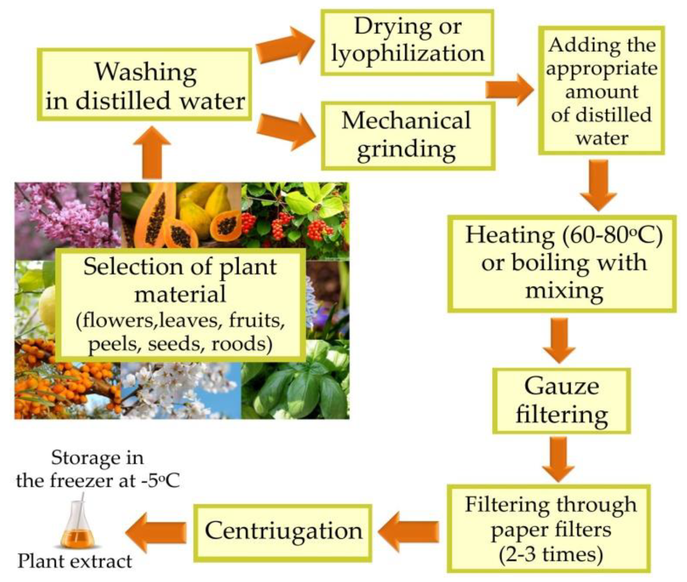
2. Biosynthesis of AuNPs Using Plants
2.1. Reduction Potential of Plant Extracts

| Compounds | Plant | Kind of Extract | References |
|---|---|---|---|
| Phenolic compounds, flavonoids | Crassocephalum rubens | Leaf; water extract | [37] |
| Anthocyanins | Cornus mas | Fruit; water extract | [47] |
| Anthocyanins | Punica granatum | Fruit; water extract | [48] |
| Cholidonic, superbine, colchicine, gloriosol, phytosterils and stigmasterin | Gloriosa superba | Leaf; water extract | [49] |
| Pectins, ribose, phenolic compounds | Papaver somniferum | Leaf; methanol extract | [52] |
| Amino acids, phenolic compounds, flavonoids | Artemisia capillaris | Whole plant; water extract | [53,54] |
| Glutanic acid, asparagine, leucine, lysine, glycine, alanine | Galaxaura elongata | Whole plant; water extract | [55,56] |
| Tannins | Black tea | Leaf; ethanol extract | [57] |
| Phenolic compounds | Medinilla speciosa | Fruit; water extract | [36] |
| Catechins, ascorbic acid | Mimosa tenuiflora | Tree bark; water/ethanol extract | [10] |
| Estragole, linalool, carvacral, urosalic acid, cirsimarin, rosmarinic acid | Ocinum sanctum | Flower and leaf; water extract | [60] |
| Genipin, genipol, geniposide, ronolazine | Genipa americana | Fruit; water extract | [61] |
| Citric and ascorbic acid | Lycopersicon esculentum | Fruit; water extract | [62] |
| Pectins | Musa paradisiaca | Fruit; water extract | [63,64,65,66,67,68,69,70] |
| Pectins | Citrus sinensis | Peels; water extract | [63,64,65,66,67,68,69,70] |
| Curcumin | Curcuma longa | Water solution | [71] |
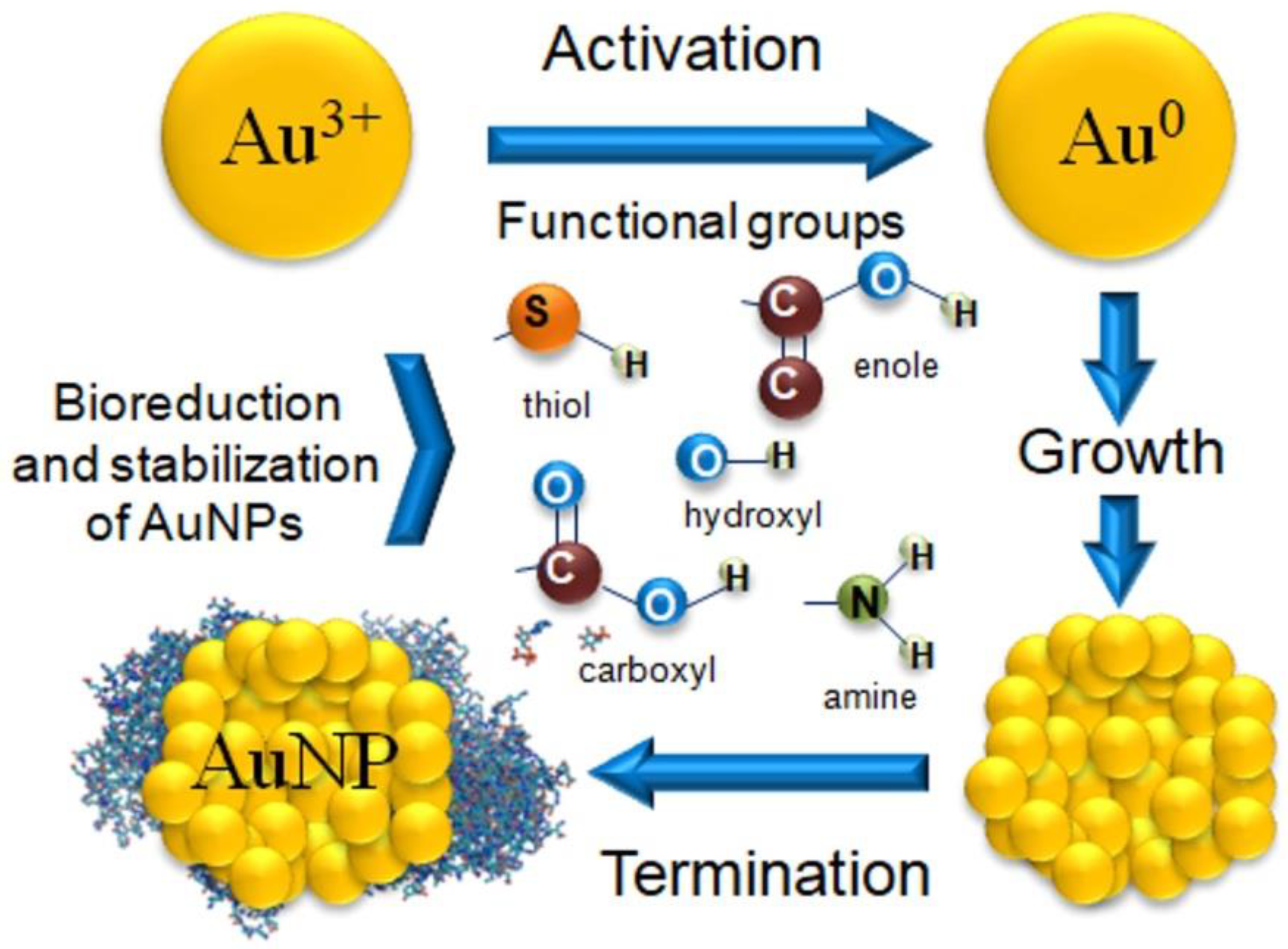
2.2. Mechanism of AuNPs Biosynthesis Using Plants
2.3. Conditions of Biosynthesis Reaction
2.3.1. Role of the Reactant Concentration
2.3.2. Role of pH
2.3.3. Role of Temperature
3. Antibacterial Activity of AuNPs Biosynthesized from Plants
3.1. Role of AuNPs Shape and Size
3.2. Role of AuNP Envelope
3.3. Composition of AuNP Envelope
3.4. Antibacterial Properties of Plant Extracts and Isolated Phytocompounds
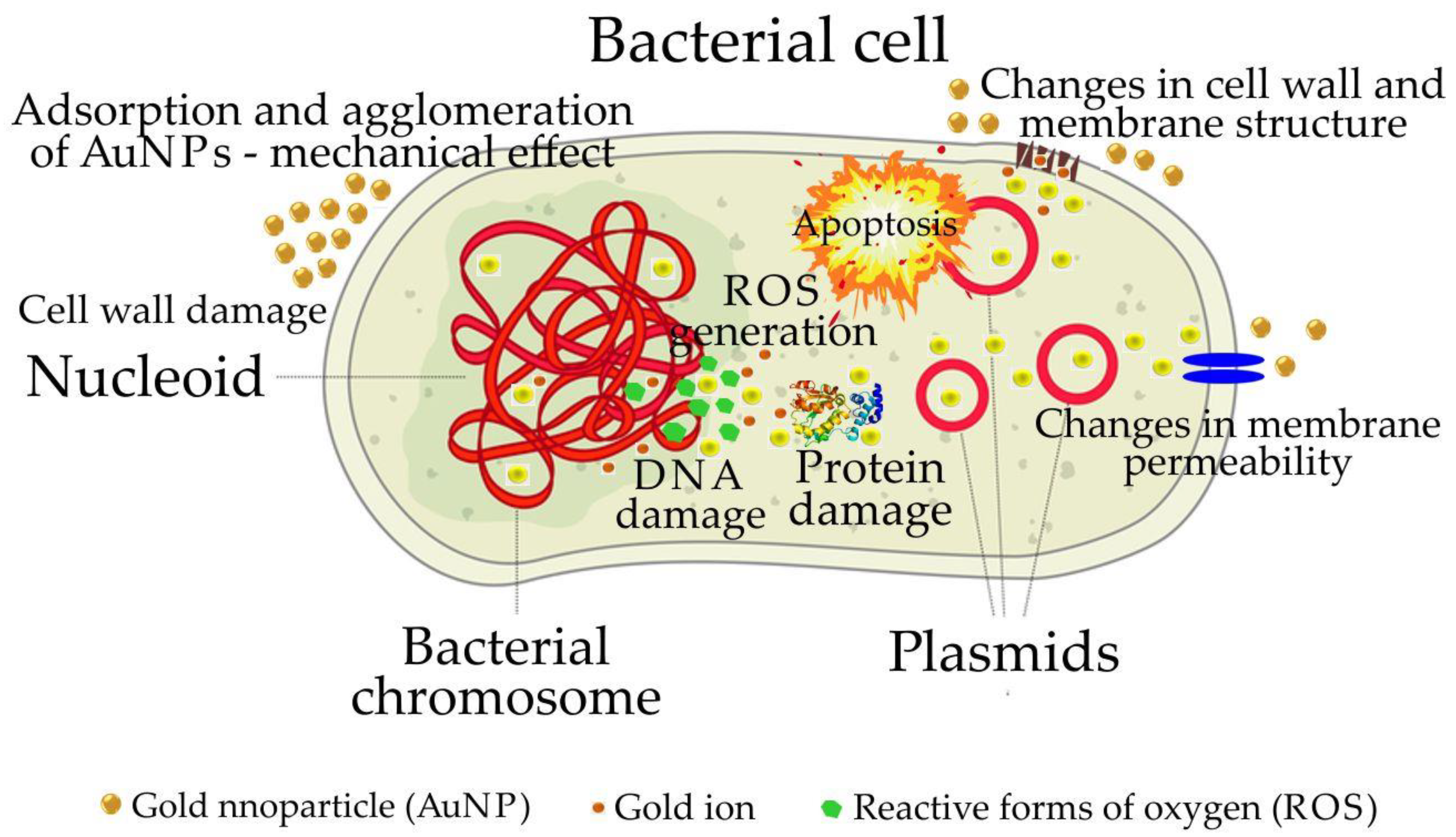
4. Mechanism of the Antibacterial Activity of AuNPs Synthesized from Plants
4.1. Damage to the Cell Wall
4.2. Damage of Proteins and DNA
4.3. Changes in the Expression of Metabolic Genes
5. Conclusions
Author Contributions
Funding
Institutional Review Board Statement
Informed Consent Statement
Data Availability Statement
Conflicts of Interest
References
- Chahardoli, A.; Karimi, N.; Sadeghi, F.; Fattahi, A. Green approach for synthesis of gold nanoparticles from Nigella arvensis leaf extract and evaluation of their antibacterial, antioxidant, cytotoxicity and catalytic activities. Artif. Cells Nanomed. Biotechnol. 2018, 46, 579–588. [Google Scholar] [CrossRef] [PubMed] [Green Version]
- Mikhailova, E.O. Gold nanoparticles: Biosynthesis and potential of biomedical application. J. Func. Biomater. 2021, 12, 70. [Google Scholar] [CrossRef] [PubMed]
- Singh, P.; Pandit, S.; Garnaes, J.; Tunjic, S.; Mokkapati, V.R.S.S.; Sultan, A.; Thygesen, A.; Mackevica, A.; Mateiu, R.V.; Daugaard, A.E.; et al. Green synthesis of gold and silver nanoparticles from Cannabis sativa (industrial hemp) and their capacity for biofilm inhibition. Int. J. Nanomed. 2018, 13, 3571–3591. [Google Scholar] [CrossRef]
- Devasvaran, K.; Lim, V. Green synthesis of metallic nanoparticles using pectin as a reducing agent: A synthetic review of the biological activities. Pharma. Biol. 2021, 59, 491–503. [Google Scholar] [CrossRef]
- Das, L.J.; Velusamy, P. Catalytic reduction of methylene blue using biegonic gold nanoparticles from Sesbania grandiflora. J. Inst. Chem. Eng. 2014, 45, 12285. [Google Scholar] [CrossRef]
- Elahi, N.; Kamali, M.; Baghersad, M.H. Recent biomedical applications of gold nanoparticles: A review. Talanta 2018, 184, 537–556. [Google Scholar] [CrossRef]
- Tian, E.-K.; Wang, Y.; Ren, R.; Zheng, W.; Liao, W. Gold nanoparticle: Recent progress on its antibacterial applications and mechanisms. J. Nanomater. 2021, 2021, 1–18. [Google Scholar] [CrossRef]
- Mobed, A.; Hasanzadeh, M.; Seidi, F. Anti-bacterial activity of gold nanoparticles as a new nanomaterial weapon to combat phatogenic agents: Recent advances and challenges. RSC Adv. 2021, 11, 34688–34698. [Google Scholar] [CrossRef]
- Timoszyk, A. A review of the biological synthesis of gold nanoparticles using fruit extracts: Scientific potential and application. Bull. Mater. Sci. 2018, 41, 154–165. [Google Scholar] [CrossRef] [Green Version]
- Rodríguez-León, E.; Rodríguez-Vázquez, B.E.; Martínez-Higuera, A.; Rodríguez-Beas, C.; Larios- Rodríguez, E.; Navarro, R.E.; López-Esparza, R.; Iñiguez-Palomares, A. Synthesis of gold nanoparticles using Mimosa tenuiflora extract, assessments of cytotoxicity, cellular uptake, and catalysis. Nanoscale Res. Lett. 2019, 14, 334. [Google Scholar] [CrossRef]
- Castro, L.; Blázquez, M.L.; Muńoz, J.A.; González, F.G.; Ballester, A. Mechanism and applications of metal nanoparticles prepared by bio-mediated process. Rev. Adv. Sci. Eng. 2014, 3, 199–216. [Google Scholar] [CrossRef]
- Stozhko, N.Y.; Bukharinova, M.A.; Khamzina, E.I.; Tarasov, A.V.; Vidrevich, M.B.; Brainina, K.Z. The effect of the antioxidant activity of plant extracts on the properties of gold nanoparticles. Nanomaterials 2019, 9, 1655. [Google Scholar] [CrossRef] [PubMed] [Green Version]
- Yazdani, E.; Talebi, M.; Zarshenas, M.M.; Moein, M. Evaluation of possible antioxidant activities of barberry solid formulation, a selected formulation from Traditional Persian Medicine (TPM) via various procedures. Biointer. Res. Appl. Chem. 2019, 9, 4517–4521. [Google Scholar] [CrossRef]
- Sathishkumar, P.; Gu, F.L.; Zhan, Q.; Palvannan, T.; Yusof, A.R.M. Flavonoids mediated ‘green’ nanomaterials: A novel nanomedicine system to treat various diseases—Current trends and future perspective. Mater. Lett. 2018, 210, 26–30. [Google Scholar] [CrossRef]
- Rao, Y.; Inwati, G.K.; Singh, M. Green synthesis of capped gold nanoparticles and their effect on Gram-positive and Gram-negative bacteria. Future Sci. OA 2017, 3, 239–254. [Google Scholar] [CrossRef] [Green Version]
- Naeem, G.A.; Jaloot, A.S.; Owaid, M.N.; Muslim, R.F. Green synthesis of gold nanoparticles from Coprinus comatus, Agaricaceae, and the effect of ultraviolent irradiation on their characteristics. Walailak J. Sci. Technol. 2021, 18, 9396. [Google Scholar] [CrossRef]
- Xu, X.Y.; Tran, T.H.M.; Perumalsamy, H.; Sanjeevram, D.; Kim, Y.-J. Biosynthetic gold nanoparticles of Hibiscus syriacus L. callus potentiates anti-inflammation efficacy via an autophagy-dependent mechanism. Mater. Sci. Eng. 2021, 124, 112035–112049. [Google Scholar] [CrossRef]
- Elia, P.; Zach, R.; Hazan, S.; Kolusheva, S.; Porat, Z.; Zeiri, Y. Green synthesis of gold nanoparticles using plant extracts as reducing agents. Int. J. Nanomed. 2014, 9, 4007–4021. [Google Scholar] [CrossRef] [Green Version]
- Ahmad, F.; Ashraf, N.; Ashraf, T.; Zhou, R.-B.; Yin, D.-C. Biological synthesis of metallic nanoparticles (MNPs) by plants and microbes: Their cellular uptake, biocompatibility, and biomedical applications. Appl. Microbiol. Biotechnol. 2019, 103, 2913–2935. [Google Scholar] [CrossRef]
- Kharissova, O.V.; Kharisov, B.I.; Oliva González, C.M.; Méndez, Y.P.; López, I. Greener synthesis of chemical compounds and materials. R. Soc. Open Sci. 2019, 6, 191378. [Google Scholar] [CrossRef]
- Ahn, S.; Singh, P.; Castro-Aceituno, V.; Yasmin Simu, S.; Kim, Y.; Mathiyalagan, R.; Yang, D. Gold nanoparticles synthesized using Panax ginseng leaves suppress inflammatory-mediators production via blockade of NF-kB activation in microphages. Artif. Cells Nanomed. Biotechnol. 2017, 45, 270–276. [Google Scholar] [CrossRef] [PubMed] [Green Version]
- Vadlapudi, V.; Kaladhar, D.S.V.G.K. Review: Green synthesis of silver and gold nanoparticles. Middle-East. J. Sci. Res. 2014, 19, 834–842. [Google Scholar] [CrossRef]
- Dahiya, D.; Nigam, P.S. The gut microbiota influenced by the intake of probiotics and functional foods with prebiotics can sustain wellness and alleviate certain ailments like gut-inflammation and colon-cancer. Microorganisms 2022, 10, 665. [Google Scholar] [CrossRef] [PubMed]
- Gupta, A.; Landis, R.F.; Rotello, V.M. Nanoparticle-based antimicrobials: Surface functionality is critical. F1000 Res. 2016, 5, 364–374. [Google Scholar] [CrossRef]
- Manivasagan, P.; Venkatesan, J.; Sivakumar, K.; Kim, S.-K. Pharmaceutically active secondary metabolites of marine actinobacteria. Microbiol. Res. 2014, 169, 262–278. [Google Scholar] [CrossRef] [PubMed]
- Zhang, J.; Mou, L.; Jiang, X. Surface chemistry of gold nanoparticles for health applications. Chem. Sci. 2020, 11, 923. [Google Scholar] [CrossRef] [Green Version]
- Princy, K.F.; Gopinath, A. Optimization of physicochemical parameters in the biofabrication of gold nanoparticles using marine macroalgae Padina tetrastromatica and its catalytic efficacy in the degradation of organic dyes. J. Nanostruc. Chem. 2018, 8, 333–342. [Google Scholar] [CrossRef] [Green Version]
- Correa, M.G.; Martínez, F.B.; Vidal, C.P.; Streitt, C.; Escrig, J.; de Dicastillo, C.L. Antimicrobial metal-based nanoparticles: A review on their synthesis, types and antimicrobial action. Beilstein J. Nanotechnol. 2020, 11, 1450–1469. [Google Scholar] [CrossRef] [PubMed]
- Li, J.; Xie, S.; Ahmed, S.; Wang, F.; Gu, Y.; Zhang, C.; Chai, X.; Wu, Y.; Cai, J.; Cheng, G. Antimicrobial activity and resistance: Influencing factors. Front. Pharmacol. 2017, 8, 364. [Google Scholar] [CrossRef] [Green Version]
- Silhavy, T.J.; Kahne, D.; Walker, S. The bacterial cell envelope. Cold Spring Harb. Perspect. Biol. 2010, 2, a000414. [Google Scholar] [CrossRef]
- Epand, R.M.; Walker, C.; Epand, R.F.; Magarvey, N.A. Molecular mechanisms of membrane targeting antibiotics. Biochim. Biophys. Acta 2016, 1858, 980–987. [Google Scholar] [CrossRef] [PubMed]
- Madkur, L.H. Ecofriendly green biosynthesized of metallic nanoparticles: Bio-reduction mechanism, characterization and pharmaceutical applications in biotechnology industry. Glob. Drug Ther. 2018, 3, 1–11. [Google Scholar] [CrossRef] [Green Version]
- Apak, R.; Özyürek, M.; Güçlü, K.; Çapanoğlu, E. Antioxidant activity/Capacity measurement. 2. Hydrogen atom transfer (HAT)-based, mixed-mode (electron transfer (ET)/HAT), and lipid peroxidation assays. J. Agric. Food Chem. 2016, 64, 1028–1045. [Google Scholar] [CrossRef] [PubMed]
- Pisoschi, A.M.; Cimpeanu, C.; Predoi, G. Electrochemical methods for total antioxidant capacity and its main contributors determination: A review. Open Chem. 2015, 13, 824–856. [Google Scholar] [CrossRef] [Green Version]
- Patil, M.P.; Kim, G.-D. Eco-friendly approach for nanoparticles synthesis and mechanism behind antibacterial activity of silver and anticancer activity of gold nanoparticles. Appl. Microbiol. Biotechnol. 2017, 101, 79–92. [Google Scholar] [CrossRef]
- Prihapsara, F.; Artanti, A.N.; Ni’mah, L.F.U. Characterization and antimicrobial activity of gold nanoparticles fruit infusion of Medinilla speciosa. J. Phys. Conf. Ser. 2022, 2190, 1–7. [Google Scholar] [CrossRef]
- Adewale, O.B.; Egbeyemi, K.A.; Onwuelu, J.O.; Potts-Johnson, S.S.; Anadozie, S.O.; Fadaka, A.O.; Osukoya, O.A.; Aluko, B.T.; Johnson, J.; Obafemi, T.O.; et al. Biological synthesis of gold and silver nanoparticles using leaf extracts of Crassocephalum rubens and their comparative in vitro antioxidant activities. Heliyon 2020, 6, e05501. [Google Scholar] [CrossRef]
- Jagessar, R.C. Plant extracts based nanoparticles, a good perspective in the development of drugs in nanomedicine. Mod. Appro. Drug Des. 2020, 3, 000556. [Google Scholar] [CrossRef]
- Javed, R.; Zia, M.; Naz, S.; Aisida, S.O.; UI Ain, N.; Ao, Q. Role of capping agents in the application of nanoparticles in biomedicine and environmental remediation: Recent trends and future prospects. J. Nanobiotechnol. 2020, 18, 172–187. [Google Scholar] [CrossRef]
- Makarov, V.V.; Love, A.J.; Sinitsyna, O.V.; Makarova, S.S.; Yaminsky, I.V.; Taliansky, M.E.; Kalinina, N.O. ‘Green’ nanotechnologies: Synthesis of metal nanoparticles using plants. Acta Nat. 2014, 6, 35–44. [Google Scholar] [CrossRef]
- Yasmin, A.; Ramesh, K.; Raje, S. Optimization and stabilization of gold nanoparticles by using herbal plant extract with microwave heating. Nano Converg. 2014, 1, 12. Available online: http://www.nanoconvergencejournal.com/content/1/1/12 (accessed on 11 April 2014). [CrossRef] [PubMed] [Green Version]
- Cherkas, A.; Holota, S.; Mdzinarashvili, T.; Gabbianelli, R.; Zarkovic, N. Glucose as a major antioxidant: When, what for and why it fails? Antioxidants 2020, 9, 140. [Google Scholar] [CrossRef] [PubMed] [Green Version]
- Ramrakhiani, L.; Ghosh, S. Metallic nanoparticle synthesized by biological route: Safer candidate for diverse applications. IET Nanobiotechnol. 2018, 12, 392–404. [Google Scholar] [CrossRef] [PubMed]
- Siddiqi, K.S.; Husen, A. Engineered gold nanoparticles and plant adaptation potential. Nanoscale Res. Lett. 2016, 11, 400–410. [Google Scholar] [CrossRef] [PubMed] [Green Version]
- Fanoro, O.T.; Oluwafemi, O.S. Bactericidal antibacterial mechanism of plant synthesized silver, gold and bimetallic nanoparticles. Pharmaceutics 2020, 12, 1044. [Google Scholar] [CrossRef] [PubMed]
- Hassanisaadi, M.; Bonjar, G.H.S.; Rahdar, A.; Pandey, S.; Hosseinipour, A.; Abdolshahi, R. Environmentally safe biosynthesis of gold nanoparticles using plant water extracts. Nanomaterials 2021, 11, 2033. [Google Scholar] [CrossRef]
- Olenic, L.; Chiorean, I. Synthesis, characterization and application of nanomaterials based on noble metallic nanoparticles and anthocyanins. Int. J. Latest Res. Sci. Technol. 2015, 4, 6–22. [Google Scholar]
- Nadagouda, M.N.; Iyanna, N.; Lalley, J.; Han, C.; Dionysiou, D.D.; Varma, R.S. Synthesis of silver and gold nanoparticles using antioxidants from blackberry, blueberry, pomegranate, and turmeric extracts. ACS Sustain. Chem. Eng. 2014, 2, 1717–1723. [Google Scholar] [CrossRef]
- Gopinath, K.; Karthika, V.; Gowri, S.; Senthilkumar, V.; Kumaresan, S.; Arumugam, A. Antibacterial activity of ruthenium nanoparticles synthesized using Gloriosa superba L. leaf extract. J. Nanosruc. Chem. 2014, 4, 83–89. [Google Scholar] [CrossRef] [Green Version]
- Lokina, S.; Narayanan, V. Antimicrobial and anticancer activity of gold nanoparticles synthesized from grapes fruit extract. Chem. Sci. Trans. 2013, 2, 105–110. [Google Scholar] [CrossRef]
- Gan, P.P.; Ng, S.H.; Huang, Y.; Li, S.F.Y. Green synthesis of gold nanoparticles using palm oil mill effluent (POME): A low-cost and eco-friendly viable approach. Bioresour. Technol. 2012, 113, 132–135. [Google Scholar] [CrossRef] [PubMed]
- Wali, M.; Sajjad, A.S.; Sumaira, S.; Muhammad, N.; Safia, H.; Muhammad, J. Green synthesis of gold nanoparticles and their characterizations using plant extract of Papaver somniferum. Nano Sci. Nano Technol. 2017, 11, 118–127. Available online: www.tsijournals.com (accessed on 7 September 2017).
- Lim, S.H.; Ahn, E.-Y.; Park, Y. Green synthesis and catalytic activity of gold nanoparticles synthesized by Artemisia capillaris water extract. Nanoscale Res. Lett. 2016, 11, 474. [Google Scholar] [CrossRef] [PubMed] [Green Version]
- Santhosh, P.B.; Genova, J.; Chamati, H. Green synthesis of gold nanoparticles: An eco-friendly approach. Chemistry 2022, 4, 345–369. [Google Scholar] [CrossRef]
- Abdel-Raouf, N.; Al-Enazi, N.M.; Ibraheem, I.B.M. Green biosynthesis of gold nanoparticle using Galaxaura elongata and characterization of their antibacterial activity. Arab. J. Chem. 2017, 10, 3029–3039. [Google Scholar] [CrossRef] [Green Version]
- Vijayaraghavan, K.; Nalini, S.P.K. Biotemplates in the green synthesis of silver nanoparticles. Biotechnol. J. 2010, 5, 1098–1110. [Google Scholar] [CrossRef]
- Teimuri-mofrad, R.; Hadi, R.; Tahmasebi, B.; Farhoudian, S.; Mehravar, M.; Nasiri, R. Green synthesis of gold nanoparticles using plant extract: Mini-review. Nanochem. Res. 2017, 2, 8–19. [Google Scholar] [CrossRef]
- Aljabali, A.A.A.; Akkam, Y.; Zoubi, M.S.A.; Al-Batayneth, K.M.; Al-Trad, B.; Alrob, O.A.; Alkilany, A.M.; Benamara, M.; Evans, D.J. Synthesis of gold nanoparticles using leaf extract of Ziziphus zizyphus and their antimicrobial activity. Nanomaterials 2018, 8, 174. [Google Scholar] [CrossRef] [Green Version]
- Boomi, P.; Ganesan, R.; Poorani, G.P.; Jegatheeswaran, S.; Balakumar, C.; Prabu, H.G.; Anand, K.; Prabhu, N.M.; Jeyakanthan, J.; Saravanan, M. Phyto-engineered gold nanoparticles (AuNPs) with potential antibacterial, antioxidant, and wound healing activities under in vitro and in vivo conditions. Int. J. Nanomed. 2020, 15, 7553–7568. [Google Scholar] [CrossRef]
- Sengani, M.; Grumezescu, A.M.; Rajeswari, V.D. Recent trends and methodologies in gold nanoparticle synthesis—A prospective review on drug delivery aspect. Open Nano 2017, 2, 37–46. [Google Scholar] [CrossRef]
- Kumar, B.; Smita, K.; Cumbal, L.; Camacho, J.; Hernández-Gallegos, E.; De Guadalupe Chávez-López, M.; Grijalva, M.; Andrade, K. One pot phytosynthesis of gold nanoparticles using Genipa americana fruit extract and its biological applications. Mater. Sci. Eng. C 2016, 62, 725–731. [Google Scholar] [CrossRef]
- Barman, G.; Maiti, S.; Laha, J.K. Bio-fabrication of gold nanoparticles using aqueous extract of red romato and its use as a colorimetric sensor. Nanoscale Res. Lett. 2013, 8, 181–190. Available online: www.nanoscalereslett.com/content/8/1/181 (accessed on 19 April 2013). [CrossRef] [PubMed]
- Ahmad, M.Z.; Abdel-Wahab, B.A.; Alam, A.; Zafar, S.; Ahmad, J.; Ahmad, F.J.; Midoux, P.; Pichon, C.; Akhter, S. Toxicity of inorganic nanoparticles used in targeted drug delivery and other biomedical application: An updated account on concern of biomedical nanotoxicology. J. Nanosci. Nanotechnol. 2016, 16, 7873–7897. [Google Scholar] [CrossRef]
- Borker, S.; Patole, M.; Moghe, A.; Pokharkar, V. Engineering of pectin-reduced gold nanoparticles for targeted delivery of an antiviral drug to macrophages: In vitro and in vivo assessment. Gold Bull. 2017, 50, 235–246. [Google Scholar] [CrossRef]
- Borker, S.; Pokharkar, V. Engineering of pectin-capped gold nanoparticles for delivery of doxorubicin to hepatocarcinoma cells: An insides into mechanism of cellular uptake. Artif. Cells Nanomed. Biotechnol. 2018, 46, 826–835. [Google Scholar] [CrossRef] [Green Version]
- Chen, H.; Dorrigan, A.; Saad, S.; Hare, D.J.; Cortie, M.B.; Valenzuela, S.M. In vivo study of spherical gold nanoparticles: Inflammatory effects and distribution in mice. PLoS ONE 2013, 8, e58208. [Google Scholar] [CrossRef] [Green Version]
- Nigoghossian, K.; dos Santos, M.V.; Barud, H.S.; da Silva, R.R.; Rocha, L.A.; Caiut, J.M.A.; de Assunção, R.M.N.; Spanhel, L.; Poulain, M.; Messaddeq, Y.; et al. Orange pectin mediated growth and stability of aqueous gold and silver nanocolloids. Appl. Surf. Sci. 2015, 341, 28–36. [Google Scholar] [CrossRef]
- Suganya, K.U.; Govindaraju, K.; Kumar, V.G.; Karthick, V.; Parthasarathy, K. Pectin mediated gold nanoparticles induces apoptosis in mammary adenocarcinoma cell lines. Int. J. Biol. Macromol. 2016, 93, 1030–1040. [Google Scholar] [CrossRef]
- Suganya, K.U.; Govindaraju, K.; Sivaraman, D.; Selvaraj, R.; Manikandan, R.; Kumar, V.G. Nanotoxicity assessment of functionalized gold nanoparticles in Sprague-Dawley rats. J. Clust. Sci. 2017, 28, 2933–2951. [Google Scholar] [CrossRef]
- Tan, H.; Chen, W.; Liu, Q.; Yang, G.; Li, K. Pectin oligosaccharides ameliorate colon cancer by regulating oxidative stress- and inflammation-activated signalling pathways. Front. Immunol. 2018, 9, 1504. [Google Scholar] [CrossRef] [Green Version]
- Patra, D.; Kurdi, R.E. Curcumin as a novel reducing and stabilizing agent for the green synthesis of metallic nanoparticles. Green Chem. Lett. Rev. 2021, 14, 474–487. [Google Scholar] [CrossRef]
- Gonelimali, F.D.; Lin, J.; Miao, W.; Xuan, J.; Charles, F.; Chen, M.; Hatab, S.R. Antimicrobial properties and mechanism of action of some plant extracts against food pathogens and spoilage microorganisms. Front. Microbiol. 2018, 9, 1639. [Google Scholar] [CrossRef] [PubMed]
- Sani, A.; Cao, C.; Cui, D. Toxicity of gold nanoparticles (AuNPs): A review. Biochem. Biophys. Rep. 2021, 26, 100991. [Google Scholar] [CrossRef] [PubMed]
- Sanchez-Mendieta, V.; Vilchis-Nestor, A.R. Green Synthesis of Noble Metal (Au, Ag, Pt) Nanoparticles, Assisted by Plant-Extracts; Su, Y.-H., Ed.; IntechOpen: Rijeka, Croatia, 2012; pp. 391–408. [Google Scholar] [CrossRef] [Green Version]
- Heuer-Jungemann, A.; Feliu, N.; Bakaimi, I.; Hamaly, M.; Alkilany, A.; Chakraborty, I.; Masood, A.; Casula, M.F.; Kostopoulou, A.; Oh, E.; et al. The role of ligands in the chemical synthesis and applications of inorganic nanoparticles. Chem. Rev. 2019, 119, 4819–4880. [Google Scholar] [CrossRef] [Green Version]
- Singh, A.K.; Srivastava, O.N. One-step green synthesis of gold nanoparticles using black cardamom and effect of pH on its synthesis. Nanoscale Res. Lett. 2015, 10, 353. [Google Scholar] [CrossRef] [Green Version]
- Rajan, A.; Vilas, V.; Philip, D. Studies on catalytic, antioxidant, antibacterial and anticancer activities of biogenic gold nanoparticles. J. Mol. Liq. 2015, 212, 331–339. [Google Scholar] [CrossRef]
- Ghodake, G.S.; Deshpande, N.G.; Lee, Y.P.; Jin, E.S. Pear fruit extract-assisted room-temperature biosynthesis of gold nanoplates. Colloids Surf. B 2010, 75, 584–589. [Google Scholar] [CrossRef]
- Amina, S.J.; Guo, B. A review on the synthesis and functionalization of gold nanoparticles as a drug delivery vehicle. Int. J. Nanomed. 2020, 15, 9823–9857. [Google Scholar] [CrossRef]
- Kumari, M.; Mishra, A.; Pandey, S.; Singh, S.P.; Chaudhry, V.; Mudiam, M.K.R.; Shukla, S.; Kakkar, P.; Nautiyal, C.S. Physico-chemical condition optimization during biosynthesis lead to development of improved and catalytically efficient gold nano particles. Sci. Rep. 2016, 6, 27575. [Google Scholar] [CrossRef] [Green Version]
- Babu, P.J.; Sharma, P.; Saranya, S.; Tamuli, R.; Bora, U. Green synthesis and characterization of biocompatible gold nanoparticles using Solanum indicum fruits. Nanomater. Nanotechnol. 2013, 3, 4. [Google Scholar] [CrossRef] [Green Version]
- Pantidos, N.; Horsfall, L.E. Biological synthesis of metallic nanoparticles by bacteria, fungi and plants. J. Nanomed. Nanotechnol. 2014, 5, 233–243. [Google Scholar] [CrossRef]
- Kuthi, N.A.; Chandren, S.; Basar, N.; Jamil, M.S.S. Biosynthesis of gold nanoisotrops using Carallia brachiata leaf extract and their catalytic application in the reduction of 4-nitrophenol. Front. Chem. 2022, 9, 800145–800160. [Google Scholar] [CrossRef] [PubMed]
- Ahmad, T.; Bustam, M.A.; Irfan, M.; Moniruzzaman, M.; Asghar, H.M.A.; Bhattacharjee, S. Effect of volume of gold chloroauric acid on size, shape and stability of biosynthesized AuNPs using aqueous Elaeis guineensis (oil palm) leaves extract. Int. J. Automot. Mech. Eng. 2018, 15, 5135–5145. [Google Scholar] [CrossRef]
- Mariychuk, R.; Grulova, D.; Grishchenko, L.M.; Linnik, R.P.; Lisnyak, V.V. Green synthesis of non-spherical gold nanoparticles using Solidago canadensis L. extract. Appl. Nanosci. 2020, 10, 4817–4826. [Google Scholar] [CrossRef]
- Mittal, J.; Batra, A.; Singh, A.; Sharma, M.M. Phytofabrication of nanoparticles through plant as nanofactories. Adv. Natural Sci. Nanosci. Nanotechnol. 2014, 5, 043002. [Google Scholar] [CrossRef]
- Vijayaraghavan, K.; Mahadevan, A.; Sathishkumar, M.; Pavagadhi, S.; Balasubramanian, R. Biosynthesis of Au(0) from Au(III) via biosorption and bioreduction using brown marine alga Turbinaria conoides. Chem. Eng. J. 2011, 167, 223–227. [Google Scholar] [CrossRef]
- Kaykhaii, M.; Haghpazir, N.; Walisadeh, J. Biosynthesis of gold nanoparticles using aqueous extract of stem of Periploca aphylla plant. J. Nanostruct. 2018, 8, 152–158. [Google Scholar] [CrossRef]
- Zhang, Y.; Dasari, T.P.S.; Deng, H.; Yu, H. Antimicrobial activity of gold nanoparticles and ionic gold. J. Environ. Sci. Health C 2015, 33, 286–327. [Google Scholar] [CrossRef]
- Wang, L.; Hu, C.; Shao, L. The antimicrobial activity of nanoparticles: Present situation and prospects for the future. Int. J. Nanomed. 2017, 2017, 1227–1249. [Google Scholar] [CrossRef] [Green Version]
- Lavaee, F.; Ranjbar, Z.; Modarezi, F.; Keshavarz, F. The effect of gold nanoparticles with different sizes on Streptococcus species. J. Dent. 2021, 4, 235–242. Available online: https://dentjods.sums.ac.ir/article_47735.html (accessed on 1 December 2021).
- Zhu, S.; Shen, Y.; Yu, Y.; Bai, X. Synthesis of antibacterial gold nanoparticles with different particle sizes using chlorogenic acid. R. Soc. Open Sci. 2020, 7, 191141–191151. [Google Scholar] [CrossRef] [PubMed] [Green Version]
- Shamaila, S.; Zafar, N.; Riaz, S.; Sharif, R.; Nazir, J.; Naseem, S. Gold nanoparticles: An efficient antimicrobial agent against enteric bacterial human pathogen. Nanomaterials 2016, 6, 71. [Google Scholar] [CrossRef] [PubMed]
- Gouyau, J.; Duval, R.E.; Boudier, A.; Lamouroux, E. Investigation of nanoparticle metallic core antibacterial activity: Gold and silver nanoparticles against Escherichia coli and Staphylococcus aureus. Int. J. Mol. Sci. 2021, 22, 1905. [Google Scholar] [CrossRef] [PubMed]
- Fatima, F.; Pathak, N.; Verma, S.R.; Bajpai, P. In vitro antimicrobicidal and cytotoxicity efficacy of gold nanoparticles synthesized from Alternaria brassicae (KF934409). SOJ Pharm. Pharm. Sci. 2016, 3, 1–6. [Google Scholar] [CrossRef] [Green Version]
- Botteon, C.E.A.; Silva, L.B.; Ccana-Ccapatinta, G.V.; Silva, T.S.; Ambrosio, S.R.; Veneziani, R.C.S.; Bastos, J.K.; Marcato, P.D. Biosynthesis and characterization of gold nanoparticles using Brazilian red propolis and evaluation of its antimicrobial and anticancer activities. Sci. Rep. 2021, 11, 1974–1990. [Google Scholar] [CrossRef]
- Srinath, B.S.; Namratha, K.; Byrappa, K. Eco-friendly synthesis of gold nanoparticles by gold mine bacteria Brevibacillus formosus and their antibacterial and biocompatible studies. IOSR J. Pharm. 2017, 7, 53–60. [Google Scholar]
- Hamelian, M.; Varmira, K.; Veisi, H. Green synthesis and characterizations of gold nanoparticles using Thyme and survey cytotoxic effect, antibacterial and antioxidant potential. J. Photochem. Photobiol. B 2018, 184, 71–79. [Google Scholar] [CrossRef]
- Wongyay, K.; Wintachai, P.; Maungchang, R.; Rattanakit, P. Exploration of the antimicrobial and catalytic properties of gold nanoparticles greenly synthesized by Cryptolepis buchanani Roem and schult extract. J. Nanomater. 2020, 2020, 1–11. [Google Scholar] [CrossRef]
- Akintelu, S.A.; Yao, B.; Folorunso, A.S. Green synthesis, characterization, and antibacterial investigation of synthesized gold nanoparticles (AuNPs) from Garcinia kola pulp extract. Plasmonics 2020, 16, 1–9. [Google Scholar] [CrossRef]
- Award, M.A.; Eisa, N.E.; Virk, P.; Hendi, A.A.; Ortashi, K.M.O.O.; Mahgoub, A.A.S.A.; Elobeid, M.A.; Eissa, F.Z. Green synthesis of gold nanoparticles: Preparation, characterization, cytotoxicity, and anti-bacterial activities. Mater. Lett. 2019, 256, 126608. [Google Scholar] [CrossRef]
- Soshnikova, V.; Kim, Y.J.; Singh, P.; Huo, Y.; Markus, J.; Ahn, S.; Castro-Aceituno, V.; Kang, J.; Chokkalingam, M.; Mathiyalagan, R.; et al. Cardamom fruits as a green resource for facile synthesis of gold and silver nanoparticles and their biological applications. Artif. Cells Nanomed. Biotechnol. 2018, 46, 108–117. [Google Scholar] [CrossRef] [PubMed] [Green Version]
- Patra, J.K.; Baek, K.H. Novel green synthesis of gold nanoparticles using Citrullus lanatus rind and investigation of proteasome inhibitory activity, antibacterial, and antioxidant potential. Int. J. Nanomed. 2015, 10, 7253–7264. [Google Scholar] [CrossRef] [Green Version]
- Vinayagam, R.; Santhoshkumar, M.; Lee, K.E.; David, E.; Kang, S.G. Bioengineered gold nanoparticles using Cynodon dactylon extract and its cytotoxicity and antibacterial activities. Bioprocess Biosys. Eng. 2021, 44, 1253–1262. [Google Scholar] [CrossRef]
- El-Borady, O.M.; Ayat, M.S.; Shabrawy, M.A.; Millet, P. Green synthesis of gold nanoparticles using Parsley leaves extract and their biological studies. Adv. Powder Technol. 2020, 31, 4390–4400. [Google Scholar] [CrossRef]
- Islam, N.U.; Jalil, K.; Shahid, M.; Rauf, A.; Muhammad, N.; Khan, A.; Shah, M.R.; Khan, M.A. Green synthesis and biological activities of gold nanoparticles functionalized with Salix alba. Arab. J. Chem. 2019, 12, 2914–2925. [Google Scholar] [CrossRef] [Green Version]
- Balasubramanian, S.; Kala, S.M.J.; Pushparaj, T.L. Biogenic synthesis of gold nanoparticles using Jasminum auriculatum leaf extract and their catalytic, antimicrobial and anticancer activities. J. Drug Deliv. Sci. Technol. 2020, 57, 101620–101630. [Google Scholar] [CrossRef]
- Muthuvel, A.; Adavallan, K.; Balamurugan, K.; Krishnakumar, N. Biosynthesis of gold nanoparticles using Solanum nigrum leaf extract and screening their free radical scavenging and antibacterial properties. Biomed. Prev. Nutr. 2014, 4, 325–332. [Google Scholar] [CrossRef]
- López-Miranda, J.L.; Esparza, R.; Rosas, G.; Pérez, R.; Estévez-González, M. Catalytic and antibacterial properties of gold nanoparticles synthesized by a green approach for bioremediation applications. 3 Biotech 2019, 9, 135–144. [Google Scholar] [CrossRef]
- Mata, R.; Bhaskaran, A.; Sadras, S.R. Green-synthesized gold nanoparticles from Plumeria alba flower extract to augment catalytic degradation of organic dyes and inhibit bacterial growth. Particuology 2016, 24, 78–86. [Google Scholar] [CrossRef]
- Anbu, P.; Gopinath, S.C.B.; Jayanthi, S. Synthesis of gold nanoparticles using Platycodon grandiflorum extract and its antipathogenic activity under optimal conditions. Nanomater. Nanotechnol. 2020, 10, 1–9. [Google Scholar] [CrossRef]
- Moustafa, N.E.; Alomari, A.A. Green synthesis and bactericidal activities of isotropic and anisotropic spherical gold nanoparticles produced using Peganum harmala L. leaf and seed extracts. Biotechnol. Appl. Biochem. 2019, 66, 664–672. [Google Scholar] [CrossRef] [PubMed]
- Hatipoğlu, A. Rapid green synthesis of gold nanoparticles: Synthesis, characterization, and antimicrobial activities. Prog. Nutr. 2021, 23, e2021242–e2021249. [Google Scholar] [CrossRef]
- Folorunso, A.; Akintelu, S.; Oyebamiji, A.K.; Ajayi, S.; Abiola, B.; Abdusalam, I.; Morakinyo, A. Biosynthesis, characterization and antimicrobial activity of gold nanoparticles from leaf extracts of Annona muricata. J. Nanostruct. Chem. 2019, 9, 111–117. [Google Scholar] [CrossRef]
- Arief, S.; Nasution, F.W.; Zulhadjri; Labanni, A. High antibacterial properties of green synthesized gold nanoparticles using Uncaria gambir Roxb. leaf extract and triethanolamine. J. Appl. Pharma. Sci. 2020, 10, 124–130. [Google Scholar] [CrossRef]
- Senthilkumar, S.; Kashinath, L.; Ashok, M.; Rajendran, A. Antibacterial properties and mechanism of gold nanoparticles obtained from Pergularia daemia leaf extract. J. Nanomed. Res. 2017, 6, 00146. [Google Scholar] [CrossRef]
- Dang, H.; Fawcett, D.; Poinern, G.E.J. Green synthesis of gold nanoparticles from waste macadamia nut shells and their antimicrobial activity against Escherichia Coli Staphylococcus Epidermis. Int. J. Res. Med. Sci. 2019, 7, 1171–1177. [Google Scholar] [CrossRef]
- Kamala Nalini, S.P.; Vijayaraghavan, K. Green synthesis of silver and gold nanoparticles using Aloe vera gel and determining its antimicrobial properties on nanoparticle impregnated cotton fabric. J. Nanotechnol. Res. 2020, 2, 042–050. [Google Scholar] [CrossRef]
- Kasabwala, H.; Ganapathy, D.; Shanmugam, R. Green preparation of gold nano particles procured from Neem and Ginger plant extract to evaluate the antimicrobial activity against oral aerobic pathogens. J. Res. Med. Dental Sci. 2021, 9, 259–262. [Google Scholar]
- Zeybek, Z.; Dogruoz, N.; Karagoz, A. Antibacterial activity of some plant extracts. IUFS J. Biol. 2008, 67, 17–21. Available online: https://dergipark.org.tr/en/pub/iufsjb/issue/9052/112897 (accessed on 11 November 2010).
- De Zoysa, M.H.N.; Rathnayake, H.; Hewawasam, R.P.; Wijayaratne, W.M.D.G.B. Determination of in vitro antimicrobial activity of five Sri Lankan medicinal plants against selected human pathogenic bacteria. Int. J. Microbiol. 2019, 2019, 7431439. [Google Scholar] [CrossRef] [Green Version]
- Moghaddam, P.Z.; Mohammadi, A.; Alesheikh, P.; Feyzi, P.; Haghbin, A.; Mollazadeh, S.; Sabeti, Z.; Nakhlband, A.; Kasaian, J. Antibacterial, antifungal, and antioxidant activity of Cleome coluteoides: An in vitro comparative study between leaves, stems, and flowers. Turk. J. Pharma. Sci. 2021, 18, 10–16. [Google Scholar] [CrossRef]
- Chamsa-ard, W.; Fawcett, D.; Fung, C.C.; Poinern, G.E.J. Biogenic synthesis of gold nanoparticles from waste watermelon and their antibacterial activity against Escherichia coli and Staphylococcus epidermidis. Int. J. Res. Med. Sci. 2019, 7, 2499–2505. [Google Scholar] [CrossRef]
- Shittu, O.K.; Stephen, D.I.; Kure, A.H. Functionalization of biosynthesized gold nanoparticle from aqueous leaf extract of Catharanthus roseus for antibacterial stadium. Afr. J. Biomed. Res. 2017, 20, 195–202. [Google Scholar]
- Bindhu, M.R.; Umadevi, M. Antibacterial activities of green synthesized gold nanoparticles. Mater. Lett. 2014, 120, 122–125. [Google Scholar] [CrossRef]
- Kureshi, A.A.; Vaghela, H.M.; Kumar, S.; Singh, R.; Kumari, P. Green synthesis of gold nanoparticles mediated by Garcinia fruits and their biological applications. Pharm. Sci. 2021, 27, 238–250. [Google Scholar] [CrossRef]
- Boruah, J.S.; Devi, C.; Hazarika, U.; Reddy, P.V.B.; Chowdhury, D.; Barthakur, M.; Kalita, P. Green synthesis of gold nanoparticles using an antiepileptic plant extract: In vitro biological and photo-catalytic activities. RSC Adv. 2021, 11, 28029. [Google Scholar] [CrossRef] [PubMed]
- Dorosti, N.; Jamshidi, F. Plant-mediated gold nanoparticles by Dracocephalum kotschyi as anticholinesterase agent: Synthesis, characterization, and evaluation of anticancer and antibacterial activity. J. Appl. Biomed. 2016, 14, 235–245. [Google Scholar] [CrossRef]
- Pandey, S.; Goswami, G.K.; Nanda, K.K. Green synthesis of polysaccharide/gold nanoparticle nanocomposite: An efficient ammonia sensor. Carbohydr. Polym. 2013, 94, 229–234. [Google Scholar] [CrossRef] [PubMed]
- Hayden, S.C.; Zhao, G.; Saha, K.; Phillips, R.L.; Li, X.; Miranda, O.R.; Rotello, V.M.; El-Sayed, M.A.; Schmidt-Krey, I.; Bunz, U.H.F. Aggregation and interaction of cationic nanoparticles on bacterial surfaces. J. Am. Chem. Soc. 2012, 134, 6920–6923. [Google Scholar] [CrossRef] [PubMed]
- Timoszyk, A.; Niedbach, J.; Śliżewska, P.; Mirończyk, A.; Kozioł, J.J. Eco-friendly and temperature dependent biosynthesis of gold nanoparticles using the bacterium Pseudomonas aeruginosa: Characterization and antibacterial activity. J. Nano Res. 2017, 48, 114–124. [Google Scholar] [CrossRef]
- Badwaik, V.D.; Vangala, L.M.; Pender, D.S.; Willis, C.B.; Aguilar, Z.P.; Gonzalez, M.S.; Paripelly, R.; Dakshinamurthy, R. Size-dependent antimicrobial properties of sugar-encapsulated gold nanoparticles synthesized by a green method. Nanoscale Res. Lett. 2012, 7, 623–634. [Google Scholar] [CrossRef] [Green Version]
- Okkeh, M.; Bloise, N.; Restivo, E.; De Vita, L.; Pallavicini, P.; Visai, L. Gold nanoparticles: Can they be the next magic bullet for multidrug-resistant bacteria? Nanomaterials 2021, 11, 312. [Google Scholar] [CrossRef] [PubMed]
- Piktel, E.; Suprewicz, Ł.; Depciuch, J.; Chmielewska, S.; Skłodowski, K.; Daniluk, T.; Król, G.; Kołat-Brodecka, P.; Bijak, P.; Pajor-Świerzy, A.; et al. Varied-shaped gold nanoparticles with nanogram killing efficiency as potential antimicrobial surface coatings for the medical devices. Sci. Rep. 2021, 11, 12546–12566. [Google Scholar] [CrossRef]
- Penders, J.; Stolzoff, M.; Hickey, D.J.; Andersson, M.; Webster, T.J. Shape-dependent antibacterial effects of non-cytotoxic gold nanoparticles. Int. J. Nanomed. 2017, 12, 2457. [Google Scholar] [CrossRef] [PubMed] [Green Version]
- Sabourian, P.; Yazdani, G.; Ashraf, S.S.; Frounchi, M.; Mashayekhan, S.; Kiani, S.; Kakkar, A. Effect of physico-chemical properties of nanoparticles on their intracellular uptake. Int. J. Mol. Sci. 2020, 21, 8019. [Google Scholar] [CrossRef]
- Xia, Q.; Li, H.; Xiao, K. Factors affecting the pharmacokinetics, biodistribution and toxicity of gold nanoparticles in drug delivery. Curr. Drug Metab. 2016, 17, 849–861. [Google Scholar] [CrossRef] [PubMed]
- Gustafson, H.H.; Holt-Casper, D.; Grainger, D.W.; Ghandehari, H. Nanoparticle uptake: The phagocyte problem. Nano Today 2015, 10, 487–510. [Google Scholar] [CrossRef] [PubMed] [Green Version]
- Mishra, A.; Tripathy, S.K.; Yun, S.-I. Bio-synthesis of gold and silver nanoparticles from Candida guilliermondii and their antimicrobial effect against pathogenic bacteria. J. Nanosci. Nanotechnol. 2011, 11, 243–248. [Google Scholar] [CrossRef]
- Tao, C. Antimicrobial activity and toxicity of gold nanoparticles: Research progress, challenges and prospects. Lett. Appl. Microbiol. 2018, 67, 537–543. [Google Scholar] [CrossRef] [PubMed] [Green Version]
- Devendiran, R.M.; Chinnaiyan, S.K.; Yadav, N.K.; Moorthy, G.K.; Ramanathan, G.; Singaravelu, S.; Sivagnanam, U.T.; Perumal, P.T. Green synthesis of folic acid-conjugated gold nanoparticles with pectin as reducing/stabilizing agent for cancer theranostics. RSC Adv. 2016, 6, 29757–29768. [Google Scholar] [CrossRef]
- Reena, K.; Prabakaran, M.; Leeba, B.; Gajendiran, M.; Antony, S.A. Green synthesis of pectin-gold-PLA-PEG-PLA nanoconjugates: In vitro cytotoxicity and anti-inflammatory activity. J. Nanosci. Nanotechnol. 2017, 17, 4549–4557. [Google Scholar] [CrossRef]
- Alsamhary, K.; Al-Enazi, N.; Alshehri, W.A.; Ameen, F. Gold nanoparticles synthesized by flavonoid tricetin as a potential antibacterial nanomedicine to treat respiratory infections causing opportunistic bacterial pathogens. Microb. Pathog. 2020, 139, 103928. [Google Scholar] [CrossRef] [PubMed]
- Avíla, S.R.R.; Schuenck, G.P.D.; Costa e Silva, L.P.; Keijok, W.S.; Xavier, L.M.; Endringer, D.C.; Oliveira, J.P.; Schuenck, R.P.; Guimarães, M.C.C. High antibacterial in vitro performance of gold nanoparticles synthesized by epigallocatechin 3-gallate. J. Mater. Res. 2021, 36, 518–532. [Google Scholar] [CrossRef]
- Niño-Martínez, N.; Salas Orozco, M.F.; Martínez-Castañón, G.-A.; Torres Méndez, F.; Ruiz, F. Molecular mechanisms of bacterial resistance to metal and metal oxide nanoparticles. Int. J. Mol. Sci. 2019, 20, 2808. [Google Scholar] [CrossRef] [PubMed] [Green Version]
- Pajerski, W.; Ochonska, D.; Brzychczy-Wloch, M.; Indyka, P.; Jarosz, M.; Golda-Cepa, M.; Sojka, Z.; Kotarba, A. Attachment efficiency of gold nanoparticles by Gram-positive and Gram-negative bacterial strains governed by surface charges. J. Nanopart. Res. 2019, 21, 186. [Google Scholar] [CrossRef] [Green Version]
- Vijilvani, C.; Bindhu, M.; Frincy, F.; AlSalhi, M.S.; Sabitha, S.; Saravanakumar, K.; Devanesan, S.; Umadevi, M.; Aljaafreh, M.J.; Atif, M. Antimicrobial and catalytic activities of biosynthesized gold, silver and palladium nanoparticles from Solanum nigrum leaves. J. Photochem. Photobiol. 2020, 202, 111713. [Google Scholar] [CrossRef]
- Hameed, S.; Wang, Y.; Zhao, L.; Ying, Y. Shape-dependent significant physical mutilation and antibacterial mechanisms of gold nanoparticles against foodborne bacterial pathogens (Escherichia coli, Pseudomonas aeruginosa and Staphylococcus aureus) at lower concentrations. Mater. Sci. Eng. C 2020, 108, 110338. [Google Scholar] [CrossRef] [PubMed]
- Sadeghi, B.; Garmaroudi, F.S.; Hashemi, M.; Nezhad, H.R.; Nasrollahi, A.; Ardalan, S. Comparison of the anti-bacterial activity on the nanosilver shapes. Adv. Powder Technol. 2012, 23, 22–26. [Google Scholar] [CrossRef]
- Etebu, E.; Arikekpar, I. Antibiotics: Classification and mechanisms of action with emphasis on molecular perspectives. Int. J. Appl. Microbiol. Biotechnol. Res. 2016, 4, 90–101. [Google Scholar]
- Aromal, S.A.; Philip, D. Green synthesis of gold nanoparticles using Trigonella foenum-graecum and its size-dependent catalytic activity. Spectrochim. Acta A 2012, 97, 1–5. [Google Scholar] [CrossRef]
- Dobrzynska, M.; Napierala, M.; Florek, E. Flavonoid nanoparticles: A promising approach for cancer therapy. Biomolecules 2020, 10, 1268. [Google Scholar] [CrossRef] [PubMed]
- Kumar, H.; Bhardwaj, K.; Cruz-Martins, N.; Nepovimova, E.; Oleksak, P.; Dhanjal, D.S.; Bhardwaj, S.; Singh, R.; Chopra, C.; Verma, R.; et al. Applications of fruit polyphenols and their functionalized nanoparticles against foodborne bacteria: A mini review. Molecules 2021, 26, 3447. [Google Scholar] [CrossRef]
- Saputra, I.S.; Suhartati, S.; Yulizar, Y.; Sudirman, S. Synthesis and characterization of gold nanoparticles (AuNPs) by utilizing bioactive compound of Imperata cylindrica (L.) Raeusch. J. Kim. Terap. Indones. 2020, 22, 1–7. [Google Scholar]
- Wahab, A.W.; Karim, A.; Asmawati; Sutapa, I.W. Bio-synthesis of gold nanoparticles through bioreduction using the aqueous extract of Muntingia calabura L. leaves. Orient. J. Chem. 2018, 34, 401–409. [Google Scholar] [CrossRef] [Green Version]
- Jasmine, R.; Selvakumar, B.N.; Daisy, P. Investigating the mechanism of action of terpenoids and the effect of interfering substances on an Indian medicinal plant extract demonstrating antibacterial activity. Int. J. Pharm. Stud. Res. 2011, 2, 19–24. [Google Scholar]
- Rajakumar, G.; Gomathi, T.; Rahuman, A.A.; Thiruvengadam, M.; Mydhili, G.; Kim, S.-H.; Lee, T.-J.; Chung, I.-M. Biosynthesis and biomedical applications of gold nanoparticles using Eclipta prostrata leaf extract. Appl. Sci. 2016, 6, 222. [Google Scholar] [CrossRef]
- Nascimento, G.G.F.; Locatelli, J.; Freitas, P.C.; Silva, G.L. Antibacterial activity of plant extracts and phytochemicals on antibiotic resistant bacteria. Braz. J. Microbiol. 2000, 31, 247–256. [Google Scholar] [CrossRef]
- Andriamampianina, H.L.; Rakoto, D.A.D.; Petit, T.; Ramanankierana, H.; Randrianarivo, H.R.; Jeannoda, V.L. Antimicrobial activity of extracts from Crotalaria bernieri Baill. (Fabaceae). Afr. J. Microbiol. Res. 2016, 10, 1229–1239. [Google Scholar] [CrossRef] [Green Version]
- Farahmandfar, R.; Kenari, R.E.; Asnaashari, M.; Shahrampour, D.; Bakhshandeh, T. Bioactive compounds, antioxidant and antimicrobial activities of Arum maculatum leaves extracts as affected by various solvents and extraction methods. Food Sci. Nutr. 2018, 7, 465–475. [Google Scholar] [CrossRef]
- Araya-Contreras, T.; Veas, R.; Escobar, C.A.; Machuca, P.; Bittner, M. Antibacterial effect of Luma apiculata (DC.) burret extracts in clinically important bacteria. Int. J. Microbiol. 2019, 2019, 1–7. [Google Scholar] [CrossRef] [Green Version]
- Viktorová, J.; Kumar, R.; Řehořová, K.; Hoang, L.; Ruml, T.; Figueroa, C.R.; Valdenegro, M.; Fuentes, L. Antimicrobial activity of extracts of two native fruits of Chile: Arrayan (Luma apiculata) and Puemo (Cryptocarya alba). Antibiotics 2020, 9, 444. [Google Scholar] [CrossRef] [PubMed]
- Suparno, O.; Afifah, A.; Panandita, T.; Marimin; Purnawati, R. The potency of plant extracts as antimicrobials for the skin or hide preservation. AIP Conf. Proc. 2017, 1823, 020098-1–020098-6. [Google Scholar] [CrossRef] [Green Version]
- Toker, G.; Baser, K.H.C.; Özek, T. The composition of the essential oils from Tilia, L. species growing in Turkey. J. Essent. Oli Res. 1999, 11, 369–374. [Google Scholar] [CrossRef]
- Elakremi, M.; Sillero, L.; Ayed, L.; Labidi, J.; Moussaoui, Y. Chemical composition of leaves and hull from Pistacia vera L. an evaluation of phenolic content and antioxidant properties of their extracts. Res. Sq. 2020, 1–16. [Google Scholar] [CrossRef]
- Ushimaru, P.I.; da Silva, M.T.N.; Di Stasi, L.C.; Barbosa, L.; Fernandes, A., Jr. Antibacterial activity of medicinal plant extracts. Braz. J. Microbiol. 2007, 38, 717–719. [Google Scholar] [CrossRef] [Green Version]
- Louis, H.; Linus, M.N.; Israt, A.; Innocent, J.; Amos, P.I.; Magu, T.O. Antimicrobial activity of stem, leave and root plant extract of Sclerocarya birrea and Sterculia setigera against some selected microorganisms. WSN Int. Sci. J. 2018, 92, 309–326. [Google Scholar]
- Restuati, M.; Pratiwi, N.; Widiyastuti, G. Effect of leaf extract Buasbuas (Premna pubescens Blume) for against of bacteria growth Staphylococcus aureus and Salmonella sp. in vitro. AIP Conf. Proc. 2017, 1868, 090007. [Google Scholar] [CrossRef] [Green Version]
- Cox, S.; Abu-Ghannam, N.; Gupta, S. An assessment of the antioxidant and antimicrobial activity of six species of edible Irish seaweeds. Int. Food Res. J. 2010, 17, 205–220. [Google Scholar]
- Dizaj, S.M.; Lotfipour, F.; Barzegar-Jalali, M.; Zarrintan, M.H.; Adibkia, K. Antimicrobial activity of the metals and metal oxide nanoparticles. Mater. Sci. Eng. C 2014, 44, 278–284. [Google Scholar] [CrossRef]
- Ortiz-Benítez, E.A.; Velázquez-Guadarrama, N.; Durán-Figueroa, N.V.; Quezada, H.; Olivares-Trejo, J.J. Antibacterial mechanism of gold nanoparticles on Streptococcus pneumonia. Metallomics 2019, 11, 1265–1276. [Google Scholar] [CrossRef]
- Katas, H.; Lim, C.S.; Azlan, A.Y.H.N.; Buang, F.; Busra, M.F.M. Antibacterial activity of biosynthesized gold nanoparticles using biomolecules from Lignosus rhinocerotis and chitosan. SPJ 2018, 27, 283–292. [Google Scholar] [CrossRef] [PubMed]
- Patra, J.K.; Kwon, Y.; Baek, K.-H. Green biosynthesis of gold nanoparticles by onion peel extract: Synthesis, characterization and biological activities. Adv. Powder Technol. 2016, 27, 2204–2213. [Google Scholar] [CrossRef]
- Rasola, A.; Bernardi, P. Mitochondrial permeability transition in Ca(2+)-dependent apoptosis and necrosis. Cell Calcium 2011, 50, 222–233. [Google Scholar] [CrossRef]
- Hajnoczky, G.; Csordas, G.; Das, S.; Garcia-Perez, C.; Saotome, M.; Sinha Roy, S.; Yi, M. Mitochondrial calcium signalling and cell death: Approaches for assessing the role of mitochondrial Ca2+ uptake in apoptosis. Cell Calcium 2006, 40, 553–560. [Google Scholar] [CrossRef] [Green Version]
- Slavin, Y.N.; Asnis, J.; Hafeli, U.O.; Bach, H. Metal nanoparticles: Understanding the mechanisms behind antibacterial activity. J. Nanobiotechnol. 2017, 15, 65. [Google Scholar] [CrossRef] [PubMed]
- Li, H.; Chen, Q.; Zhao, J.; Urmila, K. Enhancing the antimicrobial activity of natural extraction using the synthetic ultra small metal nanoparticles. Sci. Rep. 2015, 5, 11033. [Google Scholar] [CrossRef] [Green Version]
- Luan, B.; Huynh, T.; Zhou, R. Complete wetting of graphene by biological lipids. Nanoscale 2016, 8, 5750–5754. [Google Scholar] [CrossRef]
- Armentano, I.; Arciola, C.R.; Fortunati, E.; Ferrari, D.; Mattioli, S.; Amoroso, C.F.; Rizzo, J.; Kenny, J.M.; Imbriani, M.; Visai, L. The interaction of bacteria with engineered nanostructured polymeric materials: A review. Sci. World J. 2014, 410423, 1–18. [Google Scholar] [CrossRef] [Green Version]
- Linklater, D.P.; Baulin, V.A.; Le Guével, X.; Fleury, J.-B.; Hanssen, E.; Nguyen, H.P.; Juodkazis, S.; Bryant, G.; Crawford, R.J.; Stoodley, P.; et al. Antibacterial action of nanoparticles by lethal stretching of bacterial cell membranes. Adv. Mater. 2020, 32, 2005679. [Google Scholar] [CrossRef]
- Gahlawat, G.; Choudhury, A.R. A review on the biosynthesis of metal and metal salt nanoparticles by microbes. RSC Adv. 2019, 9, 12944–12967. [Google Scholar] [CrossRef] [Green Version]
- Radzig, M.A.; Nadtochenko, V.A.; Koksharova, O.A.; Kiwi, J.; Lipasova, V.A.; Khmel, I.A. Antibacterial effects of silver nanoparticles on Gram-negative bacteria: Influence on the growth and biofilms formation, mechanisms of action. Colloids Surf. B 2013, 102, 300–306. [Google Scholar] [CrossRef] [PubMed]
- Siddiqi, N.J.; Abdelhalim, M.A.; El-Ansary, A.K.; Alhomida, A.S.; Ong, W.Y. Identification of potential biomarkers of gold nanoparticle toxicity in rat brains. J. Neuroinflamm. 2012, 11, 12546. [Google Scholar] [CrossRef] [Green Version]
- Rai, A.; Prabhune, A.; Perry, C.C. Antibiotic mediated synthesis of gold nanoparticles with potent antimicrobial activity and their application in antimicrobial coatings. J. Mater. Chem. 2010, 20, 6789–6798. [Google Scholar] [CrossRef] [Green Version]
- Cui, Y.; Zhao, Y.; Tian, Y.; Zhang, W.; Lü, X.; Jiang, X. The molecular mechanism of action of bactericidal gold nanoparticles on Escherichia coli. Biomater. 2012, 33, 2327–2333. [Google Scholar] [CrossRef]
- Dash, S.S.; Sen, I.K.; Dash, S.K. A review on the plant extract mediated green syntheses of gold nanoparticles and its anti-microbial, anti-cancer and catalytic applications. Int. Nano Lett. 2022, 12, 47–66. [Google Scholar] [CrossRef]
- Zada, S.; Ahmad, A.; Khan, S.; Iqbal, A.; Ahmad, S.; Ali, H.; Fu, P. Biofabrication of gold nanoparticles by Lyptolyngbya JSC-1 extract as super reducing and stabilizing agents: Synthesis, characterization and antibacterial activity. Microb. Pathog. 2018, 114, 116–123. [Google Scholar] [CrossRef] [PubMed]
- Amini, A.; Kamali, M.; Amini, B.; Najafi, A. Enhanced antibacterial activity of imipenem immobilized on surface of spherical and rod gold nanoparticles. J. Phys. D 2018, 52, 065401. [Google Scholar] [CrossRef]
- Shah, M.; Badwaik, V.; Kherde, Y.; Waghwani, H.K.; Modi, T.; Aguilar, Z.P.; Rogers, H.; Hamilton, W.; Marutharaj, T.; Webb, C.; et al. Gold nanoparticles: Various methods of synthesis and antibacterial applications. Front. Biosci. 2014, 19, 1320–1344. [Google Scholar] [CrossRef] [Green Version]
- Perni, S.; Piccirillo, C.; Pratten, J.; Prokopovich, P.; Chrzanowski, W.; Parkin, I.P.; Wilson, M. The antimicrobial properties of light-activated polymers containing methylene blue and gold nanoparticles. Biomaterials 2009, 30, 89–93. [Google Scholar] [CrossRef]
- Dayem, A.A.; Hossain, M.K.; Lee, S.B.; Kim, K.; Saha, S.K.; Yang, G.-M.; Choi, H.Y.; Cho, S.-G. The role of reactive oxygen species (ROS) in the biological activities of metallic nanoparticles. Int. J. Mol. Sci. 2017, 18, 120. [Google Scholar] [CrossRef] [Green Version]
- Lee, H.; Lee, D.G. Gold nanoparticles induce a reactive oxygen species-independent apoptotic pathway in Escherichia coli. Colloids Surf. B 2018, 167, 1–7. [Google Scholar] [CrossRef] [PubMed]
- Schröfel, A.; Kratošová, G.; Šafarík, I.; Šafaríková, M.; Raška, I.; Shor, L.M. Applications of biosynthesized metallic nanoparticles—A review. Acta Biomater. 2014, 10, 4023–4042. [Google Scholar] [CrossRef] [PubMed]


| Plant | Kind of Extract | Reducing and Stabilizing Agent | Shape and Size of AuNPs | Bacterial Strains (MIC [μg/mL]) | Mechanism of Antibacterial Activity | Another Activity | References |
|---|---|---|---|---|---|---|---|
| Ocimum tenuiflorum | Flower and leaf; water extract | Phenolic compounds | Spherical; 20–25 nm | S. aureus, P. aeruginosa, K. pneumoniae (512–600) | Disturbing the membrane permeability and cell metabolism | - | [15] |
| Azadiriachta indica | Leaf; water extract | ||||||
| Mentha spicata | Leaf; water extract | ||||||
| Citrus sinensis | Peel; water extract | Spherical; 25–30 nm | |||||
| Nigella arvensis | Leaf; water extract | Flavones, proteins | Spherical, triangular, pentagonal, hexagonal; 3–37 nm | E. coli, B. subtilis, S. aureus (62.5–125) | Cell wall structure and cell respiration disruption | Antioxidant, cytotoxic, catalytic capacity | [1] |
| Thymus vulgaris | Whole plant | Unknown | Spherical; 6–35 nm | B. subtilis (15.62) | Unknown | Antioxidant; non-toxic | [98] |
| Cryptolepis buchanani | Leaf; water extract | Phenolic compounds | Spherical; 11.1 nm | S. aureus, A. baumannii (0.209) | Unknown | Catalytic capacity | [99] |
| Cola acuminata | Fruits; water pulp | Flavonoids, terpenoids | Spherical; 14–22 nm | S. aureus, E. coli, P. aeruginosa, B. cereus | Unknown | - | [100] |
| Olea europaea Acacia nilotica | Fruit and husk; mixture of water extracts | Phenolic compounds, terpenoids | Spherical; 45 nm | E. coli, Bacillus sp., P. aeruginosa, Klebsiella sp. | Unknown | Cytotoxic | [101] |
| Pistacia atlantica | Leaf and fruit, water extract | Unknown | Spherical; 50–60 nm | S. aureus, E. coli | Unknown | Antioxidant, cytotoxic | [98] |
| Amomum villosum | Fruit; water extract | Phenolic compounds, tannins, terpenoids | Spherical; 10–15 nm | S. aureus, E. coli | Unknown | Antioxidant, cytotoxic, catalytic capacity | [102] |
| Cannabis sativa | Stem; water extract | Cannabinoids, terpenoids, flavonoids, phenolic compounds | Spherical, triangular, elongated, hexagonal; 12–18 nm | S. epidermidis, P. aeruginosa, E. coli (25–50) | Unknown | - | [9] |
| Citrullus lanatus | Fruit; water extract | Citrulline, proteins, carotenoids | Spherical; 20–140 nm | B. cereus, S. aureus, S. typhimurium | Unknown | - | [103] |
| Medinilla speciosa | Fruit; water infusion | Phenolic compounds, amino acids | Spherical; 200–450 nm | P. aeruginosa, S. aureus | Oxidative stress | - | [36] |
| Cynodon dactylon | Whole plant; water extract | Unknown | Spherical; 200–450 nm | E. cloacae, B. cereus, S. haemolyticus, S. petrasii | Oxidative stress | Cytotoxic | [104] |
| Areca catechu | Nuts; water extract | Flavonoids, proteins | Spherical; 13.7 nm | E. coli, S. aureus, K. pneumonia, P. aeruginosa | Unknown | Antioxidant, catalytic capacity, cytotoxic | [77] |
| Petroselinum crispum | Leaf; water extract | Unknown | Spherical, semi-rod, flower-shaped; 17–50 nm | Enterobacter ludwigii | Unknown | - | [105] |
| Salix alba | Leaf; water extract | Amides, proteins | Spherical; 50–80 nm | S. aureus | Unknown | Antifungal | [106] |
| Jasminum auriculatum | Leaf; water extract | Unknown | Spherical; 8–37 nm | S. pyogenes, S. aureus, E. coli, K. pneumonia | Unknown | Catalytic capacity, antifungal, cytotoxic | [107] |
| Solanum nigrum | Leaf; water extract | Unknown | Spherical; 50 nm | S. saprophyticus, B. subtilis, E. coli, P. aeruginosa | Unknown | Antioxidant | [108] |
| Aloysia triphylla | Leaf; water extract | Flavonoids | Spherical; 40–60 nm | E. coli, S. aureus (50) | Unknown | Catalytic capacity | [109] |
| Plumeria alba | Flower; water extract | Unknown | Spherical; 28, 3.4 nm | E. coli (400) | Unknown | Catalytic capacity | [110] |
| Platycodon grandiflorum | Leaf; water extract | Flavonoids | Spherical; 15 nm | E. coli, B. subtilis (10) | Unknown | - | [111] |
| Peganum harmala | Leaf; water extract | Polyphenols, alcohols | Spherical; 43.44 nm | E. coli, S. aureus | Unknown | - | [112] |
| Allium ampeloprasum | Leaf; water extract | Phenolic, aromatic compounds, proteins | Spherical; 22.76 nm | S. aureus, B. subtilis, E. coli, P. aeruginosa | Cell wall, mitochondrial and ribosome damage | Antifungal | [113] |
| Annona muricata | Leaf; water extract | Flavonoids, terpenoids, proteins | Spherical; 25.5 nm | C. sporogenes, S. aureus, E. faecalis, K. pneumonia | Unknown | Antifungal | [114] |
| Uncaria gambir | Leaf; water extract | Unknown | Spherical, triangular, hexagonal; 32.52 nm | E. coli, S. aureus (50) | Cell wall structure disruption | - | [115] |
| Pergularia daemia | Leaf; water extract | Unknown | Spherical; 3–15 nm | E. coli, B. subtilis, P. aeruginosa (300) | Membrane permeability and cell respiration disruption | - | [116] |
| Macadamia | Nut shell; water extract | Unknown | Spherical, triangular, hexagonal; 50–200 nm | E. coli, S. epidermidis | Unknown | - | [117] |
| Aloe vera | Leaf; water extract | Alcohols, phenolic compounds | Spherical; <15 nm | E. coli, S. aureus (10) | Unknown | - | [118] |
| Azadirachta indica, Zingiber officinale | Leaf; mixed water extracts | Unknown | Spherical, triangular, hexagonal; 32.52 nm | S. mutans, S. aureus, E. faecalis | Unknown | - | [119] |
| Tilia argentea | Leaf; water: PBS extract | Unknown | Unknown | E. coli, B. subtilis, K. pneumoniae, Aeromonas sp., S. aureus (250) | Unknown | - | [120] |
| Hibiscus sabelariffa | Flower; water and ethanol extracts | Unknown | Unknown | E. coli, S. aureus, P. aeruginosa, V. parahaemolyticus, S. enteritidis, B. cereus | Changes in internal pH of bacterial cell | - | [72] |
| Syzygium aromaticum | Flower; water and ethanol extracts | ||||||
| Rosmarinus officinalis | Leaf; water and ethanol extracts | ||||||
| Epaltes divaricata | Whole plant; ethanol extract | Unknown | Unknown | S. aureus | Unknown | - | [121] |
| Whole plant; hexane extract | |||||||
| Whole plant; water extract | |||||||
| Vetiveria zizanioides | Root; ethanol extract | Unknown | Unknown | S. aureus | Unknown | - | [121] |
| Root; hexane extract | |||||||
| Cleome coluteoides | Stem; methanol extract | Polyphenols | Unknown | B. cereus, S. aureus | Unknown | - | [122] |
| Flower; dichloromethane, methanol, ethyl acetate extracts | |||||||
| Leaf; dichloromethane, methanol, ethyl acetate extracts |
| Compound/Kind of Solution | Plant | Shape and Size of AuNPs | Bacterial Strains (MIC [μg/mL]) | Mechanism of Antibacterial Activity | Other Activities | Function | References |
|---|---|---|---|---|---|---|---|
| Pectin; water solution | Musa paradisiaca | Spherical; 8 nm | - | - | Cytotoxic | Anticancer drug | [64,65,68,69] [141,142] |
| Orange peels | Spherical; 7–13 nm | Anti- inflammatory | Drug carrier | ||||
| Flavonoid, tricetin | pollen of Myrtaceae | Spherical; 12 nm | B. licheniformis, S. aureus, A. pittii, E. xiangfangensis, E. fergusonii, P. mirabilis, P. aeruginosa, A. enteropelogenes | Unknown | Biocompatibility | Chitozan carrier, antibacterial drug | [143] |
| Polyphenol, epigallocatechin 3-gallate; water solution | Green tea | Spherical | S. aureus, E. coli, E. faecalis, P. aeruginosa (15–120) | Mechanical destruction of cell wall, cell lysis | Non-cytotoxic | Antibacterial drug | [144] |
| Dextrose; water solution | Corn | Spherical; 25, 60, 120 nm | E. coli | Cell wall structure disruption, cell lysis | - | Antibacterial drug | [132] |
| Plant | Kind of Extract | Extract Composition | Bacterial Strains | Mechanism of Antibacterial Activity | References |
|---|---|---|---|---|---|
| Crotalaria bernieri | Leaf; hexane extract | Tannins, polyphenols, steroids, triterpenes, unsaturated sterols | S. aureus | Unknown | [159] |
| Leaf; methanol extract | Alkaloids, flavonoids, tannins, polyphenols | E. aerogenes, P. aeruginosa, V. parahaemolyticus, P. mirabilis, B. cereus, C. perfringens, S. aureus, S. pneumonia, S. pyogenes | Unknown | ||
| Seed; hexane extract | Tannins, polyphenols, steroids, triterpenes, unsaturated sterols | V. parahaemolyticus, S. aureus | Unknown | ||
| Seed; ethyl acetate extract | Flavonoids, tannins, polyphenols, steroids, triterpenes, unsaturated sterols | E. cloacae, S. aureus, V. parahaemolyticus, P. mirabilis, S. pneumoniae, S. pyogenes | Unknown | ||
| Seed; methanol extract | Flavonoids, tannins, polyphenols, steroids, triterpenes, unsaturated sterols | Y. enterocolitica, S. pneumoniae, S. pyogenes | Unknown | ||
| Pod; hexane extract | Steroids, triterpenes, unsaturated sterols | E. aerogenes, S. aureus, C. perfringens | Unknown | ||
| Pod; ethyl acetate extract | Polyphenols, steroids, triterpenes, unsaturated sterols | V. parahaemolyticus, S. aureus, B. cereus, C. perfringens, S. pyogenes, S. pneumoniae | Unknown | ||
| Crotalaria bernieri | Pod; methanol extract | Alkaloids, flavonoids | S. enteridis, S. flexneri, V. parahaemolyticus, P. mirabilit, P. mirabilis, S. aureus,S. pneumoniae, S. pyogenes | Unknown | [159] |
| Root; hexane extract | Tannins, polyphenols, steroids, triterpenes, unsaturated sterols | P. aeruginosa, S. enteridis, S. pyogenes | Unknown | ||
| Root; ethyl acetate extract | Tannins, polyphenols, steroids, triterpenes, unsaturated sterols | V. parahaemolyticus, B. cereus,C. Perfringens, S. aureus, S. pneumoniae, S. pyogenes | Unknown | ||
| Root; methanol extract | Saponins, tannins, polyphenols | S. enteridis, P. mirabilis, B. cereus, C. perfringens, S. aureus, S. pneumoniae, S. pyogenes | Unknown | ||
| Arum maculatum | Leaf; water extract | Phenols, tannins, tocopherols, flavonoids, beta-carotene | E. coli, S. aureus, L. monocytogene, S. enteritidis, P. aeruginosa | Unknown | [160] |
| Leaf; water: ethanol (50:50) extract | |||||
| Leaf; ethanol extract | |||||
| Luma apiculata | Leaf; hexane extract | Catechins, flavonoids, glycosyloxyflavone, triterpenoids | S. aureus, S. epidermidis, S. saprophyticus, E. coli A. boumanii, P. aeruginosa, Enterococcus sp. | Unknown | [161,162] |
| Flower; hexane extract | S. epidermidis, S. aureus, S. saprophyticus, Enterococcus sp | Unknown | |||
| Green tea; epigallocatechin 3-gallate | Leaf; water solution | Polyphenol | S. aureus, E. coli, E. faecalis, P. aeruginosa | Mechanical destruction of cell wall; cell lysis | [144] |
| Areca catechu | Nuts; water extract | Flavonoids, proteins | E. cloacae, S. haemolyticus, S. petrasii, B. cereus, Enterobacter sp. | Unknown | [77] |
| Salix alba | Leaf; hydro-alcoholic fraction | Salicin | S. aureus, K. pneumoniae, B. sublitis | Unknown | [106] |
| Thymus vulgaris | Leaf and flower; hydro-alcoholic fraction | Thymol, carvacrol, flavonoids, tannins, triterpenes | P. aeruginosa, Proteus sp. | Unknown | [158] |
| Rosmarinus officinalis | Leaf; hydro-alcoholic fraction | Flavonoids, phenolic acids (caffeic, chorogenic, rosmarinic), essential oils (camphor, cineole), diterpenes (carnosol) | B. subtilis | Unknown | |
| Syzygyum joabolanum | Leaf; hydro-alcoholic fraction | Flavonoids, tannins | S. aureus, K. pneumoniae | Unknown | |
| Punica granatum | Pericarp; hydro-alcoholic fraction | Ellagitannins, alkaloids | P. aeruginosa, B. subtilis | Unknown | |
| Psidium guajava | Leaf; hydro-alcoholic fraction | Comarins, essencial oils, flavonoids, triterpenes, ellagitannins | S. aureus | Unknown | |
| Ocimum basilicum | Leaf; hydro-alcoholic fraction | Essential oils (linalol, estragol, eugenol), tannins, flavonoids | P. aeruginosa | Unknown | |
| Hibiscus sabdariffa | Flower; water extract | Phenolic compounds, terpenoids, esters, weak and fatty acids | E. coli, V. parahaemolyticus, P. aeruginosa, S. enteritidis, B. cereus, S. aureus | Membrane potential changes | [72] |
| Flower; ethanol extract | |||||
| Syzygium aromaticum | Flower; water extract | Phenolic compounds, terpenoids, esters, weak and fatty acids | E. coli, V. parahaemolyticus, P. aeruginosa, S. enteritidis, B. cereus, S. aureus | Membrane potential changes | [72] |
| Flower; ethanol extract | |||||
| Moringa oleifera | Leaf; ethanol extract | Alkaloids, saponins, tannins, phenolics, flavonoids, triterpenoids, steroids, glycosides | S. aureus, E. coli | Unknown | [163] |
| Magnolia acuminata | Leaf; ethanol extract | Alkaloids, saponins, tannins, phenolics, flavonoids, triterpenoids, steroids, glycosides | S. aureus, E. coli | Unknown | |
| Prunus cerasus | Leaf; ethanol extract | Alkaloids, saponins, tannins, phenolics, flavonoids, triterpenoids, steroids, glycosides | S. aureus, E. coli | Unknown | |
| Leucaena leucocephala | Leaf; ethanol extract | Alkaloids, saponins, tannins, phenolics, flavonoids, triterpenoids, steroids, glycosides | S. aureus, E. coli | Unknown | |
| Tilia argentea | Leaf; water extract | Flavonoids, phenolic compounds, esters, terpenes, aliphatic acids, hydrocarbons | E. coli, S. aureus, E. pneumoniae, B. subtilis | Unknown | [120,164] |
| Anthemis pungens | Leaf; water extract | Phenolic compounds, flavonoids | E. coli, S. aureus | Unknown | [120] |
| Pistacia sp. | Leaf; water extract | Flavonoids, phenolic compounds, carboxylic acids, aromatic compounds | E. coli, B. subtilis | Unknown | [120,165] |
| Epaltes divaricata | Whole plant; water extract | Tannins, phenolic compounds, saponins, cardiac glycosides, flavonoids, alkaloids | S. aureus | Unknown | [121] |
| Asparagus falcatus | Tuber; hexane extract Tuber; ethanol extract | Tannins, phenolic compounds, saponins, cardiac glycosides, flavonoids | S. aureus | Unknown | |
| Asteracantha longifolia | Whole plant; hexane extract Whole plant; ethanol extract | Tannins, phenolic compounds, saponins, cardiac glycosides, flavonoids, alkaloids | S. aureus | Unknown | |
| Vetiveria zizanioides | Root; hexane extract Root; ethanol extract | Tannins, phenolic compounds, saponins, cardiac glycosides, flavonoids, alkaloids | S. aureus | High concentration of low-polarity compounds in the extract | |
| Coriandrum sativum | Seed; hexane extract Seed; ethanol extract | Tannins, phenolic compounds, cardiac glycosides, flavonoids, alkaloids | S. aureus | Unknown |
Publisher’s Note: MDPI stays neutral with regard to jurisdictional claims in published maps and institutional affiliations. |
© 2022 by the authors. Licensee MDPI, Basel, Switzerland. This article is an open access article distributed under the terms and conditions of the Creative Commons Attribution (CC BY) license (https://creativecommons.org/licenses/by/4.0/).
Share and Cite
Timoszyk, A.; Grochowalska, R. Mechanism and Antibacterial Activity of Gold Nanoparticles (AuNPs) Functionalized with Natural Compounds from Plants. Pharmaceutics 2022, 14, 2599. https://doi.org/10.3390/pharmaceutics14122599
Timoszyk A, Grochowalska R. Mechanism and Antibacterial Activity of Gold Nanoparticles (AuNPs) Functionalized with Natural Compounds from Plants. Pharmaceutics. 2022; 14(12):2599. https://doi.org/10.3390/pharmaceutics14122599
Chicago/Turabian StyleTimoszyk, Anna, and Renata Grochowalska. 2022. "Mechanism and Antibacterial Activity of Gold Nanoparticles (AuNPs) Functionalized with Natural Compounds from Plants" Pharmaceutics 14, no. 12: 2599. https://doi.org/10.3390/pharmaceutics14122599