Review

Gut Dysbiosis and Diabetic Foot Ulcer: Role of Probiotics

1
School of Pharmaceutical Sciences, Lovely Professional University, Phagwara 144411, India
2
Faculty of Health, Australian Research Centre in Complementary and Integrative Medicine, University of Technology Sydney, Ultimo, NSW 2007, Australia
3
Department of Life Sciences, School of Pharmacy, International Medical University, Bukit Jalil, Kuala Lumpur 57000, Malaysia
4
School of Pharmacy, Suresh Gyan Vihar University, Mahal Road, Jaipur 302017, India
5
Department of Pharmacology, Saveetha Dental College, Saveetha Institute of Medical and Technical Sciences, Saveetha University, Chennai 602105, India
6
Uttaranchal Institute of Pharmaceutical Sciences, Uttaranchal University, Dehradun 248007, India
7
School of Health Sciences, The University of Tasmania, Launceston, TAS 7248, Australia
8
Discipline of Pharmacy, Graduate School of Health, University of Technology Sydney, Ultimo, NSW 2007, Australia
*
Authors to whom correspondence should be addressed.
Pharmaceutics 2022, 14(11), 2543; https://doi.org/10.3390/pharmaceutics14112543
Submission received: 30 September 2022 / Revised: 7 November 2022 / Accepted: 19 November 2022 / Published: 21 November 2022
(This article belongs to the Special Issue Targeted Therapies in Diabetes and Its Complications)

Abstract

:
Diabetic foot ulcer (DFU) is a multifactorial disease and one of the complications of diabetes. The global burden of DFU in the health sector is increasing at a tremendous rate due to its cost management related to hospitalization, medical costs and foot amputation. Hence, to manage DFU/DWs, various attempts have been made, including treating wounds systematically/topically using synthetic drugs, herbal drugs, or tissue engineering based surgical dressings. However, less attention has been paid to the intrinsic factors that are also the leading cause of diabetes mellitus (DM) and its complications. One such factor is gut dysbiosis, which is one of the major causes of enhancing the counts of Gram-negative bacteria. These bacteria produce lipopolysaccharides, which are a major contributing factor toward insulin resistance and inflammation due to the generation of oxidative stress and immunopathy. These all lead to DM and DFU. Probiotics are the commercial form of beneficial gut microbes that are taken as nutraceuticals by people of all ages to improve gut immunity and prevent gut dysbiosis. However, the role of probiotics has been less explored in the management of DFU. Hence, the therapeutic potential of probiotics in managing DFU is fully described in the current review. This report covers the linkage between gut dysbiosis and DFU, sources of probiotics, the mechanisms of probiotics in DW healing, and the impact of probiotic supplementation in treating DFU. In addition, techniques for the stabilization of probiotics, market status, and patents related to probiotics have been also covered. The relevant data were gathered from PubMed, Scopus, Taylor and Francis, Science Direct, and Google Scholar. Our systematic review discusses the utilization of probiotic supplementation as a nutraceutical for the management of DFU.

1. Introduction

Diabetic foot ulcer (DFU) is the one of the most common complications of diabetes. The global prevalence of DFU due to diabetes is 25%. It is an open sore wound that occurs in the foot. It generally occurs due to the hypoxia and oxidative stress caused by reactive oxygen species, a decrease in the level of growth factors (GFs), nucleic acids and the lack of glycemic control. DFU has reached the 10th position in terms of the annual economic burden of diabetics [1]. this situation has arisen because of a lack of existing treatment strategies to promote wound healing. In DFU, delayed wound healing occurs [2]. The common reason for this is the extended inflammatory response that leads to impairment in keratinocyte migration, collagen synthesis, vascularization, fibroblast migration, epithelialization, collagen proliferation, differentiation and migration. Overall, these contributing factors often result in amputation and even the death of the DFU patient. The global prevalence of amputation due to DFU in 2022 is reported to be 10–15% [3].
The treatment of DFU is challenging, as it involves multiple stages, etiologies and degrees of severity that vary among the diabetic mellitus (DM) patients. The existing formulations on the market provide adequate glycemic control. However, these are unable to treat the various stages of DFU in DM patients. Therefore, this increases the burden of medications on patients suffering to DFU, because the delay in wound healing may also be dependent on the severity of the wound, rather than only glycemic control. Hence, for wound healing, the administration of antibiotics or anti-inflammatory agents is also required. Other approaches that are used to manage DFU include plastic surgery, orthopedics, vascular surgery, offloading, antibiotics (ciprofloxacin, vancomycin, clindamycin and piperacillin/tazobactam), herbal drugs (curcumin, quercetin, aloe vera, achlefan and panchavalkla), synthetic drugs (mevastatin, simvastatin, naltrexone and azelnidipine), growth factors (GFs), nucleic acids gene based delivery, novel drug delivery systems (NDDSs) such as nanostructured lipid carriers, nanoemulsion, nanoparticles and dressings such as gauze, films, foams or, hydrocolloid-based dressings as well as polysaccharide- and polymer-based dressings etc. The limitation of surgery is that in DM patients, there is a slow progression of wound healing. Once the patient has undergone surgery, the wounds take a long time to heal, leaving the patient susceptible to infections. The limitation of synthetic and herbal drugs is their poor solubility and permeability, while the limitations of GFs and nucleic acid are their high cost and low stability. The limitation associated with the NDDS is their low retainability at the injured site, if used topically; additionally, to enhance their retention, they have to be further incorporated into nanomaterials, which increases the cost of therapy. Dressings which are currently available to manage DFU have some limitations, such as the inability to absorb the exudate and high cost. Antibiotics can decrease microbial load but not heal the wound [1,2,3]. These treatment strategies are expensive and underline the need for a multi-disciplinary, cost-effective approach to control hyperglycemia with the potential to target different stages of DFU. In recent years, probiotics have gained tremendous attention for the management of various metabolic diseases due to their anti-infective, antioxidant, anti-inflammatory, anti-diabetic and immunomodulatory activities. In the case of DFU, probiotics help to maintain the levels of short chain fatty acids, gut hormones and the endocannabinoid system that helps in maintaining glucose homeostasis, decreasing inflammation and providing immunity to the DFU patients. Probiotics are part of various food products that are consumed on a daily basis. They help to manage gut microbiota function and impart immunomodulation. They also have a commercial status in the form of probiotic drinks and foods [4]. Despite having such potential, they have been clinically less explored for their potential in the management of DFU.
This review comprehensively describes the role of probiotics as multi-disciplinary agents in overcoming the clinical challenges of existing treatment strategies for DFU. Further, this review expounds on the various sources of probiotics, their mechanistic effects on DFU, stabilization techniques and relevant clinical studies, along with filed/granted patents.

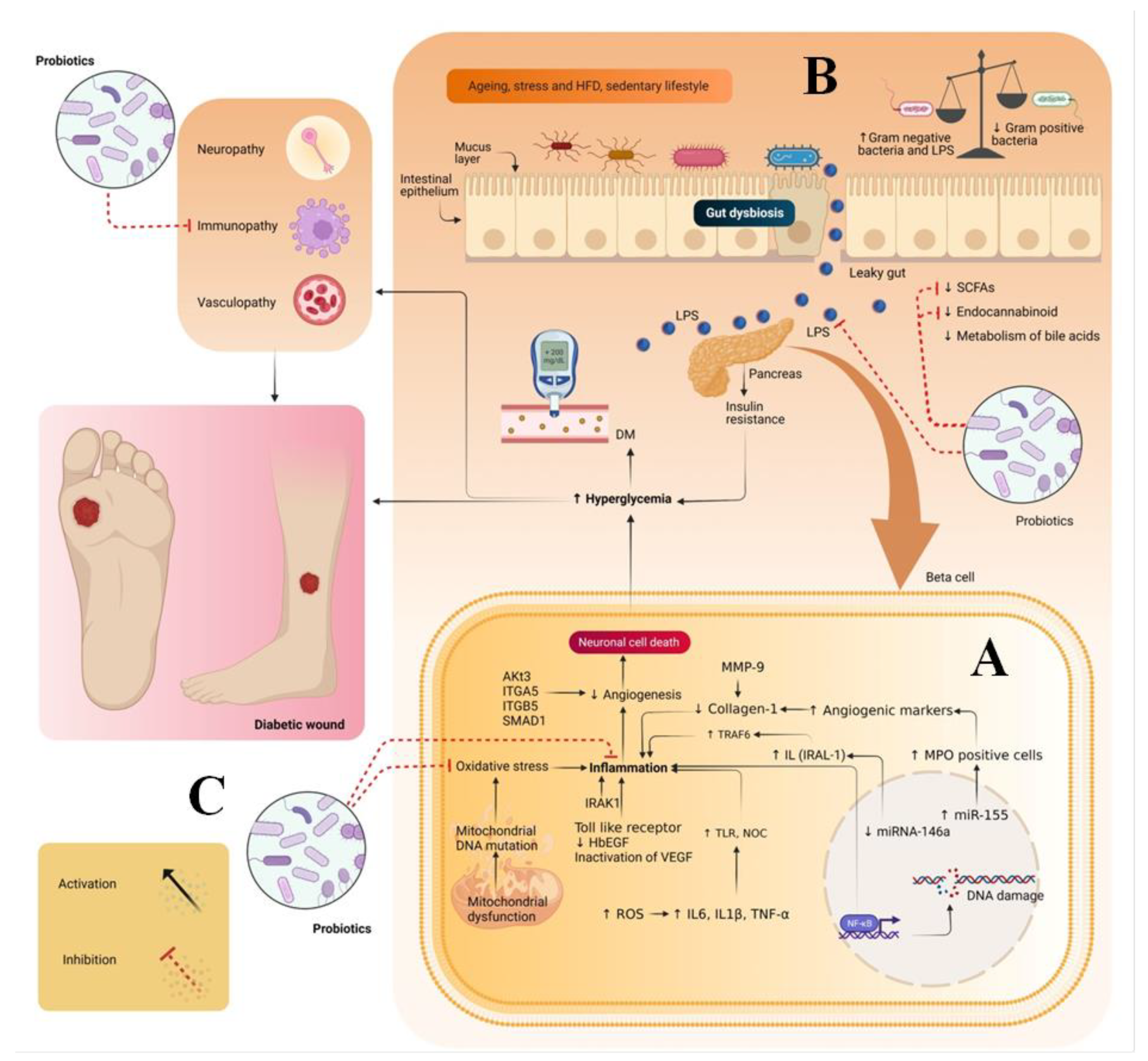
2. Pathogenesis of Diabetic Wounds

During hyperglycemia, the levels of micro-ribulose nucleic acid (miR)-155, miR-191, miR-200b, miR-15b, miR-200, and miR-205–5p are increased while those of miRNA-146a and miR-132 are decreased. The overactivation of miR-155, miR-191 and miR-200b results an increase in the level of myeloperoxidase (MPO)-positive cells and C-reactive protein levels, which, in turn, leads to impairment in angiogenic markers such as collagen 1, transforming growth factor (GF) beta-1 and alpha-smooth muscle actin. In addition, they prolong the inflammatory phase of wound healing and impede the wound healing process. Besides these factors, the overactivation of miR-15b, miR-200 and miR-205–5p results in the impairment of the vasoendothelial GF pathways and impedes the wound healing process. The decrease in the levels of miRNA-146a and miR-132 activates the tumor necrosis factor receptor-associated factor 6 (TRAF6), interleukin-1 receptor associated kinase 1 (IRAK1) and toll-like receptors. The overactivation of these pathways results in an increase in the level of inflammatory markers that prolongs the inflammatory phase and delays the wound healing process [3]. In addition to this, in DFU, the level of matrix mettalo proteinase (MMP) also gets increased, which inhibits the migration of keratinocytes toward the wound site and impairs collagen synthesis. This delays the wound healing process [1].
High blood glucose levels also result in idiopathic complications, viz. neuropathy, immunopathy and vasculopathy. Neuropathy affects sensory, motor and autonomic nerves. In sensory neuropathy, there is a loss of pain leading to unnoticed trauma, which, in turn, may lead to ulcer formation. In motor neuropathy, weakness and wasting of intrinsic foot muscles occur, which results in abnormal gait and foot deformities that can lead to ulceration. In autonomic neuropathy, sweat glands get suppressed, which results in a decrease in the sweating rate at the foot site. This makes the skin dry and brittle and leads to secondary infections and, finally, ulceration. Vasculopathy is a general term used to describe any disease affecting blood vessels. It is generally of two types: microanginopathy and macroanginopathy. Microanginopathy occurs when there is deposition of glycoproteins and blood clots on the surface of the basement of the vessels. This deposition makes the walls of the vessels thicker and causes leakage from them, leading to ulceration. Macroanginopathy includes the deposition of fats and blood clots in the blood vessels. This decreases the blood flow in the vessels, which leads to necrosis and, finally, ulceration. In the case of immunopathy, there is a decrease in immunity due to the decrease in the level of polymorpholeukocytes, intracellular killing rate and GFs, coupled with an excess of metalloproteinases. This prolongs the inflammatory phase and delays the wound healing process (Figure 1A) [2].

3. Gut Dysbiosis and DW

During hyperglycemia, there is an imbalance between Gram-positive and Gram-negative bacteria, which leads to gut dysbiosis. Imbalance in the gut microbiome ultimately results in alterations in the synthesis of short chain fatty acids (SCFA) and the secretion of gut hormones (GLP-1 and PYY). This imbalance increases the level of lipopolysaccharides (LPS) in the systemic circulation, impairs bile acid metabolism and alters circulatory branched-chain amino acids. Alterations in the SCFAs levels and gut hormones result in impairment in glucose homeostasis and lipids. Increase in the level of LPS results in metabolic endotoxemia, activates toll like receptors and causes inflammation by promoting the secretion of pro-inflammatory cytokines. Moreover, impairment in bile acid metabolism inhibits the conversion of primary bile acids such as cholic and chenodeoxycholic acids into secondary bile acid species. i.e., deoxycholic and lithocholic acids. This results in the dysregulation of glucose homeostasis. Alterations in circulating branched-chain amino acids lead to a decrease in the level of GLP-1 and impair glucose homeostasis. In addition, gut dysbiosis also diminishes the endocannabinoid system and impairs the inflammatory and immunomodulatory responses of the body. Overall, these factors result in impaired glucose homeostasis and immunity and an increase in inflammation, all of which are key contributors to DFU. To address gut dysbiosis, probiotics are suitable candidates due to their numerous health benefits (Figure 1B) [2,3,4].

4. Sources of Probiotics

Rich sources of probiotics are dairy and dairy-related products [5]. Micro-organisms, such as bifidobacteria and lactic acid bacteria (LAB), are extracted from fermented milk and have been used for centuries. It has been found that the fermented milk from Chinese yak, known as kurut, consists of 148 strains of LAB. Among these strains, Streptococcus thermophilus and Lactobacillus delbrueckii subsp bulgaricus are the most prevalent. In addition, Koumiss, Kefir grains and Masai milk are fermented milk items from which lactobacillus strains and yeast with probiotic properties may be obtained [5]. Other sources of probiotics are given in Table 1.

5. Therapeutic Potential of Probiotics in Treating DW

DW is associated with oxidative stress, inflammation and immunopathy. Hence, probiotics can play a major role in the therapy of DW. Probiotics have multiple therapeutic actions, such as antioxidant, anti-inflammatory, immunomodulatory and antidiabetic (Figure 1C) [8]. Probiotics exert antioxidant effects by decreasing the oxidative stress generated by mitochondrial dysfunction and reactive oxygen species. It is known that SOD has a short half-life and low bioavailability. They enhance the antioxidant effect by releasing antioxidant enzymes such as SOD and catalase. In mitochondrial dysfunction, oxidative stress is produced by the generation of superoxide reactive oxygen species. When probiotics are consumed, SOD enzymes are produced that help in the breakdown of superoxide ions into hydrogen peroxide and water, thereby decreasing oxidative stress. Therefore, probiotics are suitable for the local delivery of SOD in bowel-related disease. In addition, probiotics also produce catalase enzymes that help in cellular antioxidant defense and promote the decomposition of hydrogen peroxide, which, in turn, inhibits the production of hydroxyl radicals by Fenton reaction. Probiotics also produce antioxidant metabolites such as glutathione butyrate and folate. These metabolites eliminate hydrogen peroxide, peroxynitrite and hydroxyl radicals with the help of selenium-dependent glutathione peroxidase enzyme and reduce oxidative stress [9].
Nuclear factor-kappa B (NF-ĸB) is a key signaling channel which is responsible for inflammation. It is present in the cytoplasm in an inactive form, bound to an inhibitory molecule, i.e., IĸB. During inflammation, IĸB molecule breaks down, which results in the release of NF-ĸB to activate the inflammatory cascades. A probiotics strain such as Lactobacillus rhamnosus GG or Lactobacillus casei DN-114 001 inhibits the breakdown of the inhibitory molecule- IĸB and reduces the expression of proinflammatory cytokines such as IL-8. In addition, probiotics trigger toll-like receptors, which initiate beta-defensins and exert anti-inflammatory actions [10].
Probiotics exert immunomodulatory actions by interacting with antigen presenting and release chemical mediator cytokines such as interleukins (ILs), tumor necrosis factor, interferons, transforming GF and chemokines from immune cells (lymphocytes, granulocytes, macrophages, mast cells, epithelial cells, and dendritic cells (DCs)), which further regulate the innate and adaptive immune system. In addition, probiotics help in enhancing the production of cytokines, activate the tight junctions of the intestinal barrier against intercellular bacterial invasion, encourage the secretion of immunoglobulin A and production of antibacterial substances and compete with new pathogenic microorganisms for enterocyte adherence. Through these processes, probiotics regulate intestinal epithelial health. An early, innate immune response is also induced by probiotics through phagocytosis, polymorphonuclear (PMN) cell recruitment and tumor necrotic factor-alpha production [11].
Probiotics have an anti-diabetic effect because they help in the production of SCFA, which enhances the release of incretin hormones that influence glucose levels. In addition, probiotics reduce the level of LPS, making them useful for the treatment of gut dysbiosis and type 2 diabetes mellitus. Probiotics also help to increase the levels of GLP-1 and insulinotropic hormones in enteroendocrine L-cells [12]. This optimizes glucose metabolism, reduces cell damage and improves insulin sensitivity. Among several animal models used for DM, it has been reported in 91 research papers that probiotics prevent DM onset by down-regulating certain inflammatory cytokines, such as interferons (IFN) and IL-2 or IL-1, or by increasing anti-inflammatory IL-10 production. It is also claimed that probiotics produce a defensive wall that prevents pathogenic bacterial species from colonizing the epithelium [13].
Studies related to the antioxidant, anti-inflammatory, immunomodulation and anti-diabetic property of probiotics are depicted in the Table 2.
With regard to the therapeutic potential of probiotics, various studies have been carried out in the field of DW healing, which are discussed below.
In one of these studies, Peral et al. (2010) investigated the effect of Lactobacillus plantarum against chronic infected leg ulcers in diabetic patients. In their trial, 14 diabetic and 20 non-diabetic patients having venous leg ulcers were considered. For the treatment, topically Lactobacillus plantarum was applied to both diabetic and non-diabetic patients with venous leg ulcers. After 30 days of topical treatment with Lactobacillus plantarum, it was observed that 43% of diabetics and 50% of non-diabetic patients showed complete wound healing. Therefore, it was concluded that Lactobacillus plantarum accelerated wound healing in diabetic and non-diabetic patients by exerting antibacterial and anti-inflammatory actions, reducing apoptotic, neutrophils, and necrotic cells and modifying IL-8 production [40].
In another study, Majid et al. (2016) examined the effect of Lactobacillus casei and its exopolysaccharide against DW in induced male Wistar diabetic rats. The results revealed that the topical application of Lactobacillus casei and its exopolysaccharide showed 1.4-fold and 1.1-fold increase in wound contraction within 14 days as compared to negative and control groups [41].
Similarly, Mohseni et al. (2018) investigated the effect of probiotic supplementation on metabolic status and wound healing in patients with DFU. They performed a double-blind, randomized and placebo-controlled trial. In their trial, 60 patients aged 40–85 years old and having grade 3 (deep ulcer with cellulitis) DFU were considered. These 60 patients were casually distributed into two groups (30 patients on each side) to receive either placebo or oral probiotic capsule (Lactobacillus fermentum, Lactobacillus casei, Lactobacillus acidophilus, and Bifidobacterium bifidum) every day for 12 weeks. The dose of the probiotic capsule was 2 × 10⁹ CFU/g each. After 12 weeks, it was observed that compared to the placebo group, the probiotics-treated groups showed a significant reduction in ulcer length (−1.3 ± 0.9 cm for probiotic vs. −0.8 ± 0.7 cm for placebo, p = 0.01), ulcer width (−1.1 ± 0.7 cm for probiotic vs. −0.7 ± 0.7 cm for placebo, p = 0.02) and ulcer depth (−0.5 ± 0.3 cm for probiotic vs. −0.3 ± 0.3 cm for placebo, p = 0.02). Moreover, it was also observed that probiotics not only reduced the ulcer length, size and depth, but also helped in the downregulation of blood glucose level, total serum cholesterol, high sensitivity C-reactive protein (hs-CRP), malondialdehyde (MDA) levels, augmented plasma nitric oxide (NO) and total antioxidant capacity (TAC), indicating the potential of probiotics in treating DFU [42].
In another study, Gonzalez et al. (2018) explored the effect of clindamycin/cefotaxime and Lactobacillus acidophilus against micro-organisms isolated from the foot of DFU patients. The turbidimetric method was used for the bioassay. Three types of bacteria were isolated from DFUs strain, i.e., strain 1 (Pseudomonas sp.), strain 2 (yeast-like cell) and strain 3 (Enterobacter sp.). Then, clindamycin/cefotaxime and Lactobacillus acidophilus were tested against micro-organisms isolated from the foot of DFU patients. Clindamycin was used against all the strains isolated from DFU patients at concentrations of 0.15 μg/mL, 0.25 μg/mL, and 50 μg/mL. It was observed that clindamycin was only effective against strain three; the percentages of inhibition were 18, 88, and 89, respectively. Meanwhile, cefotaxime at concentrations of 0.15 μg/mL, 0.25 μg/mL, and 50 μg/mL showed an effect against all the three strains. The percentages of inhibition of cefotaxime at a dose of 0.15 μg/mL against strains 1, 2 and 3 were 85, 70 and 55, respectively. At a dose of 0.25 μg/mL cefotaxime showed a good percentage of inhibition against strains 1, 2 and 3, i.e., 87, 68, and 60, respectively. At a dose, 50 μg/mL cefotaxime showed percentages of inhibition for strains 1, 2 and 3 of 88, 65 and 76, respectively. When Lactobacillus acidophilus was tested against all these at concentrations of 40 mg/mL, 400 mg/mL, and 800 mg/mL, it was observed that it was only effective against strains 1 and 3. For strains 1 and 3, Lactobacillus acidophilus showed percentages inhibition of 3% and 9%, respectively, at a dose of 40 mg/mL. At dose of 400 mg/mL, Lactobacillus acidophilus showed percentages of inhibition against strains 1 and 3 which of 34 and 18, respectively. Similarly, at a dose of 800 mg/mL, Lactobacillus acidophilus showed 40% inhibition for strain 1 and 26% inhibition for strain 3, indicating the antibacterial potential of probiotics against the micro-organisms that are responsible for DFU [43].
Similarly, the effect of Lactobacillus plantarum gel was evaluated against burns associated DW healing in mature male Sprague-Dawley rats. The results revealed that the topical application of Lactobacillus plantarum accelerated DW healing as compared to other treated groups due to its anti-inflammatory action, increased hydroxyproline content, epithelization and angiogenesis at the site of injury [44].
In a related, Venosi et al. (2019) studied the effect of a multi-strain probiotic formulation on infected chronic ischemic wounds. This study was conducted on an 83-year old woman with a history of DM, hypertension and ischemic heart disease. The patient had critical limb ischemia and a cutaneous ulcer on the right leg. In addition, this patient was also subjected to percutaneous transluminal angioplasty (PTA) with a drug eluting balloon (DEB) ranger 5 × 100 mm in the superficial femoral artery (SFA) and right popliteal artery, followed by surgical curettage of necrotic forefoot injuries and amputation of the second toe of the right foot. To manage this, in the initial stage of treatment, the patient was given piperacillin/tazobactam 4.5 g intravenously (I.V) every eight hours. This treatment was given to the patients for 8 days. After that time, a reduction in inflammatory markers was observed, and piperacillin/tazobactam was switched to oral minocycline tablet (100 mg) every 12 h for 15 days. The patient was discharged after 21 days of hospitalization. Then local dressings and polymeric membrane (PolyMem®-Ferries Mfg) were applied at the site of injury. In spite of these treatments, the condition of the injury worsened and the patient was referred to the Department of Public Health and Infectious Diseases, University of Rome. His injury was properly examined, and multiple micro-organisms such as Proteus mirabilis, Entero faecalis and Klebsiella pneumonia were isolated. After the identification of these microorganisms, topical 10% cutaneous-iodopovidone solution (Poviderm® 10% Skin Solution) was applied. This treatment led to an improvement in wound healing. Then systemic and topical antibiotics treatment was stopped. Afterwards, it was decided to start treatment with a multi-strain probiotic formulation. The multi-strain probiotic formulation was comprised of lyophilized powder sachets, each containing 100 billion colony forming units (CFU) of Lactobacillus acidophilus NCIBMB 43030 20% in weight, Lactobacillus plantarum NCIBMB 43029 20% in weight, and Streptococcus thermophilus NCIMB 30438 40% in weight. The probiotic treatment was continued for 24 days. The results revealed that the topical application of probiotics at the site of injury led to the inhibition of multiple micro-organisms (Proteus mirabilis, Entero faecalis and Klebsiella pneumonia) and completely healed the wound [45].
Similarly, Chuang et al. (2019) studied the effect of Lactobacillus plantarum TWK10-fermented soymilk against DW in male Wistar diabetic rats. The results revealed that the topical application of Lactobacillus plantarum TWK10-fermented soymilk accelerated DW healing within 14 days by promoting collagen deposition and angiogenesis, increasing hydroxyproline content and decreasing oxidative stress, as well as by its antimicrobial action at the site of injury [46].
In another study, Kumari et al. (2019) examined the effect of Streptococcus thermophilus and low-level laser therapy on DW healing in male Albino diabetic rats. The results revealed that the topical application of saline did not lead to effective wound contraction while Streptococcus thermophilus showed a reduction in oxidative stress and promoted DW healing. However, it was observed that when Streptococcus thermophilus treatment and low-level laser therapy were used in combination, accelerated DW healing occurred. In addition, the combination promoted angiogenesis and collagen deposition at the site of injury [47].
Similarly, the effect of probiotics supplementation on DW healing was tested in male adult Wistar rats. In this study, 46 rats were used, divided into two groups, i.e., control and probiotic-treated groups. The latter received Probiatop®, while the control group received maltodextrin. The oral daily dose of both supplements was 250 mg once a day. Then, each group was further subdivided into two subgroups on the basis of euthanasia: 3rd or 10th postoperative (PO, subgroups C3 = 12 rats, P3 = 12 rats, C10 = 11 rats, P10 = 11 rats). Diabetes was induced to all rats by inducing alloxan. Supplementation was started five days before surgery and continued until euthanasia. The results revealed that the P10 group showed maximal wound contraction as compared to the C10 group. It was also observed that from the 3rd to 10th post-operative day, the probiotic treated group showed an increment in type 1 collagen deposition at the site of injury as compared to the control group. Hence, it was concluded that probiotic supplementation accelerated DW healing in rats by enhancing neovascularization and collagen deposition at the site of injury [48].
Similarly, Layus et al. (2020) studied the antibacterial activity of a probiotic containing Lactobacillus plantarum CRL 759 against microorganisms Pseudomonas aeruginosa and methicillin-resistant Staphylococcus aureus (MRSA), isolated from the foot of a DFU patient. The antimicrobial activity of the probiotic was determined by different methods, such as the modified agar slab method and the agar well diffusion method. The outcomes showed that Lactobacillus plantarum CRL 759 sans cell supernatant (SLp759) restrained both MRSA and Pseudomonas aeruginosa development. Likewise, SLp759 repressed the grip of pathogenic organisms. Furthermore, after the balance of acidic SLp759, no action against micro-organism strains was observed. In addition, treatment with proteolytic chemicals did not adjust antibacterial movement, demonstrating that no bacteriocin was available in the supernatant. Additionally, the results obtained by HPLC examination demonstrated that the inhibitory impact was the aftereffect of the creation of two natural acids, i.e., lactic and acetic [49].
In another study, Mohtashami et al. investigated the effect of Lactobacillus Plantarum against DW in alloxan-induced male Wistar diabetic rats. The results revealed that the Lactobacillus plantarum treated groups exhibited 1.14- and 1.35-fold increases in wound closure within 14 days in comparison to Lactobacillus bulgaricus and diabetic control-treated groups. In addition, the Lactobacillus plantarum treated groups showed accelerated DW healing due to the anti-inflammatory action, cell migration and proliferation at the site of injury [50].

6. Techniques Used for the Stabilization of Probiotics

Despite having various pharmacological as well as health benefits, probiotics are less commercialized due to their degradation upon exposure to sunlight, low pH, high temperatures and oxygen. It has been found that bacteria such as LAB excrete polysaccharides (EPS) that provide protection against harsh conditions. However, this protection is not sufficient. The different approaches used by the researchers to improve the stability and survival of probiotics include culture pre-exposure to the sub-lethal stresses [51] and the incorporation of micro-nutrients such as two-step fermentation [52], microencapsulation [53], the use of oxygen-impermeable containers [54] and immobilization [55]. Among these techniques, microencapsulation is the most widely used by researchers.
Microencapsulation is the process of packaging solids, liquids or gases into miniature containers. It increases stabilization and the survival rate of the probiotics at the time of processing, prevents oxidative reactions, provides sustained release at a target site and enhances shelf life [53]. Microencapsulation may be categorized into chemical and physical techniques. Both play a key role in the pharma and food sectors. Forms of physical encapsulation include spray chilling [56], suspension coating [57], fluidized bed coating [58], liposome entrapment [59], centrifugal extrusion [60], spray cooling [61], rotational suspension separation [62], annular jet, spray coating [60], spinning disk [63], air spray drying extrusion coating [60] and pan coating [64]. Chemical methods include in situ polymerization [57], interfacial polymerization [65], matrix polymerization [57] and extrusion [57]. Numerous studies on the microencapsulation technique have shown that emulsions are commonly used to enclose probiotic cultures within solid fat microcapsules, helping them to retain their vitality and activity. It is well-known that powdered foods have longer shelf-lives at normal room temperatures. Techniques that are used to dry probiotics to enhance their stability include microwave drying, spray drying, vacuum drying and lyophilization [60]. Among these, lyophilization is the best technique to maintain the viability of bacterial cells in order to use them in the preparation of starter culture cells. In addition to this, materials used for encapsulating probiotic strains include pectin [66], locust bean gum [67], rennet [68], whey protein [66], cellulose [69], к-carrageenan [70], chitosan [71] and alginate [57]. These materials act as gelling agents or support materials in the probiotic strain encapsulation. Various efforts made by the researchers to improve the stability of probiotics are listed in Table 3.
The advantages and disadvantages of commonly used techniques for the stabilization of probiotics [66,70] are discussed below.
  • Freeze drying—Advantages: (i) Easy and convenient; (ii) Does not require freezing conditions. Disadvantages: Lengthy and expensive.
  • Spray drying—Advantages: (i) Fast drying process; (ii) Powdered material obtained directly; (iii) Simple and easy to alter drying conditions; (iv) High production efficiency. Disadvantages: (i) Costly; (ii) An excessive amount of air is needed to increase the power consumption; (i) Equipment is complex; (ii) C overs large area.
  • Fluidized bed dryer—Advantages: (i) High thermal efficiency; (ii) Handling time is short; (iii) It is possible to the materials in a shorter time. Disadvantages: (i) Chance of attrition of materials; (ii) Many organic powders develop electrostatic charge during drying.
  • Extrusion—Advantages: (i) Low cost; (ii) Flexible. Disadvantages: (i) Size variances; (ii) Product limitation
  • Microencapsulation—Advantages: (i) Protects materials from external stress; (ii) It is possible to prepare sustained and controlled release formulations. Disadvantages: (i) High cost; (ii) Non uniform coating effect the release profile of the active moiety in the body.

7. Market Status of Probiotics

The health benefits and pharmacological actions of probiotics have been gaining the attention of consumers. The global market for probiotics is divided into different categories, i.e., dietary supplements, drinks, foods and animal feeds. Probiotic food may be further subdivided into baby food, yogurt, infant formula, breakfast cereals/baked goods and other probiotic foods. Additionally, probiotic drinks may be further classified into fruit-based and dairy-based drinks. Regarding distribution channels, the market for probiotics may be segmented into convenience stores, hypermarkets/supermarkets, pharmacies and drug stores, online channels and other distribution channels. In addition, the probiotics market is projected to register a CAGR rate of 7.2% during the forecast period of 2020–2030 [86]. Countries and regions which have become hubs of the probiotics market include North America (USA, Mexico, and Canada), Europe (Russia, Spain, UK, France, and Italy), Asia-Pacific (China, India, Japan, and Australia), South America (Argentina, Brazil) and the Middle East and Africa (Saudi Arabia, South Africa) [87]. Lists of probiotics that are available on the global market and patents on probiotics are depicted in Table 4, Table 5 and Table 6.

8. Conclusions

The data gathered in this review suggest that the oral consumption and topical application of probiotics bring about remarkable improvements in DFU. Moreover, the oral consumption of probiotics is much better than topical application. This is because oral probiotics have the ability to colonize the gut microbiota and improve gut dysbiosis by exerting anti-inflammatory, immunomodulatory, antioxidant and antidiabetic effects, which is restricted in topical application. The topical route will only provide a local effect decreasing the microbial load at the site of injury. Numerous preclinical as well as in vitro studies have shown the therapeutic potential of probiotics against DFU. Despite these enormous potentials, these studies are confined to academic laboratories. There are limited clinical studies on the use of probiotics against DFU. One of the leading reasons for this is the complexity in the identification and isolation of the probiotics, as well as their poor stability and high cost. Therefore, more clinical-based research is required to augment the pharmacotherapeutic potential of probiotic supplementation. Further, from a commercial perspective, it is important to seek novel techniques to enhance the stability of probiotics. Understanding the aforementioned bottlenecks and finding novel strategies to overcome them may bring about novel, effective treatments for DW.

Author Contributions

Conceptualization, S.K.S., R.D.E. and A.A.; methodology, A.A., S.V. and L.C., Data curation, S.V., A.A., B.K., M.G. L.C., G.G. and K.D.; writing—original draft preparation, A.A., L.C., S.V.; writing—review and editing, S.K.S., D.K.C., R.D.E. and K.D. All authors have read and agreed to the published version of the manuscript.

Funding

This research received no external funding.

Institutional Review Board Statement

Not applicable.

Informed Consent Statement

Not applicable.

Data Availability Statement

Not applicable.

Acknowledgments

Authors are thankful to management of Lovely Professional University for providing necessary facilities for writing this review.

Conflicts of Interest

The authors declare no conflict of interest.

References

  1. Awasthi, A.; Gulati, M.; Kumar, B.; Kaur, J.; Vishwas, S.; Khursheed, R.; Porwal, O.; Alam, A.; Kr, A.; Corrie, L.; et al. Review Article Recent Progress in Development of Dressings Used for Diabetic Wounds with Special Emphasis on Scaffolds. BioMed Res. Int. 2022, 2022, 1659338. [Google Scholar] [CrossRef] [PubMed]
  2. Awasthi, A.; Singh, S.K.; Kumar, B.; Gulati, M.; Kumar, R.; Wadhwa, S.; Khursheed, R.; Corrie, L.; KR, A.; Kumar, R.; et al. Treatment Strategies Against Diabetic Foot Ulcer: Success so Far and the Road Ahead. Curr. Diabetes Rev. 2020, 17, 421–436. [Google Scholar] [CrossRef] [PubMed]
  3. Awasthi, A.; Vishwas, S.; Gulati, M.; Corrie, L.; Kaur, J.; Khursheed, R.; Alam, A.; Alkhayl, F.F.A.; Khan, F.R.; Nagarethinam, S.; et al. Expanding Arsenal against Diabetic Wounds Using Nanomedicines and Nanomaterials: Success so Far and Bottlenecks. J. Drug Deliv. Sci. Technol. 2022, 74, 103534. [Google Scholar] [CrossRef]
  4. Yahfoufi, N.; Mallet, J.F.; Graham, E.; Matar, C. Role of Probiotics and Prebiotics in Immunomodulation. Curr. Opin. Food Sci. 2018, 20, 82–91. [Google Scholar] [CrossRef]
  5. Swain, M.R.; Anandharaj, M.; Ray, R.C.; Parveen Rani, R. Fermented Fruits and Vegetables of Asia: A Potential Source of Probiotics. Biotechnol. Res. Int. 2014, 2014, 250424. [Google Scholar] [CrossRef]
  6. James, A.; Wang, Y. Characterization, Health Benefits and Applications of Fruits and Vegetable Probiotics. CYTA—J. Food 2019, 17, 770–780. [Google Scholar] [CrossRef] [Green Version]
  7. Patel, A.R. Probiotic Fruit and Vegetable Juices- Recent Advances and Future Perspective. Int. Food Res. J. 2017, 24, 1850–1857. [Google Scholar]
  8. Plaza-Diaz, J.; Ruiz-Ojeda, F.J.; Gil-Campos, M.; Gil, A. Mechanisms of Action of Probiotics. Adv. Nutr. 2019, 10, S49–S66. [Google Scholar] [CrossRef] [Green Version]
  9. Tavakoli, M.; Habibi Najafi, M.B.; Mohebbi, M. Effect of the Milk Fat Content and Starter Culture Selection on Proteolysis and Antioxidant Activity of Probiotic Yogurt. Heliyon 2019, 5, e01204. [Google Scholar] [CrossRef] [Green Version]
  10. Sharma, S.; Singh, A.; Sharma, S.; Kant, A.; Sevda, S.; Taherzadeh, M.J.; Garlapati, V.K. Functional Foods as a Formulation Ingredients in Beverages: Technological Advancements and Constraints. Bioengineered 2021, 12, 11055–11075. [Google Scholar] [CrossRef]
  11. Azad, M.A.K.; Sarker, M.; Wan, D. Immunomodulatory Effects of Probiotics on Cytokine Profiles. Biomed Res. Int. 2018, 2018, 8063647. [Google Scholar] [CrossRef]
  12. Pegah, A.; Abbasi-Oshaghi, E.; Khodadadi, I.; Mirzaei, F.; Tayebinia, H. Probiotic and Resveratrol Normalize GLP-1 Levels and Oxidative Stress in the Intestine of Diabetic Rats. Metab. Open 2021, 10, 100093. [Google Scholar] [CrossRef]
  13. Denkova, R.; Goranov, B.; Teneva, D.; Kostov, G. Antimicrobial Activity of Probiotic Microorganisms: Mechanisms of Interaction and Methods of Examination, 1st ed.; Z.-Mendez Villas: Polvdiv, Bulgaria, 2017; pp. 201–212. [Google Scholar]
  14. Oh, B.T.; Jeong, S.Y.; Velmurugan, P.; Park, J.H.; Jeong, D.Y. Probiotic-Mediated Blueberry (Vaccinium Corymbosum L.) Fruit Fermentation to Yield Functionalized Products for Augmented Antibacterial and Antioxidant Activity. J. Biosci. Bioeng. 2017, 124, 542–550. [Google Scholar] [CrossRef]
  15. Helmy, S.A. Chemical Composition and Antimicrobial Activity of Some Essential Oils and Their Major Constituents. Int. J. Acad. Res. 2012, 4, 124–137. [Google Scholar] [CrossRef]
  16. Li, A.; Wang, Y.; Li, Z.; Qamar, H.; Mehmood, K.; Zhang, L.; Liu, J.; Zhang, H.; Li, J. Probiotics Isolated from Yaks Improves the Growth Performance, Antioxidant Activity, and Cytokines Related to Immunity and Inflammation in Mice. Microb. Cell Fact. 2019, 18, 112. [Google Scholar] [CrossRef] [Green Version]
  17. Abdhul, K.; Ganesh, M.; Shanmughapriya, S.; Kanagavel, M.; Anbarasu, K.; Natarajaseenivasan, K. Antioxidant activity of exopolysaccharide from probiotic strain Enterococcus faecium (BDU7) from Ngari. Int. J. Biol. Macromol. 2014, 70, 450–454. [Google Scholar] [CrossRef]
  18. Shori, A.B.; Aljohani, G.S.; Al-zahrani, A.J.; Al-sulbi, O.S.; Baba, A.S. Viability of Probiotics and Antioxidant Activity of Cashew Milk-Based Yogurt Fermented with Selected Strains of Probiotic lactobacillus Spp. LWT 2022, 153, 112482. [Google Scholar] [CrossRef]
  19. Das, D.; Goyal, A. Antioxidant Activity and γ-Aminobutyric Acid (GABA) Producing Ability of Probiotic Lactobacillus plantarum DM5 Isolated from Marcha of Sikkim. LWT—Food Sci. Technol. 2015, 61, 263–268. [Google Scholar] [CrossRef]
  20. Uugantsetseg, E.; Batjargal, B. Antioxidant Activity of Probiotic Lactic Acid Bacteria Isolated from Mongolian Airag. Mong. J. Chem. 2014, 15, 73–78. [Google Scholar] [CrossRef] [Green Version]
  21. Mäkelä, S.M.; Forssten, S.D.; Kailajärvi, M.; Langén, V.L.; Scheinin, M.; Tiihonen, K.; Ouwehand, A.C. Effects of Bifidobacterium animalis Ssp. Lactis 420 on Gastrointestinal Inflammation Induced by a Nonsteroidal Anti-Inflammatory Drug: A Randomized, Placebo-Controlled, Double-Blind Clinical Trial. Br. J. Clin. Pharmacol. 2021, 87, 4625–4635. [Google Scholar] [CrossRef]
  22. Shadnoush, M.; Hosseini, R.S.; Khalilnezhad, A.; Navai, L.; Goudarzi, H.; Vaezjalali, M. Effects of Probiotics on Gut Microbiota in Patients with Inflammatory Bowel Disease: A Double-Blind, Placebo-Controlled Clinical Trial. Korean J. Gastroenterol. 2015, 65, 215–221. [Google Scholar] [CrossRef]
  23. Babadi, M.; Khorshidi, A.; Aghadavood, E.; Samimi, M.; Kavossian, E.; Bahmani, F.; Mafi, A.; Shafabakhsh, R.; Satari, M.; Asemi, Z. The Effects of Probiotic Supplementation on Genetic and Metabolic Profiles in Patients with Gestational Diabetes Mellitus: A Randomized, Double-Blind, Placebo-Controlled Trial. Probiotics Antimicrob. Proteins 2019, 11, 1227–1235. [Google Scholar] [CrossRef]
  24. Zaharuddin, L.; Mokhtar, N.M.; Muhammad Nawawi, K.N.; Raja Ali, R.A. A Randomized Double-Blind Placebo-Controlled Trial of Probiotics in Post-Surgical Colorectal Cancer. BMC Gastroenterol. 2019, 19, 131. [Google Scholar] [CrossRef] [Green Version]
  25. Trial, A.A.R.P. Nutrients Effects of 12-Week Ingestion of Yogurt Containing Lactobacillus Plantarum OLL2712 on Glucose Metabolism and Chronic Inflammation in Prediabetic Adults: A Randomized Placebo-Controlled Trial. Nutrients. 2020, 12, 374. [Google Scholar] [CrossRef] [Green Version]
  26. Choi, M.; Lee, Y.; Lee, N.K.; Bae, C.H.; Park, D.C.; Paik, H.D.; Park, E. Immunomodulatory Effects by Bifidobacterium longum KACC 91563 in Mouse Splenocytes and Macrophages. J. Microbiol. Biotechnol. 2019, 29, 1739–1744. [Google Scholar] [CrossRef]
  27. Enani, S.M.; Childs, C.E.; Przemska, A.; Maidens, C.; Dong, H.; Rowland, I.; Tuohy, K.; Todd, S.; Gosney, M.; Yaqoob, P. Effects of a Novel Probiotic, Bifidobacterium longum Bv. Infantis CCUG 52486 with Prebiotic on the B-Cell Response to Influenza Vaccination. Proc. Nutr. Soc. 2014, 73, 52486. [Google Scholar] [CrossRef] [Green Version]
  28. Dong, H.; Rowland, I.; Thomas, L.V.; Yaqoob, P. Immunomodulatory Effects of a Probiotic Drink Containing Lactobacillus casei Shirota in Healthy Older Volunteers. Eur. J. Nutr. 2013, 52, 1853–1863. [Google Scholar] [CrossRef]
  29. Villena, J.; Salva, S.; Agüero, G.; Alvarez, S. Immunomodulatory and Protective Effect of Probiotic Lactobacillus casei against Candida Albicans Infection in Malnourished Mice. Microbiol. Immunol. 2011, 55, 434–445. [Google Scholar] [CrossRef]
  30. D’ambrosio, S.; Ventrone, M.; Fusco, A.; Casillo, A.; Dabous, A.; Cammarota, M.; Corsaro, M.M.; Donnarumma, G.; Schiraldi, C.; Cimini, D. Limosilactobacillus fermentum from Buffalo Milk Is Suitable for Potential Biotechnological Process Development and Inhibits Helicobacter Pylori in a Gastric Epithelial Cell Model. Biotechnol. Rep. 2022, 34, e00732. [Google Scholar] [CrossRef]
  31. Zhang, Y.; Guo, X.; Guo, J.; He, Q.; Li, H.; Song, Y.; Zhang, H. Lactobacillus casei Reduces Susceptibility to Type 2 Diabetes via Microbiota-Mediated Body Chloride Ion Influx. Sci. Rep. 2014, 4, 5654. [Google Scholar] [CrossRef] [Green Version]
  32. Sharma, P.; Bhardwaj, P.; Singh, R. Administration of Lactobacillus casei and Bifidobacterium Bifidum Ameliorated Hyperglycemia, Dyslipidemia, and Oxidative Stress in Diabetic Rats. Int. J. Prev. Med. 2016, 7, 102. [Google Scholar] [CrossRef]
  33. Asgharzadeh, F.; Tanomand, A.; Ashoori, M.R.; Asgharzadeh, A.; Zarghami, N. Investigating the Effects of Lactobacillus casei on Some Biochemical Parameters in Diabetic Mice. J. Endocrinol. Metab. Diabetes S. Afr. 2017, 22, 47–50. [Google Scholar] [CrossRef] [Green Version]
  34. Wang, G.; Li, X.; Zhao, J.; Zhang, H.; Chen, W. Lactobacillus casei CCFM419 Attenuates Type 2 Diabetes via a Gut Microbiota Dependent Mechanism. Food Funct. 2017, 8, 3155–3164. [Google Scholar] [CrossRef]
  35. Niibo, M.; Shirouchi, B.; Umegatani, M.; Morita, Y.; Ogawa, A.; Sakai, F.; Kadooka, Y.; Sato, M. Probiotic Lactobacillus gasseri SBT2055 Improves Insulin Secretion in a Diabetic Rat Model. J. Dairy Sci. 2019, 102, 997–1006. [Google Scholar] [CrossRef] [Green Version]
  36. Li, X.; Wang, N.; Yin, B.; Fang, D.; Jiang, T.; Fang, S.; Zhao, J.; Zhang, H.; Wang, G.; Chen, W. Effects of Lactobacillus plantarum CCFM0236 on Hyperglycaemia and Insulin Resistance in High-Fat and Streptozotocin-Induced Type 2 Diabetic Mice. J. Appl. Microbiol. 2016, 121, 1727–1736. [Google Scholar] [CrossRef]
  37. Lee, E.; Jung, S.R.; Lee, S.Y.; Lee, N.K.; Paik, H.D.; Lim, S. Il Lactobacillus plantarum Strain Ln4 Attenuates Diet-Induced Obesity, Insulin Resistance, and Changes in Hepatic MRNA Levels Associated with Glucose and Lipid Metabolism. Nutrients 2018, 10, 643. [Google Scholar] [CrossRef] [Green Version]
  38. Balakumar, M.; Prabhu, D.; Sathishkumar, C.; Prabu, P.; Rokana, N.; Kumar, R.; Raghavan, S.; Soundarajan, A.; Grover, S.; Batish, V.K.; et al. Improvement in Glucose Tolerance and Insulin Sensitivity by Probiotic Strains of Indian Gut Origin in High-Fat Diet-Fed C57BL/6J Mice. Eur. J. Nutr. 2018, 57, 279–295. [Google Scholar] [CrossRef]
  39. Bagarolli, R.A.; Tobar, N.; Oliveira, A.G.; Araújo, T.G.; Carvalho, B.M.; Rocha, G.Z.; Vecina, J.F.; Calisto, K.; Guadagnini, D.; Prada, P.O.; et al. Probiotics Modulate Gut Microbiota and Improve Insulin Sensitivity in DIO Mice. J. Nutr. Biochem. 2017, 50, 16–25. [Google Scholar] [CrossRef] [Green Version]
  40. Peral, M.C.; Rachid, M.M.; Gobbato, N.M.; Huaman Martinez, M.A.; Valdez, J.C. Interleukin-8 Production by Polymorphonuclear Leukocytes from Patients with Chronic Infected Leg Ulcers Treated with Lactobacillus plantarum. Clin. Microbiol. Infect. 2010, 16, 281–286. [Google Scholar] [CrossRef] [Green Version]
  41. Ahmadi Majd, S.; Khorasgani, M.R.; Talebi, A. Study of Diabetic Cutaneous Wound Healing in Rats Treated with Lactobacillus casei and Its Exopolysaccharide. Int. J. Adv. Biotechnol. Res. 2016, 7, 2083–2092. [Google Scholar]
  42. Mohseni, S.; Bayani, M.; Bahmani, F.; Tajabadi-Ebrahimi, M.; Bayani, M.A.; Jafari, P.; Asemi, Z. The Beneficial Effects of Probiotic Administration on Wound Healing and Metabolic Status in Patients with Diabetic Foot Ulcer: A Randomized, Double-Blind, Placebo-Controlled Trial. Diabetes. Metab. Res. Rev. 2018, 34, e2970. [Google Scholar] [CrossRef]
  43. González, M.P.B.; Quiñones-Gutiérrez, Y. Antibiosis of Cefotaxime/Clindamycin and Lactobacillus acidophilus on Related Bacteria to Diabetic Foot Ulcer. Food Nutr. Sci. 2018, 9, 277–289. [Google Scholar] [CrossRef]
  44. Salaran, M.; Oryan, A.; Nikahval, B.; Kamali, A.; Ghaemi, M.; Abbasi-Teshnizi, F.; Azizzadeh, M. Topical Application of Lactobacillus plantarum on Burn Wound Healing in Diabetic Rats. Iran. J. Vet. Surg. 2019, 14, 60–72. [Google Scholar] [CrossRef]
  45. Venosi, S.; Ceccarelli, G.; De Angelis, M.; Laghi, L.; Bianchi, L.; Martinelli, O.; Maruca, D.; Cavallari, E.N.; Toscanella, F.; Vassalini, P.; et al. Infected Chronic Ischemic Wound Topically Treated with a Multi-Strain Probiotic Formulation: A Novel Tailored Treatment Strategy. J. Transl. Med. 2019, 17, 364. [Google Scholar] [CrossRef] [Green Version]
  46. Chuang, Y.C.; Cheng, M.C.; Lee, C.C.; Chiou, T.Y.; Tsai, T.Y. Effect of Ethanol Extract from Lactobacillus plantarum TWK10-Fermented Soymilk on Wound Healing in Streptozotocin-Induced Diabetic Rat. AMB Express 2019, 9, 163. [Google Scholar] [CrossRef] [Green Version]
  47. Kumari, D.; Khan, H.; Jiskani, A.R.; Rafique, M.; Asif, M.; Kumar, V.; Maqsood, S. Neovascularization: Topical Effects of Streptococcus thermophilus and Low Level Laser Therapy in Treatment of Diabetic Wound in Rats. Int. J. Res. Med. Sci. 2019, 7, 3357–3361. [Google Scholar] [CrossRef]
  48. Campos, L.F.; Tagliari, E.; Casagrande, T.A.C.; de Noronha, L.; Campos, A.C.L.; Matias, J.E.F. Effects of Probiotics Supplementation on Skin Wound Healing in Diabetic Rats. Arq. Bras. Cir. Dig. 2020, 33, 1–6. [Google Scholar] [CrossRef]
  49. Layus, B.I.; Gerez, C.L.; Rodriguez, A.V. Antibacterial Activity of Lactobacillus plantarum CRL 759 Against Methicillin-Resistant Staphylococcus Aureus and Pseudomonas Aeruginosa. Arab. J. Sci. Eng. 2020, 45, 4503–4510. [Google Scholar] [CrossRef]
  50. Mohtashami, M.; Mohamadi, M.; Azimi-Nezhad, M.; Saeidi, J.; Nia, F.F.; Ghasemi, A. Lactobacillus bulgaricus and Lactobacillus plantarum Improve Diabetic Wound Healing through Modulating Inflammatory Factors. Biotechnol. Appl. Biochem. 2021, 68, 1421–1431. [Google Scholar] [CrossRef]
  51. Kulkarni, S.; Haq, S.F.; Samant, S.; Sukumaran, S. Adaptation of Lactobacillus acidophilus to Thermal Stress Yields a Thermotolerant Variant Which Also Exhibits Improved Survival at PH 2. Probiotics Antimicrob. Proteins 2018, 10, 717–727. [Google Scholar] [CrossRef]
  52. Seyoum, Y.; Humblot, C.; Baxter, B.A.; Nealon, N.J.; Weber, A.M.; Ryan, E.P. Metabolomics of Rice Bran Differentially Impacted by Fermentation with Six Probiotics Demonstrates Key Nutrient Changes for Enhancing Gut Health. Front. Nutr. 2022, 8, 795334. [Google Scholar] [CrossRef]
  53. Rokka, S.; Rantamäki, P. Protecting Probiotic Bacteria by Microencapsulation: Challenges for Industrial Applications. Eur. Food Res. Technol. 2010, 231, 1–12. [Google Scholar] [CrossRef]
  54. Shah, N.P. Probiotic Bacteria: Selective Enumeration and Survival in Dairy Foods. J. Dairy Sci. 2000, 83, 894–907. [Google Scholar] [CrossRef]
  55. Krisch, J.; Kerekes, E.B.; Takó, M.; Vágvölgyi, C. Cell Immobilization for the Dairy Industry. In Microbial Fermentation and Enzyme Technology; Thatoi, H., Mohapatra, P.K.D., Mohapatra, S., Mondal, K.C., Eds.; CRC Press: Boca Raton, FL, USA, 2020; pp. 115–127. [Google Scholar] [CrossRef]
  56. Bampi, G.B.; Backes, G.T.; Cansian, R.L.; de Matos, F.E.; Ansolin, I.M.A.; Poleto, B.C.; Corezzolla, L.R.; Favaro-Trindade, C.S. Spray Chilling Microencapsulation of Lactobacillus acidophilus and Bifidobacterium animalis Subsp. Lactis and Its Use in the Preparation of Savory Probiotic Cereal Bars. Food Bioprocess Technol. 2016, 9, 1422–1428. [Google Scholar] [CrossRef]
  57. Rajagopal, V.; Ramaiyan, B. Encapsulation “The Future of Probiotics”—A Review. Adv. Biol. Res. 2021, 3, 96–103. [Google Scholar]
  58. Nag, A.; Das, S. Improving Ambient Temperature Stability of Probiotics with Stress Adaptation and Fluidized Bed Drying. J. Funct. Foods 2013, 5, 170–177. [Google Scholar] [CrossRef]
  59. Htwe, M.M.; Teanpaisan, R.; Khongkow, P.; Amnuaikit, T. Liposomes of Probiotic’s Lyophilized Cell Free Supernatant; A Potential Cosmeceutical Product. Pharmazie 2019, 74, 462–466. [Google Scholar] [CrossRef]
  60. Arepally, D.; Reddy, R.S.; Goswami, T.K.; Coorey, R. A Review on Probiotic Microencapsulation and Recent Advances of Their Application in Bakery Products. Food Bioprocess Technol. 2022, 15, 1677–1699. [Google Scholar] [CrossRef]
  61. Liliana, S.C.; Vladimir, V.C. Probiotic Encapsulation. Afr. J. Microbiol. Res. 2013, 7, 4743–4753. [Google Scholar] [CrossRef]
  62. Trilokia, M.; Campus Chatha, M.; Julie Bandral, I.D.; Ankita Chib, I.; Preeti Choudhary Agromet Observer, I.; Corresponding Author, I.; Bandral, J.D.; Chib, A.; Choudhary, P. Microencapsulation for Food: An Overview. Pharma Innov. J. 2022, 11, 1174–1180. [Google Scholar]
  63. Nedovic, V.; Kalusevic, A.; Manojlovic, V.; Levic, S.; Bugarski, B. An Overview of Encapsulation Technologies for Food Applications. Procedia Food Sci. 2011, 1, 1806–1815. [Google Scholar] [CrossRef] [Green Version]
  64. Oberoi, K.; Tolun, A.; Sharma, K.; Sharma, S. Microencapsulation: An Overview for the Survival of Probiotic Bacteria. J. Microbiol. Biotechnol. Food Sci. 2019, 9, 280–287. [Google Scholar] [CrossRef]
  65. Mutukumira, A.N.; Ang, J.; Lee, S.J. Microencapsulation of Probiotic Bacteria. Microbiol. Monographs 2015, 4, 63–80. [Google Scholar] [CrossRef]
  66. Šipailienė, A.; Petraitytė, S. Encapsulation of Probiotics: Proper Selection of the Probiotic Strain and the Influence of Encapsulation Technology and Materials on the Viability of Encapsulated Microorganisms. Probiotics Antimicrob. Proteins 2018, 10, 1–10. [Google Scholar] [CrossRef] [PubMed]
  67. Damodharan, K.; Palaniyandi, S.A.; Yang, S.H.; Suh, J.W. Co-Encapsulation of Lactic Acid Bacteria and Prebiotic with Alginate-Fenugreek Gum-Locust Bean Gum Matrix: Viability of Encapsulated Bacteria under Simulated Gastrointestinal Condition and during Storage Time. Biotechnol. Bioprocess Eng. 2017, 22, 265–271. [Google Scholar] [CrossRef]
  68. Santillo, A.; Albenzio, M.; Bevilacqua, A.; Corbo, M.R.; Sevi, A. Encapsulation of Probiotic Bacteria in Lamb Rennet Paste: Effects on the Quality of Pecorino Cheese. J. Dairy Sci. 2012, 95, 3489–3500. [Google Scholar] [CrossRef] [PubMed] [Green Version]
  69. Moreno, J.S.; Dima, P.; Chronakis, I.S.; Mendes, A.C. Electrosprayed Ethyl Cellulose Core-Shell Microcapsules for the Encapsulation of Probiotics. Pharmaceutics 2022, 14, 7. [Google Scholar] [CrossRef]
  70. Borooah, B. Overview of Materials and Techniques for Encapsulation of Natural Products: A Mini-Review. Int. J. Pharm. Sci. Res. 2022, 13, 621–627. [Google Scholar] [CrossRef]
  71. Calinoiu, L.F.; Ştefanescu, B.E.; Pop, I.D.; Muntean, L.; Vodnar, D.C. Chitosan Coating Applications in Probiotic Microencapsulation. Coatings 2019, 9, 194. [Google Scholar] [CrossRef] [Green Version]
  72. Giraffa, G.; Gatti, M.; Rossetti, L.; Senini, L.; Neviani, E. Molecular Diversity within Lactobacillus helveticus as Revealed by Genotypic Characterization. Appl. Environ. Microbiol. 2000, 66, 1259–1265. [Google Scholar] [CrossRef] [Green Version]
  73. Wang, A.; Lin, J.; Zhong, Q. Spray-Coating as a Novel Strategy to Supplement Broiler Feed Pellets with Probiotic Lactobacillus salivarius NRRL B-30514. LWT 2021, 137, 110419. [Google Scholar] [CrossRef]
  74. Muhammad, Z.R.R.R.Z.M.Z. Resistant Starch-Based Edible Coating Composites for Digestion and Physicochemical Characteristics. Coatings 2021, 11, 587. [Google Scholar] [CrossRef]
  75. Mirzamani, S.S.; Bassiri, A.R.; Tavakolipour, H.; Azizi, M.H.; Kargozari, M. Survival of Fluidized Bed Encapsulated Lactobacillus acidophilus under Simulated Gastro-Intestinal Conditions and Heat Treatment during Bread Baking. J. Food Meas. Charact. 2021, 15, 5477–5484. [Google Scholar] [CrossRef]
  76. Tavakolipour, H.; Kargozari, M. Fluidized Bed Microencapsulation of Lactobacillus sporogenes with Some Selected Hydrocolloids for Probiotic Bread Production. J. Food Biosci. Technol. 2021, 11, 23–34. [Google Scholar]
  77. Meng, J.; Wang, Y.Y.; Hao, Y.P.; Zhang, S.B.; Ding, C.H.; You, Y.Z. Coating Function and Stabilizing Effects of Surface Layer Protein from Lactobacillus acidophilus ATCC 4356 on Liposomes. Int. J. Biol. Macromol. 2021, 183, 457–462. [Google Scholar] [CrossRef] [PubMed]
  78. Sultana, M.; Chan, E.S.; Pushpamalar, J.; Choo, W.S. Advances in Extrusion-Dripping Encapsulation of Probiotics and Omega-3 Rich Oils. Trends Food Sci. Technol. 2022, 123, 69–86. [Google Scholar] [CrossRef]
  79. Afzaal, M.; Saeed, F.; Hussain, S.; Mohamed, A.A.; Alamri, M.S.; Ahmad, A.; Ateeq, H.; Tufail, T.; Hussain, M. Survival and Storage Stability of Encapsulated Probiotic under Simulated Digestion Conditions and on Dried Apple Snacks. Food Sci. Nutr. 2020, 8, 5392–5401. [Google Scholar] [CrossRef]
  80. Bhagwat, A.; Bhushette, P.; Annapure, U.S. Spray Drying Studies of Probiotic Enterococcus Strains Encapsulated with Whey Protein and Maltodextrin. Beni-Suef Univ. J. Basic Appl. Sci. 2020, 9, 33. [Google Scholar] [CrossRef]
  81. Zhou, H.; Li, S.; Chen, Y.; Zhang, Q.; Bai, X.; Zhu, C.; Liu, H.; Wang, L.; Wu, C.; Pan, X.; et al. Evaluation of Streptococcus thermophilus IFFI 6038 Microcapsules Prepared Using an Ultra-Fine Particle Processing System. AAPS PharmSciTech 2018, 19, 1020–1028. [Google Scholar] [CrossRef]
  82. Moghaddas Kia, E.; Ghasempour, Z.; Ghanbari, S.; Pirmohammadi, R.; Ehsani, A. Development of Probiotic Yogurt by Incorporation of Milk Protein Concentrate (MPC) and microencapsulated Lactobacillus paracasei in Gellan-Caseinate Mixture. Br. Food J. 2018, 120, 1516–1528. [Google Scholar] [CrossRef]
  83. Ester, B.; Noelia, B.; Laura, C.J.; Francesca, P.; Cristina, B.; Rosalba, L.; Marco, D.R. Probiotic Survival and in Vitro Digestion of L Salivarius Spp. Salivarius Encapsulated by High Homogenization Pressures and Incorporated into a Fruit Matrix. LTW 2019, 111, 883–888. [Google Scholar] [CrossRef]
  84. Thangrongthong, S.; Puttarat, N.; Ladda, B.; Itthisoponkul, T.; Pinket, W.; Kasemwong, K.; Taweechotipatr, M. Microencapsulation of Probiotic lactobacillus Brevis ST-69 Producing GABA Using Alginate Supplemented with Nanocrystalline Starch. Food Sci. Biotechnol. 2020, 29, 1475–1482. [Google Scholar] [CrossRef] [PubMed]
  85. Lin, S.; Mukherjee, S.; Li, J.; Hou, W.; Pan, C.; Liu, J. Mucosal Immunity–Mediated Modulation of the Gut Microbiome by Oral Delivery of Probiotics into Peyer’s Patches. Sci. Adv. 2021, 7, abf0677. [Google Scholar] [CrossRef] [PubMed]
  86. Available online: https://www.grandviewresearch.com/industry-analysis/probiotics-market (accessed on 10 August 2022).
  87. Available online: https://www.marketdataforecast.com/market-reports/north-america-probiotics-market (accessed on 10 August 2022).
  88. Available online: https://www.researchgate.net/figure/1-List-of-probiotic-products-available-in-the-market_tbl1_324371121 (accessed on 11 August 2022).
  89. Available online: https://www.livestrong.com/article/411592-ingredients-of-activia-yogurt/ (accessed on 11 August 2022).
  90. Available online: https://brainreference.com/adult-formula-cp-1-review/#:~:text=Adult%20Formula%20Cp-1%20contains%20the%20following%20ingredients%3A%20Adult,capsules.%203%29%20What%20Does%20Adult%20Formula%20Cp-1%20Do%3F (accessed on 11 August 2022).
  91. Available online: https://www.alignprobiotics.com/en-us/why-align/how-to-take-align-probiotic (accessed on 11 August 2022).
  92. Available online: https://celiac-disease.com/review-attune-probiotic-bars/ (accessed on 11 August 2022).
  93. Available online: https://biokplus.com/ (accessed on 11 August 2022).
  94. Available online: https://biokplus.com/products/bio-k-probiotic-capsules-daily-care-50b (accessed on 14 August 2022).
  95. Available online: https://in.iherb.com/pr/culturelle-probiotics-digestive-daily-probiotic-10-billion-cfus-30-once-daily-vegetarian-capsules/56049#:~:text=Take%20charge%20of%20your%20digestive%20health%20with%20Culturelle%C2%AE,balance%20your%20digestive%20system%2C%20plus%20the%20prebiotic%20inulin (accessed on 14 August 2022).
  96. Available online: https://oribalt.lt/en/25/gefilus (accessed on 14 August 2022).
  97. Available online: https://medical.gerber.com/faqs/infant-formula-faq#:~:text=Gerber%20%C2%AE%20Good%20Start%20%C2%AE%20Gentle%20formula%20has,the%20developing%20immune%20system%20in%20formula%20fed%20infants (accessed on 16 August 2022).
  98. Available online: https://www.verywellhealth.com/goodbelly-probiotic-juice-drinks-1945005 (accessed on 16 August 2022).
  99. Available online: https://scottspharmacy1.com/probiotic-by-one-wellness-place/ (accessed on 16 August 2022).
  100. Available online: https://swansoneurope.com/en/swanson-ultimate-probiotic-formula.htmL (accessed on 16 August 2022).
  101. Available online: https://shop.vsl3.com/products/vsl-3%C2%AE-450-packets-30-count (accessed on 18 August 2022).
  102. Available online: https://oureverydaylife.com/facts-about-yoplait-yogurt-12564843.htmL (accessed on 19 August 2022).
  103. Available online: https://patents.google.com/patent/WO2019180748A1/en?q=probiotic+wound+healing&oq=probiotic+for+wound+healing (accessed on 19 August 2022).
  104. Available online: https://link.springer.com/chapter/10.1007/978-981-15-8214-1_15 (accessed on 19 August 2022).
  105. Available online: https://patents.google.com/patent/US20180280312A1/en?q=probiotic+wound&oq=probiotic+for+wound (accessed on 21 August 2022).
  106. Available online: https://patents.google.com/patent/RU2401116C2/en?q=probiotic+wound+healing&oq=probiotic+for+wound+healing (accessed on 21 August 2022).
  107. Available online: https://patents.google.com/patent/CN102946891A/en?q=probiotic+wound+healing&oq=probiotic+for+wound+healing&page=14 (accessed on 21 August 2022).
  108. Available online: https://www.bing.com/search?q=KR20090023626A&cvid=81dc8b1b619244cd921c08e1e1848cb2&aqs=edge..69i57.922j0j4&FORM=ANAB01&PC=U531 (accessed on 21 August 2022).
  109. Available online: https://patents.google.com/patent/KR20090023626A/en?q=probiotic+wound&oq=probiotic+for+wound&page=4 (accessed on 21 August 2022).
  110. Available online: https://patents.google.com/patent/EP2450062A1/en?q=probiotic+wound&oq=probiotic+for+wound (accessed on 24 August 2022).
  111. Available online: https://patents.google.com/patent/KR101885403B1/en?q=probiotic+foot+ulcer&oq=probiotic+for+foot+ulcer (accessed on 24 August 2022).
  112. Available online: https://patents.google.com/patent/EP2837292A1/en (accessed on 24 August 2022).
  113. Available online: https://patents.google.com/patent/US20030017192A1/en (accessed on 26 August 2022).
  114. Available online: https://patents.google.com/patent/KR102083002B1/en?q=probiotic+wounds&oq=probiotic+on+wounds (accessed on 3 September 2022).
  115. Available online: https://patents.google.com/patent/WO2020261055A1/en?q=probiotic+wound+healing&oq=probiotic+for+wound+healing (accessed on 3 September 2022).
  116. Available online: https://patents.google.com/patent/JP6944399B2/en?q=probiotic+wound+healing&oq=probiotic+for+wound+healing (accessed on 7 September 2022).
  117. Available online: https://patents.google.com/patent/WO2017099559A1/en (accessed on 7 September 2022).
  118. Available online: https://patents.google.com/patent/CN107438666B/en?q=probiotic+wound+healing&oq=probiotic+for+wound+healing (accessed on 10 September 2022).
  119. Available online: https://patents.google.com/patent/EP1607096B1/en (accessed on 13 September 2022).
  120. Available online: https://patents.google.com/patent/WO1996008261A1/en (accessed on 13 September 2022).
  121. Available online: https://patents.google.com/patent/US6468525B1/en (accessed on 15 September 2022).
  122. Available online: https://patents.google.com/patent/WO2002065840A3/en (accessed on 16 September 2022).
Figure 1. (A) Pathogenesis of DFU (B) Gut dysbiosis and its relation with pathogenesis of DFU and (C) the role of probiotics in the treatment of DFU. ↑ indicates upregulation and symbol ↓ indicates downregulation.
Pharmaceutics 14 02543 g001
Table 1. Fruit and vegetable-based source of probiotics [5,6,7].
SourceFermented
Product
Micro-Organism Isolated
Bamboo shootsSoidonLactococcus lactis, Lactobacillus brevis and Leuconostoc fallax
Black mustard seedsHardlineLactobacillus sanfranciscensis, Lactobacillus casei, Lactobacillus brevis, Lactobacillus acetotolerans, Lactobacillus paracasei and Lactobacillus pontis
BroccoliYan-tsai-shinLeuconostoc Mesenteroides, Weissella cibaria, Lactobacillus plantarum, Enterococcus sulfurous and Weissella paramesenteroides,
CabbageDhamuoiLeuconostoc mesenteroides and Lactobacillus plantarum
Celery, cabbage, radish,
and cucumber
PascalLactobacillus brevis, Lactobacillus plantarum, Lactobacillus lactis, Leuconostoc mesenteroides, Lactobacillus fermentum, and Lactobacillus pentosus
CherriesCherries juiceEnterococcus gallinarum and Pediococcus pentosaceus
Chinese cabbageKimchiWeissella koreensis, Lactobacillus lactis, Lactobacillus plantarum, Leuconostoc gasicomitatum, Lactobacillus brevis, Lactobacillus curvatus, Leuconostoc citreum, Pediococcus pentosaceus, Lactobacillus sakei, Weissella confusa, and Leuconostoc mesenteroides
CucumberKhalpiLeuconostoc fallax, Lactobacillus brevis and Lactobacillus plantarum
CucumberJiang-guaisEnterococcus casseliflavus, Weissella hellenica, Leuconostoc lactis, Lactobacillus Plantarum and Weissella cibaria
CummingcordiaPobuzihiWeissella cibaria, Pediococcus pentosaceus, Lactobacills plantarum, Lactobacillus pobuzihii and Weissella paramesenteroides
Durian fruitTempoyakLactobacills durianis Lactobacillus brevis Leuconostoc mesenteroides Lactobacillus fermentum and Liquorilactobacillus mali
Field mustardNozawana-zukeLeuconostoc and Lactobacillus
Fresh cabbageSauerkrautLactobacillus spp., Leuconostoc spp. and Pediococcus spp.
Fresh peachesYan-taozihWeissella cibaria, Lactobacillus brevis, Weissella minor, Leuconostoc mesenteroides, Enterococcus faecalis, Lactobacillus lactis and Weissella paramesenteroides
GingerYan-jiangisLactobacillus plantarum and Weissella cibaria
GrapesWine (red)Lactobacillus Plantarum, Pediococcus parvulus, Oenococcus oeni and Lactobacillus casei
Green peppers and green tomatoesTursuPediococcus pentosaceus, Leuconostoc mesenteroides, Lactobacillus brevis and Lactobacillus plantarum
MaganesaagGoyangLactobacillus Brevis, Pediococcus pentosaceus, Lactococcus lactis, yeasts Candida spp., Enterococcus faecium and Lactobacillus plantarum
Mustard leavesInziangsangPediococcus Lactobacillus plantarum and Lactobacillus brevis
Mustard cabbage leafSayur asinLactobacillus confusus, Lactobacillus plantarum, Leuconostoc mesenteroides and Pediococcus pentosaceus
Rayosag, mustard leaves, cauliflower
leaves, and cabbages
GundrukPediococcus pentosaceus, Lactobacillus casei, Lactobacillus plantarum and Lactobacillus fermentum
Radish taprootSinkiLactobacillus casei, Leuconostoc fallax, Lactobacillus brevis and Lactobacillus plantarum
TurnipsShalgam juiceLactobacillus paracasei, Pediococcus pentosaceus, Lactobacillus brevis and Lactobacillus buchneri
Wax gourdYan-Dong-GuaWeissella cibaria and Weissella paramesenteroides
Table 2. Probiotic compositions, indicating their pharmacological activity and their outcomes.
Probiotic StrainAssayResultsReferences
Antioxidant effect
Bacillus amyloliquefaciens,
Starmerella bombicola, and
Lactobacillus brevis
DPPH, ABTS
  • ABTS antioxidant activity tests of Bacillus amyloliquefaciens (400 µg/mL) showed 1.01-, 1.03- and 1.05-fold increases in antioxidant activity in comparison to Lactobacillus brevis, Starmerella bombicola and blueberry fruit extract without probiotic bacteria
  • A DPPH radical assay revealed that Bacillus amyloliquefaciens (1600 µg/mL) led to an increase in antioxidant activity by 1.01-, 1- and 1.23-fold as compared to Lactobacillus brevis, Starmerella bombicola, and blueberry fruit extract without probiotic bacteria
[14]
Bifidobacterium breve, Rhamnosus GG, Probionebacterium freudenreichii and Lactobacillus retueria,DPPH, ABTS
  • A DPPH antioxidant scavenging assay revealed that Probionebacterium freudenreichii (100 µg/mL) strain led to 1.01-, 1.12-, 1.06-, 1.05- and 1.04-fold increases in antioxidant activity in comparison to Lactobacillus retueria, Bifidobacterium breve and Lactobacillus rhamnosus, ascorbic acid, and butylated hydroxytoluene
  • ABTS antioxidant activity tests of Probionebacterium freudenreichii ( (100 µg/mL) strain revealed an increase in antioxidant activity by 1-, 1-, 1.06-, 1.01- and 1.01-fold as compared to Lactobacillus rhamnosus, Lactobacillus retueria, Bifidobacterium breve, ascorbic acid, and Butylated hydroxytoluene
[15]
BS1, BS2, BVTAOC, MDA, SOD
  • TAOC results revealed that BV led to 1.17-, 1.11- and 2.5-fold increase in antioxidant activity in comparison to BS2, BS1and saline-treated group (Control)
  • MDA study: BS2 treated groups showed 3.6-, 1.05- and 1.11-fold decreases in MDA level as compared to control, BS1 and BV1 treated groups
  • SOD study showed that BS2 treated groups exhibited an increase in antioxidant activity by 1.7-, 1.2- and 1.4-fold in comparison to control, BS1 and BV1 treated groups
[16]
Enterococcus faeciumDPPH, Superoxide, Hydroxyl scavenging assay
  • DPPH assay showed that Enterococcus faecium (10 mg/mL) led to a 1.08-fold increase in antioxidant activity as compared to ascorbic acid
  • Superoxide scavenging assay revealed Enterococcus faecium (10 mg/mL) led to a 1.13-fold increase in antioxidant activity in comparison to ascorbic acid
  • Hydroxyl scavenging assay result revealed that Enterococcus faecium (10 mg/mL) led to a 1.42-fold in antioxidant activity as compared to ascorbic acid
[17]
Lactobacillus acidophilusDPPH
  • SY (0.2 mg/mL) led to a 1.16-, 1- and 1.04-fold increase in antioxidant activity in comparison to control, SWY and WY, respectively
[9]
Lactobacillus plantarum, Lactobacillus rhamnosus, Lactobacillus casei,DPPH
  • DPPH assay revealed that Lactobacillus rhamnosus (0.1 mg/mL) led to a 1.21-, 1.19- and 1.46-fold increase in antioxidant activity as compared to Lactobacillus casei, Lactobacillus plantarum and cashew milk-yoghurt without probiotic strain
[18]
Lactobacillus plantarum DM5DPPH, Superoxide anion, Hydroxyl
  • Lactobacillus plantarum DM5 (1010 CFU/mL) has 20% and 30% higher hydroxyl radical activity than Lactobacillus acidophilus and Lactobacillus plantarum
  • Lactobacillus plantarum DM5 (1010 CFU/mL) showed 31% and 22% higher superoxide anion scavenging activity than Lactobacillus Plantarum and Lactobacillus acidophilus
  • Lactobacillus plantarum DM5 (1010 CFU/mL) exhibited an increase in DPPH scavenging activity by 43% and 33%, as compared to Lactobacillus plantarum and Lactobacillus acidophilus
[19]
Lactobacillus paracasei A-4, Lactobacillus plantarum A-7, Lactobacillus paracasei BL-12, Lactobacillus paracasei DU-8, Lactococcus lactis T-8DPPH
  • Lactobacillus plantarum A-7 1 mg/mL) exhibited increase in antioxidant activity by 1.22-, 2.81-, 3.19-, 1.01-, 3.47- and 5.41-fold as compared to Lactobacillus paracasei A-4, Lactobacillus paracasei BL-12, Lactobacillus paracasei DU-8, Lactobacillus brevis O-9, Lactococcus lactis T-8 and Control milk respectively
[20]
Anti-inflammatory
Probiotic strainDesign/
participants
ResultsReferences
Bifidobacterium animalis ssp.
lactis 420 (900 billion CFU/day)
Randomized/50
  • Improved bacterial dysbiosis and immunity
  • Reconstructed the balance of intestinal flora
[21]
Lactobacillus acidophilus La-5
and Bifidobacterium BB-12 (106 CFU/g each)
Randomized double-blind/210
  • Decreased inflammation
  • Increased bacterial count in the intestine and colon
[22]
Lactobacillus acidophilus, Lactobacillus casei, Bifidobacterium bifidum, Lactobacillus fermentum (2 × 10⁹ CFU/g each)Randomized double-blind/48
  • Improved glucose homeostasis.
  • Decreased oxidative stress and inflammation
[23]
Lactobacillus acidophilus, Lactobacillus infantis, Bifidobacterium bifidum, Lactobacillus fermentum and Bifidobacterium longum (6 billion CFU each)Randomized double-blind/
52
  • Decreased proinflammatory mediators of inflammation
[24]
Lactobacillus plantarum OLL2712 (5 × 10⁹ CFU)Randomized/
130
  • Decreased chronic inflammation
  • Decreased HbA1c level
[25]
Immunomodulatory effect
Probiotics strainAnimal model/otherResultsReferences
Bifidobacterium longum KACC 91563(100 billion CFU/g)Male BALB/c mice
  • Improved systemic immunity
  • Regulated T and B-cell proliferation
  • Inhibited the Th1cytokine imbalance and immune cytokine production
[26]
Bifidobacterium longum CCUG 52486 (5 × 10⁸ CFU/day) Human
  • Increased NK cell activity
  • Increased the number of IgG+ memory B-cells
[27]
Lactobacillus casei Shirota (1.3 × 10¹⁰ CFU/day)Human
  • Increased innate immunity by increasing levels of natural killer cell activity
  • Increased inflammatory status by promoting IL-10/IL-12 ratio
[28]
Lactobacillus casei; CRL 431 (109 cells/day) Female BALB/c mice
  • Increased mucosal activity
  • Maintain homeostasis at the mucosal level
  • Increased phagocytosis
  • Increased IL-10 levels
[29]
Limosilactobacillus fermentum
(109 CFU/mL)
Female Balb/c mice
  • Modulated inflammatory cytokines
  • Stimulated response of the immune system
[30]
Antidiabetic effect
Probiotic strainAnimal modelResultsReferences
Lactobacillus casei (4.0 × 10⁹
CFU/rat/day)
Rat
  • ↓BGL
[31]
Lactobacillus casei and Bifidio bifidum (1 × 10⁷ cfu/mL)Wistar rat
  • ↓ BGL, ↓ HbA1c, ↓ TC, ↓ TGs
  • ↓ LDL, ↓ VLDL, ↑ HDL
[32]
Lactobacillus.casei (109 CFU/mL) Mice
  • ↓ BGL, ↓ insulin
  • ↓ insulin-like growth factor I, ↓ C-peptide
[33]
Lactobacillus casei CCFM419 (109 CFU)Mice
  • ↓ Fasting and postprandial blood glucose
  • ↓ glucose intolerance, ↓ IR, ↓ TNFα, ↓ IL-6, ↑ GLP-1
[34]
Lactobacillus. Gasseri (6 × 10⁷ cfu/g)Rat
  • ↓ BGL, ↓ IR, ↓ inflammation
  • ↑ SCFA, ↑ insulin secretion
[35]
Lactobacillus plantarum CCFM0236 (8 × 10⁹ cfu/mL)Mice
  • ↓ Food intake, ↓ BGL, ↓ HbA1c, ↓ leptin level, ↓ insulin level
  • ↓ TNFα, ↓ HOMA-IR index, ↑ activities of GPx
[36]
Lactobacillus.plantarum, strain Ln4 (5 × 10⁸ cfu/day)Male mice
  • ↓ Weight gain, ↓ epididymal fat mass, ↓ total plasma TG level
  • ↓ HOMA-IR, ↑ glucose tolerance, ↑ insulin response
[37]
Lactobacillus.plantarum MTCC5690 and Lactobacillus fermentum MTCC5689 (1.5 × 10⁹ colonies/day)C57BL/6J male mice
  • ↓ IR, ↓ glucose intolerance, ↓ glucose level, ↓ lipid level, ↓ TNFα ↓IL6
  • ↑ gene expression patterns of intestinal tight junction
[38]
Lactobacillus.rhamnoss, Lactobacillus.acidophilus, Bifidio bifidumi (6 × 10⁸ CFU each)Mice
  • ↓ Intestinal permeability, ↓ LPS translocation, ↓ low-grade systemic inflammation
  • ↓ glucose tolerance, ↓ hyperphagic behavior, ↓ hypothalamic insulin, and leptin resistance
[39]
ABTS 2,2′-azino-bis(3-ethylbenzothiazoline-6-sulfonic) acid, CFU/g; Colony forming units/gram, TAOC; Total antioxidant capacity, MDA; maleic dialdehyde, GSH-PX; Glutathione peroxidase, SOD; Superoxide dismutase, BS1; Bacillus subtilis1, BS2; Bacillus subtilis2, BV; Bacillus velezensisis, SY; Probiotic fat-free yogurt, SWY; Probiotic semi-fat yogurt, WY; Probiotic full fat yogurt; DPPH; 2,2-DiPhenyl-2-Picryl hydrazyl hydrate. Here sign ↓ indicates decrease in the level and ↑ indicates increase in the level.
Table 3. Different stabilization techniques for probiotics.
Probiotic
Strains
Microencapsulation
Technique
Parameters TestObservationReferences
LA and BLSpray chillingViability count
  • Stability of probiotics was enhanced for 4 months
  • A microencapsulated blend of probiotics containing BL and LA exhibited a 5.2-fold increase in cell viability on the 120th day as compared to non-encapsulated probiotics blend
[56]
LRIMC-501Spray chillingViability count
  • The blend of probiotics showed stability of LRIMC-501 for 12 months
  • Microencapsulated LR IMC 501 exhibited 100-fold increase in cell viability as compared to its non-encapsulated form
[72]
LsSpray coating using
Sucrose
Viability count
  • Stability of probiotics was enhanced for 24 months
  • The sucrose coating improved the bacterial viability by 4.28-fold as compared to non-coated probiotics blend
[73]
LASpray coating using maize and potatoViability count
  • Stability of probiotics was enhanced for 42 days
  • Maize coated probiotics exhibited an increase in cell viability by 1.11-fold and 1.03-fold as compared to non-encapsulated and rice coated probiotics
[74]
LAFluidized bed coatingThermal stability
  • Fluidized bed coated probiotics showed a 6.3-fold increase in cell viability at 90 °C for 30 min as compared to non-coated probiotics
[75]
LSFluidized bed coatingThermal stability
  • Fluidized bed-coated probiotics showed a 15.22% increase in cell viability as compared non-encapsulated probiotics
[76]
LALiposomeThermal stability
  • A probiotic blend was able to bear a thermal stress of 50 °C
  • Surface layer protein-based liposomes exhibited 1.56-fold decrease in carboxyfluorescein leakage as compared to control liposomes
[77]
LP-PR01Extrusion-dripping techniqueThermal stability
  • Encapsulated probiotics showed greater stability than non-encapsulated probiotics at 4 °C
[78]
LA-ATCC-4356Extrusion-dripping techniqueThermal stability
  • Encapsulated probiotics exhibited higher cell viability at 65 °C as compared to non-encapsulated probiotics
  • The encapsulation of probiotics prolonged their shelf life up to 15 days
[79]
EnterococcusSpray dryingStability
  • Spray drying protected probiotics against degradation from bile salts
  • Stability of probiotics was enhanced for 60 days
  • Spray dried probiotic powder exhibited a 2.56-fold increase in cell viability at 4 °C as compared to non-coated probiotic powder kept at room temperature
[80]
ST IFFI 6038 ExtrusionViability count
  • Extrusion-based probiotic microcapsules exhibited a 3.5-fold increase in viable count as compared to ST IFFI 6038 powder
[81]
LP pH induced gelationViability count
  • LP microencapsulated probiotics exhibited 1.14-fold increase in cell viability within 21 days as compared to non-encapsulated probiotics
[82]
Ls Alginate coating by homogenization pressureViability count
  • Microencapsulated probiotics exhibited 1.1-fold increase in cell viability as compared to non-encapsulated probiotics
[83]
LB-ST-69Matrix polymerizationViability count
  • Microencapsulated probiotics exhibited 1.26-fold increase in cell viability as compared to non-encapsulated probiotics
  • At room temperature microencapsulated probiotics showed 1.31-fold increase in cell survival rate as compared to non-encapsulated probiotics within 28 days
[84]
YEPCo-extrusionViability count
  • Encapsulated probiotics exhibited 1.8-fold increase in cell1viability as compared to non-encapsulated probiotics at 4 °C
[85]
BL; Bifibobacterium lactis, LA; Lactobacillus acidophilus, LB-ST-69; Lactobacillus brevis ST-69, LP; Lactobacillus paracasei, Ls; Lactobacillus salivarius, LS; Lactobacillus sporogenes, ST-IFFI-6038; Streptococcus thermophilus IFFI 6038, LA-ATCC-4356; Lactobacillus acidophilus ATCC-4356, LP-PR01; Lactobacillus pentosus PR01, YEP; Yeast extracted probiotics.
Table 4. List of commercialized probiotics as nutraceutical.
Brand and
Trade Name
ManufacturerCountryStains IsolatedFood TypeReferences
AciforceBiohormaThe NetherlandsEnterococcus faecium, Lactobacillus acidophilus, Bifidobacterium bifidum, Lactococcus lactisLyophilized products
ActiviaDanoneFranceBifidus actiregularisCreamy yoghurt
ActimelDanoneFranceLactobacillus casei ImmunitasProbiotic yoghurt drink
BacilacTHTBelgiumLactobacillus acidophilus,
Lactobacillus rhamnosus
Lyophilized product
BactisubtilSynthelaboBelgiumBacillus sp. strain IP5832Lyophilized product
HellusTallinna Piimatööstuse ASEstoniaLactobacillus fermentum ME-3Dairy product
Jovita ProbiotischH & J BruggenGermanyLactobacillus strainProbiotic yoghurt[88]
ProfloraChefaroBelgiumLactobacillus delbrueckii subsp. bulgaricus, Lactobacillus acidophilus, Bifidobacterium, Streptococcus thermophilusLyophilized product
ProvieSkanemejerierSwedenLactobacillus plantarumFruit drink
ProVivaSkanemejerierSwedenLactobacillus plantarumFruit drink
RelaIngman FoodsFinlandLactobacillus reuteriCultured milk
Revital ActiveOlma Czech RepublicLactobacillus acidophilusyoghurt drink
YakultYakultJapanLactobacillus casei ShirotaMilk drink
YosaBiofermeFinlandBifidobacterium lactis, Lactobacillus acidophilusYoghurt-like oat product
VitamelCampinaThe NetherlandsLactobacillus casei GG, Lactobacillus acidophilus, Bifidobacterium bifidumDairy products
VifitCampinaThe NetherlandsLactobacillus strainYoghurt drink
ActiviaDanoneFranceBifidus actiregularisCreamy yoghurt
Table 5. List of probiotics under clinical investigation.
Probiotic NameManufacturerStrainColony Forming
Units (CFUs)
Health ClaimsReferences
Activa yogurtDannon IncLactobacillus bulgaricus, Streptococcus thermophilus, Bifidobacterium regularis, Bifidobacterium animalis DN-173010 10 billion
  • Antibacterial activity
  • Lipid lowering activity
  • Maintain gut microflora
[89]
Adult Formula CP-1Custom Probiotics IncLactobacillus rhamnosus, Lactobacillus acidophilus, Bifidobacterium bifidum, Bifidobacterium lactis50 billion
  • Immunomodulatory effect
  • Maintain gut microflora
  • Antibacterial activity
  • Improve pancreatitis
  • Lipid lowering action
[90]
Align capsulesProctor & GambleBifidobacterium. infantis 356241 billion
  • Increased immunity
[91]
Attune nutrition barsAttune FoodsLactobacillus casei Lc-11, Bifidobacterium lactis HN019, Lactobacillus acidophilus NCFM6.1 billion
  • Antitumor activity
[92]
Bio-K+ cultured milk-based probioticBio-K+ Int Inc.Lactobacillus casei LBC804, Lactobacillus acidophilus CL128550 billion
  • Antibacterial activity
[93]
Bio-K+ probiotic capsulesBio-K+ Int Inc.Lactobacillus casei LBC804, Lactobacillus acidophilus CL128550 billion
  • Antibacterial activity
[94]
Culturelle
capsules
Amerifit Nutrition, IncLactobacillus rhamnosus GG10 billion
  • Immunomodulatory effect
  • Activity against toxins
  • Inhibit reactive oxygen species
  • Action against inflammatory bowel disease
[95]
Gefilus juiceValio Ltd.Lactobacillus rhamnosus GG5 million
  • Immunomodulatory effect
  • Activity against toxins
  • Inhibit reactive oxygen species
[96]
Gerber Good Start Protect Plus powdered infant milk formulaNestleBifidobacterium lactis Bb-1210 billion
  • Anticancer effect
  • Maintain gut microflora
[97]
Good Belly fruit drinkNext FoodsLactobacillus plantarum 299v20 billion
  • Antimicrobial action
  • Improve pancreatitis
[98]
OWP probioticsOne Wellness PlaceBifidobacterium breve, Bifidobacterium longum, Bifidobacterium infantis, Lactobacillus acidophilus, Lactobacillus plantarum, Lactobacillus rhamnosus15 billion
  • Immunomodulatory effect
  • Maintain gut microflora
  • Action against inflammatory bowel disease
  • Antibacterial activity
  • Improve pancreatitis
[99]
Ultimate Probiotic FormulaSwanson Health ProductsBifidobacterium longum, Bifidobacterium lactis, Lactobacillus plantarum, Lactobacillus casei, Lactobacillus sylvarius, Lactobacillus bulgaricus, Lactobacillus sporogenes + Prebiotic NutraFlora FOS60 billion
  • Immunomodulatory effect
  • Maintain gut microflora
  • Action against inflammatory bowel disease
  • Antibacterial activity
  • Improve pancreatitis
  • Improve arthritis
[100]
VSL#3 saketSigma-Tau
Pharmaceuticals
Bifidobacterium breve, Bifidobacterium longum, Bifidobacterium infantis, Lactobacillus acidophilus, Streptococcus thermophilus, Lactobacillus casei450 billion
  • Lipid lowering action
  • Improve pancreatitis
  • Antibacterial activity
  • Action against inflammatory bowel disease
[101]
Yo-Plus yogurtYoplait IncBifidobacterium animalis subsp Bb-12, Streptococcus thermophilus, Lactobacillus bulgaricus + Prebiotics >5 billion
  • Immunomodulatory effect
  • Maintain gut microflora
  • Action against inflammatory bowel disease
  • Antibacterial activity
  • Improve pancreatitis
[102]
Table 6. List of various patents filed on probiotics.
Probiotic Formulation
Composition
Patent NumberBeneficial ClaimsReferences
Therapeutic potential
A61K35/741—ProbioticsWO2019180748A1
  • Immunomodulatory, antibacterial and anti-inflammatory action
[103]
Bacillus circulans ATCC PTA-5614,
5615, 5616
US 7361497 B2
  • Treat Salmonellosis in food production animals
[104]
Bacillus strain, Saccharomyces cerevisiae,
Saccharomyces boulardii, LAB
US20180280312A1
  • Enhance stability and antibacterial action at wound site
[105]
Bacillus subtilis, Lactobacillus plantarumRU2401116C2
  • Treatment of burn related wounds and antibacterial action
Bifidobacterium strain AH1714CN102946891A
  • Immunomodulatory effect
[106]
Enterococcus faeciumEP0508701A2
  • Treat inflammatory bowel disease
[107]
Enterococcus mundtiiKR20090023626A
  • Antibacterial activity
[108]
Lactobacillus acidophilus LPV 31EP2450062A1
  • Treat burn and ulcer related wounds
[109]
LABKR101885403B1
  • Antimicrobial activity against Pseudomonas aeurogonisa and Staphylococcus aureus
[110]
Lactobacillus casei, Lactobacillus
rhamnosus
+ tagatose
EP2837292 A1
  • Increase growth of Lactobacillus spp. in the intestine
[111]
Lactobacillus genera, Bifidobacterium
genera
US20030017192 A1
  • Improve gut dysbiosis
[112]
Lactobacillus plantarum,
Lactobacillus brevis
KR102083002B1
  • Ensure probiotic stability and provide wound healing
[113]
Lactobacillus plantarum, Lactobacillus acidophilusWO2020261055A1
  • Re-epithelization and antibacterial action
[114]
Lactobacillus plantarum, Lactobacillus acidophilus, Bifidobacterium longumJP6944399B2
  • Wound healing action
[115]
Probiotic bacteria + sodium laureth sulfate + alkyl polyglycozide + cocamide DEA + glycerol + orange terpenes + fragrance + D-pantenol + ethyl hydroxy ethyl cellulose + orange terpenes + citric acidWO2017099559A1
  • Increase stability and the survival rate of probiotic strain
[116]
Probiotic + valproic acidUS20190282523A1
  • Treat acne, wounds and MRSA infections
[117]
Recombinant probioticCN107438666B
  • Treatment of inflammatory skin dysfunction
[118]
Nutraceutical
Bacillus coagulans, clostridium, Bacillus subtilis or Lactobacillus sporogenes + arabinogalactanEP1607096B1
  • Increase the colonization of gut microflora
[119]
Bifidobacterium, Lactococcus and Staphylococcus, Saccharomyces, Clostridium, Lactobacillus, Enteroccus, Peptostreptococcus, Eubacterium, Streptococcus,WO 1996008261 A1
  • Provides health benefits
[120]
Bifidobacterium longum, Bfidobacterium bifidum, Lactobacillus salivarius, Lactobacillus acidophilus, Bifidobacterium infantis, L-glutamine, fructooligosaccharides and N-acetyl glucosamineUS6468525B1
  • Maintain the gut microflora
[121]
Probiotic foodWO2002065840A3
  • Improve stability and make them as a consumable product
[122]
Publisher’s Note: MDPI stays neutral with regard to jurisdictional claims in published maps and institutional affiliations.

