
Quercetin Loaded Cationic Solid Lipid Nanoparticles in a Mucoadhesive In Situ Gel—A Novel Intravesical Therapy Tackling Bladder Cancer
Abstract
:1. Introduction
2. Materials and Methods
2.1. Materials
2.2. Methods
2.2.1. Preparation and In Vitro Characterization of SLNs
Solid Lipids Screening
Preparation of Plain, QCT-Loaded and Fluorescently Labelled SLNs
Preparation of Chitosan-Coated Nanoparticles
Colloidal Characteristics and Morphology of Solid Lipid Nanoparticles
Entrapment Efficiency and Drug Loading
In Vitro Drug Release and Release Kinetics
Storage Stability
Differential Scanning Calorimetry (DSC)
Cell Viability Assay
- Cell Culture
- 2.
- Cell Viability Assay
Cellular Drug Uptake and QCT Analysis Using High Performance Liquid Chromatography (HPLC)
2.2.2. Preparation and Characterization of Mucoadhesive In Situ Gel
Preparation of Plain and SLNs Loaded Mucoadhesive In Situ Gels
Characterization of Poloxamer Based In Situ Gels
2.2.3. Ex Vivo Studies
Preparation of Artificial Urine
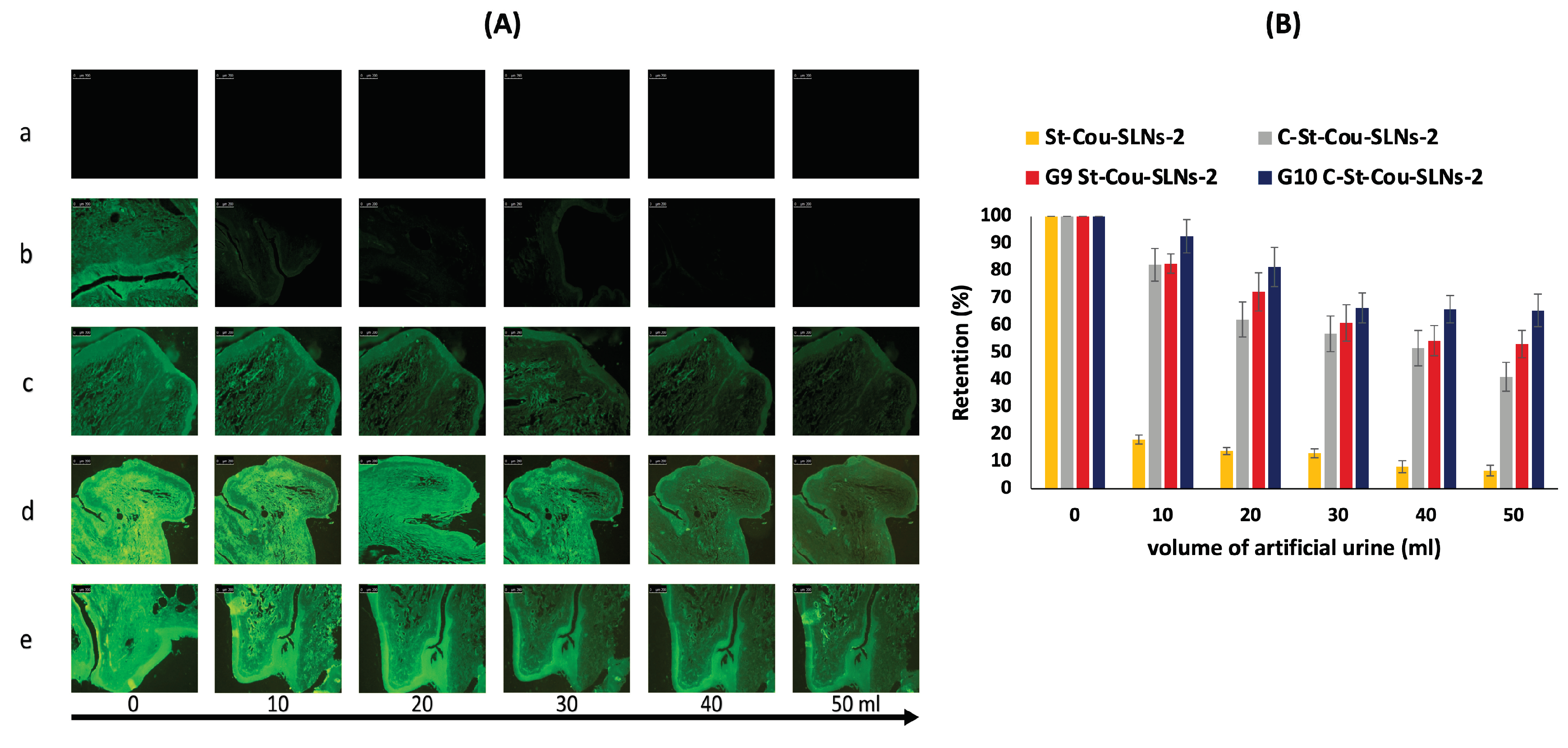
Retention on Bladder Mucosa
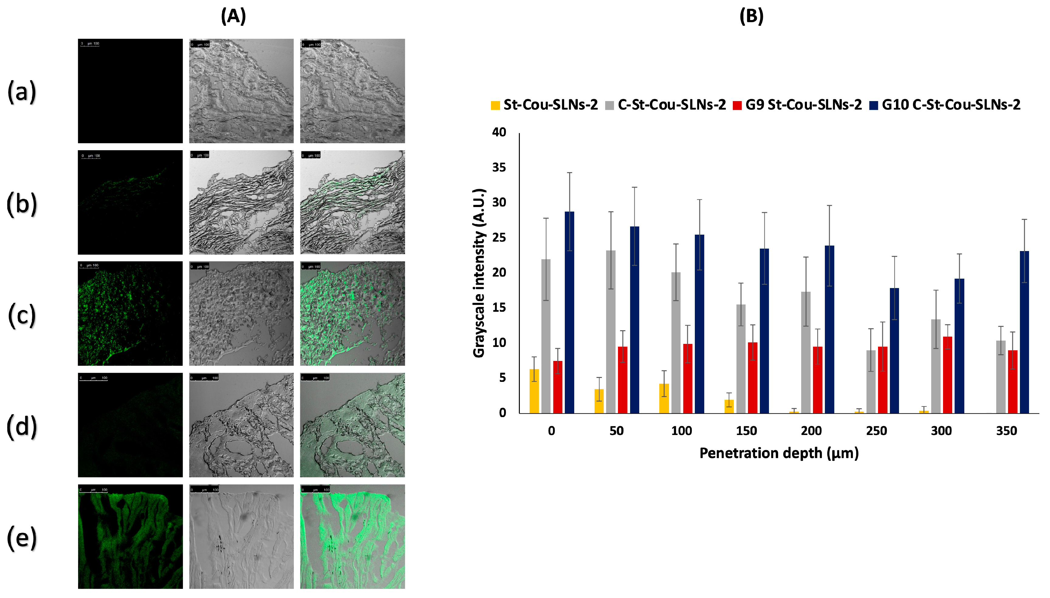
Mucosal Penetration
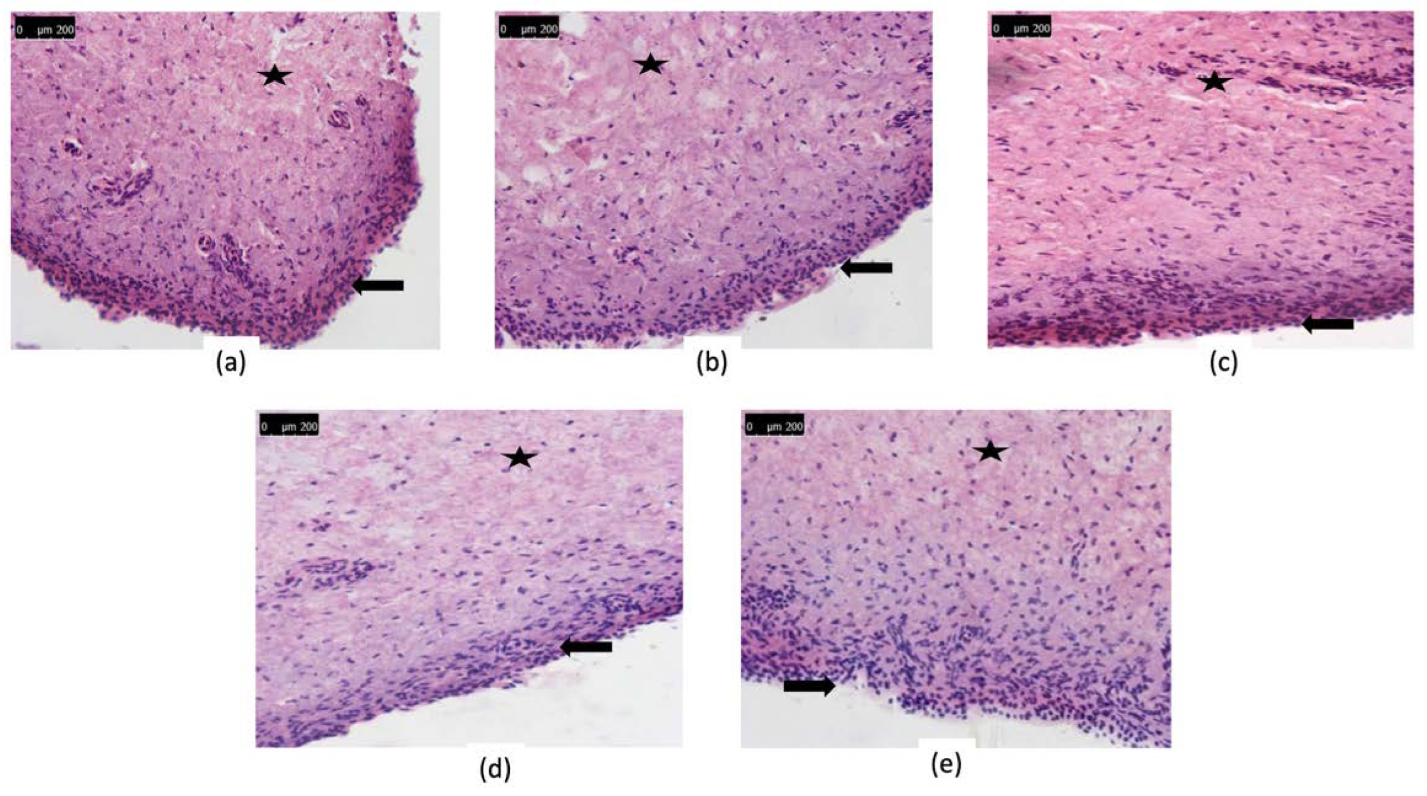
Histopathological Evaluation
2.2.4. Statistical Analysis
3. Results and Discussion
3.1. Preparation and In Vitro Characterization of SLNs
3.1.1. Effect of Formulation Variables on % EE and PS of SLNs
Effect of Stabilizer Type and Concentration on % EE and PS of SLNs
Effect of Lipid to Drug Ratio on %EE and PS of SLNs
3.1.2. Colloidal Characteristics, EE and DL of Selected Formulations
3.1.3. Chitosan Coating
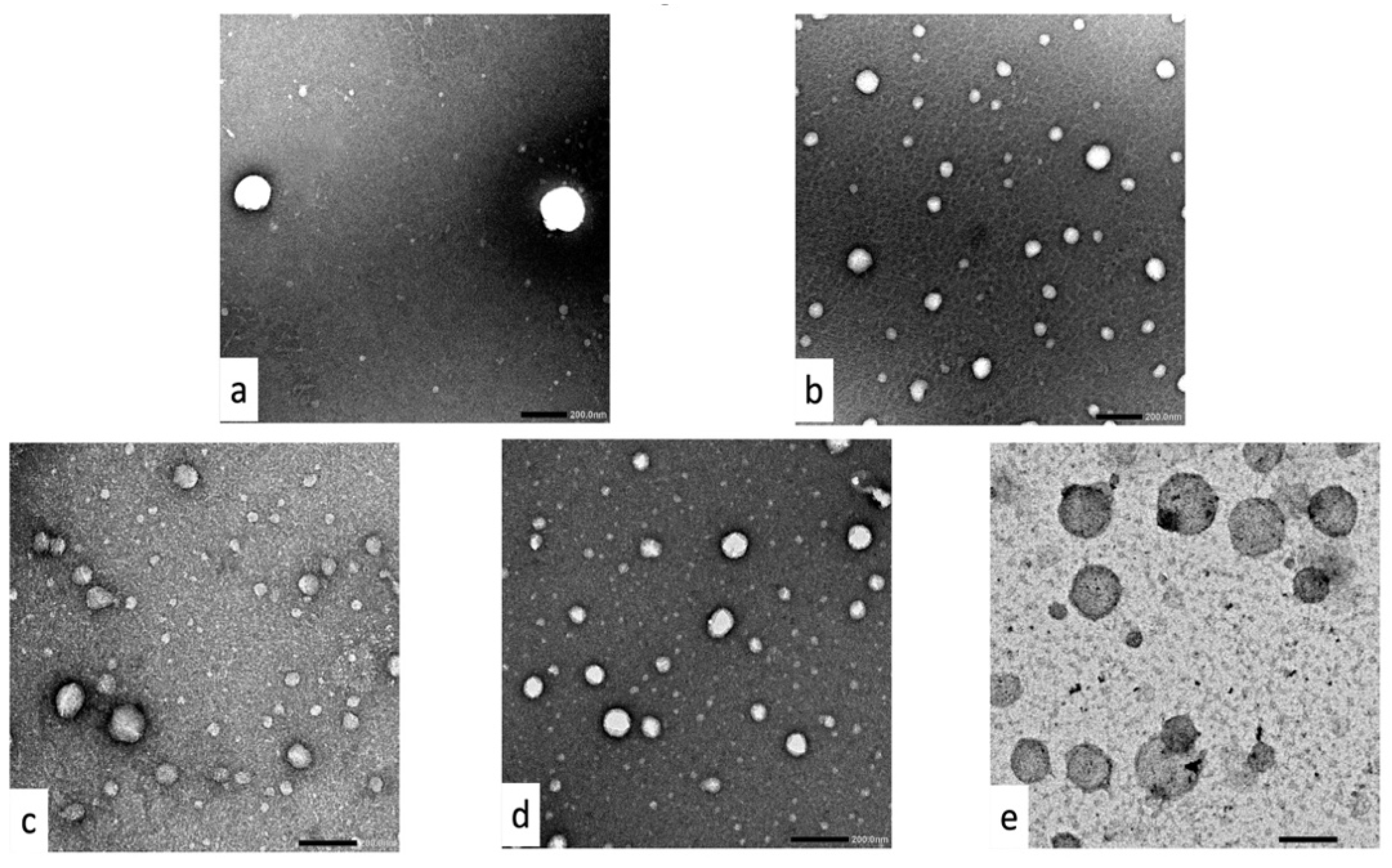
3.1.4. Morphology of Plain, Coated and Uncoated SLNs
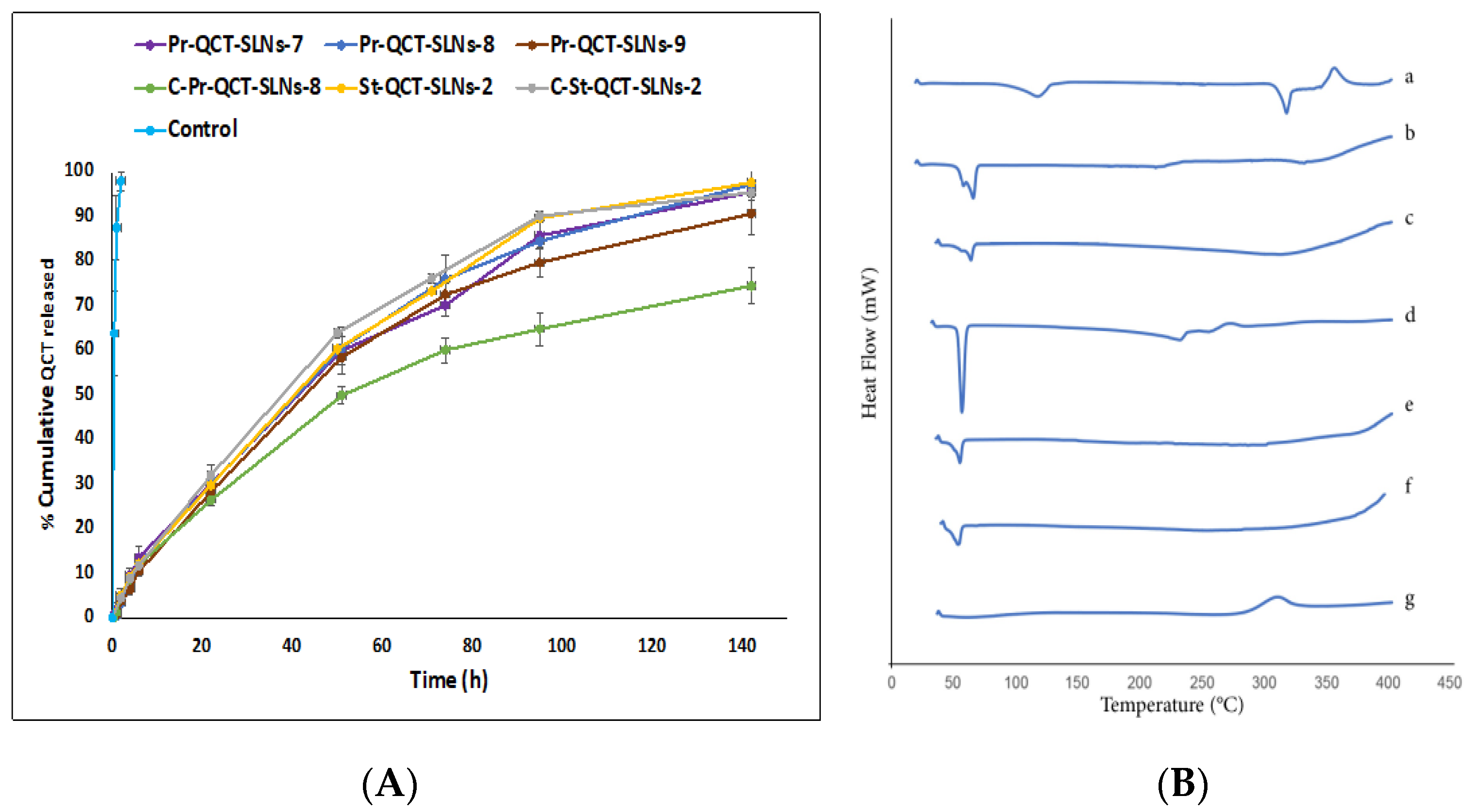
3.1.5. In Vitro Release of QCT from Nanoparticles
3.1.6. Stability Study
3.1.7. Thermal Properties of SLNs Using DSC
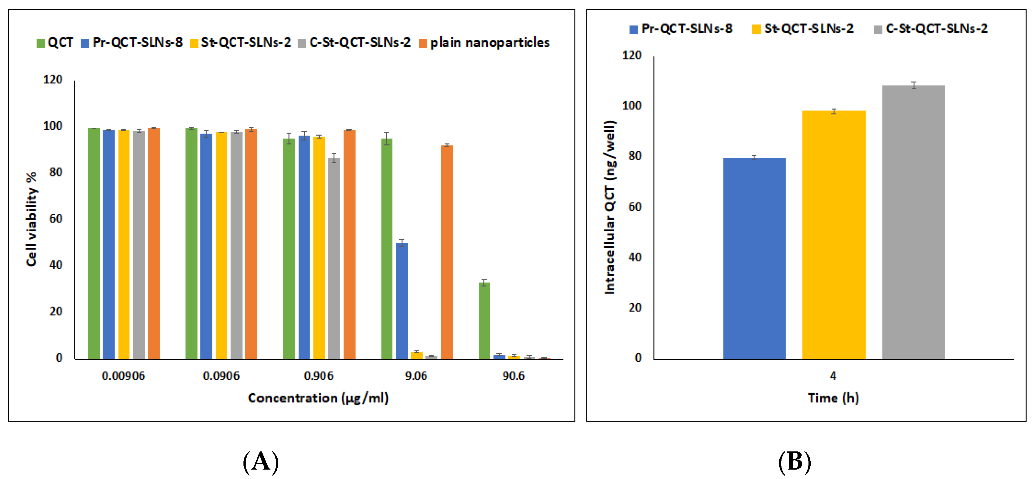
3.1.8. Cytotoxicity Assay
3.1.9. Cellular Drug Uptake
3.2. Characterization and Optimization of the Mucoadhesive In Situ Gel Formulations
3.2.1. Gelation Temperature
3.2.2. Gelation Time
3.2.3. Erosion Time
3.2.4. Muco-Adhesion
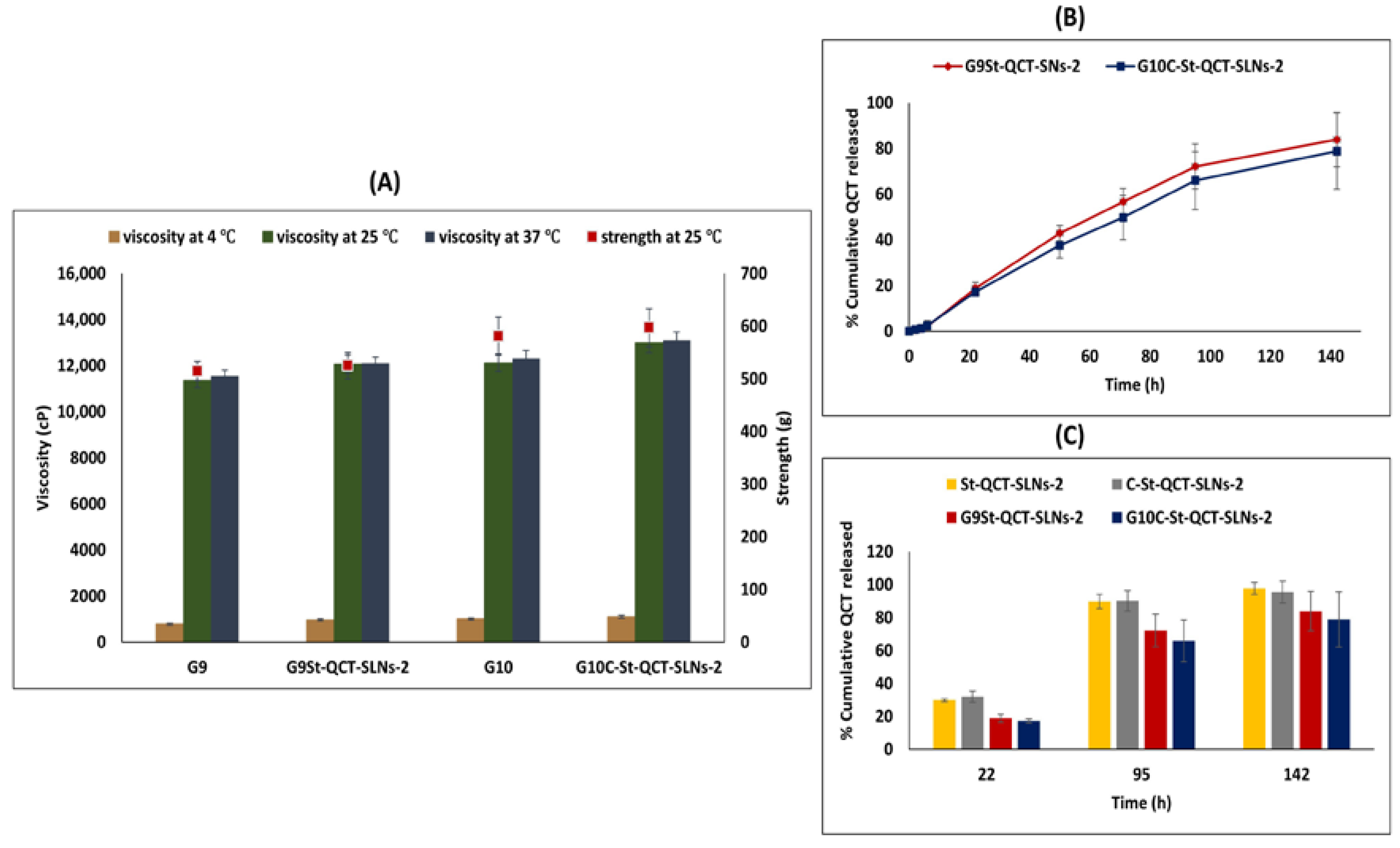
3.2.5. Rheological and Mechanical Characteristics
3.2.6. Determination of the Percentage of Gels That Can Be Pushed through a Catheter
3.2.7. In Vitro Release of QCT from Gel Formulations
3.3. Ex Vivo Studies
3.3.1. Retention of Selected Formulations on Mucosal Surfaces of Bovine Urinary Bladder
3.3.2. Mucosal Penetration
3.3.3. Histopathological Evaluation
4. Conclusions
Author Contributions
Funding
Institutional Review Board Statement
Informed Consent Statement
Data Availability Statement
Conflicts of Interest
References
- Huang, C.; Liao, X.; Jin, H.; Xie, F.; Zheng, F.; Li, J.; Zhou, C.; Jiang, G.; Wu, X.R.; Huang, C. MEG3, as a Competing Endogenous RNA, Binds with miR-27a to Promote PHLPP2 Protein Translation and Impairs Bladder Cancer Invasion. Mol. Ther. Nucleic Acids 2019, 16, 51–62. [Google Scholar] [CrossRef] [PubMed] [Green Version]
- Yoon, H.Y.; Yang, H.M.; Kim, C.H.; Goo, Y.T.; Kang, M.J.; Lee, S.; Choi, Y.W. Current status of the development of intravesical drug delivery systems for the treatment of bladder cancer. Expert Opin. Drug Deliv. 2020, 17, 1555–1572. [Google Scholar] [CrossRef] [PubMed]
- GuhaSarkar, S.; Banerjee, R. Intravesical drug delivery: Challenges, current status, opportunities and novel strategies. J. Control. Release 2010, 148, 147–159. [Google Scholar] [CrossRef] [PubMed]
- Elstad, N.L.; Fowers, K.D. OncoGel (ReGel/paclitaxel)—Clinical applications for a novel paclitaxel delivery system. Adv. Drug Deliv. Rev. 2009, 61, 785–794. [Google Scholar] [CrossRef]
- Kolawole, O.M.; Lau, W.M.; Mostafid, H.; Khutoryanskiy, V.V. Advances in intravesical drug delivery systems to treat bladder cancer. Int. J. Pharm. 2017, 532, 105–117. [Google Scholar] [CrossRef] [Green Version]
- Xu, X.; Liu, K.; Jiao, B.; Luo, K.; Ren, J.; Zhang, G.; Yu, Q.; Gan, Z. Mucoadhesive nanoparticles based on ROS activated gambogic acid prodrug for safe and efficient intravesical instillation chemotherapy of bladder cancer. J. Control. Release 2020, 324, 493–504. [Google Scholar] [CrossRef]
- Suo, N.; Wang, M.; Jin, Y.; Ding, J.; Gao, X.; Sun, X.; Zhang, H.; Cui, M.; Zheng, J.; Li, N.; et al. Magnetic multiwalled carbon nanotubes with controlled release of epirubicin: An intravesical instillation system for bladder cancer. Int. J. Nanomed. 2019, 14, 1241–1254. [Google Scholar] [CrossRef] [Green Version]
- Iacopetta, D.; Grande, F.; Caruso, A.; Mordocco, R.A.; Plutino, M.R.; Scrivano, L.; Ceramella, J.; Muià, N.; Saturnino, C.; Puoci, F.; et al. New insights for the use of quercetin analogs in cancer treatment. Future Med. Chem. 2017, 9, 2011–2028. [Google Scholar] [CrossRef]
- Hidalgo, M.; Sánchez-Moreno, C.; de Pascual-Teresa, S. Flavonoid–flavonoid interaction and its effect on their antioxidant activity. Food Chem. 2010, 121, 691–696. [Google Scholar] [CrossRef]
- Rogerio, A.P.; Dora, C.L.; Andrade, E.L.; Chaves, J.S.; Silva, L.F.; Lemos-Senna, E.; Calixto, J.B. Anti-inflammatory effect of quercetin-loaded microemulsion in the airways allergic inflammatory model in mice. Pharmacol. Res. 2010, 61, 288–297. [Google Scholar] [CrossRef]
- Fang, X.K.; Gao, J.; Zhu, D.N. Kaempferol and quercetin isolated from Euonymus alatus improve glucose uptake of 3T3-L1 cells without adipogenesis activity. Life Sci. 2008, 82, 615–622. [Google Scholar] [CrossRef] [PubMed]
- Hirpara, K.V.; Aggarwal, P.; Mukherjee, A.J.; Joshi, N.; Burman, A.C. Quercetin and its derivatives: Synthesis, pharmacological uses with special emphasis on anti-tumor properties and prodrug with enhanced bio-availability. Anticancer Agents Med. Chem. 2009, 9, 138–161. [Google Scholar] [CrossRef] [PubMed]
- Liu, Y.; Tang, Z.G.; Lin, Y.; Qu, X.G.; Lv, W.; Wang, G.B.; Li, C.L. Effects of quercetin on proliferation and migration of human glioblastoma U251 cells. Biomed. Pharmacother. 2017, 92, 33–38. [Google Scholar] [CrossRef] [PubMed]
- Murakami, A.; Ashida, H.; Terao, J. Multitargeted cancer prevention by quercetin. Cancer Lett. 2008, 269, 315–325. [Google Scholar] [CrossRef] [PubMed]
- Shih, H.; Pickwell, G.V.; Quattrochi, L.C. Differential effects of flavonoid compounds on tumor promoter-induced activation of the human CYP1A2 enhancer. Arch. Biochem. Biophys. 2000, 373, 287–294. [Google Scholar] [CrossRef] [PubMed]
- Brito, A.F.; Ribeiro, M.; Abrantes, A.M.; Pires, A.S.; Teixo, R.J.; Tralhão, J.G.; Botelho, M.F. Quercetin in Cancer Treatment, Alone or in Combination with Conventional Therapeutics? Curr. Med. Chem. 2015, 22, 3025–3039. [Google Scholar] [CrossRef] [PubMed] [Green Version]
- Oršolić, N.; Karač, I.; Sirovina, D.; Kukolj, M.; Kunštić, M.; Gajski, G.; Garaj-Vrhovac, V.; Štajcar, D. Chemotherapeutic potential of quercetin on human bladder cancer cells. J. Environ. Sci. Health Part A Toxic Hazard. Subst. Environ. Eng. 2016, 51, 776–781. [Google Scholar] [CrossRef]
- Pralhad, T.; Rajendrakumar, K. Study of freeze-dried quercetin–cyclodextrin binary systems by DSC, FT-IR, X-ray diffraction and SEM analysis. J. Pharm. Biomed. 2004, 34, 333–339. [Google Scholar] [CrossRef]
- Tyagi, P.; Chancellor, M.B.; Li, Z.; de Groat, W.C.; Yoshimura, N.; Fraser, M.O.; Huang, L. Urodynamic and Immunohistochemical Evaluation of Intravesical Capsaicin Delivery Using Thermosensitive Hydrogel and Liposomes. J. Urol. 2004, 171, 483–489. [Google Scholar] [CrossRef]
- Chuang, Y.C.; Tyagi, P.; Huang, C.C.; Yoshimura, N.; Wu, M.; Kaufman, J.; Chancellor, M.B. Urodynamic and Immunohistochemical Evaluation of Intravesical Botulinum Toxin A Delivery Using Liposomes. J. Urol. 2009, 182, 786–792. [Google Scholar] [CrossRef]
- Frangos, D.N.; Killion, J.J.; Fan, D.; Fishbeck, R.; Von Eschenbach, A.c.; Fidler, I.J. The Development of Liposomes Containing Interferon Alpha for the Intravesical Therapy of Human Superficial Bladder Cancer. J. Urol. 1990, 143, 1252–1256. [Google Scholar] [CrossRef]
- Chang, L.C.; Wu, S.C.; Tsai, J.W.; Yu, T.J.; Tsai, T.R. Optimization of epirubicin nanoparticles using experimental design for enhanced intravesical drug delivery. Int. J. Pharm. 2009, 376, 195–203. [Google Scholar] [CrossRef] [PubMed]
- Martin, D.T.; Hoimes, C.J.; Kaimakliotis, H.Z.; Cheng, C.J.; Zhang, K.; Liu, J.; Wheeler, M.A.; Kelly, W.K.; Tew, G.N.; Saltzman, W.M. Nanoparticles for urothelium penetration and delivery of the histone deacetylase inhibitor belinostat for treatment of bladder cancer. Nanomedicine 2013, 9, 1124–1134. [Google Scholar] [CrossRef] [PubMed] [Green Version]
- Martin, D.T.; Steinbach, J.M.; Liu, J.; Shimizu, S.; Kaimakliotis, H.Z.; Wheeler, M.A.; Hittelman, A.B.; Mark Saltzman, W.; Weiss, R.M. Surface-modified nanoparticles enhance transurothelial penetration and delivery of survivin siRNA in treating bladder cancer. Mol. Cancer Ther. 2014, 13, 71–81. [Google Scholar] [CrossRef] [Green Version]
- Yuan, Q.; Han, J.; Cong, W.; Ge, Y.; Ma, D.; Dai, Z.; Li, Y.; Bi, X. Docetaxel-loaded solid lipid nanoparticles suppress breast cancer cells growth with reduced myelosuppression toxicity. Int. J. Nanomed. 2014, 9, 4829–4846. [Google Scholar] [CrossRef] [Green Version]
- Harivardhan Reddy, L.; Sharma, R.K.; Chuttani, K.; Mishra, A.K.; Murthy, R.S. Influence of administration route on tumor uptake and biodistribution of etoposide loaded solid lipid nanoparticles in Dalton’s lymphoma tumor bearing mice. J. Control. Release 2005, 105, 185–198. [Google Scholar] [CrossRef]
- Subedi, R.K.; Kang, K.W.; Choi, H.K. Preparation and characterization of solid lipid nanoparticles loaded with doxorubicin. Eur. J. Pharm. Sci. 2009, 37, 508–513. [Google Scholar] [CrossRef]
- Wong, H.L.; Bendayan, R.; Rauth, A.M.; Li, Y.; Wu, X.Y. Chemotherapy with anticancer drugs encapsulated in solid lipid nanoparticles. Adv. Drug Deliv. Rev. 2007, 59, 491–504. [Google Scholar] [CrossRef]
- Makled, S.; Nafee, N.; Boraie, N. Nebulized solid lipid nanoparticles for the potential treatment of pulmonary hypertension via targeted delivery of phosphodiesterase-5-inhibitor. Int. J. Pharm. 2017, 517, 312–321. [Google Scholar] [CrossRef]
- Di Stasi, S.M.; Giannantoni, A.; Stephen, R.L.; Capelli, G.; Navarra, P.; Massoud, R.; Vespasiani, G. Intravesical electromotive mitomycin C versus passive transport mitomycin C for high risk superficial bladder cancer: A prospective randomized study. J. Urol. 2003, 170, 777–782. [Google Scholar] [CrossRef]
- Men, K.; Liu, W.; Li, L.; Duan, X.; Wang, P.; Gou, M.; Wei, X.; Gao, X.; Wang, B.; Du, Y.; et al. Delivering instilled hydrophobic drug to the bladder by a cationic nanoparticle and thermo-sensitive hydrogel composite system. Nanoscale 2012, 4, 6425–6433. [Google Scholar] [CrossRef] [PubMed]
- Qiu, H.; Guo, H.; Li, D.; Hou, Y.; Kuang, T.; Ding, J. Intravesical Hydrogels as Drug Reservoirs. Trends Biotechnol. 2020, 38, 579–583. [Google Scholar] [CrossRef] [PubMed]
- Lee, H.; Cima, M.J. An intravesical device for the sustained delivery of lidocaine to the bladder. J. Control. Release 2011, 149, 133–139. [Google Scholar] [CrossRef] [PubMed]
- Tyagi, P.; Kashyap, M.; Hensley, H.; Yoshimura, N. Advances in intravesical therapy for urinary tract disorders. Expert Opin. Drug Deliv. 2016, 13, 71–84. [Google Scholar] [CrossRef] [PubMed] [Green Version]
- Şenyiğit, Z.A.; Karavana, S.Y.; İlem-Özdemir, D.; Çalışkan, Ç.; Waldner, C.; Şen, S.; Bernkop-Schnürch, A.; Baloğlu, E. Design and evaluation of an intravesical delivery system for superficial bladder cancer: Preparation of gemcitabine HCl-loaded chitosan-thioglycolic acid nanoparticles and comparison of chitosan/poloxamer gels as carriers. Int. J. Nanomed. 2015, 10, 6493–6507. [Google Scholar] [CrossRef] [Green Version]
- Lin, T.; Wu, J.; Zhao, X.; Lian, H.; Yuan, A.; Tang, X.; Zhao, S.; Guo, H.; Hu, Y. In Situ Floating Hydrogel for Intravesical Delivery of Adriamycin Without Blocking Urinary Tract. J. Pharm. Sci. 2014, 103, 927–936. [Google Scholar] [CrossRef]
- Diniyanti, S. Preparation of solid lipid nanoparticles containing mangosteen pericarp extract. Asian J. Pharm. Clin. Res. 2018, 11, 80–84. [Google Scholar] [CrossRef]
- Bose, S.; Du, Y.; Takhistov, P.; Michniak-Kohn, B. Formulation optimization and topical delivery of quercetin from solid lipid based nanosystems. Int. J. Pharm. 2013, 441, 56–66. [Google Scholar] [CrossRef]
- Patra, A.; Satpathy, S.; Shenoy, A.K.; Bush, J.A.; Kazi, M.; Hussain, M.D. Formulation and evaluation of mixed polymeric micelles of quercetin for treatment of breast, ovarian, and multidrug resistant cancers. Int. J. Nanomed. 2018, 13, 2869–2881. [Google Scholar] [CrossRef] [Green Version]
- Wang, W.; Chen, T.; Xu, H.; Ren, B.; Cheng, X.; Qi, R.; Liu, H.; Wang, Y.; Yan, L.; Chen, S.; et al. Curcumin-Loaded Solid Lipid Nanoparticles Enhanced Anticancer Efficiency in Breast Cancer. Molecules 2018, 23, 1578. [Google Scholar] [CrossRef]
- Schmolka, I.R. Artificial skin I. Preparation and properties of pluronic F-127 gels for treatment of burns. J. Biomed. Mater. Res. 1972, 6, 571–582. [Google Scholar] [CrossRef] [PubMed]
- Singh, R.M.P.; Kumar, A.; Pathak, K. Thermally Triggered Mucoadhesive In Situ Gel of Loratadine: β-Cyclodextrin Complex for Nasal Delivery. AAPS PharmSciTech 2013, 14, 412–424. [Google Scholar] [CrossRef] [PubMed] [Green Version]
- Youssef, N.; Kassem, A.A.; Farid, R.M.; Ismail, F.A.; El-Massik, M.A.E.; Boraie, N.A. A novel nasal almotriptan loaded solid lipid nanoparticles in mucoadhesive in situ gel formulation for brain targeting: Preparation, characterization and in vivo evaluation. Int. J. Pharm. 2018, 548, 609–624. [Google Scholar] [CrossRef] [PubMed]
- Khan, S.; Patil, K.; Bobade, N.; Yeole, P.; Gaikwad, R. Formulation of intranasal mucoadhesive temperature-mediated in situ gel containing ropinirole and evaluation of brain targeting efficiency in rats. J. Drug Target. 2010, 18, 223–234. [Google Scholar] [CrossRef] [PubMed]
- Nakamura, F.; Ohta, R.; Machida, Y.; Nagai, T. In vitro and in vivo nasal mucoadhesion of some water-soluble polymers. Int. J. Pharm. 1996, 134, 173–181. [Google Scholar] [CrossRef]
- Chatta, D.; Cottrell, L.; Burnett, B.; Laverty, G.; McConville, C. The use of water-soluble mucoadhesive gels for the intravesical delivery of epirubicin to the bladder for the treatment of non-muscle-invasive bladder cancer. J Pharm. Pharmacol. 2015, 67, 1355–1362. [Google Scholar] [CrossRef] [Green Version]
- Mun, E.A.; Williams, A.C.; Khutoryanskiy, V.V. Adhesion of thiolated silica nanoparticles to urinary bladder mucosa: Effects of PEGylation, thiol content and particle size. Int. J. Pharm. 2016, 512, 32–38. [Google Scholar] [CrossRef] [Green Version]
- Mansfield, E.D.H.; de la Rosa, V.R.; Kowalczyk, R.M.; Grillo, I.; Hoogenboom, R.; Sillence, K.; Hole, P.; Williams, A.C.; Khutoryanskiy, V.V. Side chain variations radically alter the diffusion of poly(2-alkyl-2-oxazoline) functionalised nanoparticles through a mucosal barrier. Biomater. Sci. 2016, 4, 1318–1327. [Google Scholar] [CrossRef] [Green Version]
- GuhaSarkar, S.; More, P.; Banerjee, R. Urothelium-adherent, ion-triggered liposome-in-gel system as a platform for intravesical drug delivery. J. Control. Release 2017, 245, 147–156. [Google Scholar] [CrossRef]
- Jensen, L.B.; Magnussson, E.; Gunnarsson, L.; Vermehren, C.; Nielsen, H.M.; Petersson, K. Corticosteroid solubility and lipid polarity control release from solid lipid nanoparticles. Int. J. Pharm. 2010, 390, 53–60. [Google Scholar] [CrossRef]
- Mehnert, W.; Mäder, K. Solid lipid nanoparticles: Production, characterization and applications. Adv. Drug Deliv. Rev. 2012, 64, 83–101. [Google Scholar] [CrossRef]
- Zewail, M.; Nafee, N.; Helmy, M.W.; Boraie, N. Coated nanostructured lipid carriers targeting the joints—An effective and safe approach for the oral management of rheumatoid arthritis. Int. J. Pharm. 2019, 567, 118447. [Google Scholar] [CrossRef] [PubMed]
- Tanvir, S.; Qiao, L. Surface tension of nanofluid-type fuels containing suspended nanomaterials. Nanoscale Res. Lett. 2012, 7, 226. [Google Scholar] [CrossRef] [PubMed] [Green Version]
- Han, F.; Li, S.; Yin, R.; Liu, H.; Xu, L. Effect of surfactants on the formation and characterization of a new type of colloidal drug delivery system: Nanostructured lipid carriers. Colloids Surf. A Physicochem. Eng. Asp. 2008, 315, 210–216. [Google Scholar] [CrossRef]
- Üner, M.; Wissing, S.A.; Yener, G.; Müller, R.H. Influence of surfactants on the physical stability of solid lipid nanoparticle (SLN) formulations. Pharmazie 2004, 59, 331–332. [Google Scholar]
- Singh, S.; Dobhal, A.K.; Jain, A.; Pandit, J.K.; Chakraborty, S. Formulation and Evaluation of Solid Lipid Nanoparticles of a Water Soluble Drug: Zidovudine. Chem. Pharm. Bull. 2010, 58, 650–655. [Google Scholar] [CrossRef] [PubMed] [Green Version]
- Lv, Q.; Yu, A.; Xi, Y.; Li, H.; Song, Z.; Cui, J.; Cao, F.; Zhai, G. Development and evaluation of penciclovir-loaded solid lipid nanoparticles for topical delivery. Int. J. Pharm. 2009, 372, 191–198. [Google Scholar] [CrossRef]
- Shah, B.; Khunt, D.; Bhatt, H.; Misra, M.; Padh, H. Application of quality by design approach for intranasal delivery of rivastigmine loaded solid lipid nanoparticles: Effect on formulation and characterization parameters. Eur. J. Pharm. Sci. 2015, 78, 54–66. [Google Scholar] [CrossRef]
- Rahman, Z.; Zidan, A.S.; Khan, M.A. Non-destructive methods of characterization of risperidone solid lipid nanoparticles. Eur. J. Pharm. Biopharm. 2010, 76, 127–137. [Google Scholar] [CrossRef]
- Fernandes, A.; Pydi, C.; Verma, R.; Jose, J.; Kumar, L. Design, preparation and in vitro characterizations of fluconazole loaded nanostructured lipid carriers. Braz. J. Pharm. Sci. 2020, 56, e18069. [Google Scholar] [CrossRef]
- Salem, S.G.; Gardouh, A.R.; Gad, S. C:Parameter Optimization of Solid Lipid Nanoparticles Formulation. Rec. Pharm. Biomed. Sci. 2020, 4, 1–7. [Google Scholar] [CrossRef]
- Reddy, L.H.; Murthy, R.S. Etoposide-loaded nanoparticles made from glyceride lipids: Formulation, characterization, in vitro drug release, and stability evaluation. AAPS PharmSciTech 2005, 6, E158–E166. [Google Scholar] [CrossRef] [PubMed]
- Anton, N.; Benoit, J.P.; Saulnier, P. Design and production of nanoparticles formulated from nano-emulsion templates-a review. J. Control. Release 2008, 128, 185–199. [Google Scholar] [CrossRef] [PubMed]
- Aditya, N.P.; Macedo, A.S.; Doktorovova, S.; Souto, E.B.; Kim, S.; Chang, P.S.; Ko, S. Development and evaluation of lipid nanocarriers for quercetin delivery: A comparative study of solid lipid nanoparticles (SLN), nanostructured lipid carriers (NLC), and lipid nanoemulsions (LNE). LWT-Food Sci. Technol. 2014, 59, 115–121. [Google Scholar] [CrossRef]
- Ay, M.; Charli, A.; Jin, H.; Anantharam, V.; Kanthasamy, A.; Kanthasamy, A.G. Quercetin. In Nutraceuticals; Gupta, R.C., Ed.; Academic Press: Boston, MA, USA, 2016; Chapter 32; pp. 447–452. [Google Scholar] [CrossRef]
- Nzekwea, I.T.; Azodo, V.I.; Agubata, C.O.; Naicker, B.; Okore, V.O.; Esimone, C.O. Preliminary formulation and characterization of solid lipid nanoparticles containing chloroquine and a P-glycoprotein inhibitor: Influences of lipid-surfactant ratios. J. Chem. Pharm. Res. 2015, 7, 932–939. [Google Scholar]
- Trotta, M.; Debernardi, F.; Caputo, O. Preparation of solid lipid nanoparticles by a solvent emulsification–diffusion technique. Int. J. Pharm. 2003, 257, 153–160. [Google Scholar] [CrossRef]
- Dantas, I.L.; Bastos, K.T.S.; Machado, M.; Galvao, J.G.; Lima, A.D.; Gonsalves, J.K.M.C.; Almeida, E.D.P.; Araújo, A.A.S.; de Meneses, C.T.; Sarmento, V.H.V. Influence of stearic acid and beeswax as solid lipid matrix of lipid nanoparticles containing tacrolimus. J. Therm. Anal. Calorim. 2018, 132, 1557–1566. [Google Scholar] [CrossRef]
- Talarico, L.; Consumi, M.; Leone, G.; Tamasi, G.; Magnani, A. Solid Lipid Nanoparticles Produced via a Coacervation Method as Promising Carriers for Controlled Release of Quercetin. Molecules 2021, 26, 2694. [Google Scholar] [CrossRef]
- González-Mira, E.; Nikolić, S.; García, M.L.; Egea, M.A.; Souto, E.B.; Calpena, A.C. Potential Use of Nanostructured Lipid Carriers for Topical Delivery of Flurbiprofen. J. Pharm. Sci. 2011, 100, 242–251. [Google Scholar] [CrossRef]
- Han, S.B.; Kwon, S.S.; Jeong, Y.M.; Yu, E.R.; Park, S.N. Physical characterization and in vitro skin permeation of solid lipid nanoparticles for transdermal delivery of quercetin. Int. J. Cosmet. Sci. 2014, 36, 588–597. [Google Scholar] [CrossRef]
- Liu, C.H.; Huang, Y.C.; Jhang, J.W.; Liu, Y.H.; Wu, W.C. Quercetin delivery to porcine cornea and sclera by solid lipid nanoparticles and nanoemulsion. RSC Adv. 2015, 5, 100923–100933. [Google Scholar] [CrossRef]
- Pinheiro, R.G.R.; Granja, A.; Loureiro, J.A.; Pereira, M.C.; Pinheiro, M.; Neves, A.R.; Reis, S. Quercetin lipid nanoparticles functionalized with transferrin for Alzheimer’s disease. Eur. J. Pharm. Sci. 2020, 148, 105314. [Google Scholar] [CrossRef]
- Kean, T.; Thanou, M. Biodegradation, biodistribution and toxicity of chitosan. Adv. Drug Deliv. Rev. 2010, 62, 3–11. [Google Scholar] [CrossRef] [PubMed]
- Wang, F.Z.; Zhang, M.W.; Zhang, D.S.; Huang, Y.; Chen, L.; Jiang, S.M.; Shi, K.; Li, R. Preparation, optimization, and characterization of chitosan-coated solid lipid nanoparticles for ocular drug delivery. J. Biomed. Res. 2018, 32, 411–423. [Google Scholar] [CrossRef] [PubMed]
- Grabnar, I.; Bogataj, M.; Mrhar, A. Influence of chitosan and polycarbophil on permeation of a model hydrophilic drug into the urinary bladder wall. Int. J. Pharm. 2003, 256, 167–173. [Google Scholar] [CrossRef]
- Luo, Y.; Teng, Z.; Li, Y.; Wang, Q. Solid lipid nanoparticles for oral drug delivery: Chitosan coating improves stability, controlled delivery, mucoadhesion and cellular uptake. Carbohydr. Polym. 2015, 122, 221–229. [Google Scholar] [CrossRef]
- Vieira, A.C.; Chaves, L.L.; Pinheiro, S.; Pinto, S.; Pinheiro, M.; Lima, S.C.; Ferreira, D.; Sarmento, B.; Reis, S. Mucoadhesive chitosan-coated solid lipid nanoparticles for better management of tuberculosis. Int. J. Pharm. 2018, 536, 478–485. [Google Scholar] [CrossRef] [PubMed]
- Kumar, S.; Randhawa, J.K. Preparation and characterization of Paliperidone loaded solid lipid nanoparticles. Colloids Surf. B Biointerfaces 2013, 102, 562–568. [Google Scholar] [CrossRef] [PubMed]
- Makled, S.; Boraie, N.; Nafee, N. Nanoparticle-mediated macrophage targeting—A new inhalation therapy tackling tuberculosis. Drug Deliv. Transl. Res. 2021, 11, 1037–1055. [Google Scholar] [CrossRef]
- Hazzah, H.A.; Farid, R.M.; Nasra, M.M.A.; Hazzah, W.A.; El-Massik, M.A.; Abdallah, O.Y. Gelucire-Based Nanoparticles for Curcumin Targeting to Oral Mucosa: Preparation, Characterization, and Antimicrobial Activity Assessment. J. Pharm. Sci. 2015, 104, 3913–3924. [Google Scholar] [CrossRef]
- Wang, J.Y.; Wang, Y.; Meng, X. Chitosan Nanolayered Cisplatin-Loaded Lipid Nanoparticles for Enhanced Anticancer Efficacy in Cervical Cancer. Nanoscale Res. Lett. 2016, 11, 524. [Google Scholar] [CrossRef] [PubMed]
- Borges, A.; Freitas, V.d.; Mateus, N.; Fernandes, I.; Oliveira, J. Solid Lipid Nanoparticles as Carriers of Natural Phenolic Compounds. Antioxidants 2020, 9, 998. [Google Scholar] [CrossRef] [PubMed]
- Müller, R.H.; Mäder, K.; Gohla, S. Solid lipid nanoparticles (SLN) for controlled drug delivery—A review of the state of the art. Eur. J. Pharm. Biopharm. 2000, 50, 161–177. [Google Scholar] [CrossRef]
- Rishitha, N.; Muthuraman, A. Therapeutic evaluation of solid lipid nanoparticle of quercetin in pentylenetetrazole induced cognitive impairment of zebrafish. Life Sci. 2018, 199, 80–87. [Google Scholar] [CrossRef] [PubMed]
- Li, H.; Zhao, X.; Ma, Y.; Zhai, G.; Li, L.; Lou, H. Enhancement of gastrointestinal absorption of quercetin by solid lipid nanoparticles. J. Control. Release 2009, 133, 238–244. [Google Scholar] [CrossRef] [PubMed]
- Shah, R.M.; Eldridge, D.S.; Palombo, E.A.; Harding, I.H. Microwave-assisted formulation of solid lipid nanoparticles loaded with non-steroidal anti-inflammatory drugs. Int. J. Pharm. 2016, 515, 543–554. [Google Scholar] [CrossRef]
- Costa Silva, L.; Kasten Korgaard, G.; Campos, C.; Chinelatto, A.; Senna, E. Preparation and characterization of quercetin-loaded solid lipid microparticles for pulmonary delivery. Powder Technol. 2013, 239, 183–192. [Google Scholar] [CrossRef]
- Wang, T.; Ma, X.; Lei, Y.; Luo, Y. Solid lipid nanoparticles coated with cross-linked polymeric double layer for oral delivery of curcumin. Colloids Surf. B Biointerfaces 2016, 148, 1–11. [Google Scholar] [CrossRef]
- Nafee, N.; Makled, S.; Boraie, N. Nanostructured lipid carriers versus solid lipid nanoparticles for the potential treatment of pulmonary hypertension via nebulization. Eur. J. Pharm. Sci. 2018, 125, 151–162. [Google Scholar] [CrossRef]
- Dey, S.; Al-Amin, M.; Rashid, T.; Sultan, Z.; Ashaduzzaman, M.; Sarker, M.; Shamsuddin, S. Preparation, characterization and performance evaluation of chitosan as an adsorbent for remazol red. Int. J. Latest Res. Eng. Technol. 2016, 2, 52–62. [Google Scholar]
- Rajesh Kumar, S.; Priyatharshni, S.; Babu, V.N.; Mangalaraj, D.; Viswanathan, C.; Kannan, S.; Ponpandian, N. Quercetin conjugated superparamagnetic magnetite nanoparticles for in-vitro analysis of breast cancer cell lines for chemotherapy applications. J. Colloid Interface Sci. 2014, 436, 234–242. [Google Scholar] [CrossRef] [PubMed]
- Choi, E.J.; Bae, S.M.; Ahn, W.S. Antiproliferative effects of quercetin through cell cycle arrest and apoptosis in human breast cancer MDA-MB-453 cells. Arch. Pharm. Res 2008, 31, 1281–1285. [Google Scholar] [CrossRef] [PubMed]
- Kakran, M.; Sahoo, N.G.; Li, L. Dissolution enhancement of quercetin through nanofabrication, complexation, and solid dispersion. Colloids Surf. B Biointerfaces 2011, 88, 121–130. [Google Scholar] [CrossRef] [PubMed]
- Huang, J.; Si, L.; Jiang, L.; Fan, Z.; Qiu, J.; Li, G. Effect of pluronic F68 block copolymer on P-glycoprotein transport and CYP3A4 metabolism. Int. J. Pharm. 2008, 356, 351–353. [Google Scholar] [CrossRef]
- Baksi, R.; Singh, D.P.; Borse, S.P.; Rana, R.; Sharma, V.; Nivsarkar, M. In vitro and in vivo anticancer efficacy potential of Quercetin loaded polymeric nanoparticles. Biomed. Pharmacother. 2018, 106, 1513–1526. [Google Scholar] [CrossRef]
- Wei, Z.; Hao, J.; Yuan, S.; Li, Y.; Juan, W.; Sha, X.; Fang, X. Paclitaxel-loaded Pluronic P123/F127 mixed polymeric micelles: Formulation, optimization and in vitro characterization. Int. J. Pharm. 2009, 376, 176–185. [Google Scholar] [CrossRef]
- Affram, K.O.; Smith, T.; Ofori, E.; Krishnan, S.; Underwood, P.; Trevino, J.G.; Agyare, E. Cytotoxic effects of gemcitabine-loaded solid lipid nanoparticles in pancreatic cancer cells. J. Drug Deliv. Sci. Technol. 2020, 55, 101374. [Google Scholar] [CrossRef]
- Schöler, N.; Hahn, H.; Müller, R.H.; Liesenfeld, O. Effect of lipid matrix and size of solid lipid nanoparticles (SLN) on the viability and cytokine production of macrophages. Int. J. Pharm. 2002, 231, 167–176. [Google Scholar] [CrossRef]
- Marslin, G.; Siram, K.; Liu, X.; Khandelwal, V.K.M.; Xiaolei, S.; Xiang, W.; Franklin, G. Solid Lipid Nanoparticles of Albendazole for Enhancing Cellular Uptake and Cytotoxicity against U-87 MG Glioma Cell Lines. Molecules 2017, 22, 2040. [Google Scholar] [CrossRef] [Green Version]
- Huang, M.; Khor, E.; Lim, L.Y. Uptake and Cytotoxicity of Chitosan Molecules and Nanoparticles: Effects of Molecular Weight and Degree of Deacetylation. Pharm. Res. 2004, 21, 344–353. [Google Scholar] [CrossRef]
- Ma, J.J.; Yu, Y.G.; Yin, S.W.; Tang, C.H.; Yang, X.Q. Cellular Uptake and Intracellular Antioxidant Activity of Zein/Chitosan Nanoparticles Incorporated with Quercetin. J. Agric. Food Chem. 2018, 66, 12783–12793. [Google Scholar] [CrossRef] [PubMed]
- Frank, L.; Onzi, G.; Morawski, A.; Pohlmann, A.; Guterres, S.; Contri, R. Chitosan as a coating material for nanoparticles intended for biomedical applications. React. Funct. Polym. 2020, 147, 104459. [Google Scholar] [CrossRef]
- Vigani, B.; Valentino, C.; Sandri, G.; Listro, R.; Fagiani, F.; Collina, S.; Lanni, C.; Bonferoni, M.; Caramella, C.; Rossi, S.; et al. A Composite Nanosystem as a Potential Tool for the Local Treatment of Glioblastoma: Chitosan-Coated Solid Lipid Nanoparticles Embedded in Electrospun Nanofibers. Polymers 2021, 13, 1371. [Google Scholar] [CrossRef] [PubMed]
- Alban, L.; Monteiro, W.F.; Diz, F.M.; Miranda, G.M.; Scheid, C.M.; Zotti, E.R.; Morrone, F.B.; Ligabue, R. New quercetin-coated titanate nanotubes and their radiosensitization effect on human bladder cancer. Mater. Sci. Eng. C 2020, 110, 110662. [Google Scholar] [CrossRef]
- Zhang, Y.; Tang, L.; Sun, L.; Bao, J.; Song, C.; Huang, L.; Liu, K.; Tian, Y.; Tian, G.; Li, Z.; et al. A novel paclitaxel-loaded poly(ε-caprolactone)/Poloxamer 188 blend nanoparticle overcoming multidrug resistance for cancer treatment. Acta Biomater. 2010, 6, 2045–2052. [Google Scholar] [CrossRef]
- Ersoz, M.; Erdemir, A.; Derman, S.; Arasoglu, T.; Mansuroglu, B. Quercetin-loaded nanoparticles enhance cytotoxicity and antioxidant activity on C6 glioma cells. Pharm. Dev. Technol. 2020, 25, 757–766. [Google Scholar] [CrossRef]
- Gou, M.; Li, X.; Dai, M.; Gong, C.; Wang, X.; Xie, Y.; Deng, H.; Chen, L.; Zhao, X.; Qian, Z.; et al. A novel injectable local hydrophobic drug delivery system: Biodegradable nanoparticles in thermo-sensitive hydrogel. Int. J. Pharm. 2008, 359, 228–233. [Google Scholar] [CrossRef]
- Sherif, A.Y.; Mahrous, G.M.; Alanazi, F.K. Novel in-situ gel for intravesical administration of ketorolac. Saudi Pharm. J. 2018, 26, 845–851. [Google Scholar] [CrossRef]
- Klouda, L.; Mikos, A.G. Thermoresponsive hydrogels in biomedical applications. Eur. J. Pharm. Biopharm. 2008, 68, 34–45. [Google Scholar] [CrossRef] [Green Version]
- Gratieri, T.; Gelfuso, G.M.; Rocha, E.M.; Sarmento, V.H.; de Freitas, O.; Lopez, R.F.V. A poloxamer/chitosan in situ forming gel with prolonged retention time for ocular delivery. Eur. J. Pharm. Biopharm. 2010, 75, 186–193. [Google Scholar] [CrossRef]
- Wei, G.; Xu, H.; Ding, P.T.; Li, S.M.; Zheng, J.M. Thermosetting gels with modulated gelation temperature for ophthalmic use: The rheological and gamma scintigraphic studies. J. Control. Release 2002, 83, 65–74. [Google Scholar] [CrossRef]
- Ban, E.; Park, M.; Jeong, S.; Kwon, T.; Kim, E.-H.; Jung, K.; Kim, A. Poloxamer-Based Thermoreversible Gel for Topical Delivery of Emodin: Influence of P407 and P188 on Solubility of Emodin and Its Application in Cellular Activity Screening. Molecules 2017, 22, 246. [Google Scholar] [CrossRef] [PubMed]
- Liu, Y.; Yang, F.; Feng, L.; Yang, L.; Chen, L.; Wei, G.; Lu, W. In vivo retention of poloxamer-based in situ hydrogels for vaginal application in mouse and rat models. Acta Pharm. Sin. B 2017, 7, 502–509. [Google Scholar] [CrossRef]
- Balakrishnan, P.; Park, E.K.; Song, C.K.; Ko, H.J.; Hahn, T.W.; Song, K.W.; Cho, H.J. Carbopol-incorporated thermoreversible gel for intranasal drug delivery. Molecules 2015, 20, 4124–4135. [Google Scholar] [CrossRef] [Green Version]
- Yuan, Y.; Cui, Y.; Zhang, L.; Zhu, H.P.; Guo, Y.S.; Zhong, B.; Hu, X.; Zhang, L.; Wang, X.h.; Chen, L. Thermosensitive and mucoadhesive in situ gel based on poloxamer as new carrier for rectal administration of nimesulide. Int. J. Pharm. 2012, 430, 114–119. [Google Scholar] [CrossRef]
- Zaki, N.M.; Awad, G.A.; Mortada, N.D.; Abd ElHady, S.S. Enhanced bioavailability of metoclopramide HCl by intranasal administration of a mucoadhesive in situ gel with modulated rheological and mucociliary transport properties. Eur. J. Pharm. Sci. 2007, 32, 296–307. [Google Scholar] [CrossRef]
- Dorraj, G.; Moghimi, H.R. Preparation of SLN-containing Thermoresponsive In-situ Forming Gel as a Controlled Nanoparticle Delivery System and Investigating its Rheological, Thermal and Erosion Behavior. Iran. J. Pharm. Res. 2015, 14, 347–358. [Google Scholar]
- Fathalla, Z.; Mustafa, W.W.; Abdelkader, H.; Moharram, H.; Sabry, A.M.; Alany, R.G. Hybrid thermosensitive-mucoadhesive in situ forming gels for enhanced corneal wound healing effect of L-carnosine. Drug Deliv. 2022, 29, 374–385. [Google Scholar] [CrossRef] [PubMed]
- Giuliano, E.; Paolino, D.; Fresta, M.; Cosco, D. Mucosal Applications of Poloxamer 407-Based Hydrogels: An Overview. Pharmaceutics 2018, 10, 159. [Google Scholar] [CrossRef] [Green Version]
- Yang, H.M.; Yoon, H.Y.; Kim, C.H.; Goo, Y.T.; Choi, I.J.; Park, S.G.; Chang, I.H.; Choi, Y.W. Poloxamer 407-based Floating Hydrogels for Intravesical Instillation: Statistical Optimization Using Central Composite Design, Gel Erosion, and Drug Release. Bull Korean Chem. Soc. 2021, 42, 72–79. [Google Scholar] [CrossRef]
- Khairnar, P.S.; Walke, P.S.; Narkhede, M.R.; Nehete, J. Formulation and in-vitro evaluation thermoreversible rizatriptan benzoate nasal gel. J. Pharm. Pharm. Sci. 2011, 3, 250–256. [Google Scholar]
- Cao, F.; Zhang, X.; Ping, Q. New method for ophthalmic delivery of azithromycin by poloxamer/carbopol-based in situ gelling system. Drug Deliv. 2010, 17, 500–507. [Google Scholar] [CrossRef] [PubMed]
- El-Kamel, A.; Sokar, M.; Naggar, V.; Al Gamal, S. Chitosan and sodium alginate—Based bioadhesive vaginal tablets. AAPS PharmSci 2002, 4, 224–230. [Google Scholar] [CrossRef] [PubMed] [Green Version]
- Deacon, M.P.; McGURK, S.; Roberts, C.J.; Williams, P.M.; Tendler, S.J.; Davies, M.C.; Davis, S.; HARDING, S.E. Atomic force microscopy of gastric mucin and chitosan mucoadhesive systems. Biochem. J. 2000, 348, 557–563. [Google Scholar] [CrossRef] [PubMed]
- Buwalda, S.J.; Boere, K.W.; Dijkstra, P.J.; Feijen, J.; Vermonden, T.; Hennink, W.E. Hydrogels in a historical perspective: From simple networks to smart materials. J. Control. Release 2014, 190, 254–273. [Google Scholar] [CrossRef]
- Garala, K.; Joshi, P.; Shah, M.; Ramkishan, A.; Patel, J. Formulation and evaluation of periodontal in situ gel. Int. J. Pharm. Investig. 2013, 3, 29–41. [Google Scholar] [CrossRef] [PubMed] [Green Version]
- Abdeltawab, H.; Svirskis, D.; Sharma, M. Formulation strategies to modulate drug release from poloxamer based in situ gelling systems. Expert Opin. Drug Deliv. 2020, 17, 495–509. [Google Scholar] [CrossRef]
- Hemelryck, S.V.; Dewulf, J.; Niekus, H.; van Heerden, M.; Ingelse, B.; Holm, R.; Mannaert, E.; Langguth, P. In vitro evaluation of poloxamer in situ forming gels for bedaquiline fumarate salt and pharmacokinetics following intramuscular injection in rats. Int. J. Pharm. X 2019, 1, 100016. [Google Scholar] [CrossRef]
- Khan, M.F.A.; Ur.Rehman, A.; Howari, H.; Alhodaib, A.; Ullah, F.; Mustafa, Z.u.; Elaissari, A.; Ahmed, N. Hydrogel Containing Solid Lipid Nanoparticles Loaded with Argan Oil and Simvastatin: Preparation, In Vitro and Ex Vivo Assessment. Gels 2022, 8, 277. [Google Scholar] [CrossRef]
- Štorha, A.; Mun, E.A.; Khutoryanskiy, V.V. Synthesis of thiolated and acrylated nanoparticles using thiol-ene click chemistry: Towards novel mucoadhesive materials for drug delivery. Rsc Adv. 2013, 3, 12275–12279. [Google Scholar] [CrossRef] [Green Version]
- Ways, T.M.M.; Lau, W.M.; Khutoryanskiy, V.V. Chitosan and Its Derivatives for Application in Mucoadhesive Drug Delivery Systems. Polymers 2018, 10, 267. [Google Scholar] [CrossRef] [PubMed] [Green Version]
- Sogias, I.A.; Williams, A.C.; Khutoryanskiy, V.V. Why is Chitosan Mucoadhesive? Biomacromolecules 2008, 9, 1837–1842. [Google Scholar] [CrossRef] [PubMed]
- Kaldybekov, D.B.; Tonglairoum, P.; Opanasopit, P.; Khutoryanskiy, V.V. Mucoadhesive maleimide-functionalised liposomes for drug delivery to urinary bladder. Eur. J. Pharm. Sci. 2018, 111, 83–90. [Google Scholar] [CrossRef] [PubMed] [Green Version]
- Kolawole, O.M.; Lau, W.M.; Khutoryanskiy, V.V. Methacrylated chitosan as a polymer with enhanced mucoadhesive properties for transmucosal drug delivery. Int. J. Pharm. 2018, 550, 123–129. [Google Scholar] [CrossRef]
- Rossetti, F.; Depieri, L.; Bentley, M.V.L.B. Confocal laser scanning microscopy as a tool for the investigation of skin drug delivery systems and diagnosis of skin disorders. In Confocal Laser Microscopy—Principles and Applications in Medicine, Biology, and the Food Sciences; Lagali, N., Ed.; IntechOpen: Rijeka, Croatia, 2013; pp. 99–140. [Google Scholar]


 and lamina propria
  . Abbreviations: G9: carbapol/poloxamer 407 gel, G10: chitosan/poloxamer 407 gel, QCT: quercetin, St: stearic acid, C: coated, SLNs: solid lipid nanoparticles.
and lamina propria . Abbreviations: G9: carbapol/poloxamer 407 gel, G10: chitosan/poloxamer 407 gel, QCT: quercetin, St: stearic acid, C: coated, SLNs: solid lipid nanoparticles.
. Abbreviations: G9: carbapol/poloxamer 407 gel, G10: chitosan/poloxamer 407 gel, QCT: quercetin, St: stearic acid, C: coated, SLNs: solid lipid nanoparticles.
and lamina propria . Abbreviations: G9: carbapol/poloxamer 407 gel, G10: chitosan/poloxamer 407 gel, QCT: quercetin, St: stearic acid, C: coated, SLNs: solid lipid nanoparticles.

| Formulation Code | Lipid Type | Lipid to QCT Ratio | Stabilizer Type | Stabilizer Concentration (%w/v) | PS (nm) | PDI | ZP (mV) | EE (%) | DL (%) |
|---|---|---|---|---|---|---|---|---|---|
| Pr-QCT-SLNs-1 | Precirol | 15:1 | Tween 80 | 1.25 | 182 ± 3 | 0.33 ± 0.01 | - | 65 ± 1.2 | - |
| Pr-QCT-SLNs-2 | Precirol | 15:1 | Tween 80 | 2.5 | 131 ± 2 | 0.27 ± 0.02 | - | 60.3 ± 1 | - |
| Pr-QCT-SLNs-3 | Precirol | 15:1 | Tween 80 | 5 | 114 ± 2 | 0.22 ± 0.01 | - | 45.6 ± 1.5 | - |
| Pr-QCT-SLNs-4 | Precirol | 30:1 | P188 | 0.1 | 691 ± 14 | 0.38 ± 0.07 | - | 98.2 ± 0.6 | - |
| Pr-QCT-SLNs-5 | Precirol | 30:1 | P188 | 0.5 | 341 ± 3 | 0.18 ± 0.01 | - | 98.4 ± 0.6 | - |
| Pr-QCT-SLNs-6 | Precirol | 30:1 | P188 | 0.75 | 364 ± 9 | 0.25 ± 0.01 | - | 98.8 ± 0.8 | - |
| Pr-QCT-SLNs-7 | Precirol | 5:1 | P188 | 0.5 | 346 ± 21 | 0.46 ± 0.04 | - | 98.6 ± 1 | 19.2 ± 1 |
| Pr-QCT-SLNs-8 | Precirol | 10:1 | P188 | 0.5 | 220 ± 2 | 0.32 ± 0.02 | −33 ± 7 | 99 ± 1.1 | 8 ± 0.5 |
| Pr-QCT-SLNs-9 | Precirol | 15:1 | P188 | 0.5 | 304 ± 1 | 0.33 ± 0.02 | - | 98.4 ± 0.7 | 5.5 ± 0.4 |
| Pr-SLNs-8 | Precirol | 10:1 | P188 | 0.5 | 264 ± 4 | 0.25 ± 0.02 | - | - | - |
| C-Pr-QCT-SLNs-8 | Precirol | 10:1 | P188 | 0.5 | 243 ± 3 | 0.33 ± 0.03 | 20 ± 4 | 98.4 ± 1 | 6.7 ± 0.4 |
| St-QCT-SLNs-1 | Stearic acid | 5:1 | P188 | 0.5 | 323 ± 7 | 0.32 ± 0.03 | - | 98 ± 0.7 | 18.1 ± 1.1 |
| St-QCT-SLNs-2 | Stearic acid | 10:1 | P188 | 0.5 | 262 ± 7 | 0.23 ± 0.01 | −34 ± 7 | 98.3 ± 1.5 | 8.87 ± 0.3 |
| St-QCT-SLNs-3 | Stearic acid | 15:1 | P188 | 0.5 | 359 ± 7 | 0.19 ± 0.02 | −37 ± 7 | 98 ± 1 | 6.27 ± 0.1 |
| St-SLNs-2 | Stearic acid | 10:1 | P188 | 0.5 | 240 ± 5 | 0.13 ± 0.03 | - | - | - |
| C-St-QCT-SLNs-2 | Stearic acid | 10:1 | P188 | 0.5 | 275 ± 3 | 0.21 ± 0.02 | 29 ± 6 | 99 ± 1.2 | 8.85 ± 0.6 |
| Formulation Code | Zero-Order | First-Order | Hixson-Crowell | Higuchi | Korsmeyer-Peppas | |
|---|---|---|---|---|---|---|
| r2 | r2 | r2 | r2 | r2 | n | |
| Pr-QCT-SLNs-7 | 0.901 | 0.995 | 0.995 | 0.975 | 0.997 | 0.60 |
| Pr-QCT-SLNs-8 | 0.909 | 0.996 | 0.997 | 0.967 | 0.998 | 0.82 |
| Pr-QCT-SLNs-9 | 0.901 | 0.998 | 0.995 | 0.964 | 0.998 | 0.62 |
| C-Pr-QCT-SLNs-8 | 0.868 | 0.981 | 0.962 | 0.979 | 0.995 | 0.68 |
| St-QCT-SLNs-2 | 0.890 | 0.993 | 0.997 | 0.971 | 0.998 | 0.78 |
| C-St-QCT-SLNs-2 | 0.860 | 0.996 | 0.997 | 0.967 | 0.999 | 0.81 |
| Formulation Code | Colloidal Properties and %EE | Zero Time | One Month | Three Months |
|---|---|---|---|---|
| Pr-QCT-SLNs-8 | Particle size (nm) | 220 ± 2 | 204 ± 3 | 225 ± 5 |
| PDI | 0.32 ± 0.02 | 0.38 ± 0.02 | 0.28 ± 0.02 | |
| EE (%) | 99 ± 1.1 | 98.8 ± 1.2 | 98.5 ± 1.5 | |
| C-Pr-QCT-SLNs-8 | Particle size (nm) | 243 ± 3 | 255 ± 5 | 243 ± 3 |
| PDI | 0.33 ± 0.03 | 0.35 ± 0.02 | 0.28 ± 0.003 | |
| EE (%) | 98.4 ± 1 | 97.9 ± 1.2 | 98 ± 2 | |
| St-QCT-SLNs-2 | Particle size (nm) | 262 ± 7 | 287 ± 5 | 297 ± 3 |
| PDI | 0.23 ± 0.01 | 0.33 ± 0.03 | 0.27 ± 0.01 | |
| EE (%) | 98.3 ± 1.5 | 98 ± 1 | 98.5 ± 1.2 | |
| C-St-QCT-SLNs-2 | Particle size (nm) | 275 ± 8 | 292 ± 3 | 311 ± 6 |
| PDI | 0.21 ± 0.03 | 0.27 ± 0.002 | 0.24 ± 0.01 | |
| EE (%) | 99 ± 1.2 | 98.2 ± 1 | 98.6 ± 0.9 |
| Sample | Peak Onset (°C) | Melting Point (°C) | Enthalpy (J/g) |
|---|---|---|---|
| QCT | 75.75 | 117.29 | −159.64 |
| QCT | 305.17 | 317.6 | −100.81 |
| QCT | 345.23 | 356.16 | +131.47 |
| Precirol | 47.2 | 64.28 | −175.2 |
| Pr-QCT-SLNs-8 | 45.1 | 63.66 | −67.5 |
| Stearic acid | 46 | 56.06 | −196 |
| St-QCT-SLNs-2 | 44.3 | 54.5 | −76.87 |
| C-St-QCT-SLNs-2 | 43.2 | 53.04 | −54.6 |
| Chitosan | 273.83 | 310.33 | +204.17 |
| Gel Code | Poloxamers | Mucoadhesive Polymers | Gelation Temp. (°C) | Gelation Time (s) | Erosion Time (h) | Mucoadhesion | % Gel Pushed through Catheter | |
|---|---|---|---|---|---|---|---|---|
| P407 (%w/w) | P188 (%w/w) | |||||||
| G1 | 15 | - | - | 30.7 ± 0.75 | 30 ± 3.25 | 3.5 ± 0.25 | NA | - |
| G2 | 16 | - | - | 29 ± 0.25 | 30 ± 2 | 6 ± 0.25 | NA | - |
| G3 | 18 | - | - | 26 ± 0.3 | 25 ± 3 | 10 ± 0.25 | NA | - |
| G4 | 25 | - | - | 22.1 ± 0.17 | 10 ± 2.5 | 48 ± 0.5 | NA | - |
| G5 | 20 | - | - | 25 ± 0.15 | 11 ± 2 | 20.5 ± 0.25 | NA | - |
| G6 | 20 | 2.5 | - | 27 ± 0.2 | 20 ± 2.5 | 18 ± 0.5 | NA | - |
| G7 | 20 | 5 | - | 31 ± 0.15 | 24 ± 3 | 15 ± 0.25 | NA | - |
| G8 | 20 | - | HPMC (1%) | 20 ± 0.25 | 7 ± 1 | - | NA | - |
| G9 | 20 | - | Cb (1%) | 24.7 ± 0.3 | 9.6 ± 1.5 | 23 ± 0.25 | Good | 96.1 ± 0.7 |
| G9St-QCT-SLNs-2 | 20 | - | Cb (1%) | 24.7 ± 0.15 | 10.3 ± 1.5 | 24 ± 0.25 | Good | 96.53 ± 1.1 |
| G10 | 20 | - | Cs (1%) | 23.5 ± 0.25 | 8.3 ± 1.25 | 23.5 ± 0.25 | Good | 96.42 ± 1 |
| G10C-St-QCT-SLNs-2 | 20 | - | Cs (1%) | 23 ± 0.11 | 8 ± 1 | 27 ± 0.25 | Good | 95.59 ± 0.8 |
Publisher’s Note: MDPI stays neutral with regard to jurisdictional claims in published maps and institutional affiliations. |
© 2022 by the authors. Licensee MDPI, Basel, Switzerland. This article is an open access article distributed under the terms and conditions of the Creative Commons Attribution (CC BY) license (https://creativecommons.org/licenses/by/4.0/).
Share and Cite
Shawky, S.; Makled, S.; Awaad, A.; Boraie, N. Quercetin Loaded Cationic Solid Lipid Nanoparticles in a Mucoadhesive In Situ Gel—A Novel Intravesical Therapy Tackling Bladder Cancer. Pharmaceutics 2022, 14, 2527. https://doi.org/10.3390/pharmaceutics14112527
Shawky S, Makled S, Awaad A, Boraie N. Quercetin Loaded Cationic Solid Lipid Nanoparticles in a Mucoadhesive In Situ Gel—A Novel Intravesical Therapy Tackling Bladder Cancer. Pharmaceutics. 2022; 14(11):2527. https://doi.org/10.3390/pharmaceutics14112527
Chicago/Turabian StyleShawky, Sylvia, Shaimaa Makled, Ashraf Awaad, and Nabila Boraie. 2022. "Quercetin Loaded Cationic Solid Lipid Nanoparticles in a Mucoadhesive In Situ Gel—A Novel Intravesical Therapy Tackling Bladder Cancer" Pharmaceutics 14, no. 11: 2527. https://doi.org/10.3390/pharmaceutics14112527