
Photo-Stimuli-Responsive CuS Nanomaterials as Cutting-Edge Platform Materials for Antibacterial Applications

Abstract
:
1. Introduction
2. Phototherapy and Antibacterial Activity
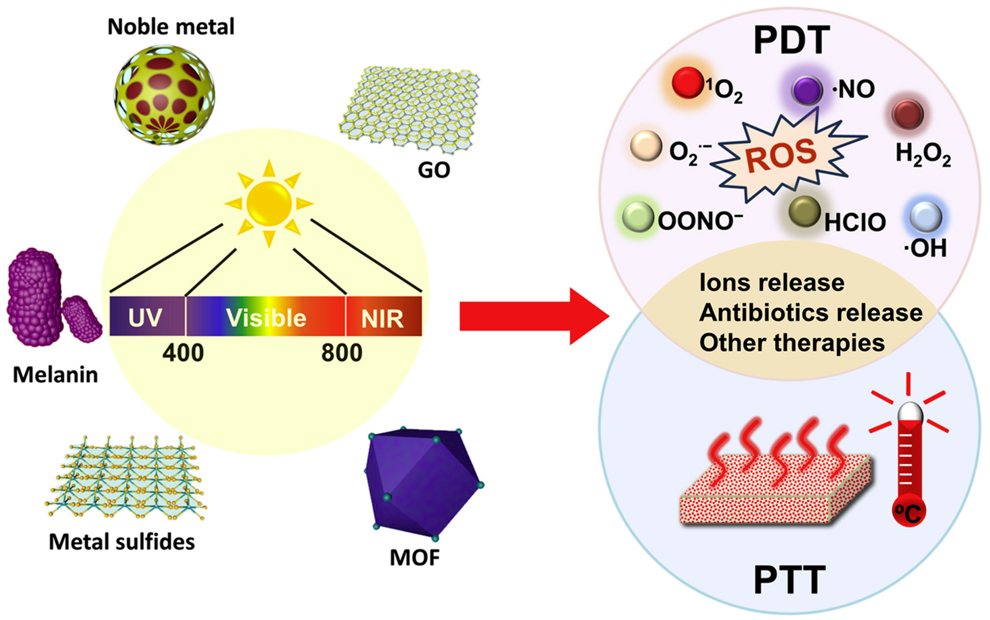
2.1. Importance of Photo-Stimuli-Responsive Nanomaterials with Antibacterial Activity
2.2. Different Types of Photo-Stimuli-Responsive Nanomaterials with Antibacterial Activity
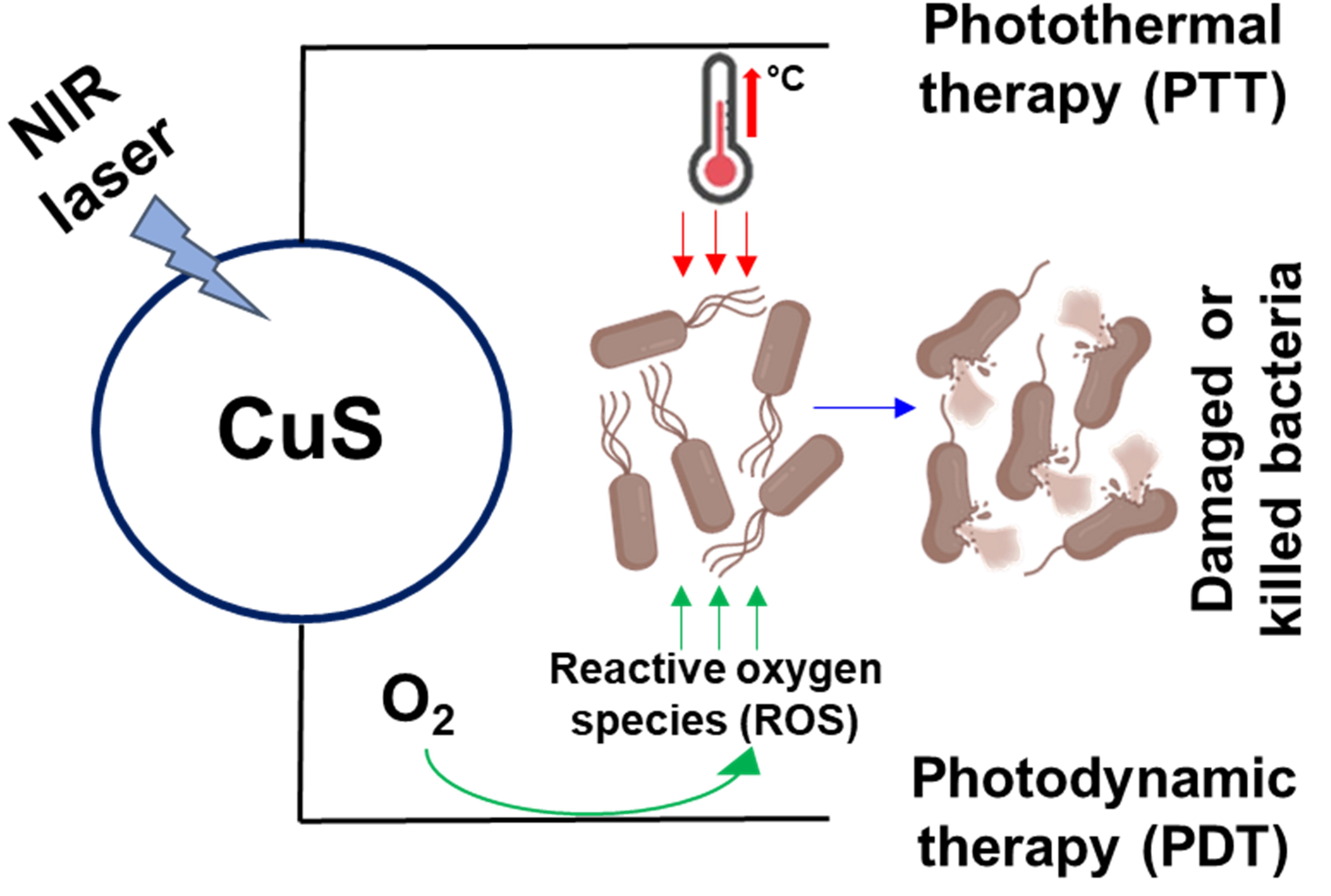
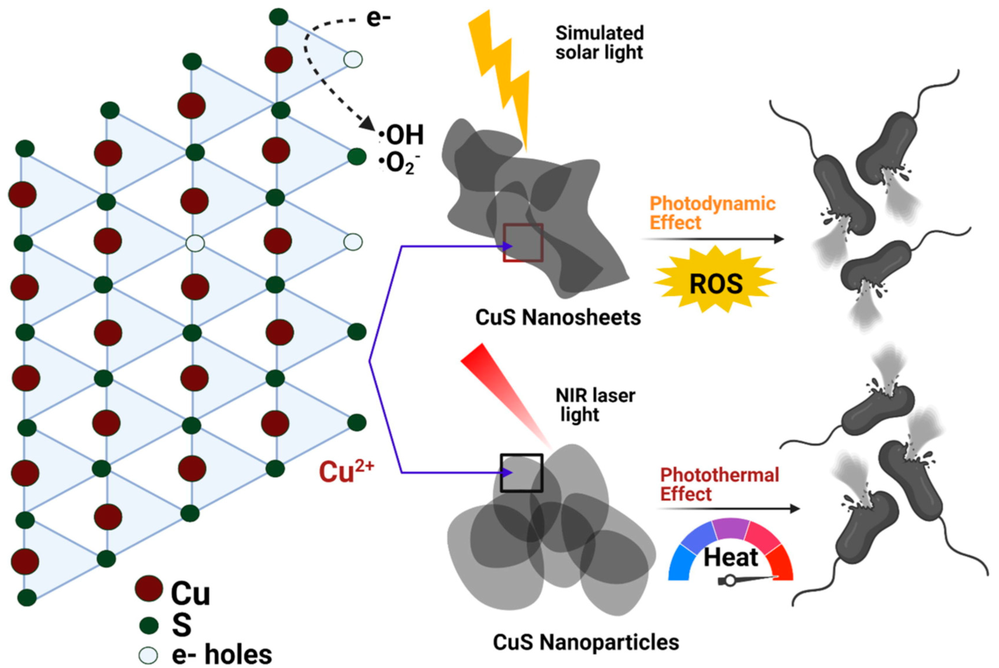
2.3. Photo-Stimuli-Responsive CuS Nanomaterials with Antibacterial Potential
3. Controllable Synthesis of CuS Nanomaterials
3.1. Synthesis of Zero-Dimensional (0D) CuS Nanomaterials
3.2. Synthesis of One-Dimensional (1D) CuS Nanomaterials
3.3. Synthesis of Two-Dimensional (2D) CuS Nanomaterials
3.4. Synthesis of Three-Dimensional (3D) CuS Nanomaterials
4. Photo-Stimuli-Responsive Antibacterial Applications of CuS and Its Nanocomposites
4.1. CuS Nanomaterials
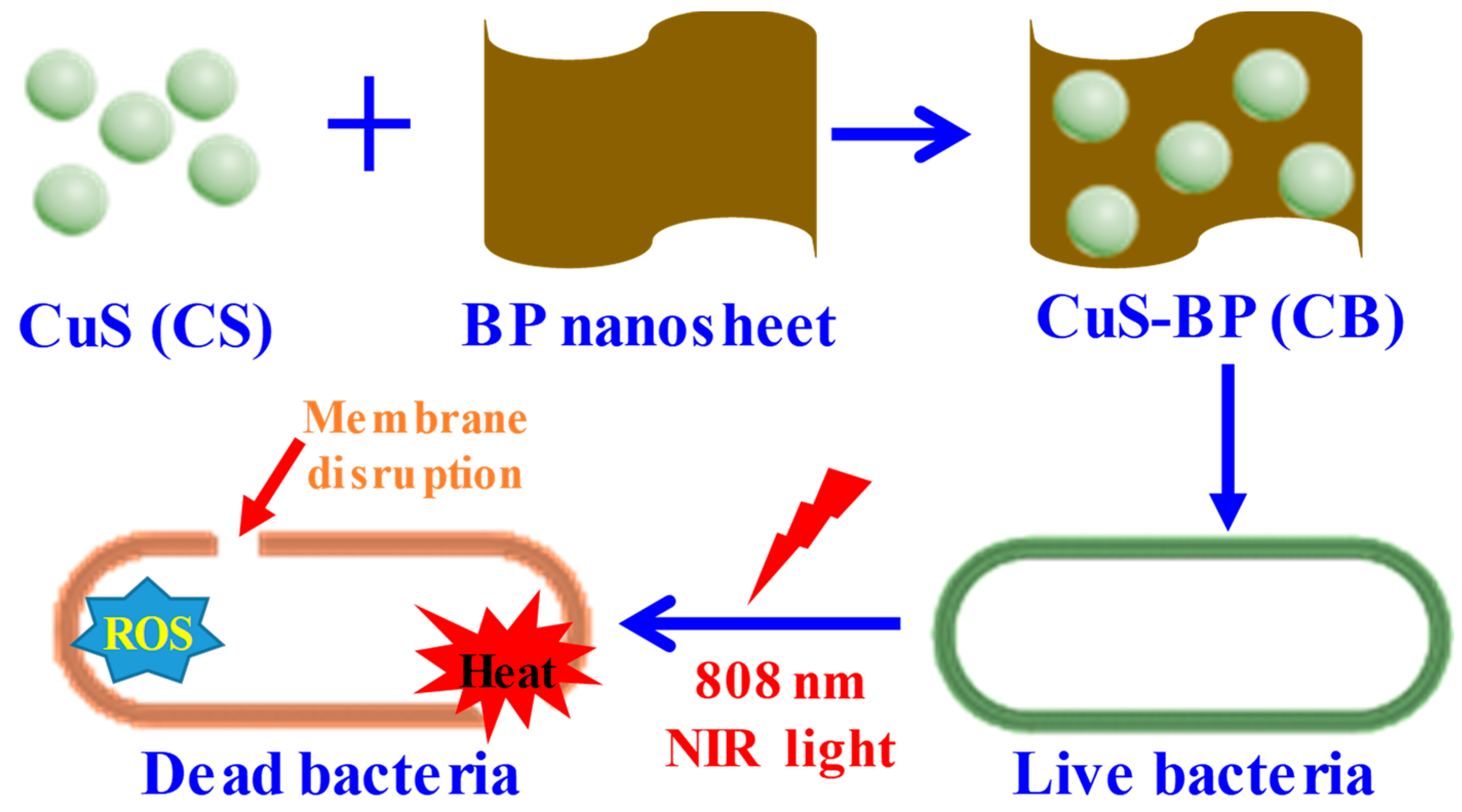
4.2. CuS with 2D Nanomaterials
4.3. CuS with Polymers
4.4. CuS with Protein
4.5. CuS with Metals
5. Conclusions and Future Perspectives
Author Contributions
Funding
Institutional Review Board Statement
Informed Consent Statement
Data Availability Statement
Acknowledgments
Conflicts of Interest
References
- Mendelson, M.; Matsoso, M.P. The World Health Organization Global Action Plan for antimicrobial resistance. S. Afr. Med. J. 2015, 105, 325. [Google Scholar] [CrossRef] [PubMed] [Green Version]
- Naskar, A.; Kim, K.-S. Nanomaterials as delivery vehicles and components of new strategies to combat bacterial infections: Advantages and Limitations. Microorganisms 2019, 7, 356. [Google Scholar] [CrossRef] [PubMed] [Green Version]
- Lee, H.P.; Gaharwar, A.K. Light-Responsive Inorganic Biomaterials for Biomedical Applications. Adv. Sci. 2020, 7, 2000863. [Google Scholar] [CrossRef] [PubMed]
- Ren, Y.; Liu, H.; Liu, X.; Zheng, Y.; Li, Z.; Li, C.; Yeung, K.W.K.; Zhu, S.; Liang, Y.; Cui, Z.; et al. Photoresponsive Materials for Antibacterial Applications. Cell Rep. Phys. Sci. 2020, 1, 100245. [Google Scholar] [CrossRef]
- Huo, J.; Jia, Q.; Huang, H.; Zhang, J.; Li, P.; Dong, X.; Huang, W. Emerging photothermal-derived multimodal synergistic therapy in combating bacterial infections. Chem. Soc. Rev. 2021, 50, 8762–8789. [Google Scholar] [CrossRef]
- Ain, N.U.; Abdul Nasir, J.; Khan, Z.; Butler, I.S.; Rehman, Z. Copper sulfide nanostructures: Synthesis and biological applications. RSC Adv. 2022, 12, 7550–7567. [Google Scholar] [CrossRef]
- Mutalik, C.; Okoro, G.; Krisnawati, D.I.; Jazidie, A.; Rahmawati, E.Q.; Rahayu, D.; Hsu, W.T.; Kuo, T.R. Copper sulfide with morphology-dependent photodynamic and photothermal antibacterial activities. J. Colloid Interface Sci. 2022, 607, 1825–1835. [Google Scholar] [CrossRef]
- Li, Q.; Wang, W.; Feng, H.; Cao, L.; Wang, H.; Wang, D.; Chen, S. NIR-triggered photocatalytic and photothermal performance for sterilization based on copper sulfide nanoparticles anchored on Ti3C2Tx MXene. J. Colloid Interface Sci. 2021, 604, 810–822. [Google Scholar] [CrossRef]
- Dai, X.; Zhao, Y.; Yu, Y.; Chen, X.; Wei, X.; Zhang, X.; Li, C. Single Continuous Near-Infrared Laser-Triggered Photodynamic and Photothermal Ablation of Antibiotic-Resistant Bacteria Using Effective Targeted Copper Sulfide Nanoclusters. ACS Appl. Mater. Interfaces. 2017, 9, 30470–30479. [Google Scholar] [CrossRef]
- Gao, D.Y.; Ji, X.; Wang, J.L.; Wang, Y.T.; Li, D.L.; Liu, Y.B.; Chang, K.W.; Qu, J.L.; Zheng, J.; Yuan, Z. Engineering a protein-based nanoplatform as an antibacterial agent for light activated dual-modal photothermal and photodynamic therapy of infection in both the NIR I and II windows. J. Mater. Chem. B 2018, 6, 732–739. [Google Scholar] [CrossRef]
- Yang, X.; Liu, G.; Shi, Y.; Huang, W.; Shao, J.; Dong, X. Nano-black phosphorus for combined cancer phototherapy: Recent advances and prospects. Nanotechnology 2018, 29, 222001. [Google Scholar] [CrossRef] [PubMed]
- Zhang, B.; Wang, Y.; Liu, J.; Zhai, G. Recent Developments of Phototherapy Based on Graphene Family Nanomaterials. Curr. Med. Chem. 2017, 24, 268–291. [Google Scholar] [CrossRef] [PubMed]
- Gøtzsche, P.C. Niels Finsen’s treatment for lupus vulgaris. J. R. Soc. Med. 2011, 104, 41–42. [Google Scholar] [CrossRef] [PubMed] [Green Version]
- Moller, K.I.; Kongshoj, B.; Philipsen, P.A.; Thomsen, V.O.; Wulf, H.C. How Finsen’s light cured lupus vulgaris. Photodermatol. Photoimmunol. Photomed. 2005, 21, 118–124. [Google Scholar] [CrossRef] [PubMed]
- Zhang, X.; Wang, S.; Cheng, G.; Yu, P.; Chang, J. Light-Responsive Nanomaterials for Cancer Therapy. Engineering 2022, 13, 18–30. [Google Scholar] [CrossRef]
- Xu, J.W.; Yao, K.; Xu, Z.K. Nanomaterials with a photothermal effect for antibacterial activities: An overview. Nanoscale 2019, 11, 8680–8691. [Google Scholar] [CrossRef]
- Memar, M.Y.; Ghotaslou, R.; Samiei, M.; Adibkia, K. Antimicrobial use of reactive oxygen therapy: Current insights. Infect. Drug. Resist. 2018, 11, 567–576. [Google Scholar] [CrossRef] [Green Version]
- Sun, Y.D.; Zhu, Y.X.; Zhang, X.; Jia, H.R.; Xia, Y.; Wu, F.G. Role of Cholesterol Conjugation in the Antibacterial Photodynamic Therapy of Branched Polyethylenimine-Containing Nanoagents. Langmuir 2019, 35, 14324–14331. [Google Scholar] [CrossRef]
- Yang, M.; Qiu, S.; Coy, E.; Li, S.; Załęski, K.; Zhang, Y.; Pan, H.; Wang, G. NIR-Responsive TiO2 Biometasurfaces: Toward In Situ Photodynamic Antibacterial Therapy for Biomedical Implants. Adv. Mater. 2022, 34, e2106314. [Google Scholar] [CrossRef]
- Gupta, J.; Mohapatra, J.; Bahadur, D. Visible light driven mesoporous Ag-embedded ZnO nanocomposites: Reactive oxygen species enhanced photocatalysis, bacterial inhibition and photodynamic therapy. Dalton Trans. 2017, 46, 685–696. [Google Scholar] [CrossRef]
- Duan, G.; Chen, L.; Jing, Z.; De Luna, P.; Wen, L.; Zhang, L.; Zhao, L.; Xu, J.; Li, Z.; Yang, Z.; et al. Robust Antibacterial Activity of Tungsten Oxide (WO3-x) Nanodots. Chem. Res. Toxicol. 2019, 32, 1357–1366. [Google Scholar] [CrossRef] [PubMed]
- Huang, J.; Ho, W.; Wang, X. Metal-free disinfection effects induced by graphitic carbon nitride polymers under visible light illumination. Chem. Commun. 2014, 50, 4338–4340. [Google Scholar] [CrossRef] [PubMed]
- Amos-Tautua, B.M.; Songca, S.P.; Oluwafemi, O.S. Application of Porphyrins in Antibacterial Photodynamic Therapy. Molecules 2019, 24, 2456. [Google Scholar] [CrossRef] [Green Version]
- Xu, Z.; Mei, L.; Shi, Y.; Yun, M.; Luan, Y.; Miao, Z.; Liu, Z.; Li, X.M.; Jiao, M. Multivalent Phthalocyanine-Based Cationic Polymers with Enhanced Photodynamic Activity for the Bacterial Capture and Bacteria-Infected Wound Healing. Biomacromolecules 2022, 23, 2778–2784. [Google Scholar] [CrossRef] [PubMed]
- Vara, J.; Gualdesi, M.S.; Aiassa, V.; Ortiz, C.S. Evaluation of physicochemical properties and bacterial photoinactivation of phenothiazine photosensitizers. Photochem. Photobiol. Sci. 2019, 18, 1576–1586. [Google Scholar] [CrossRef]
- Liang, G.; Wang, H.; Shi, H.; Wang, H.; Zhu, M.; Jing, A.; Li, J.; Li, G. Recent progress in the development of upconversion nanomaterials in bioimaging and disease treatment. J. Nanobiotechnol. 2020, 18, 154. [Google Scholar] [CrossRef]
- Mohamed, M.M.; Fouad, S.A.; Elshoky, H.A.; Mohammed, G.M.; Salaheldin, T.A. Antibacterial effect of gold nanoparticles against Corynebacterium pseudotuberculosis. Int. J. Vet. Sci. Med. 2017, 5, 23–29. [Google Scholar] [CrossRef] [Green Version]
- Sun, P.; Ye, L.; Tan, X.; Peng, J.; Zhao, L.; Zhou, Y. Silver Nanoparticle-Assisted Photodynamic Therapy for Biofilm Eradication. ACS Appl. Nano Mater. 2022, 5, 8251–8259. [Google Scholar] [CrossRef]
- Han, D.; Ma, M.; Han, Y.; Cui, Z.; Liang, Y.; Liu, X.; Li, Z.; Zhu, S.; Wu, S. Eco-friendly Hybrids of Carbon Quantum Dots Modified MoS2 for Rapid Microbial Inactivation by Strengthened Photocatalysis. ACS Sustain. Chem. Eng. 2020, 8, 534–542. [Google Scholar] [CrossRef]
- Nie, X.; Jiang, C.; Wu, S.; Chen, W.; Lv, P.; Wang, Q.; Liu, J.; Narh, C.; Cao, X.; Ghiladi, R.A.; et al. Carbon quantum dots: A bright future as photosensitizers for in vitro antibacterial photodynamic inactivation. J. Photochem. Photobiol. B 2020, 206, 111864. [Google Scholar] [CrossRef]
- Garin, C.; Alejo, T.; Perez-Laguna, V.; Prieto, M.; Mendoza, G.; Arruebo, M.; Sebastian, V.; Rezusta, A. Chalcogenide nanoparticles and organic photosensitizers for synergetic antimicrobial photodynamic therapy. J. Mater. Chem. B 2021, 9, 6246–6259. [Google Scholar] [CrossRef] [PubMed]
- Ye, L.; Cao, Z.; Liu, X.; Cui, Z.; Li, Z.; Liang, Y.; Zhu, S.; Wu, S. Noble metal-based nanomaterials as antibacterial agents. J. Alloys Compd. 2022, 904, 164091. [Google Scholar] [CrossRef]
- Lv, Y.K.; Mei, L.; Zhang, L.X.; Yang, D.H.; Yin, Z.Y. Multifunctional graphene-based nanocomposites for simultaneous enhanced photocatalytic degradation and photothermal antibacterial activity by visible light. Environ. Sci. Pollut. Res. Int. 2021, 28, 49880–49888. [Google Scholar] [CrossRef] [PubMed]
- Qie, X.; Zan, M.; Gui, P.; Chen, H.; Wang, J.; Lin, K.; Mei, Q.; Ge, M.; Zhang, Z.; Tang, Y.; et al. Design, Synthesis, and Application of Carbon Dots With Synergistic Antibacterial Activity. Front Bioeng. Biotechnol. 2022, 10, 894100. [Google Scholar] [CrossRef] [PubMed]
- Zhu, K.; Qian, S.; Guo, H.; Wang, Q.; Chu, X.; Wang, X.; Lu, S.; Peng, Y.; Guo, Y.; Zhu, Z.; et al. pH-Activatable Organic Nanoparticles for Efficient Low-Temperature Photothermal Therapy of Ocular Bacterial Infection. ACS Nano 2022, 16, 11136–11151. [Google Scholar] [CrossRef]
- Naskar, A.; Lee, S.; Kim, K.-s. Au-ZnO Conjugated Black Phosphorus as a Near-Infrared Light-Triggering and Recurrence-Suppressing Nanoantibiotic Platform against Staphylococcus aureus. Pharmaceutics 2021, 13, 52. [Google Scholar] [CrossRef]
- Pan, W.Y.; Huang, C.C.; Lin, T.T.; Hu, H.Y.; Lin, W.C.; Li, M.J.; Sung, H.W. Synergistic antibacterial effects of localized heat and oxidative stress caused by hydroxyl radicals mediated by graphene/iron oxide-based nanocomposites. Nanomedicine 2016, 12, 431–438. [Google Scholar] [CrossRef]
- Nguyen, T.K.; Duong, H.T.; Selvanayagam, R.; Boyer, C.; Barraud, N. Iron oxide nanoparticle-mediated hyperthermia stimulates dispersal in bacterial biofilms and enhances antibiotic efficacy. Sci. Rep. 2015, 5, 18385. [Google Scholar] [CrossRef] [Green Version]
- Wang, Y.; Jin, Y.; Chen, W.; Wang, J.; Chen, H.; Sun, L.; Li, X.; Ji, J.; Yu, Q.; Shen, L.; et al. Construction of nanomaterials with targeting phototherapy properties to inhibit resistant bacteria and biofilm infections. Chem. Eng. J. 2019, 358, 74–90. [Google Scholar] [CrossRef]
- Meeker, D.G.; Jenkins, S.V.; Miller, E.K.; Beenken, K.E.; Loughran, A.J.; Powless, A.; Muldoon, T.J.; Galanzha, E.I.; Zharov, V.P.; Smeltzer, M.S.; et al. Synergistic Photothermal and Antibiotic Killing of Biofilm-Associated Staphylococcus aureus Using Targeted Antibiotic-Loaded Gold Nanoconstructs. ACS Infect Dis. 2016, 2, 241–250. [Google Scholar] [CrossRef]
- Huang, J.; Zhou, J.; Zhuang, J.; Gao, H.; Huang, D.; Wang, L.; Wu, W.; Li, Q.; Yang, D.P.; Han, M.Y. Strong Near-Infrared Absorbing and Biocompatible CuS Nanoparticles for Rapid and Efficient Photothermal Ablation of Gram-Positive and -Negative Bacteria. ACS Appl Mater. Interfaces 2017, 9, 36606–36614. [Google Scholar] [CrossRef] [PubMed]
- Wang, P.; Yuan, Y.; Xu, K.; Zhong, H.; Yang, Y.; Jin, S.; Yang, K.; Qi, X. Biological applications of copper-containing materials. Bioact. Mater. 2020, 6, 916–927. [Google Scholar] [CrossRef] [PubMed]
- Gargioni, C.; Borzenkov, M.; D’Alfonso, L.; Sperandeo, P.; Polissi, A.; Cucca, L.; Dacarro, G.; Grisoli, P.; Pallavicini, P.; D‘Agostino, A.; et al. Self-Assembled Monolayers of Copper Sulfide Nanoparticles on Glass as Antibacterial Coatings. Nanomaterials 2020, 10, 352. [Google Scholar] [CrossRef] [PubMed] [Green Version]
- Ding, H.; Han, D.; Han, Y.; Liang, Y.; Liu, X.; Li, Z.; Zhu, S.; Wu, S. Visible light responsive CuS/ protonated g-C3N4 heterostructure for rapid sterilization. J. Hazard. Mater. 2020, 393, 122423. [Google Scholar] [CrossRef] [PubMed]
- Zhou, L.; Chen, F.; Hou, Z.; Chen, Y.; Luo, X. Injectable self-healing CuS nanoparticle complex hydrogels with antibacterial, anti-cancer, and wound healing properties. Chem. Eng. J. 2021, 409, 128224. [Google Scholar] [CrossRef]
- Qiao, Y.; Ping, Y.; Zhang, H.; Zhou, B.; Liu, F.; Yu, Y.; Xie, T.; Li, W.; Zhong, D.; Zhang, Y.; et al. Laser-Activatable CuS Nanodots to Treat Multidrug-Resistant Bacteria and Release Copper Ion to Accelerate Healing of Infected Chronic Nonhealing Wounds. ACS Appl. Mater. Interfaces 2019, 11, 3809–3822. [Google Scholar] [CrossRef] [Green Version]
- Naskar, A.; Cho, H.; Kim, K.S. Black phosphorus-based CuS nanoplatform: Near-infrared-responsive and reactive oxygen species-generating agent against environmental bacterial pathogens. J. Environ. Chem. Eng. 2022, 10, 108226. [Google Scholar] [CrossRef]
- Naskar, A.; Cho, H.; Lee, S.; Kim, K.-s. Biomimetic Nanoparticles Coated with Bacterial Outer Membrane Vesicles as a New-Generation Platform for Biomedical Applications. Pharmaceutics 2021, 13, 1887. [Google Scholar] [CrossRef]
- Naskar, A.; Kim, K.S. Recent Advances in Nanomaterial-Based Wound-Healing Therapeutics. Pharmaceutics 2020, 12, 499. [Google Scholar] [CrossRef]
- Li, Y.; Lu, W.; Huang, Q.; Huang, M.; Li, C.; Chen, W. Copper sulfide nanoparticles for photothermal ablation of tumor cells. Nanomedicine 2010, 5, 1161–1171. [Google Scholar] [CrossRef]
- Li, M.; Wang, Y.; Lin, H.; Qu, F. Hollow CuS nanocube as nanocarrier for synergetic chemo/photothermal/photodynamic therapy. Mater. Sci. Eng. C Mater. Biol. Appl. 2019, 96, 591–598. [Google Scholar] [CrossRef] [PubMed]
- Wang, Y.; Xiao, Y.; Zhou, H.; Chen, W.; Tang, R. Ultra-high payload of doxorubicin and pH-responsive drug release in CuS nanocages for a combination of chemotherapy and photothermal therapy. RSC Adv. 2013, 3, 23133–23138. [Google Scholar] [CrossRef]
- Raj, S.I.; Jaiswal, A.; Uddin, I. Ultrasmall aqueous starch-capped CuS quantum dots with tunable localized surface plasmon resonance and composition for the selective and sensitive detection of mercury(ii) ions. RSC Adv. 2020, 10, 14050–14059. [Google Scholar] [CrossRef] [PubMed]
- Kalimuldina, G.; Nurpeissova, A.; Adylkhanova, A.; Adair, D.; Taniguchi, I.; Bakenov, Z. Morphology and Dimension Variations of Copper Sulfide for High-Performance Electrode in Rechargeable Batteries: A Review. ACS Appl. Energy Mater. 2020, 3, 11480–11499. [Google Scholar] [CrossRef]
- Bekhit, M.; Abo El Naga, A.O.; El Saied, M.; Abdel Maksoud, M.I.A. Radiation-induced synthesis of copper sulfide nanotubes with improved catalytic and antibacterial activities. Environ. Sci. Pollut. Res. Int. 2021, 28, 44467–44478. [Google Scholar] [CrossRef] [PubMed]
- Zhang, X.; Wang, G.; Gu, A.; Wei, Y.; Fang, B. CuS nanotubes for ultrasensitive nonenzymatic glucose sensors. Chem. Commun. 2008, 45, 5945–5947. [Google Scholar] [CrossRef] [PubMed]
- Manikandan, T.; Padmalaya, G.; Mahalakshmi, S.; Nivethitha, A.S.; Pavithran, V.; Perumal, S.; Sreeja, B.S.; Senthil Kumar, P. Facile hydrothermal bio-synthesis of cellulose acetate templated CuS nanorods like fibres: Antibacterial, cytotoxicity effects and DNA cleavage properties against A549 lung cancer cells. IET Nanobiotechnol. 2020, 14, 47–52. [Google Scholar] [CrossRef]
- Kwon, Y.T.; Ryu, S.H.; Shin, J.W.; Yeo, W.H.; Choa, Y.H. Electrospun CuS/PVP Nanowires and Superior Near-Infrared Filtration Efficiency for Thermal Shielding Applications. ACS Appl. Mater. Interfaces 2019, 11, 6575–6580. [Google Scholar] [CrossRef] [PubMed]
- Mirzaei, A.; Peymanfar, R.; Javanshir, S.; Fallahi, R.; Karimi, J. Antibacterial Characteristics of CuS Nanoplates Anchored onto g-C3N4 Nanosheets, Suspended in PMMA Matrix. Int. J. Nanosci. Nanotechnol. 2021, 17, 249–260. [Google Scholar]
- Nishi, H.; Asami, K.; Tatsuma, T. CuS nanoplates for LSPR sensing in the second biological optical window. Opt. Mater. Express 2016, 6, 1043–1048. [Google Scholar] [CrossRef]
- Palanisamy, S.; Velmurugan, S.; Yang, T.C.K. One-pot sonochemical synthesis of CuS nanoplates decorated partially reduced graphene oxide for biosensing of dopamine neurotransmitter. Ultrason. Sonochem. 2020, 64, 105043. [Google Scholar] [CrossRef] [PubMed]
- Wang, K.-J.; Li, G.-D.; Li, J.-X.; Wang, Q.; Chen, J.-S. Formation of Single-Crystalline CuS Nanoplates Vertically Standing on Flat Substrate. Cryst Growth Des. 2007, 7, 2265–2267. [Google Scholar] [CrossRef]
- Han, L.; Hao, Y.N.; Wei, X.; Chen, X.W.; Shu, Y.; Wang, J.H. Hollow Copper Sulfide Nanosphere-Doxorubicin/Graphene Oxide Core-Shell Nanocomposite for Photothermo-chemotherapy. ACS Biomater. Sci. Eng. 2017, 3, 3230–3235. [Google Scholar] [CrossRef] [PubMed]
- Wu, S.; Zhang, P.; Jiang, Z.; Zhang, W.; Gong, X.; Wang, Y. Enhanced Peroxidase-like Activity of CuS Hollow Nanocages by Plasmon-Induced Hot Carriers and Photothermal Effect for the Dual-Mode Detection of Tannic Acid. ACS Appl. Mater. Interfaces 2022, 14, 40191–40199. [Google Scholar] [CrossRef] [PubMed]
- Cui, Y.; Li, J.; Liu, M.; Tong, H.; Liu, Z.; Hu, J.; Qian, D. Convenient synthesis of three-dimensional hierarchical CuS@Pd core-shell cauliflowers decorated on nitrogen-doped reduced graphene oxide for non-enzymatic electrochemical sensing of xanthine. Mikrochim. Acta 2020, 187, 589. [Google Scholar] [CrossRef]
- Deng, X.; Wang, C.; Yang, H.; Shao, M.; Zhang, S.; Wang, X.; Ding, M.; Huang, J.; Xu, X. One-pot hydrothermal synthesis of CdS decorated CuS microflower-like structures for enhanced photocatalytic properties. Sci. Rep. 2017, 7, 3877. [Google Scholar] [CrossRef] [Green Version]
- Fang, J.; Zhang, P.; Chang, H.; Wang, X. Hydrothermal synthesis of nanostructured CuS for broadband efficient optical absorption and high-performance photo-thermal conversion. Sol. Energy Mater. Sol. Cells 2018, 185, 456–463. [Google Scholar] [CrossRef]
- Shu, Q.W.; Li, C.M.; Gao, P.F.; Gao, M.X.; Huang, C.Z. Porous hollow CuS nanospheres with prominent peroxidase-like activity prepared in large scale by a one-pot controllable hydrothermal step. RSC Adv. 2015, 5, 17458–17465. [Google Scholar] [CrossRef]
- AL-Jawad, S.M.H.; Taha, A.A.; Redha, A.M. Studying the structural, morphological, and optical properties of CuS:Ni nanostructure prepared by a hydrothermal method for biological activity. J. Sol.-Gel. Sci. Technol. 2019, 91, 310–323. [Google Scholar] [CrossRef]
- Thongtem, T.; Phuruangrat, A.; Thongtem, S. Characterization of copper sulfide nanostructured spheres and nanotubes synthesized by microwave-assisted solvothermal method. Mater. Lett. 2010, 64, 136–139. [Google Scholar] [CrossRef]
- Krajnik, B.; Golacki, L.W.; Fiedorczyk, E.; Bański, M.; Noculak, A.; Hołodnik, K.M.; Podhorodecki, A. Quantitative comparison of luminescence probes for biomedical applications. Methods Appl. Fluoresc. 2021, 9, 045001. [Google Scholar] [CrossRef] [PubMed]
- Ren, Y.; Yan, B.; Wang, P.; Yu, Y.; Cui, L.; Zhou, M.; Wang, Q. Construction of a Rapid Photothermal Antibacterial Silk Fabric via QCS-Guided In Situ Deposition of CuSNPs. ACS Sustain. Chem. Eng. 2022, 10, 2192–2203. [Google Scholar] [CrossRef]
- Lv, R.; Liang, Y.-Q.; Li, Z.-Y.; Zhu, S.-L.; Cui, Z.-D.; Wu, S.-L. Flower-like CuS/graphene oxide with photothermal and enhanced photocatalytic effect for rapid bacteria-killing using visible light. Rare Met. 2022, 41, 639–649. [Google Scholar] [CrossRef]
- Liu, X.; Li, X.; Shan, Y.; Yin, Y.; Liu, C.; Lin, Z.; Kumar, S.S. CuS nanoparticles anchored to g-C3N4 nanosheets for photothermal ablation of bacteria. RSC Adv. 2020, 10, 12183–12191. [Google Scholar] [CrossRef] [Green Version]
- Wang, H.Y.; Hua, X.W.; Wu, F.G.; Li, B.; Liu, P.; Gu, N.; Wang, Z.; Chen, Z. Synthesis of ultrastable copper sulfide nanoclusters via trapping the reaction intermediate: Potential anticancer and antibacterial applications. ACS Appl. Mater. Interfaces 2015, 7, 7082–7092. [Google Scholar] [CrossRef]
- Zhao, Y.; Cai, Q.; Qi, W.; Jia, Y.; Xiong, T.; Fan, Z.; Liu, S.; Yang, J.; Li, N.; Chang, B. BSA-CuS Nanoparticles for Photothermal Therapy of Diabetic Wound Infection In Vivo. ChemistrySelect 2018, 3, 9510–9516. [Google Scholar] [CrossRef]
- Swaidan, A.; Ghayyem, S.; Barras, A.; Addad, A.; Szunerits, S.; Boukherroub, R. Enhanced Antibacterial Activity of CuS-BSA/Lysozyme under Near Infrared Light Irradiation. Nanomaterials 2021, 11, 2156. [Google Scholar] [CrossRef]
- Addae, E.; Dong, X.; McCoy, E.; Yang, C.; Chen, W.; Yang, L. Investigation of antimicrobial activity of photothermal therapeutic gold/copper sulfide core/shell nanoparticles to bacterial spores and cells. J. Biol. Eng. 2014, 8, 11. [Google Scholar] [CrossRef] [Green Version]
- Derakhshi, M.; Daemi, S.; Shahini, P.; Habibzadeh, A.; Mostafavi, E.; Ashkarran, A.A. Two-Dimensional Nanomaterials beyond Graphene for Biomedical Applications. J. Funct. Biomater. 2022, 13, 27. [Google Scholar] [CrossRef]
- Naskar, A.; Kim, S.; Kim, K.-s. A nontoxic biocompatible nanocomposite comprising black phosphorus with Au-γ-Fe2O3 nanoparticles. RSC Adv. 2020, 10, 16162–16167. [Google Scholar] [CrossRef] [Green Version]
- Naskar, A.; Lee, S.; Ko, D.; Kim, S.; Kim, K.-s. Bovine Serum Albumin-Immobilized Black Phosphorus-Based γ-Fe2O3 Nanocomposites: A Promising Biocompatible Nanoplatform. Biomedicines 2021, 9, 858. [Google Scholar] [CrossRef] [PubMed]
- Ansari, M.O.; Gauthaman, K.; Essa, A.; Bencherif, S.A.; Memic, A. Graphene and Graphene-Based Materials in Biomedical Applications. Curr. Med. Chem. 2019, 26, 6834–6850. [Google Scholar] [CrossRef] [PubMed]
- Naskar, A.; Khan, H.; Sarkar, R.; Kumar, S.; Halder, D.; Jana, S. Anti-biofilm activity and food packaging application of room temperature solution process based polyethylene glycol capped Ag-ZnO-graphene nanocomposite. Mater. Sci. Eng. C Mater. Biol. Appl. 2018, 91, 743–753. [Google Scholar] [CrossRef] [PubMed]
- Zheng, K.; Li, S.; Jing, L.; Chen, P.Y.; Xie, J. Synergistic Antimicrobial Titanium Carbide (MXene) Conjugated with Gold Nanoclusters. Adv. Healthc. Mater. 2020, 9, e2001007. [Google Scholar] [CrossRef] [PubMed]
- Li, M.; Wang, S.; Li, R.; Wang, Y.; Fan, X.; Gong, W.; Ma, Y. The Mechanical and Antibacterial Properties of Boron Nitride/Silver Nanocomposite Enhanced Polymethyl Methacrylate Resin for Application in Oral Denture Bases. Biomimetics 2022, 7, 138. [Google Scholar] [CrossRef] [PubMed]
- Naskar, A.; Shin, J.; Kim, K.-s. A MoS2 based silver-doped ZnO nanocomposite and its antibacterial activity against β-lactamase expressing Escherichia coli. RSC Adv. 2022, 12, 7268–7275. [Google Scholar] [CrossRef] [PubMed]
- Naskar, A.; Kim, K.-s. Black phosphorus nanomaterials as multi-potent and emerging platforms against bacterial infections. Microb. Pathog. 2019, 137, 103800. [Google Scholar] [CrossRef]
- Cho, H.; Naskar, A.; Lee, S.; Kim, S.; Kim, K.-s. A New Surface Charge Neutralizing Nano-Adjuvant to Potentiate Polymyxins in Killing Mcr-1 Mediated Drug-Resistant Escherichia coli. Pharmaceutics 2021, 13, 250. [Google Scholar] [CrossRef]
- Park, E.; Selvaraj, R.; Kim, Y. High-efficiency photothermal sterilization on PDMS film with Au@CuS yolk-shell nanoparticles. J. Ind. Eng. Chem. 2022, 113, 522–529. [Google Scholar] [CrossRef]


| Photoactivated CuS | Antibacterial Mechanism | Light Source | Bacteria | Efficacy | Ref. |
|---|---|---|---|---|---|
| CuS NDs | PTT & Cu2+ release | NIR (808 nm, 2.5 W·cm−2) | ESBL E. coli, MRSA | ~100% | [46] |
| CuS NPs | PTT | UV (400 mW·cm−2) | E. coli, S. aureus | 99.99% | [72] |
| CuS NPs | PTT | NIR (800, 900, and 1000 nm, 0.26 W·cm−2) | E. coli, S. aureus | 95% (>5 h) | [43] |
| CuS MSs, NSs, NPs | PDT, PTT | Simulated solar light and NIR (808 nm, 1.5 W·cm−2) | E. coli | Effective | [7] |
| CuS/GO | PTT | Visible light (0.2 W·cm−2) | E. coli, S. aureus | 99.98% | [73] |
| PEG-CuS@g-C3N4 | PTT | NIR (808 nm, 2.5 W) | E. coli, S. aureus | ~99% | [74] |
| Ti3C2Tx@CuS | PDT, PTT | NIR (808 nm, 1.5 W) | E. coli, S. aureus | ~99% | [8] |
| CuS-BP | PDT, PTT | NIR (808 nm, 2.5 W) | P. aeruginosa, S. aureus | ~100% | [47] |
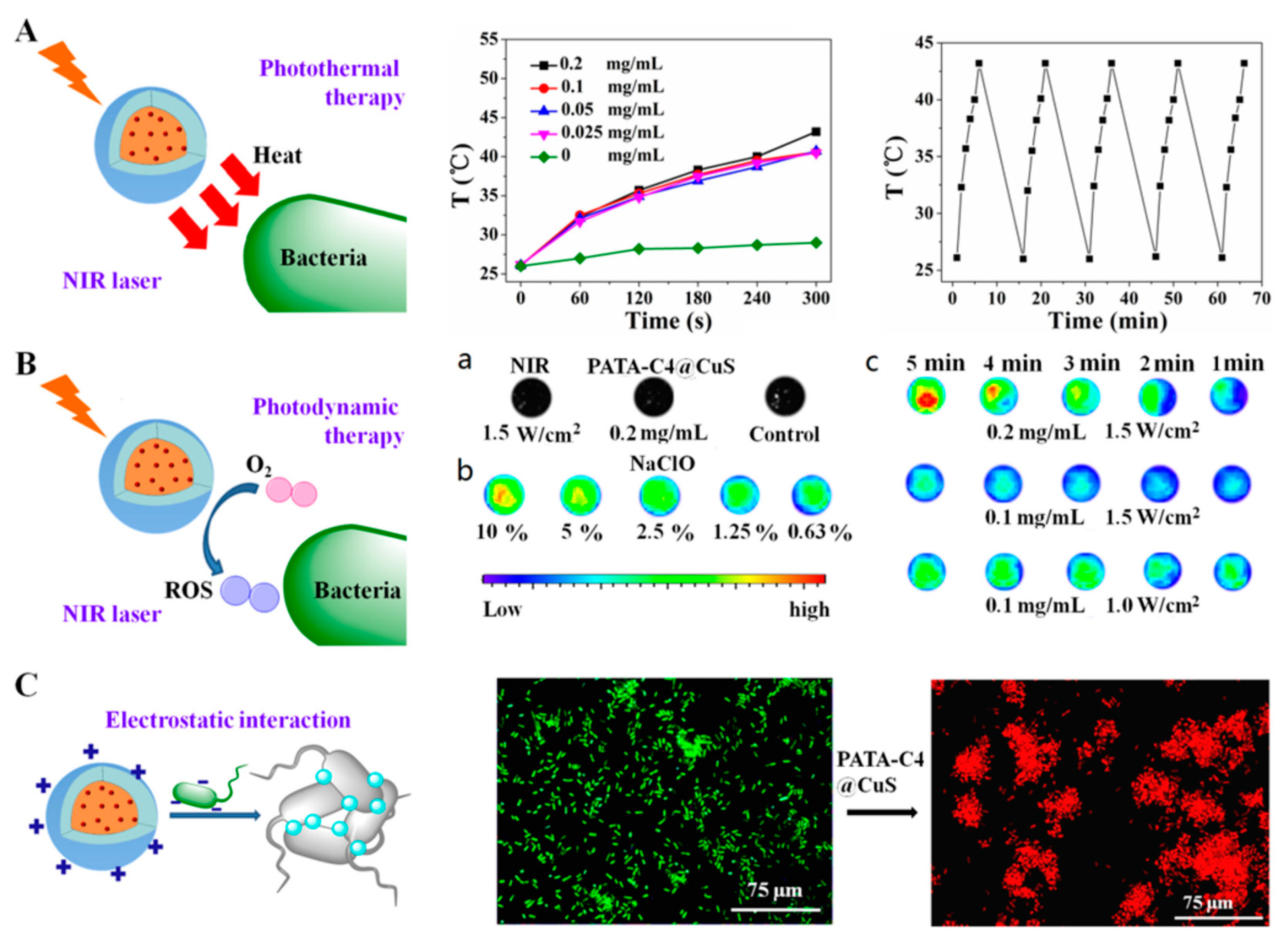
| PATAC4@CuS | PDT, PTT | NIR (980 nm, 1.5 W·cm−2) | Bacillus amyloliquefaciens, E. coli, P. aeruginosa, Levofloxacin-resistant S. aureus | Effective | [9] |
| MUH@CuS NCs | PTT | NIR | E. coli | ~100% | [75] |
| BSA-CuS NPs | PTT | NIR (808, 8.0 W·cm−2) | A. baumannii, S. aureus, S. haemolyticus | Effective | [76] |
| Ce6-labeled BSA-CuS NPs | PDT, PTT | NIR (980 nm, 1.59 W·cm−2) | E. coli, S. aureus | 97% | [10] |
| CuS-BSA/lysozyme | PTT | NIR (980 nm, 0.7 W·cm−2) | B. subtilis, E. coli | ~100% | [77] |
| Au@CuS | PTT | NIR | E. coli | ~100% | [78] |
Publisher’s Note: MDPI stays neutral with regard to jurisdictional claims in published maps and institutional affiliations. |
© 2022 by the authors. Licensee MDPI, Basel, Switzerland. This article is an open access article distributed under the terms and conditions of the Creative Commons Attribution (CC BY) license (https://creativecommons.org/licenses/by/4.0/).
Share and Cite
Naskar, A.; Kim, K.-s. Photo-Stimuli-Responsive CuS Nanomaterials as Cutting-Edge Platform Materials for Antibacterial Applications. Pharmaceutics 2022, 14, 2343. https://doi.org/10.3390/pharmaceutics14112343
Naskar A, Kim K-s. Photo-Stimuli-Responsive CuS Nanomaterials as Cutting-Edge Platform Materials for Antibacterial Applications. Pharmaceutics. 2022; 14(11):2343. https://doi.org/10.3390/pharmaceutics14112343
Chicago/Turabian StyleNaskar, Atanu, and Kwang-sun Kim. 2022. "Photo-Stimuli-Responsive CuS Nanomaterials as Cutting-Edge Platform Materials for Antibacterial Applications" Pharmaceutics 14, no. 11: 2343. https://doi.org/10.3390/pharmaceutics14112343