Article

Synthesis, Characterization, and Biological Evaluation of Tetrahydropyrimidines: Dual-Activity and Mechanism of Action

1
University of Kragujevac, Institute for Information Technologies Kragujevac, Department of Sciences, Jovana Cvijića bb, 34000 Kragujevac, Serbia
2
University of Kragujevac, Faculty of Science, Department of Chemistry, Radoja Domanovića 12, 34000 Kragujevac, Serbia
3
University of Kragujevac, Faculty of Science, Department of Biology and Ecology, Radoja Domanovića 12, 34000 Kragujevac, Serbia
4
Institute of Oncology and Radiology of Serbia, Pasterova 14, 11000 Belgrade, Serbia
5
University of Novi Sad, Faculty of Science, Department of Physics, Trg Dositeja Obradovića 3, 21000 Novi Sad, Serbia
6
Institute of Meat Hygiene and Technology, Kaćanskog 13, 11000 Belgrade, Serbia
*
Author to whom correspondence should be addressed.
Pharmaceutics 2022, 14(10), 2254; https://doi.org/10.3390/pharmaceutics14102254
Submission received: 26 September 2022 / Revised: 17 October 2022 / Accepted: 19 October 2022 / Published: 21 October 2022

Abstract

:
In this paper, the synthesis, characterization, and biological evaluation of the novel tetrahydropyrimidines—THPMs are described. THPMs are well-known for wide pharmacological activities such as antimicrobial, anticancer, antiviral, etc. This research includes obtained results of in vitro antimicrobial, anticancer, and α-glucosidase inhibitory activities of the eleven novel THPMs. An antibiotic assessment was done against five bacteria (two Gram-positive and three Gram-negative) and five fungi by determining the minimal inhibitory concentration (MIC), using the broth tube dilution method. The most active antibacterial compounds were 4a, 4b, and 4d, while the best antifungal activity was shown by 4e, 4f, and 4k. The lowest MIC value (0.20 mg/mL) was measured for 4e, 4f, and 4k against the Trichophyton mentagrophytes. Moreover, examining the α-glucosidase inhibitory activity revealed the compound 4g as the one with the best activity. The cytotoxic activity was performed on the tumor cell lines (HeLa, K562, and MDA-MB-231) and normal cells (MRC-5). The best antitumor activity was shown by compounds 4b and 4k against HeLa cell lines. The influence on cell cycle and mechanism of action of the most active compounds were examined too. Compound 4b had good antibacterial and anticancer activities, while 4k showed promising antifungal and anticancer activities.

1. Introduction

Pietro Giacomo Biginelli (1860–1937) was an Italian chemist who discovered a one-pot multicomponent reaction. He invented the procedure that would subsequently be named Biginelli synthesis in his honor [1,2]. Despite being the first multicomponent reaction, the scientific community dismisses the Biginelli reaction as outmoded. Nevertheless, since the finding of monastrol as a kinesin-5 inhibitor [3], as well as its anticancer activity, Biginelli chemistry has expanded [4,5,6]. Huge numbers of tetrahydropyrimidines–THPMs (former name 3,4-dihydropyrimidine-2(1H)-(thi)ones-DHPMs) have been synthesized using the Biginelli reaction to date. THPMs derivatives have a wide range of biological effects, including anti-inflammatory, antiviral, anticancer, calcium channel inhibition, antibacterial, antifungal, and antioxidant [7,8]. Significant anti-inflammatory activity of the Biginelli thioxo-hybrids was denoted towards various inflammatory diseases [9,10,11]. Although compounds delivered from a Biginelli reaction could possess broad spectra of antiviral activities, among them the most important were those that had inhibitory potential against HIV [12,13,14]. However, it was shown that thio-Biginelli adducts are significantly better calcium channel inhibitors compared to appropriate oxo- and aza-hybrids. In that sense, scientists have found that the (R)- was more potent than the (S)-enantiomer, which implies that the configuration at the C4 stereocenter plays a key role in this kind of activity [15,16]. Some Biginelli compounds bearing a pyrazole scaffold exhibited good-to-high potency against Mycobacterium tuberculosis [17,18]. In addition, the presence of 1,3-dihydro-2H-indol-2-one core in the structure of oxo- and thioxo-Biginelli’s analogs also displayed good antibacterial activities against Bacillus subtilis, E. coli, and S. aureus [19]. Indeed, the most useful aza-nucleophile in Biginelli’s chemistry is urea or N-methylurea. However, over the years were shown as quite hard to apply thiourea or thiourea-based nucleophiles in Biginelli’s chemistry [20,21,22,23,24,25]. This fact lies predominantly in its behavior and huge sensitivity towards strong acids and bases, even the Lewis acid/base salt type. In the above-mentioned conditions with heating, it is very hard to apply thioureas in the Biginelli reaction with success. Furthermore, efficient synthesis of Biginelli’s hybrids containing thiourea-based nucleophiles was and is quite challenging. Considering this, our goal was to synthesize a small library of novel THPMs derivatives containing N-methylthiourea functionality. Specific objectives were an investigation of their potential for dual biological activities against various microbes and cancer cell lines.

2. Materials and Methods

2.1. Chemistry

All solvents and substrates (acetyl, benzoyl, and p-toluoyl chloride, methyl acetoacetate, N-methylthiourea, vanillin, 5-bromovanillin, 5-iodovanillin, 5-nitrovanillin) were purchased from Sigma. Vanillic derivatives 1d–k were prepared following the procedures described previously in the literature [26]. We determined melting points (Mp) on a Mel-Temp apparatus. For recording IR spectra, we used a Perkin–Elmer Spectrum One FT-IR spectrometer on KBr pellet. A Varian Gemini 200 MHz NMR spectrometer (1H at 200 and 13C at 50 MHz) was utilized for NMR characterization of compounds 4a–k. The compounds were dissolved in DMSO-d6. 1H and 13C NMR spectra are presented in Supplementary Materials (see Figures S1–S22).

Experimental Procedure for Synthesis of 4ak

A total of 6 mmol of N-methylthiourea and 2 mmol of aldehyde were dissolved in the mixture of dioxane/CHCl3 (4/1, v/v) in a 25 mL round-bottom flask. Ten minutes later, 3 drops of HCl (36.5%) were added at room temperature. In all cases, yellow color appeared. Then, two hours later, 6 mmol of methyl acetoacetate and 10 mol% of 2-amino-1-(4-nitrophenyl)-1,3-propanediol (ANP) were loaded. After one day, the solvent was evaporated. Crude solid had been dissolved in ethanol/water mixture (2/1, v/v). The product was then put in the fridge; after 24 h in those conditions, a precipitate appeared. The formed powder was filtered, and washed with acetone and water. Recrystallization from acetone or ethanol/water was done to reach the desired product with a good purity grade (>95%).

2.2. Sample Preparation for MS Analysis

Having in mind that the experiment was not quantitative in nature, care was taken to introduce about 10 ng of the compound into the MS system; a quantity that will provide adequate response without saturating the detector of the instrument. Solid samples were weighted on an analytical balance into the 10 mL volumetric flask up to cca. 10 mg and dissolved in HPLC-grade acetonitrile, solutions were vigorously shaken and subsequently sonicated for 10 min at 40 °C in order to dissolve completely. Further dilution to the final concentration of cca. 10 ng/uL (in acetonitrile) was prepared directly into the autosampler vials.
Confirmation of theoretical masses was carried out on a Shimadzu (Kyoto, Japan) LC-MS/MS system consisting of two LC-40D × 3 pumps, DGU-405 degassing unit, CTO-40S column oven, SIL-40C × 3 autosampler, and LCMS-8050cl mass spectrometer with ESI interface. The flow injection analysis (FIA) technique was used for direct introduction of the sample into the inlet system avoiding chromatographic separation. Stainless steel tubing (4 m × 0.1 mm ID) was attached instead of the column to the LC pumps/autosampler and mass spectrometer providing a retention time of about 25 s. The mobile phase was 0.1% formic acid in acetonitrile, flow rate was set to 0.2 mL/min.
Positive ESI was used for all compounds with the following parameters: nebulizing gas flow 3 L/min; drying gas flow 10 L/min; heating gas flow10 L/min; interface (capillary) voltage 4.5 kV; interface temperature 350 °C, desolvation temperature 600 °C; desolvation line temperature 250 °C; heat block temperature 300 °C.
The mass spectrometer operated in Q3 scan mode (unit resolution), the scanning range was set from 200 to 800, the scanning speed was 600 u/s.
All materials and methods used for biological examination (antibacterial, antifungal, anticancer, and α-glucosidase inhibitory activities) and crystal structure determination are presented in the Supplementary Materials, File S1.

3. Results and Discussion

3.1. Chemistry

At the beginning of our research, we were seeking the best reaction conditions to synthesize tetrahydropyrimidine 4a under a one-pot manner by applying various acid-based catalysts. For this part of the investigation, methanol, n-butanol, and dioxane were utilized as solvents. Under classical Biginelli conditions with hydrochloric acid as a catalyst, the desired product was isolated in a lower yield than expected in all applied solvents. When amidosulfonic acid was used as a catalyst, 4a was obtained in the yield of 35–50% in the mentioned solvents under reflux. Among the applied solvents, the reaction in dioxane gave the best yield of 4a. However, reactants 1a, 2, and 3 were not fully converted into the expected product. At room temperature, the yield was lower. We decided to apply another strategy preparing an imine intermediate that would allow better production of 4a. First, we mixed the aldehyde with N-methylthiourea, and 3 drops of 36.5% HCl at room temperature in the mixture of dioxane and CHCl3. Based on TLC, one hour later, we added active methylene 3, and substituted 2-amino-1,3-propanediol. The targeted product 4a was obtained in 63% yield. The reaction without 2-amino-1,3-propanediol only produced imine intermediate. Going forward, different substituted aldehydes were employed to broaden the substrate scope (Scheme 1). The usage of thioureas and their derivatives is quite challenging in Biginelli chemistry, and there are a couple of described procedures in the literature [27,28]; in this research, we performed a reaction at room temperature in a specific mixture of solvents with good yields.
The structures of the isolated Biginelli hybrids (4ak) were confirmed by IR, NMR, and ESI-MS spectroscopy. The compound 4j was suitable for crystallographic analysis. Benzylic proton appeared as a doublet in the range 5.10–5.40 ppm. Thioamide proton and carbon were detected at around 10 ppm, and 178 ppm, respectively. In the ESI-MS spectrum of 4a, a molecular ion was found at 401 [M]+ and 403 [M + 2]+ m/z.

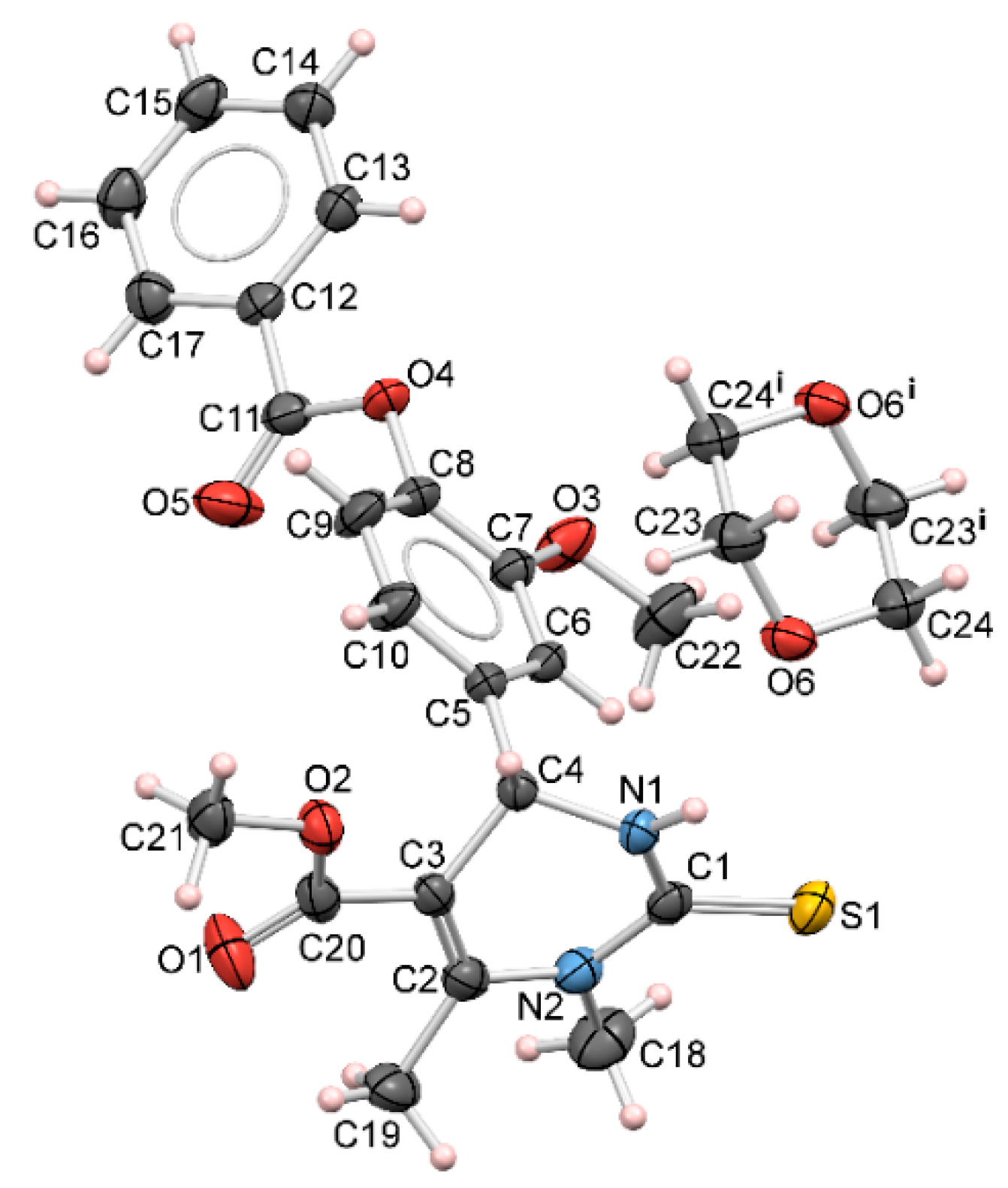
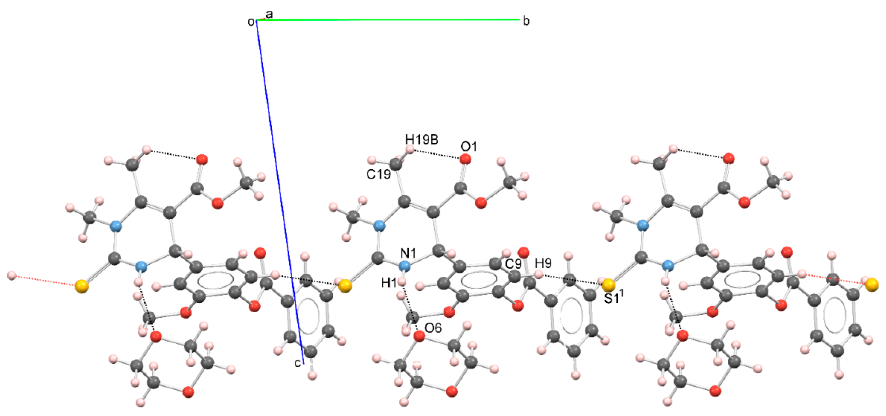
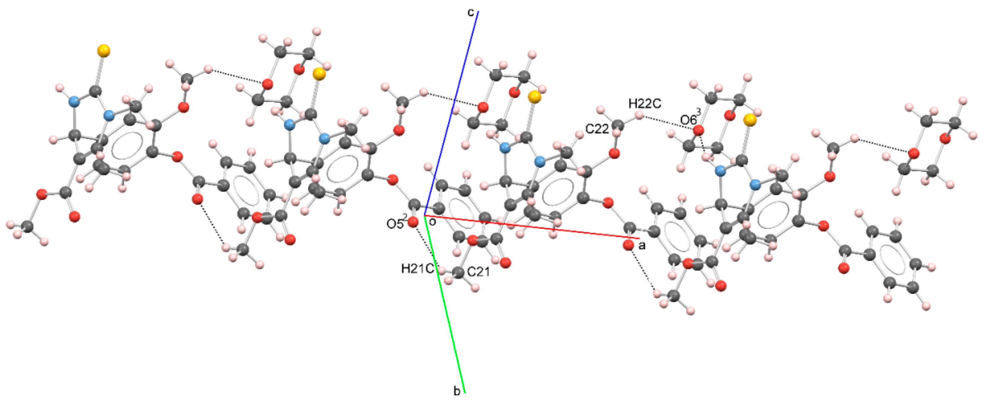
3.2. X-ray Crystallography

Figure 1 shows a perspective view of the molecular structure of methyl 4-(4′-benzoyloxy-3′-methoxyphenyl)-1,2,3,4-tetrahydro-1,6-dimethyl-2-thioxopyrimidine-5-carboxylate (4j). A list of selected torsion angles, bond angles, and bond lengths is tabulated in Table 1.
The asymmetric unit of compound 4j consists of two moieties: one methyl 4-(4′-benzoyloxy-3′-methoxyphenyl)-1,2,3,4-tetrahydro-1,6-dimethyl-2-thioxopyrimidine-5-carboxylate molecule and half of 1,4-dioxane molecule. The conformation of 4j is best defined by the torsion angles ∠C11—O4—C8—C7 = 103.7 (2)°, ∠C11—O4—C8—C9 = −80.8 (3)°, ∠C10—C5—C4—C3 = 81.9 (2)°, and ∠C6—C5—C4—C3 = −97.22 (18)° (Table 1). Two phenyl moieties are in the nearly gauche position, with the angle between the mean planes of both phenyl rings of 69.63 (10)°. On the other hand, both phenyl rings are nearly perpendicular with the pyrimidine moiety, whereas the angle between the least-squares planes through N1/C1/N2 and C5/C6/C7/C8/C9/C10 atoms is 88.67 (19)°, and through N1/C1/N2 and C12/C13/C14/C15/C16/C17 atoms, it is 82.8 (2)°.
Conformation of molecular structure of 4j is stabilized by N—H•••O hydrogen bond and C—H•••O contacts (Table 2 and Figure 2).
Molecular arrangement in the crystal packing of 4j is governed by a network of C—H•••S and C—H•••O contacts (Table 2) in a head-to-tail manner, whereas C—H•••S form a path of interactions in the [010] direction, forming a thread, as seen in Figure 2. Moreover, the molecules are linked by weak C—H•••O interactions with C21 and C22 methyl atoms as donors and ester atom O5 (x−1, y, z) and dioxane atom O6 (x+1, y, z) as acceptors in these linkages in the [100] direction (Figure 3).

3.3. Biology

3.3.1. Antimicrobial Activity

The obtained MICs of the studied compounds are summarized in Table 3 and Table 4. As shown in the tables, the tested substances manifested relatively strong antimicrobial efficiency. They inhibited growth of all the used microorganisms, except 4e which acted selectively (this compound had no inhibitory effect on the Proteus mirabilis). The measured MICs for the tested components against the used bacteria and fungi ranged from 0.20 to 3.25 mg/mL. The maximum antimicrobial activity showed 4e, 4f, and 4k against the Trichophyton mentagrophytes (MIC value was 0.20 mg/mL). The strongest antibacterial effect was found in 4b and 4d, while 4f and 4k demonstrated the best antifungal effect. The most susceptible microorganism towards all components was Trichophyton mentagrophytes.

3.3.2. Cytotoxic Activity of the Compounds

The cytotoxic activity of eleven novel compounds was examined against HeLa, K562, MDA-MB-231, and MRC-5. The tested compounds exerted concentration-dependent cytotoxic effects on cell lines. Among the tested compounds, 4k and 4b showed the strongest cytotoxicity against HeLa cells with IC50 values of 43.63 µM and 52.59 µM, as presented in Table 5. Compounds 4j and 4a exerted lower cytotoxic effects on HeLa cells with IC50 values of 78.11 µM and 97.40 µM when compared with the cytotoxicity of the two most active compounds. Compared to compound 4j, 4k has a methyl group in para position of benzoyl fragment; so, probably this structural difference is responsible for better activity and selectivity of compound 4k on HeLa (43.63 ± 1.49 µM) and K562 (39.11 ± 2.90 µM). In addition, the difference between 4a and 4b in position C5′ (4a has bromine, and 4b iodine) provides significantly stronger activity of 4b on HeLa cell lines. The compounds 4e, 4f, 4g, 4h, and 4i exhibited cytotoxic activity against HeLa cells with IC50 values ranging from 120.85 µM-197.49 µM, while compounds 4c and 4d showed very low cytotoxicity at concentrations up to 200 µM. K562 cells were the most sensitive to the cytotoxic activity of compound 4k with an IC50 value of 39.11 µM.
The four tested compounds 4a, 4b, 4h and 4j showed moderate cytotoxic activity against K562 cells (IC50 values in the range from 67.97 µM–79.94 µM). The observed intensities of cytotoxic activity against K562 cells for the other six examined compounds were in the range from 99.36 µM to 180.21 µM, presented as IC50 values. Compound 4k was the most active against MDA-MB-231 cells (IC50 value of 74.12 µM). Cytotoxicity of compounds 4a, 4b, 4f, 4h, and 4j on MDA-MB-231 cells was weaker in comparison with activity of 4k (determined IC50 values were in the range from 114.02 µM-161.29 µM). Compounds 4c, 4d, 4e, 4g, and 4i had very low cytotoxic activity, with IC50 values higher than 200 µM. Each of the eleven examined compounds showed cytotoxic effects on normal fibroblasts MRC-5 (determined IC50 values ranging from 77.82 µM–196.08 µM).
Regarding selectivity in the cytotoxic activity, compounds 4a, 4b, 4j, and 4k exerted higher intensity of cytotoxic activity against HeLa and K562 cells when compared with activity against normal MRC-5 cells. In addition, compound 4a showed higher cytotoxicity on MDA-MB-231 cells in comparison with cytotoxicity on MRC-5 cells. It is noteworthy that compound 4k exerted the strongest cytotoxic activity against all tested cancer cell lines. This compound showed good selectivity in the cytotoxic action with selectivity coefficients 2 and 2.23 for HeLa and K562 cells, respectively. The selectivity in the cytotoxic activity against HeLa cells, when compared with activity against MRC-5 cells, was observed for compound 4b (selectivity coefficient 2.13). Compounds 4k and 4b, which showed the strongest selective cytotoxic activity against HeLa cells, were chosen for further analysis of mechanisms of cytotoxic activity.

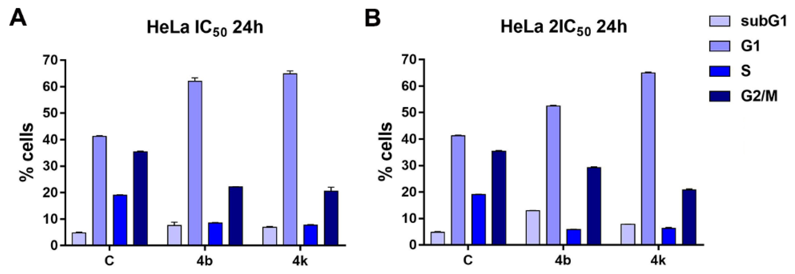
3.3.3. Effects of the Compounds 4b and 4k on Cell Cycle

The treatment of HeLa cells for 24 h with IC50 and 2IC50 concentrations of compounds 4b and 4k induced an increase in the percentage of dead cells within subG1 phase of the cell cycle when compared with that percentage in the control, untreated cell sample, as it can be seen in Figure 4. In addition, both tested compounds applied at IC50 and 2IC50 concentrations triggered a pronounced increase in the percentage of HeLa cells in the G1 phase. The observed changes in the amounts of treated cells within subG1 and G1 phases of the cell cycle were accompanied with decreased percentages of cells within the S and G2/M phases. The effect on the cell-cycle phase distribution in HeLa cell samples was concentration-dependent for compound 4b, while compound 4k showed similar effects on HeLa cells at both tested concentrations. These results indicate that cytotoxic effects of compounds 4b and 4k against HeLa cells could be attributed to an increase in the percentage of cells in the subG1 phase and G1 cell cycle arrest.

3.3.4. Morphological Evaluation of HeLa Cell Death Mode Induced by the Compounds 4b and 4k

The photomicrographs of control HeLa cells and HeLa cells incubated for 24 h with 2IC50 concentrations of the compounds 4b and 4k and stained with a mixture of acridine orange/ethidium bromide are presented in Figure 5. Green-stained rounded cells with condensed chromatin and shrunk nuclei were observed in HeLa cell samples treated with compound 4b, pointing to the ability of this compound to induce apoptotic cell death. Orange-red-stained shrunken cells with condensed chromatin in later stages of apoptosis were detected as well. Compound 4k also showed the ability to activate apoptosis in HeLa cells after 24 h treatment, as demonstrated by rounded green cells with condensed nuclei. The proapoptotic effect of compound 4b was to some extent stronger when comparing with effect of compound 4k. This result is in accordance with the higher amount of subG1 cells detected in the HeLa cell sample exposed to 2IC50 concentration of compound 4b in comparison with the amount of subG1 cells in cells exposed to compound 4k.

3.3.5. Inhibitory Effects of Compounds on α-Glucosidase Enzymatic Activity

The tested compounds showed the ability to inhibit α-glucosidase enzymatic activity, with the exception of compounds 4c and 4j, as can be seen in Table 6. The obtained IC50 values were in the range from 191.80 µM to 1121.91 µM. Among the tested compounds, the compound 4g exerted the best α-glucosidase inhibitory activity with an IC50 value of 191.80 µM. Its activity was stronger than the activity of the anti-diabetic drug acarbose with an IC50 value of 304.21 µM. The compounds 4a and 4b showed inhibitory activities similar to acarbose. The inhibitory effects of the other examined compounds on α-glucosidase enzymatic activity were weaker when compared with the effect of acarbose.

4. Conclusions

Eleven new THPM derivatives were synthesized under mild conditions, while good yields were obtained. For the reaction, methyl acetoacetate, vanillic aldehydes, and N-methylthiourea were used. The obtained compounds were characterized by IR, NMR, ES-MS, while molecular structure of compound 4j has been determined by single-crystal X-ray diffraction analysis. Moreover, their biological activities—antimicrobial, anticancer, and α-glucosidase inhibitory potential—were investigated. The antimicrobial activities of the tested compounds were evaluated against five strains of bacteria and fungi. Compounds 4a, 4b, and 4d have the most effect on bacteria, while the compounds 4e, 4f, and 4k showed the best antifungal activity. Generally, all tested compounds have very good antimicrobial activities which make them worthy of potential pharmacological usage.
The cytotoxic activity was examined against HeLa, K562, MDA-MB-231 cells, and normal human lung fibroblasts MRC-5. The compounds 4k and 4b presented the strongest cytotoxicity against HeLa cells with IC50 values of 43.63 µM and 52.59 µM. In addition, the effects of the 4k and 4b on the cell cycle were examined. Cytotoxic effects of 4k and 4b against HeLa cells could be attributed to an increase in the percentage of cells in the subG1 phase and G1 cell cycle arrest. Furthermore, the investigation of the α-glucosidase inhibitory activity revealed compound 4g as the one with the best activity among all the tested ones. It possesses stronger α-glucosidase inhibitory activity than the anti-diabetic drug acarbose. Further, compounds 4a, 4b, and 4i showed similar activity as acarbose.
Compound 4b had good antibacterial and anticancer activities, compound 4k showed promising antifungal and anticancer activities, while compound 4a has very good α-glucosidase inhibitory activity and antibacterial activity, and compound 4b possesses similar activity as acarbose, and has good anticancer activity. Therefore, we confirmed the potential of some THPMs to be effective agents for more diseases at the same time. All the results have shown the importance of this type of compound as one with the most diverse, and interesting biological activities. The mentioned facts make THPMs excellent candidates for further investigations.

Supplementary Materials

The following supporting information can be downloaded at: https://www.mdpi.com/article/10.3390/pharmaceutics14102254/s1, Supplementary Materials File S1: Materials and methods for biological examination and crystal structure determination; Figures S1–S22: NMR spectra; Table S1: Crystallographic data and refinement parameters for 4j; Supplementary Materials File S2: MS spectra of 4ak. Refs. [30,31,32,33,34,35,36,37,38,39,40] are cited in Supplementary Materials File S1.

Author Contributions

Conceptualization and supervision, N.J. (Nenad Janković); synthesis, E.M., J.P., N.J. (Nenad Joksimović) and N.J. (Nenad Janković); antimicrobial activity, M.K.; cytotoxic analysis, T.S., I.M. and N.G.; crystallographic studies, O.K.; MS analysis, S.S.; writing—original draft preparation, E.M., M.K., I.M., O.K., N.J. (Nenad Joksimović) and N.J. (Nenad Janković); writing—review and editing, N.J. (Nenad Janković) and E.M.; visualization, E.M. and J.P. All authors have read and agreed to the published version of the manuscript.

Funding

This research was funded by the Ministry of Education, Science and Technological Development of the Republic of Serbia (Agreements no. 451-03-68/2022-14/200378, 451-03-68/2022-14/200043, and 451-03-68/2022-14/200122). The authors also acknowledge the partial financial support of the APV Provincial Secretariat for Higher Education and Scientific Research (Project no. 142-451-2635/2021-01/2).

Institutional Review Board Statement

Not applicable.

Informed Consent Statement

Not applicable.

Data Availability Statement

Not applicable.

Acknowledgments

We would like to thank Julijana Tadić and Sanja Rackov for IR analysis.

Conflicts of Interest

The authors declare no conflict of interest.

References

  1. Biginelli, P. Derivati Aldeiduredici Degli Eteri Acetile Dossal-Acetico. Gazz. Chim. Ital. 1893, 23, 360–416. [Google Scholar]
  2. Biginelli, P. Ueber Aldehyduramide des Acetessigäthers. Ber. Dtsch. Chem. Ges. 1891, 24, 1317–1319. [Google Scholar]
  3. Mayer, T.U.; Kapoor, T.M.; Haggarty, S.J.; King, R.W.; Schreiber, S.L.; Mitchison, T.J. Small molecule inhibitor of mitotic spindle bipolarity identified in a phenotype-based screen. Science 1999, 286, 971–974. [Google Scholar] [CrossRef] [Green Version]
  4. Russowsky, D.; Canto, R.F.S.; Sanches, S.A.A.; D’oca, M.G.M.; de Fatima, A.; Pilli, R.A.; Kohn, L.K.; Antonio, M.A.; de Carvalho, J.E. Synthesis and differential antiproliferative activity of Biginelli compounds against cancer cell lines: Monastrol, oxo-monastrol and oxygenated analogues. Bioorg. Chem. 2006, 34, 173–182. [Google Scholar] [CrossRef]
  5. Prokopcova, H.; Dallinger, D.; Uray, G.; Kaan, H.Y.K.; Ulaganathan, V.; Kozielski, F.; Lagner, C.; Kappe, C.O. Structure-activity relationships and molecular docking of novel dihydropyrimidine-based mitotic Eg5 inhibitors. ChemMedChem 2010, 10, 1760–1769. [Google Scholar] [CrossRef]
  6. Kumar, B.R.P.; Sankar, G.; Baig, R.B.N.; Chandrashekaram, S. Novel Biginelli dihydropyrimidines with potential anticancer activity: A parallel synthesis and CoMSIA study. Eur. J. Med. Chem. 2009, 44, 4192–4198. [Google Scholar] [CrossRef]
  7. de Fatima, A.; Braga, T.C.; da Silva Neto, L.; Terra, B.S.; Oliveira, B.G.F.; da Silva, D.L.; Modolo, L.V. A mini-review on Biginelli adducts with notable pharmacological properties. J. Adv. Res. 2015, 6, 363–373. [Google Scholar] [CrossRef] [Green Version]
  8. Kappe, C.O. Biologically active dihydropyrimidones of the Biginelli-type—A literature survey. Eur. J. Med. Chem. 2000, 35, 1043–1062. [Google Scholar] [CrossRef]
  9. Tale, R.H.; Rodge, A.H.; Hatnapure, G.D.; Keche, A.P.; Patil, K.M.; Pawar, R.P. The synthesis, anti-inflammatory and antimicrobial activity evaluation of novel thioanalogs of 3,4-dihydrotyopyrimidin-2(1H)-one derivatives of N-aryl urea. Med. Chem. Res. 2012, 21, 4252–4260. [Google Scholar] [CrossRef]
  10. Gireesh, T.; Kamble, R.R.; Kattimani, P.P.; Dorababu, A.; Manikantha, M.; Hoskeri, J.H. Synthesis of sydnone substituted Biginelli derivatives as hyaluronidase inhibitors. Arch. Pharm. Chem. Life Sci. 2013, 346, 645–653. [Google Scholar] [CrossRef]
  11. Viveka, S.; Nagaraja, G.K.; Shama, P.; Basavarajaswamy, G.; Rao, K.P.; Sreenivasa, M.Y. One pot synthesis of thiazolo[2,3-b]dihydropyrimidinone possessing pyrazole moiety and evaluation of their antiinflammatory and antimicrobial activities. Med. Chem. Res. 2018, 27, 171–185. [Google Scholar] [CrossRef]
  12. Wadhwa, P.; Jain, P.; Jadhav, H.R. Design, synthesis and in vitro evaluation of 4-oxo-6-substituted phenyl-2-thioxo-1,2,3,4-tetrahydropyrimidine-5-carbonitrile derivatives as HIV integrase strand transfer inhibitors. Lett. Drug Des. Discov. 2018, 18, 387–395. [Google Scholar] [CrossRef]
  13. Sepehri, S.; Soleymani, S.; Zabihollahi, R.; Aghasadeghi, M.R.; Sadat, M.; Saghaie, L.; Memarian, H.R.; Fassihi, A. Design, synthesis, and anti-HIV-1 evaluation of a novel series of 1,2,3,4-tetrahydropyrimidine-5-carboxylic acid derivatives. Chem. Biodivers. 2018, 15, 1700502. [Google Scholar] [CrossRef]
  14. Senapathi, J.; Bommakanti, A.; Kusuma, V.; Vangara, S.; Kondapi, A.K. Design, synthesis, and antiviral activity of 1,2,3,4-Tetrahydropyrimidine derivatives acting as novel entry inhibitors to target at “Phe43 cavity” of HIV-1 gp120. Bioorg. Med. Chem. 2021, 52, 116526. [Google Scholar] [CrossRef]
  15. Atwal, K.S.; Rovnyak, G.C.; Kimball, S.D.; Floyd, D.M.; Moreland, S.; Swanson, B.N.; Gougoutas, J.Z.; Schwartz, J.; Smillie, K.M.; Malley, M.F. Dihydropyrimidine calcium channel blockers, 2. 3-Substituted-4-aryl-1,4-dihydro-6-methyl-5-pyrimidinecarboxylic acid esters as potent mimics of dihydropyridines. J. Med. Chem. 1990, 33, 2629–2635. [Google Scholar] [CrossRef]
  16. Zorkun, I.S.; Sarac, S.; Celebib, S.; Erol, K. Synthesis of 4-aryl-3,4-dihydropyrimidin-2(1H)-thione derivatives as potential calciumchannel blockers. Bioorg. Med. Chem. 2006, 14, 8582–8589. [Google Scholar] [CrossRef]
  17. Trivedi, A.R.; Bhuva, V.R.; Dholariya, B.H.; Dodiya, D.K.; Kataria, V.B.; Shah, V.H. Novel dihydropyrimidines as a potential new class of antitubercular agents. Bioorg. Med. Chem. Lett. 2010, 20, 6100–6102. [Google Scholar] [CrossRef]
  18. Yadlapalli, R.K.; Chourasia, O.P.; Vemuri, K.; Sritharan, M.; Perali, R.S. Synthesis and in vitro anticancer and antitubercular activity of diarylpyrazole ligated dihydropyrimidines possessing lipophilic carbamoyl group. Bioorg. Med. Chem. Lett. 2012, 22, 2708–2711. [Google Scholar] [CrossRef]
  19. Akhaja, T.N.; Raval, J.P. 1,3-Dihydro-2H-indol-2-ones derivatives: Design, synthesis, in vitro antibacterial, antifungal and antitubercular study. Eur. J. Med. Chem. 2011, 46, 5573–5579. [Google Scholar] [CrossRef]
  20. Milović, E.; Petronijević, J.; Joksimović, N.; Beljkaš, M.; Ružić, D.; Nikolić, K.; Vraneš, M.; Tot, A.; Đorđić Crnogorac, M.; Stanojković, T.; et al. Anticancer evaluation of the selected tetrahydropyrimidines: 3D-QSAR, cytotoxic activities, mechanism of action, DNA, and BSA interactions. J. Mol. Struct. 2022, 1257, 132621. [Google Scholar] [CrossRef]
  21. Ristovski Trifunović, J.; Minorics, R.; Bartha, S.; Janković, N.; Zupkó, I. The evaluation of anticancer activity of the Biginelli hybrids and pharmacokinetic profiling based on their retention parameters. J. Mol. Struct. 2022, 1254, 132373. [Google Scholar] [CrossRef]
  22. Milović, E.; Janković, N.; Bogdanović, G.; Petronijević, J.; Joksimović, N. On water synthesis of the novel 2-oxo-1,2,3,4-tetrahydropyrimidines. Tetrahedron 2021, 78, 131790. [Google Scholar] [CrossRef]
  23. Janković, N.; Trifunović, J.; Vraneš, M.; Tot, A.; Petronijević, J.; Joksimović, N.; Stanojković, T.; Đorđić Crnogorac, M.; Petrović, N.; Boljević, I.; et al. Discovery of the Biginelli hybrids as novel caspase-9 activators in apoptotic machines: Lipophilicity, molecular docking study, influence on angiogenesis gene and miR-21 expression levels. Bioorg. Chem. 2019, 86, 569–582. [Google Scholar] [CrossRef]
  24. Janković, N.; Stefanović, S.; Petronijević, J.; Joksimović, N.; Novaković, S.B.; Bogdanović, G.A.; Muškinja, J.; Vraneš, M.; Ratković, Z.; Bugarčić, Z. Water-tuned tautomer-selective tandem synthesis of the 5,6-Dihydropyrimidin-4(3H)-ones, driven under the umbrella of sustainable chemistry. ACS Sustain. Chem. Eng. 2018, 6, 13358–13366. [Google Scholar] [CrossRef]
  25. Janković, N.; Bugarčić, Z.; Marković, S. Double catalytic effect of (PhNH3)2CuCl4 in a novel, highly efficient synthesis of 2-oxo and thioxo-1,2,3,4-tetrahydropyrimidines. J. Serb. Chem. Soc. 2015, 80, 595–604. [Google Scholar] [CrossRef]
  26. Spurg, A.; Waldvogel, S.R. High-yielding cleavage of (aryloxy)acetates. Eur. J. Org. Chem. 2008, 2, 337. [Google Scholar] [CrossRef]
  27. Ryabukhin, S.V.; Plaskon, A.S.; Ostapchuk, E.N.; Volochnyuk, D.M.; Tolmachev, A.A. N-Substituted ureas and thioureas in Biginelli reaction promoted by chlorotrimethylsilane: Convenient synthesis of N1-alkyl-, N1-aryl-, and N1, N3-dialkyl-3,4-dihydropyrimidin-2(1H)-(thi)ones. Synthesis 2007, 3, 417. [Google Scholar] [CrossRef]
  28. Pani, M.S.; Arjun, M.; Sridhar, D.; Srinivas, K.; Raviprasad, T. N-Substituted benzoxazolyl ureas and thioureas in Biginelli reaction promoted by trifluoromethane sulfonic acid: An efficient and convenient synthesis of substituted benzoxazolyl 3,4-dihydropyrimidine (1H)-(thio)-ones. Chin. Chem. Lett. 2009, 20, 909. [Google Scholar] [CrossRef]
  29. Bruno, I.J.; Cole, J.C.; Edgington, P.R.; Kessler, M.K.; Macrae, C.F.; McCabe, P.; Pearson, J.; Taylor, R. New software for searching the Cambridge Structural Database and visualizing crystal structures. Acta Crystallogr. Sect. B Struct. Sci. Cryst. Eng. Mater. 2002, 58, 389–397. [Google Scholar] [CrossRef]
  30. Sarker, S.D.; Nahar, L.; Kumarasamy, Y. Microtitre plate-based antibacterial assay incorporating resazurin as an indicator of cell growth, and its application in the in vitro antibacterial screening of phytochemicals. Methods. 2007, 42, 321–324. [Google Scholar] [CrossRef]
  31. Mosmann, T. Rapid colorimetric assay for cellular growth and survival: Application to proliferation and cytotoxicity assays. J. Immunol. Methods 1983, 65, 55–63. [Google Scholar] [CrossRef]
  32. Ohno, M.; Abe, T. Rapid colorimetric assay for the quantification of leukemia inhibitory factor (LIF) and interleukin-6 (IL-6). J. Immunol. Methods 1991, 145, 199–203. [Google Scholar] [CrossRef]
  33. Ormerod, M.G. Flow Cytometry. A Practical Approach; Oxford University Press: Oxford, UK; p. 2000.
  34. McCue, P.; Kwon, Y.I.; Shetty, C. Anti-amylase, anti-glucosidase and anti-angiotensin i-converting enzyme potential of selected foods. J. Food Biochem. 2005, 29, 278–294. [Google Scholar] [CrossRef]
  35. Grozdanić, N.; Zdunić, G.; Šavikin, K.; Đuričić, I.; Kosanić, M.; Mačić, V.; Matić, I.Z.; Stanojković, T. Seasonal variation in biopharmaceutical activity and fatty acid content of endemic Fucus virsoidesalgae from Adriatic sea. Acta Pol. Pharm. 2019, 76, 833–844. [Google Scholar] [CrossRef]
  36. Rigaku, O.D. CrysAlis PRO; Rigaku Oxford Diffraction Ltd.: Oxfordshire, UK, 2015. [Google Scholar]
  37. Blessing, R.H. An empirical correction for absorption anisotropy. Acta Crystallogr. A 1995, 51, 33–38. [Google Scholar] [CrossRef] [PubMed]
  38. Sheldrick, G.M. SHELXT – Integrated Space-Group and Crystal-Structure Determination. Acta Crystallogr. A Found. Adv. 2015, 71, 3–8. [Google Scholar] [CrossRef] [Green Version]
  39. Sheldrick, G.M. Crystal Structure Refinement with SHELXL. Acta Crystallogr. Sect. C Struct. Chem. 2015, 71, 3–8. [Google Scholar] [CrossRef] [Green Version]
  40. Dolomanov, O.V.; Bourhis, L.J.; Gildea, R.J.; Howard, J.A.K.; Puschmann, H. OLEX2: A complete structure solution, refinement and analysis program. J. Appl. Cryst. 2009, 42, 339–341. [Google Scholar] [CrossRef]
Scheme 1. Substrate scope in the synthesis of tetrahydropyrimidines 4a–k. Reaction conditions: (a) aldehyde and N-methylthiourea, dioxane/CHCl3, room temperature, (b) methyl acetoacetate, HCl, 2-amino-1-(4-nitrophenyl)-1,3-propanediol (ANP), room temperature.
Pharmaceutics 14 02254 sch001
Figure 1. MERCURY [29] drawing of the molecular structure of compound 4j with labeled non-H atoms. Displacement ellipsoids are shown at 30% probability. Symmetry code: (i) −x, −y+1, −z+2.
Pharmaceutics 14 02254 g001
Figure 2. MERCURY [29] drawing showing crystal packing of 4-(4′-benzoyloxy-3′-methoxyphenyl)- 1,2,3,4-tetrahydro-1,6-dimethyl-2-thioxopyrimidine-5-carboxylate (4j) viewed along the a axis. Intramolecular N—H•••O and C—H•••O contacts are shown as dashed lines. Intermolecular C9—H9•••S11(x, y+1, z) contacts (dashed line) connect molecules in a head-to-tail manner along the b axis.
Pharmaceutics 14 02254 g002
Figure 3. MERCURY [29] drawing of crystal packing of 4-(4′-benzoyloxy-3′-methoxyphenyl)- 1,2,3,4-tetrahydro-1,6-dimethyl-2-thioxopyrimidine-5-carboxylate (4j) showing intermolecular C—H•••O contacts (dashed line) connecting molecules in a head-to-tail manner along the a axis. Symmetry codes: 2: x−1, y, z; 3: x+1, y, z.
Pharmaceutics 14 02254 g003
Figure 4. Cell-cycle phase distribution of control HeLa cells and HeLa cells incubated with IC50 (A) and 2 IC50 (B) concentrations of compounds 4b and 4k during 24 h. The data shown represent the average ± standard deviation of two independent experiments.
Pharmaceutics 14 02254 g004
Figure 5. Photomicrographs of acridine orange/ethidium bromide-stained control HeLa cells and HeLa cells incubated with 2IC50 concentrations of compounds 4b and 4k during 24 h (magnification 20×). Representative photomicrographs are shown.
Pharmaceutics 14 02254 g005
Table 1. Selected geometrical parameters for methyl 4-(4′-benzoyloxy-3′-methoxyphenyl)- 1,2,3,4-tetrahydro-1,6-dimethyl-2-thioxopyrimidine-5-carboxylate (4j).
Bond Lengths [Å]
O1—C201.203 (3)N1—C11.339 (2)
O2—C201.334 (3)N1—C41.453 (2)
O2—C211.442 (2)N2—C11.373 (3)
O3—C71.354 (2)N2—C21.410 (3)
O3—C221.424 (3)N2—C181.472 (3)
O4—C111.354 (2)C3—C21.336 (3)
O4—C81.402 (2)C4—C31.517 (2)
O5—C111.196 (3)S1—C11.6733 (19)
O6—C241.423 (3)O6—C231.425 (3)
Bond angles [°]
C11—O4—C8116.71 (15)C1—N1—C4122.60 (14)
C24—O6—C23110.08 (16)N1—C1—N2115.90 (16)
C20—O2—C21116.98 (17)N1—C1—S1120.25 (14)
Torsion angles [°]
C4—N1—C1—N2−23.7 (2)C2—C3—C20—O1−19.9 (3)
N1—C4—C3—C2−31.1 (2)C4—C3—C20—O1157.6 (2)
C2—N2—C1—N1−10.4 (2)C2—C3—C20—O2162.28 (16)
N1—C4—C3—C20151.29 (15)C4—C3—C20—O2−20.2 (2)
C18—N2—C1—S1−8.3 (3)O3—C7—C8—O4−3.0 (3)
C2—N2—C1—S120.2 (3)C11—O4—C8—C9−80.8 (3)
C1—N2—C2—C3169.09 (13)C11—O4—C8—C7103.7 (2)
C5—C4—C3—C292.44 (18)C17—C12—C11—O59.3 (3)
Table 2. Intramolecular and intermolecular contact parameters in the crystal structure of methyl 4-(4′-benzoyloxy-3′-methoxyphenyl)-1,2,3,4-tetrahydro-1,6-dimethyl-2-thioxopyrimidine-5-carboxylate (4j).
D—H•••AD—HH•••AD•••AD—H•••A
N1—H1•••O60.862.232.897 (2)134.9
C19—H19B•••O10.932.953.809 (2)154.0
C9—H9•••S1 10.962.613.363 (3)135.7
C21—H21C•••O5 20.962.613.363 (3)135.7
C22—H22C•••O6 30.962.623.407 (3)139.5
Symmetry codes: 1 x, y + 1, z; 2 x − 1, y, z; 3 x + 1, y, z.
Table 3. The antibacterial activity of the tested compounds.
Tested CompoundsStaphylococcus aureusBacillus subtilisKlebsiella oxytocaProteus
mirabilis
Escherichia coli
MIC (mg/mL)
4a1.623.251.620.811.62
4b1.623.251.620.811.62
4c1.623.251.621.621.62
4d0.813.251.620.811.62
4e3.253.253.25ND1.62
4f3.253.253.251.623.25
4g3.253.251.620.811.62
4h3.253.253.253.253.25
4i3.253.251.621.621.62
4j3.253.253.251.623.25
4k3.253.253.253.253.25
Streptomycin0.0310.0160.0080.0620.062
ND—not detected.
Table 4. The antifungal activity of the tested compounds.
Tested CompoundsTrichophyton
mentagrophytes
Mucor mucedoPenicillium italicumAspergillus flavusAspergillus niger
MIC (mg/mL)
4a0.401.621.621.621.62
4b0.401.621.621.620.81
4c0.401.621.620.400.81
4d0.401.621.621.621.62
4e0.201.621.621.621.62
4f0.201.621.621.620.81
4g0.401.621.621.621.62
4h0.811.621.621.621.62
4i0.811.621.621.621.62
4j0.401.621.621.621.62
4k0.201.621.620.811.62
Fluconazole0.251110.5
Table 5. Cytotoxic activity of compounds.
Tested CompoundsHeLaK562MDA-MB-231MRC-5
IC50 [µM] Average ± SD
4a97.40 ± 5.4078.98 ± 7.49144.50 ± 8.29186.92 ± 4.66
4b52.59 ± 4.4576.83 ± 5.01115.65 ± 8.71111.87 ± 10.85
4c>200180.21 ± 9.15>200195.83 ± 5.89
4d>200164.66 ± 8.50>200192.71 ± 10.32
4e197.22 ± 3.93149.08 ± 5.74>200127.98 ± 3.20
4f135.34 ± 9.32122.91 ± 8.52161.29 ± 9.69196.08 ± 5.54
4g197.49 ± 3.55152.68 ± 2.58>200193.22 ± 9.59
4h120.85 ± 9.9679.94 ± 6.73114.02 ± 12.8677.82 ± 2.57
4i169.06 ± 4.1299.36 ± 9.38>200152.34 ± 10.15
4j78.11 ± 5.7867.97 ± 6.53122.61 ± 2.19104.17 ± 8.61
4k43.63 ± 1.4939.11 ± 2.9074.12 ± 1.2587.23 ± 7.31
cisPt4.91 ± 0.746.89 ± 0.2114.74 ± 0.369.35 ± 1.29
The results are presented as average ± standard deviation of three independent experiments performed in triplicate.
Table 6. α-glucosidase inhibitory activity of the compounds.
CompoundsIC50 [µM]
4a312.91 ± 7.73
4b291.77 ± 6.67
4cND
4d1121.91 ± 20.59
4e674.81 ± 5.65
4f767.91 ± 5.91
4g191.80 ± 5.95
4h932.75 ± 25.16
4i418.02 ± 5.68
4jND
4k1083.41 ± 54.54
acarbose304.21 ± 14.62
The results are presented as average ± standard deviation of two experiments. ND—non-determined
Publisher’s Note: MDPI stays neutral with regard to jurisdictional claims in published maps and institutional affiliations.

