Article

77Se-Enriched Selenoglycoside Enables Significant Enhancement in NMR Spectroscopic Monitoring of Glycan–Protein Interactions

1
Department of Organic Chemistry, University of Debrecen, Egyetem tér 1, H-4032 Debrecen, Hungary
2
Department of Inorganic and Analytical Chemistry, University of Debrecen, Egyetem tér 1, H-4032 Debrecen, Hungary
3
Molecular Recognition and Interaction Research Group, Hungarian Academy of Sciences, University of Debrecen, Egyetem tér 1, H-4032 Debrecen, Hungary
*
Authors to whom correspondence should be addressed.
Pharmaceutics 2022, 14(1), 201; https://doi.org/10.3390/pharmaceutics14010201
Submission received: 16 December 2021 / Revised: 7 January 2022 / Accepted: 10 January 2022 / Published: 15 January 2022

Abstract

:
Detailed investigation of ligand–protein interactions is essential for better understanding of biological processes at the molecular level. Among these binding interactions, the recognition of glycans by lectins is of particular importance in several diseases, such as cancer; therefore, inhibition of glycan-lectin/galectin interactions represents a promising perspective towards developing therapeutics controlling cancer development. The recent introduction of 77Se NMR spectroscopy for monitoring the binding of a selenoglycoside to galectins prompted interest to optimize the sensitivity by increasing the 77Se content from the natural 7.63% abundance to 99%. Here, we report a convenient synthesis of 77Se-enriched selenodigalactoside (SeDG), which is a potent ligand of the medically relevant human galectin-3 protein, and proof of the expected sensitivity gain in 2D 1H, 77Se correlation NMR experiments. Our work opens perspectives for adding isotopically enriched selenoglycans for rapid monitoring of lectin-binding of selenated as well as non-selenated ligands and for ligand screening in competition experiments.

Graphical Abstract

1. Introduction

Mounting awareness in recent decades has highlighted the importance of molecular events in how lectins, a class of carbohydrate-binding proteins, recognize cell surface glycans [1]. Lectin–glycan interactions are involved in cellular processes such as adhesion, intercellular communication, growth and differentiation, cell cycle, and apoptosis. As a result, lectin-mediated processes are implicated in wide range of diseases such as cancer, inflammation and fibrosis, heart disease, and stroke [2,3,4,5,6,7]. Galectins, Gal-1, -3, -7, and -9 in particular, have been implicated in multiple ways in malignant cell proliferation processes [4,5,7,8]. Inhibition of glycan–galectin interactions thus offers a promising perspective towards developing therapeutics influencing these processes. Research on small-molecule- or multivalent glycomimetics as lectin inhibitors is especially worth mentioning in this aspect [9,10,11].
Several analytical techniques (surface plasmon resonance—SPR, isothermal titration calorimetry—ITC, nuclear magnetic resonance—NMR, or X-ray crystallography) are available to investigate these interactions. Among these methods, NMR spectroscopy stands out as it allows studying protein–glycan interactions in solution, i.e., close to physiological conditions at the molecular level [12,13]. Observation of NMR signals of the ligand molecules binding to protein receptors is one of the most powerful approaches to gain insight into structural and dynamic aspects of these interactions [14,15]. NMR spectroscopy is plagued, however, with sensitivity issues, representing a double challenge because of low concentrations and typically weak interactions. Signal overlap is another problem to be dealt with, especially with carbohydrate ligands typically having narrow 1H chemical shift ranges with consequent serious overlaps in the spectra [16]. Spectral overcrowding quickly becomes unmanageable with oligosaccharide ligands or with carbohydrate mixtures, such as in studies of potential competition with different ligands. 13C- or 15N-based NMR approaches are much less affected by spectral overlap, but suffer even more from low sensitivity at natural abundance of these NMR-active isotopes even in sensitivity-enhanced 1H-detected versions. A continuous methodological challenge in NMR-spectroscopical monitoring is to expand the range of techniques and, here, the introduction of distinct NMR sensors beyond the commonly used 1H, 13C, and 15N isotopes comes into play. Application of 19F as a reporter nucleus has been proposed as an alternative, combining high sensitivity (0.83% compared with that of 1H) with superior selectivity owing to a large chemical shift range, as well as specific labeling used to introduce this label just to a few predefined positions of the molecule of interest (typically substituting OH for F) [17,18,19,20]. It is to be noted, however, that fluorinated derivatives are chemically distinct from their natural counterparts; still, very often, they turn out to be good mimics of the parent molecules in terms of structure and binding properties.
The success of the 19F-approach prompted us to look for a further isotope to be introduced as an NMR reporter into lectin–oligosaccharide interaction analysis. Inspired by the discovery that thiodigalactoside (TDG) is a potent ligand for adhesion/growth-regulatory galactose-binding lectins [21,22,23], we have tested selenodigalactoside (SeDG) as a binding partner and its 77Se isotope as an NMR-spectroscopical sensor. As we have detected rather equal affinities of TDG and SeDG to human galectins-1 and -3 (Gal-1/-3) [24], we initiated 77Se NMR-based monitoring of SeDG binding to these lectins [25,26]. 77Se is a spin-½ NMR isotope with broad chemical shift range (ca. 3000 ppm), which makes it promising as a selective probe [25,27,28,29]. Direct 77Se NMR detection [30,31] is, however, unfavorable owing to the low natural abundance (7.63%) and decreased sensitivity (0.7 of 1H) of this nucleus. To improve sensitivity, we proposed a 2D 1H-77Se HSQMBC (heteronuclear single quantum multiple-bond correlation) experiment via indirect 77Se detection using CPMG-INEPT (Carr–Purcell–Meiboom–Gill insensitive nuclei enhanced by polarization transfer) out-and-back 1H→77Se→1H polarization transfer. Theoretically, this approach can yield up to 60-fold ((γHSe)2.5) sensitivity enhancement, but, owing to competing relaxation and other transfer processes, a sensitivity gain about 20 can be realized in practice [25,28,32].
A next step toward optimizing 77Se sensor capacity is to prepare the lectin ligand as an isotopically enriched probe, that is, bringing isotope representation from the natural 7.63% abundance to close to 100%. To exploit this attractive potential, we provide an example by the synthesis of a sample of di(β-D-galactopyranosyl)selenide enriched to 99% in 77Se isotope ([77Se]DGal). Exploratory experiments reported here with this 77Se-enriched material indeed confirmed our expectations for a spectacular increase in NMR detection sensitivity. We will furthermore demonstrate that combining our advanced 1H-77Se HSQMBC NMR method with 77Se enrichment enables rapid monitoring of lectin-binding of selenated as well as non-selenated ligands and high-throughput screening in competition experiments.

2. Materials and Methods

2.1. Synthetic Procedures

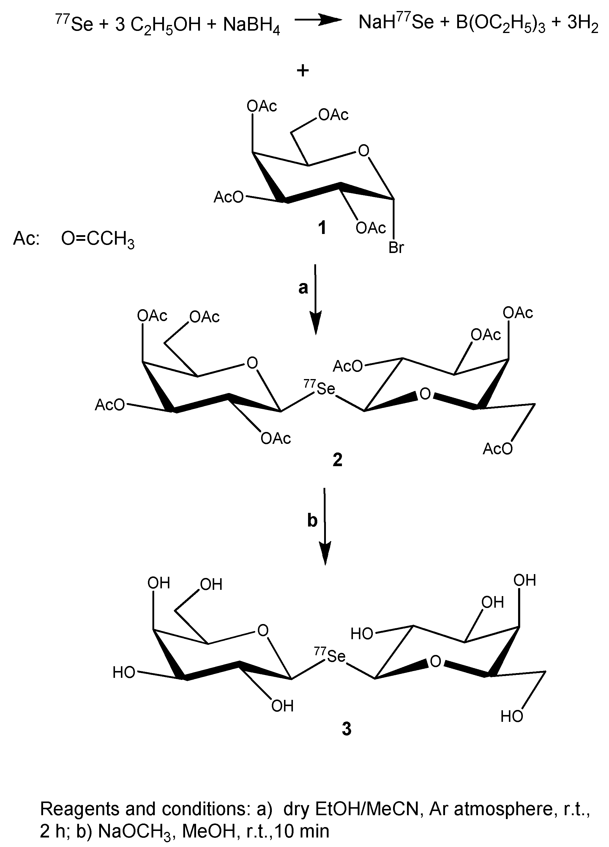
2.1.1. 77Se-Enriched Di(2,3,4,6-tetra-O-acetyl-β-D-galactopyranosyl)selenide (2)

An ethanolic solution of NaH77Se was prepared following a procedure described by Klayman and Griffin [33]. In a flame-dried flask, isotopically enriched (99%) 77-selenium powder (Laboratory Standards Kft. Budapest, Hungary, lot no. SELM-2445-PK) (50 mg, 0.65 mmol) was stirred in dry ethanol (5 mL) under argon atmosphere (Note). After cooling in an ice bath, sodium borohydride (50 mg, 1.3 mmol) was added and stirring continued until the selenium was dissolved and gas evolution subsided (ca. 5 min). To this clear solution, 1-Br-2,3,4,6-tetra-O-acetyl-α-D-galactopyranose (1, 520 mg, 1.3 mmol) dissolved in dry methyl cyanide (2 mL) was added and stirring continued at room temperature until TLC (hexane/EtOAc 1:1) indicated consumption of 1 (2 h). The reaction mixture was evaporated under reduced pressure, the syrupy residue dissolved in dichloromethane, it extracted with water twice, the organic phase dried (MgSO4), and the solvent was removed in vacuo to yield 2 as a pale-yellow solid at 323 mg (69%). According to its 1H NMR spectrum, this product was judged to be sufficiently pure, apart from some residual signals from ethanol (1.23/3.70 ppm, see Figure S1, bottom), to be deacetylated in the next step (2.1.2). [α]D22—5.2 (c 0.2 CHCl3); 1H NMR (CDCl3, 500 MHz): δ (ppm) 5.46 (br.d, 1H, H-4, J3.4 3.0 Hz); 5.30 (t, 1H, H-2, J1,2 = J2,3 10.3 Hz); 5.06 (d, 1H, H-1); 5.05 (br.d, 1H, H-3); 4.17 (dd, 1H, H-6a, J6a,6b 11.2 Hz, J5,6a 6.7 Hz); 4.12 (dd, 1H, H-6b, J5,6b 6.5 Hz); 3.90 (m, 1H, H-5); 1.99; 2.05; 2.06; 2.17 (12H, 4 CH3(CO)); 13C NMR (CDCl3, 125 MHz): δ (ppm) 169.7; 169.9; 170.1; 170.3 (4 CH3(CO)); 77.0 (C-1, 1JSe,C1 88.7 Hz); 75.8 (C-5); 71.6 (C-3, 3JSe,C3 5.4 Hz); 68.1 (C-2, 2JSe,C2 16.2 Hz); 67.2 (C-4); 61.6 (C-6); 20.74, 20.65(2x), 20.74 (4xCH3(CO)); HRMS: calc. for C28H38O1877Se: [M + Na]+:762.116, found: 762.115.

2.1.2. 77Se-Enriched Di(β-D-galactopyranosyl)selenide (3)

To a solution of 77Se-enriched di(2,3,4,6-tetra-O-acetyl-β-D-galactopyranosyl)selenide (2) (180 mg) in dry methanol (50 mL), a 25% solution of sodium methoxide in methanol (0.06 mL) was added. After 15 min at room temperature, 0.5 g of Amberlist® 15H ion exchange resin was added, the resin filtered off after 5 min of stirring, and the filtrate evaporated to dryness under reduced pressure. Recrystallization from methanol furnished 94 mg of white solid (96%). 1H NMR (Figure S2) and 13C NMR (Figure S3) spectra of this product (3) are provided in the Supplementary Material. [α]D22—30.9 (c 0.2 H2O); 1H NMR (DMSO-d6, 500 MHz): δ (ppm) 4.78 (dd, 1H, H-1, J1,2 9.8 Hz, 2JH2,Se 6.1 Hz); 3.70 (m, 1H, H-4); 3.45–3.53 overlapping signals (3H, H-2, H-6a, H-6b); 3.32 (m, 1H, H-5); 3.28 (m, 1H, H-3); 13C NMR (DMSO-d6, 125 MHz): δ (ppm) 80.4 (C-5); 79.8 (C-1, 1JSe,C1 80.4 Hz); 74.6 (C-3, 3JSe,C3 4.6 Hz); 71.0 (C-2, 2JSe,C2 12.8 Hz); 68.4 (C-4); 60.4 (C-6); HRMS: calc. for C12H22O1077Se [M + Na]+: 426.030, found: 426.032.
Note: Exclusion of traces of water and of oxygen is important to avoid losses of selenium via formation of hydrogen selenide and/or oxidation. Any hydrogen selenide (which is very poisonous) that might have formed was trapped by passing it into a 5% aqueous solution of lead acetate.

2.2. Sample Preparation for NMR Measurements

For 77Se-NMR-based monitoring of ligand binding to hGal-3, 10 μL of a 120 mM [77Se]DGal ligand stock solution (in a 10 mM phosphate buffer in D2O, pH 7.4, containing 0.5 M NaCl) and 590 μl 10 mM phosphate buffer in D2O, pH 7.4, containing 0.5 M NaCl were pipetted into a 5 mm NMR tube to obtain a 2.0 mM final concentration of [77Se]DGal ligand. In a second step, hGal-3 protein was added as lyophilized powder to reach a hGal-3/[77Se]DGal ratio of 0.0145:1 (chGal-3 = 29 μM) in the NMR tube. This sample was used for binding studies with the 1H-77Se CPMG-HSQMBC experiment. Thiodigalactoside (TDG) was added as a solid material (0.43 mg) to the previous sample, resulting in a hGal-3/[77Se]DGal/TDG ratio of 0.0145:1:1 (29 μM/2 mM/2 mM) in the NMR sample measured in the third step of the 1H-77Se CPMG-HSQMBC binding experiments. 1H NMR spectra obtained on samples of each step of binding experiments can be seen in Figure S4. It is noteworthy that the protein–ligand concentration ratio in these NMR experiments should always be optimized in accordance with the pertinent binding affinity of the system being studied.

2.3. NMR Measurements

1H-77Se CPMG-HSQMBC NMR experiments were performed on a Bruker Avance I spectrometer (Bruker BioSpin GmbH, Rheinstetten, Germany) operating at 400 MHz 1H frequency equipped with a BBI z-gradient probe. All experiments were carried out at 303 K, and NMR data were processed with TopSpin 2.1 or 3.5 (Bruker Biospin GmbH, Karlsruhe, Germany).
1H-77Se correlation spectra were measured by the refocused 2D 1H-77Se CPMG-HSQMBC pulse sequence with 77Se decoupling [25]. Simultaneous composite π pulses on the 1H and 77Se channels were applied with an equal duration of 90° pulses (18 μs) achieved with careful adjustment of power levels. The CPMG-INEPT delay Δ for long-range heteronuclear coupling evolution was adjusted to 45.1 ms. For 77Se CPD decoupling during FID acquisition (183 ms), the WALTZ16 scheme with a 90° pulse length of 400 μs was used. 2D 1H-77Se CPMG-HSQMBC spectra were recorded for sensitivity comparison on [77Se]DGal and SeDGal samples, with 1024 total data points in the 1H (t2) dimension and 32 total points in the 77Se (t1) dimension, using spectral windows of 6.99 ppm (2796 Hz) for 1H and 6.00 ppm (458 Hz) for 77Se. On [77Se]DGal sample, 8 scans per t1 increment were accumulated; in contrast, 360 scans per t1 increment were used for SeDGal sample. The polarization recovery delay between consecutive scans, d1, was set to 1.7 s. In binding experiments, each 2D 1H-77Se CPMG-HSQMBC spectrum was recorded with 1024 total data points in the 1H (t2) dimension and 2 total points in the 77Se (t1) dimension, using spectral windows of 6.99 ppm (2796 Hz) for 1H and 6.00 ppm (458 Hz) for 77Se. 32 scans per t1 increment were accumulated and the polarization recovery delay between consecutive scans, d1, was set to 1.7 s.

3. Results and Discussion

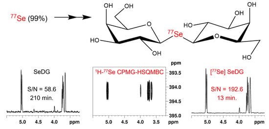
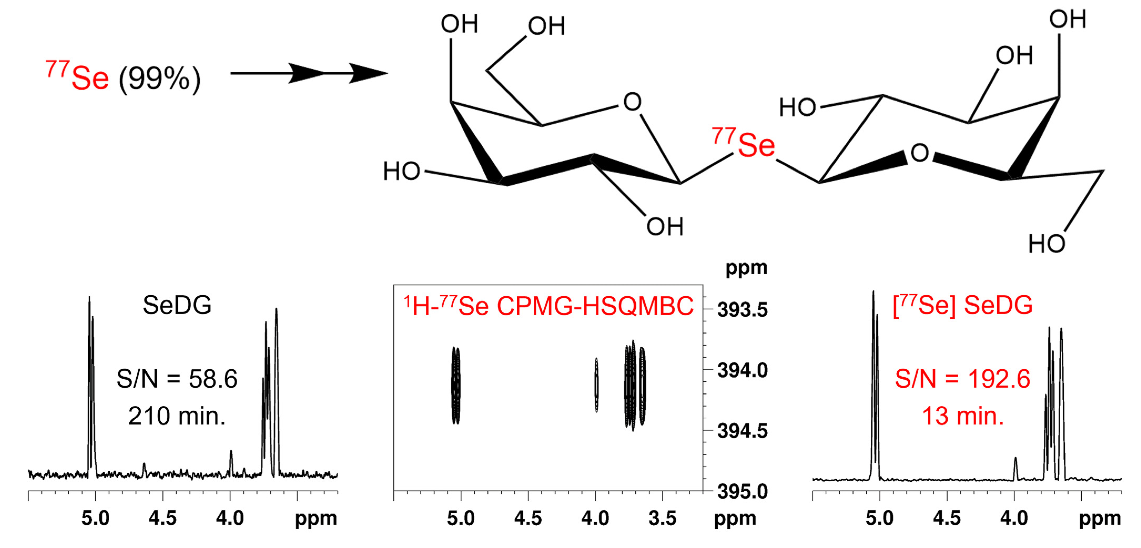
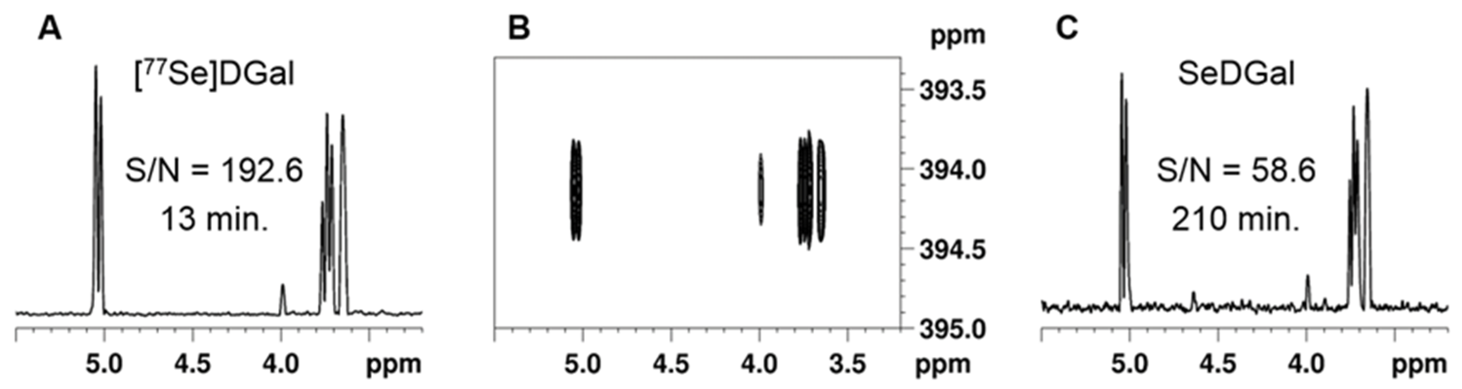
3.1. Synthesis of 77Se-Enriched Di(β-D-galactopyranosyl)selenide, [77Se]DGal

Preparation of this compound was based on the reaction of 1-Br-2,3,4,6-tetra-O-acetyl-α-D-galactopyranose (1) with NaH77Se, with the latter obtained by NaBH4 reduction of elemental selenium [33] 99% enriched in 77Se isotope. Reaction of (1) with NaH77Se furnished 77Se-enriched octa-O-acetyl-di(β-D-galactopyranosyl)selenide (2), which gave the desired product, 77Se-enriched selenodigalactoside ([77Se]DGal 3), upon Zemplén deacetylation (Scheme 1). This two-step synthesis was carried out with a good overall yield; further details of the reactions are given in Section 2.

3.2. NMR Experiments

As mentioned in the introduction, we have recently proposed a 2D 1H-77Se heteronuclear correlation experiment to enhance the detection sensitivity of 77Se NMR by replacing direct NMR observation on 77Se for indirect observation on 1H nuclei [25,26]. Our 2D 1H-77Se CPMG-HSQMBC pulse sequence is based on out-and-back 1H→77Se→1H polarization transfer via 2,3J(1H,77Se) long-range couplings. CPMG-INEPT long-range transfer eliminates signal phase modulation and significantly reduces intensity losses from co-evolving J(1H, 1H) couplings. Furthermore, it can suppress line broadening from chemical exchange, ensuring maximum detection sensitivity and clean signal phases, as was described earlier [25,26,32].
The sensitivity gain using 99% 77Se-enriched SeDGal ([77Se]DGal) versus the nonenriched specimen (SeDGal) was checked via comparative 2D 1H-77Se CPMG-HSQMBC experiments. Figure 1 shows two sets of data corresponding to [77Se]DGal (A) and SeDGal (C), respectively, in terms of signal to noise (S/N) ratios and times of data acquisition (Acq) for samples of equal concentrations. The overall sensitivity improvement calculated from the experimental data in Figure 1 is 13.21 in terms of unit acquisition time (Equation (1)):
[S/N(A)/S/N(C)] ∗ [(Acq(C)/Acq(A)]0.5 = (192.6/58.6) ∗ (210/13)0.5 = 13.21
This figure compares well with the theoretical value of 12.98 based on isotopic abundances [(99.0/7.63)]. Conversion of this ratio into overall NMR experimental times results in a remarkable ~170-fold [(12.98)2] reduction in favor of the enriched specimen for samples of equal concentrations.
To demonstrate the usefulness of 77Se-enriched ligand in binding experiments, Figure 2A shows the 1D cross section from a 3-minute 2D 1H-77Se CPMG-HSQMBC experiment of [77Se]DGal (2 mM). This spectrum conveys the reference (100%) signal intensity. It is noteworthy that the S/N ratio is more than adequate in this 3-minute spectrum, leaving room to reduce the concentration of the ligand (and, consequently, that of the protein) in these experiments. The addition of human galectin-3 (hGal-3, 29 μM) to this sample resulted in a decrease in signal intensity (from 100% to 76%, Figure 2B) by line broadening, which indicated binding of the ligand to the protein [25]. The line broadening in Figure 2B was then partially reversed (from 76% to 86%, Figure 2C) with the introduction of thiodigalactoside (TDG, 2 mM) to the sample. This phenomenon is a consequence of reduced relaxation enhancement of [77Se]DGal resonances via competition between the two ligands (TDG and SeDGal) for the same recognition site on hGal-3, as explained earlier [25]. It also illustrates that [77Se]DGal can be used via its competitive displacement to indirectly monitor the binding of even non-selenated compounds to hGal-3 (or any other target proteins) by our sensitivity-optimized 1H-77Se CPMG-HSQMBC method.
In sum, we have demonstrated that the enhanced detection sensitivity inherent in our original 1H-77Se CPMG-HSQMBC sequence receives a further significant boost by using 77Se-enriched ligands, such as [77Se]DGal, described in this article. The illustrated straightforward introduction of 77Se into the glycosidic bond opens wide applicability of 77Se-enriched selenoglycosides for biomedically relevant tissue lectins and beyond. In terms of NMR experimental time, this enhancement allows to obtain data in much less time (a factor of about 170 (12.982) between solutions of identical concentrations), and this gain is further documented by the results of a ligand competition experiment. The significant sensitivity gain offered by 77Se-enrichment, such as in [77Se]DGal, enables, furthermore, a fast and specific detection approach for screening the binding of non-selenated ligands as well to hGal-3. The combined sensitivity advantage of this approach will likely aid NMR to become competitive for binding/screening studies at low concentrations nearing physiological/cellular levels with selenated and non-selenated ligands too. Our present study is a first step toward allowing 77Se-enriched selenoglycosides to become versatile tools in interaction analysis by NMR spectroscopy, and inspires new approaches to combine it with synthetic carbohydrate chemistry, as illustrated herein. The current work opens perspectives for using isotopically enriched selenoglycans alone or in combination of 77Se with 2D, 13C, 15N, 19F, and/or 31P for analyzing the structure and dynamics of ligand binding to proteins by taking advantage of different NMR time scales offered by distinct NMR frequencies of the individual isotopes. Therefore, our method has the potential to support drug development through the rapid and detailed investigation of glycan–lectin interactions.

Supplementary Materials

The following are available online at https://www.mdpi.com/article/10.3390/pharmaceutics14010201/s1, Figure S1: 500 MHz 1H NMR spectrum of di(2,3,4,6-tetra-O-acetyl-β-D-galactopyranosyl)selenide (2) (99% 77Se-enriched, bottom), compared with the spectrum of the same compound with 77Se in natural abundance (top) in CDCl3; Figure S2: 500 MHz 1H NMR spectrum of [77Se]DG, 3 (99% 77Se-enriched, bottom), compared with the spectrum of SeDG (77Se in natural abundance, top) in D2O; Figure S3: 125 MHz 13C NMR spectrum of [77Se]DG, 3 (99% 77Se-enriched, bottom), compared with the spectrum of SeDG (77Se in natural abundance, top) in DMSO-d6; Figure S4: 400 MHz 1H NMR spectra obtained on samples of [77Se]DGal (2 mM) in the absence of hGal-3 (bottom, blue), [77Se]DGal (2 mM), and hGal-3 (29 μM, i.e., molar ratio = 1:0.0145) (middle, red), as well as [77Se]DGal (2 mM), TDG (2 mM), and hGal-3 (29 μM, i.e., molar ratio = 1:1:0.0145) (top, green), in D2O.

Author Contributions

Conceptualization—I.T., K.E.K., and L.S.; chemical synthesis—S.B. and L.S.; NMR measurements and analysis—I.T. and K.E.K.; interpretation of the data and writing of the manuscript—I.T., K.F., K.E.K., and L.S.; funding acquisition—I.T. and K.E.K. All authors have read and agreed to the published version of the manuscript.

Funding

Our studies were supported by the National Research, Development, and Innovation Office of Hungary (grant numbers: NN 128368 (to L.S. and K.E.K.) and PD 135034 (to I.T.)) and co-financed by the European Regional Development Fund (projects GINOP-2.3.3-15-2016-00004 and GINOP-2.3.2-15-2016-00008). The research of I.T. was supported by the János Bolyai Research Scholarship of the Hungarian Academy of Sciences (BO/00372/20/7) and the ÚNKP-21-5-DE-471 New National Excellence Program of the Ministry for Innovation and Technology from the source of the National Research, Development, and Innovation Fund.

Data Availability Statement

All data can be directly obtained by contacting the authors.

Acknowledgments

This article is dedicated to the memory of Hans-Joachim Gabius, deceased in August 2021. We are indebted to the laboratory of the late Hans-Joachim Gabius for the donation of galectin specimens used in this research.

Conflicts of Interest

The authors declare no conflict of interest.

References

  1. Gabius, H.-J. (Ed.) The Sugar Code. Fundamentals of Glycosciences; Wiley-VCH Verlag GmbH & Co. KGaA: Weinheim, Germany, 2009; p. 569. [Google Scholar]
  2. Delaine, T.; Collins, P.; MacKinnon, A.; Sharma, G.; Stegmayr, J.; Rajput, V.K.; Mandal, S.; Cumpstey, I.; Larumbe, A.; Salameh, B.A.; et al. Galectin-3-Binding glycomimetics that strongly reduce bleomycin-induced lung fibrosis and modulate intracellular glycan recognition. ChemBioChem 2016, 17, 1759–1770. [Google Scholar] [CrossRef]
  3. Liu, F.T.; Rabinovich, G.A. Galectins: Regulators of acute and chronic inflammation. Ann. N. Y. Acad. Sci. 2019, 1183, 158–182. [Google Scholar] [CrossRef] [PubMed]
  4. Bartolazzi, A. Galectins in cancer and translational medicine. Int. J. Mol. Sci. 2018, 19, 2934. [Google Scholar] [CrossRef] [Green Version]
  5. Chou, F.-C.; Chen, H.-Y.; Kuo, C.-C.; Sytwu, H.-K. Role of galectins in tumors and in clinical immunotherapy. Int. J. Mol. Sci. 2018, 19, 430. [Google Scholar] [CrossRef] [Green Version]
  6. Mirandola, L.; Yu, Y.F.; Cannon, M.J.; Jenkins, M.R.; Rahman, R.L.; Nguyen, D.D.; Grizzi, F.; Cobos, E.; Figueroa, J.A.; Chiriva-Internati, M. Galectin-3 inhibition suppresses drug resistance, motility, invasion and angiogenic potential in ovarian cancer. Gynecol. Oncol. 2014, 135, 573–579. [Google Scholar] [CrossRef]
  7. Girotti, M.R.; Salatino, M.; Dalotto-Moreno, T.; Rabinovich, G.A. Sweetening the hallmarks of cancer: Galectins as multifunctional mediators of tumor progression. J. Exp. Med. 2020, 217, e20182041. [Google Scholar] [CrossRef]
  8. Compagno, D.; Tiraboschi, C.; Garcia, J.D.; Rondón, Y.; Corapi, E.; Velazquez, C.; Laderach, D.J. Galectins as checkpoints of the immune system in cancers, their clinical relevance, and implication in clinical trials. Biomolecules 2020, 10, 750. [Google Scholar] [CrossRef]
  9. Bertuzzi, S.; Quintana, J.I.; Arda, A.; Gimeno, A.; Jimenez-Barbero, J. Targeting galectins with glycomimetics. Front. Chem. 2020, 8, 593. [Google Scholar] [CrossRef]
  10. Denavit, V.; Laine, D.; Tremblay, T.; St-Gelais, J.; Giguere, D. Synthetic inhibitors of galectins: Structures and syntheses. Trends Glycosci. Glycotechnol. 2018, 30, SE21–SE40. [Google Scholar] [CrossRef] [Green Version]
  11. Stegmayr, J.; Zetterberg, F.; Carlsson, M.C.; Huang, X.L.; Sharma, G.; Kahl-Knutson, B.; Schambye, H.; Nilsson, U.J.; Oredsson, S.; Leffler, H. Extracellular and intracellular small-molecule galectin-3 inhibitors. Sci. Rep. 2019, 9, 2186. [Google Scholar] [CrossRef] [PubMed] [Green Version]
  12. Arda, A.; Jimenez-Barbero, J. The recognition of glycans by protein receptors. Insights from NMR spectroscopy. Chem. Commun. 2018, 54, 4761–4769. [Google Scholar] [CrossRef] [PubMed]
  13. Valverde, P.; Quintana, J.I.; Santos, J.I.; Ardá, A.; Jiménez-Barbero, J. Novel NMR avenues to explore the conformation and interactions of glycans. ACS Omega 2019, 4, 13618–13630. [Google Scholar] [CrossRef] [Green Version]
  14. Cala, O.; Guilliere, F.; Krimm, I. NMR-based analysis of protein-ligand interactions. Anal. Bioanal. Chem. 2014, 406, 943–956. [Google Scholar] [CrossRef] [PubMed]
  15. Wagstaff, J.L.; Taylor, S.L.; Howard, M.J. Recent developments and applications of saturation transfer difference nuclear magnetic resonance (STD NMR) spectroscopy. Mol. Biosyst. 2013, 9, 571–577. [Google Scholar] [CrossRef]
  16. Fehér, K.; Groves, P.; Batta, G.; Jimenez-Barbero, J.; Muhle-Goll, C.; Kövér, K.E. Competition saturation transfer difference experiments improved with isotope editing and filtering schemes in NMR-based screening. J. Am. Chem. Soc. 2008, 130, 17148–17153. [Google Scholar] [CrossRef] [PubMed]
  17. Fittolani, G.; Shanina, E.; Guberman, M.; Seeberger, P.H.; Rademacher, C.; Delbianco, M. Automated glycan assembly of F-19-labeled glycan probes enables high-throughput NMR studies of protein-glycan interactions. Angew. Chem. Int. Ed. 2021, 60, 13302–13309. [Google Scholar] [CrossRef]
  18. Martinez, J.D.; Manzano, A.I.; Calvino, E.; de Diego, A.; de Francisco, B.R.; Romano, C.; Oscarson, S.; Millet, O.; Gabius, H.J.; Jimenez-Barbero, J.; et al. Fluorinated carbohydrates as lectin ligands: Simultaneous screening of a monosaccharide library and chemical mapping by F-19 NMR spectroscopy. J. Org. Chem. 2020, 85, 16072–16081. [Google Scholar] [CrossRef] [PubMed]
  19. Troelsen, N.S.; Shanina, E.; Gonzalez-Romero, D.; Dankova, D.; Jensen, I.S.A.; Sniady, K.J.; Nami, F.; Zhang, H.X.; Rademacher, C.; Cuenda, A.; et al. The 3F library: Fluorinated Fsp(3)-rich fragments for expeditious F-19 NMR based screening. Angew. Chem. Int. Ed. 2020, 59, 2204–2210. [Google Scholar] [CrossRef]
  20. Linclau, B.; Arda, A.; Reichardt, N.C.; Sollogoub, M.; Unione, L.; Vincent, S.P.; Jimenez-Barbero, J. Fluorinated carbohydrates as chemical probes for molecular recognition studies. Current status and perspectives. Chem. Soc. Rev. 2020, 49, 3863–3888. [Google Scholar] [CrossRef]
  21. Teichberg, V.I.; Silman, I.; Beitsch, D.D.; Resheff, G. A beta-D-galactoside binding protein from electric organ tissue of Electrophorus electricus. Proc. Natl. Acad. Sci. USA 1975, 72, 1383–1387. [Google Scholar] [CrossRef] [Green Version]
  22. Sparrow, C.P.; Leffler, H.; Barondes, S.H. Multiple soluble beta-galactoside-binding lectins from human lung. J. Biol. Chem. 1987, 262, 7383–7390. [Google Scholar] [CrossRef]
  23. Ahmad, N.; Gabius, H.J.; Kaltner, H.; Andre, S.; Kuwabara, I.; Liu, F.T.; Oscarson, S.; Norberg, T.; Brewer, C.F. Thermodynamic binding studies of cell surface carbohydrate epitopes to galectins-1,-3, and-7: Evidence for differential binding specificities. Can. J. Chem. 2002, 80, 1096–1104. [Google Scholar] [CrossRef]
  24. André, S.; Kövér, K.E.; Gabius, H.J.; Szilágyi, L. Thio- and selenoglycosides as ligands for biomedically relevant lectins: Valency-activity correlations for benzene-based dithiogalactoside clusters and first assessment for (di)selenodigalactosides. Bioorg. Med. Chem. Lett. 2015, 25, 931–935. [Google Scholar] [CrossRef] [PubMed] [Green Version]
  25. Raics, M.; Timári, I.; Diercks, T.; Szilágyi, L.; Gabius, H.J.; Kövér, K.E. Selenoglycosides as Lectin Ligands: Se-77-Edited CPMG-HSQMBC NMR Spectroscopy To Monitor Biomedically Relevant Interactions. ChemBioChem 2019, 20, 1688–1692. [Google Scholar] [CrossRef] [PubMed] [Green Version]
  26. Raics, M.; Timári, I.; Szilágyi, L.; Gabius, H.-J.; Kövér, K.E. Introducing 77Se NMR Spectroscopy to Analyzing Galectin–Ligand Interaction. In Galectins: Methods and Protocols, Methods in Molecular Biology; Stowell, S.R., Arthur, C.M., Cummings, R.D., Eds.; Springer Science+Business Media, LLC, Part of Springer Nature: Berlin Germany, 2022; Volume 2442, p. 22. [Google Scholar]
  27. Duddeck, H. 77Se NMR Spectroscopy and Its Applications in Chemistry. In Annual Reports on NMR Spectroscopy; Webb, G.A., Ed.; Elsevier Inc.: Amsterdam, The Netherlands, 2004; Volume 52, pp. 105–166. [Google Scholar]
  28. Diercks, T.; Medrano, F.J.; FitzGerald, F.G.; Beckwith, D.; Pedersen, M.J.; Reihill, M.; Ludwig, A.-K.; Romero, A.; Oscarson, S.; Cudic, M.; et al. Galectin–Glycan Interactions: Guidelines for Monitoring by 77Se NMR Spectroscopy, and Solvent (H2O/D2O) Impact on Binding. Chem. Eur. J. 2021, 27, 316–325. [Google Scholar] [CrossRef]
  29. Silva, M.S.; Alves, D.; Hartwig, D.; Jacob, R.G.; Perin, G.; Lenardao, E.J. Selenium-NMR Spectroscopy in Organic Synthesis: From Structural Characterization Toward New Investigations. Asian J. Org. Chem. 2021, 10, 91–128. [Google Scholar] [CrossRef]
  30. Hamark, C.; Landstrom, J.; Widmalm, G. SEAL by NMR: Glyco-Based Selenium-Labeled Affinity Ligands Detected by NMR Spectroscopy. Chem. Eur. J. 2014, 20, 13905–13908. [Google Scholar] [CrossRef] [Green Version]
  31. Perez-Victoria, I.; Boutureira, O.; Claridge, T.D.W.; Davis, B.G. Glycosyldiselenides as lectin ligands detectable by NMR in biofluids. Chem. Commun. 2015, 51, 12208–12211. [Google Scholar] [CrossRef] [PubMed] [Green Version]
  32. Fehér, K.; Timári, I.; Rákosi, K.; Szolomajer, J.; Illyés, T.Z.; Bartók, A.; Varga, Z.; Panyi, G.; Tóth, G.K.; Kövér, K.E. Probing pattern and dynamics of disulfide bridges using synthesis and NMR of an ion channel blocker peptide toxin with multiple diselenide bonds. Chem. Sci. 2016, 7, 2666–2673. [Google Scholar] [CrossRef] [Green Version]
  33. Klayman, D.L.; Griffin, T.S. Reaction of selenium with sodium borohydride in protic solvents. A Facile Method for the introduction of selenium into organic molecules. J. Am. Chem. Soc. 1973, 95, 197–199. [Google Scholar] [CrossRef]
Scheme 1. Synthesis of 77Se-enriched selenodigalactoside ([77Se]DGal, 3).
Pharmaceutics 14 00201 sch001
Figure 1. Comparison of signal-to-noise (S/N) ratios of 2D 1H-77Se CPMG-HSQMBC spectra obtained on [77Se]DGal and SeDGal samples of 2 mM each. (A) 1H (F2) projection of 2D 1H-77Se correlation spectrum of [77Se]DGal recorded in 13 min, resulting in an S/N = 192.6; (B) representative 2D 1H-77Se correlation spectrum of [77Se]DGal; and (C) 1H (F2) projection of 2D 1H-77Se correlation spectrum of nonenriched SeDGal recorded in 3.5 h, resulting in an S/N = 58.6.
Pharmaceutics 14 00201 g001
Figure 2. 77Se-enriched selenodigalactoside ([77Se]DGal) binding to hGal-3 and its competitive displacement by thio-di-galactoside (TDG) monitored with the 1H-77Se CPMG-HSQMBC method. 1D 1H (F2) cross sections of 2D 1H-77Se CPMG-HSQMBC experiments (3 min each) performed on samples of (A) [77Se]DGal (2 mM) in the absence of hGal-3; (B) [77Se]DGal (2 mM) and hGal-3 (29 μM, i.e., molar ratio = 1:0.0145); and (C) [77Se]DGal (2 mM), TDG (2 mM), and hGal-3 (29 μM, i.e., molar ratio = 1:1:0.0145). Each spectrum was measured and plotted with identical parameters. At the top of the spectra, dashed lines indicate the overall intensity change of correlation signals of [77Se]DGal. In the presence of hGal-3 (B), the signal intensity decreased compared with that of free [77Se]DGal in (A). The addition of TDG causes a rebound of the attenuated [77Se]DGal signals (C), indicating its competitive displacement by TDG.
Pharmaceutics 14 00201 g002
Publisher’s Note: MDPI stays neutral with regard to jurisdictional claims in published maps and institutional affiliations.

