
Nanostructured Lipid Carriers (NLCs) for Oral Peptide Drug Delivery: About the Impact of Surface Decoration


Abstract
:
1. Introduction
2. Materials and Methods
2.1. Materials
2.2. HPLC Method
2.3. Formation of Hydrophobic Ion Pairs of Insulin
2.4. Determination of Lipophilicity
2.5. Preparation of NLC Formulations
2.6. Basic Characterization of NLCs
2.7. Cytotoxicity Study
2.8. In Vitro Lipolysis Study
2.8.1. Preparation of Digestion Medium
2.8.2. Digestion Experimental Procedure
2.9. Degradation Studies
2.10. Data Analysis
3. Results and Discussion
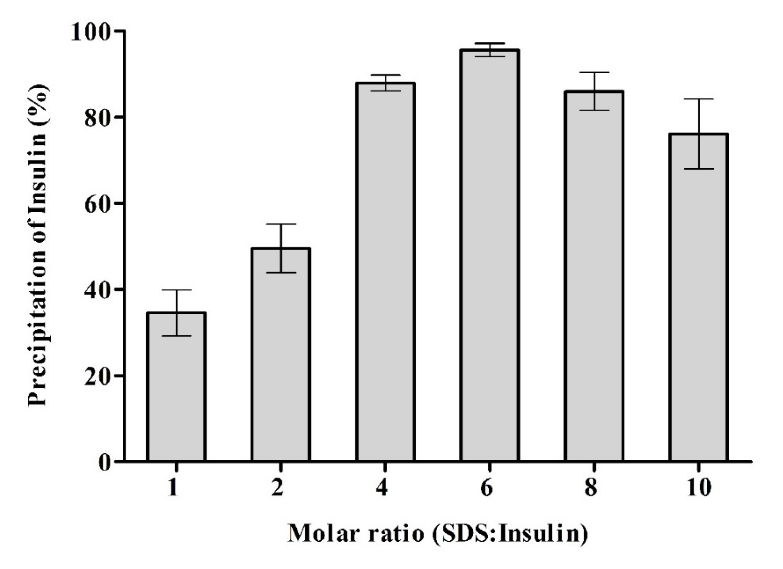
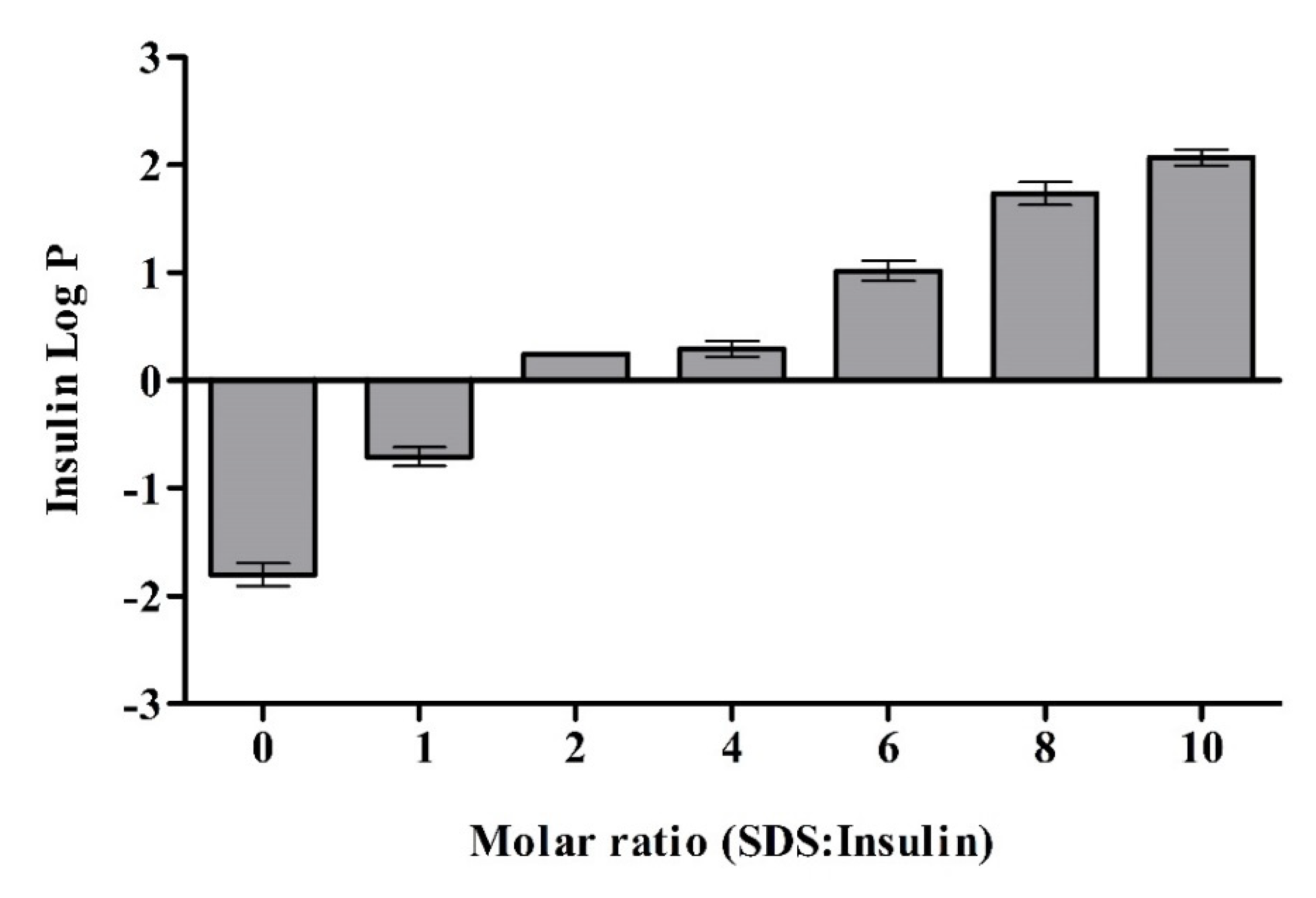
3.1. Synthesis of Insulin INS-SDS Ionic Complex
3.2. NLC Formulations
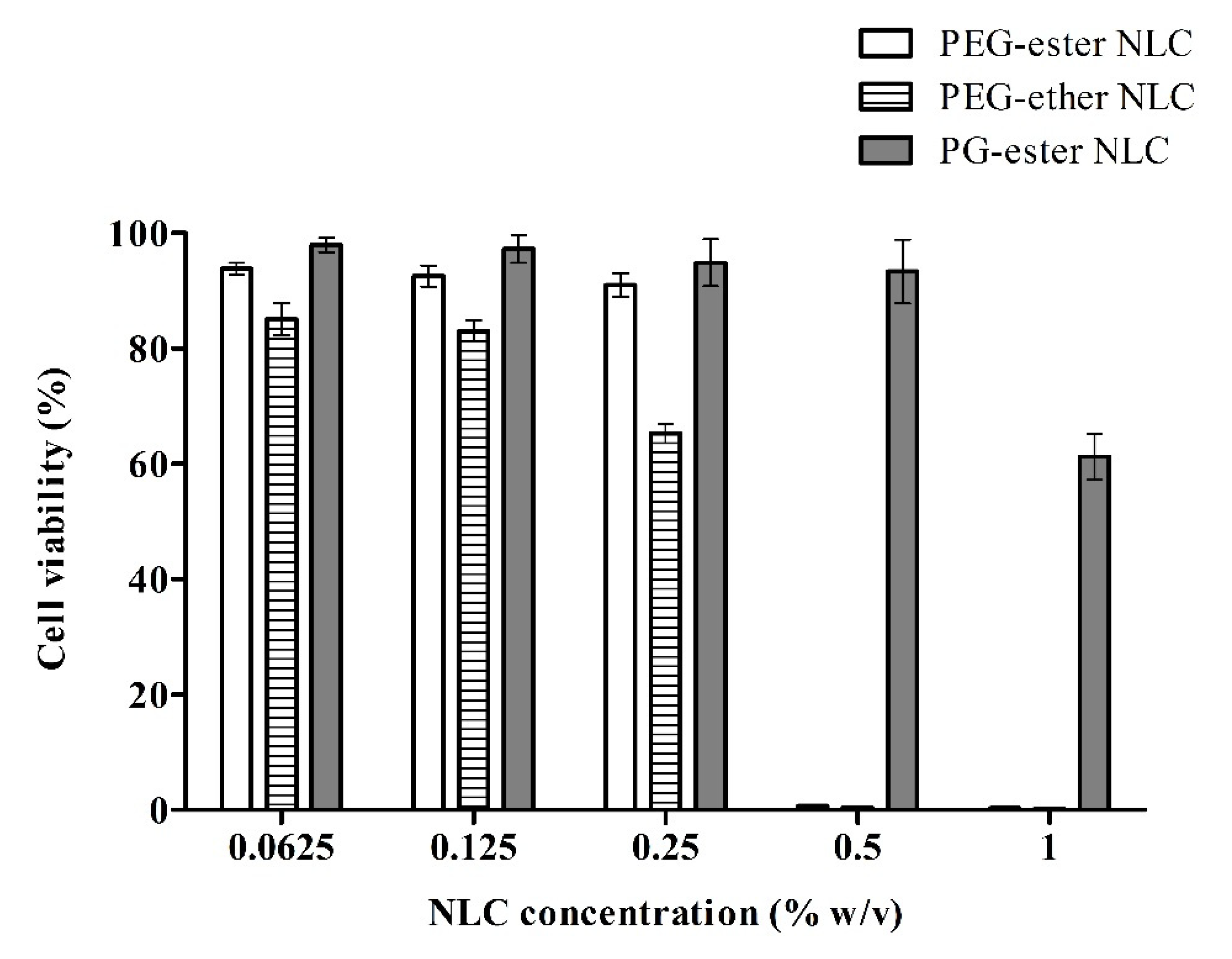
3.3. Cytotoxicity
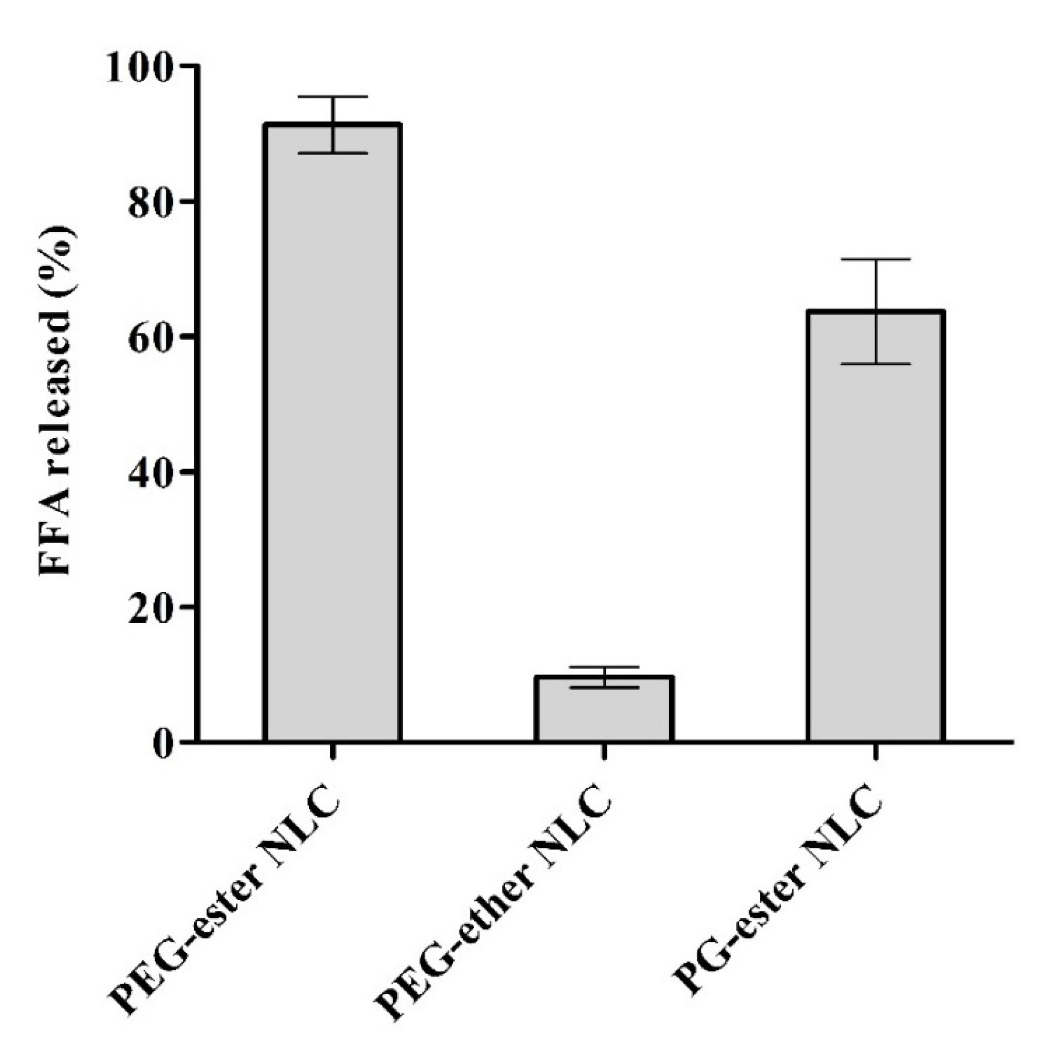
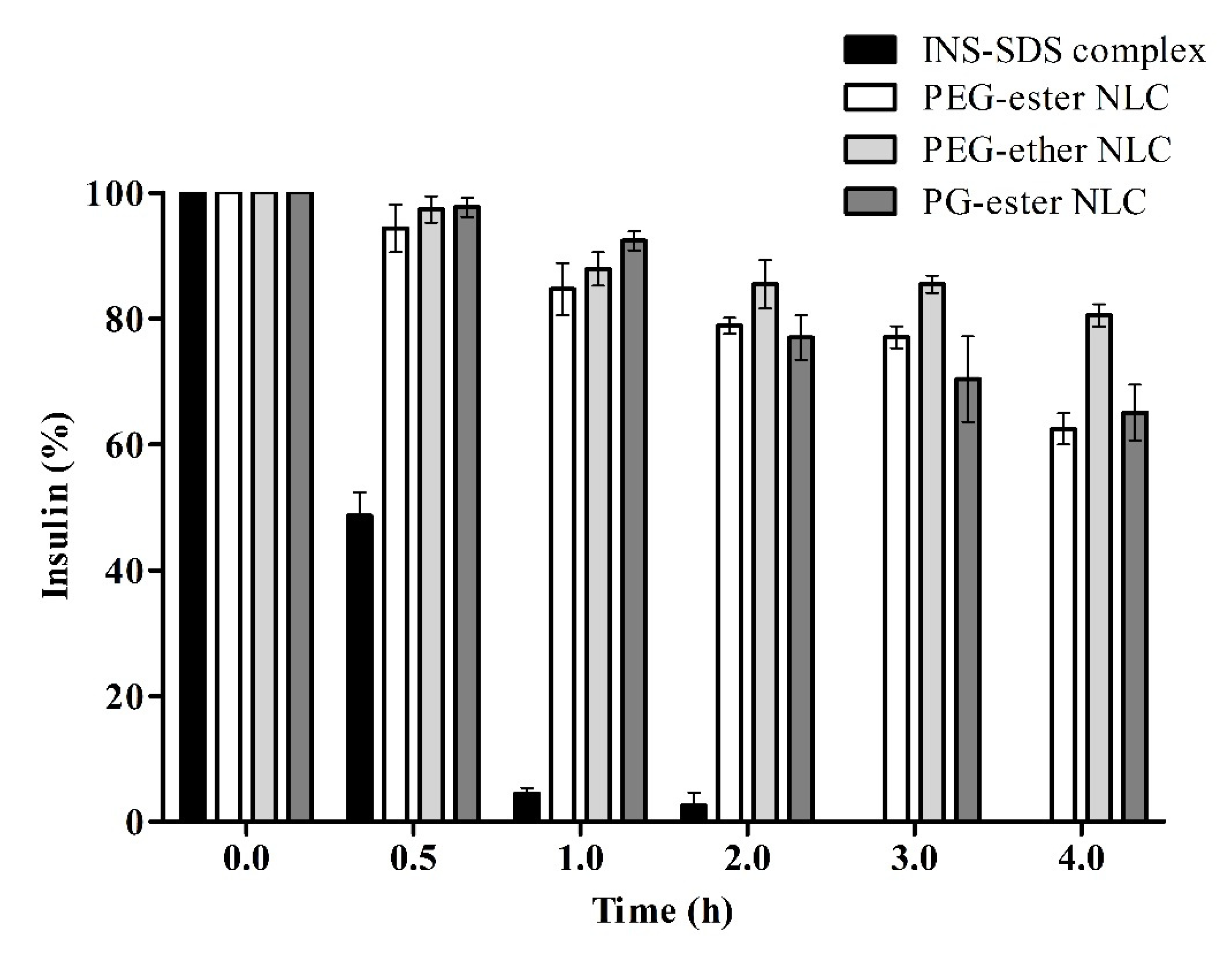
3.4. Lipolysis
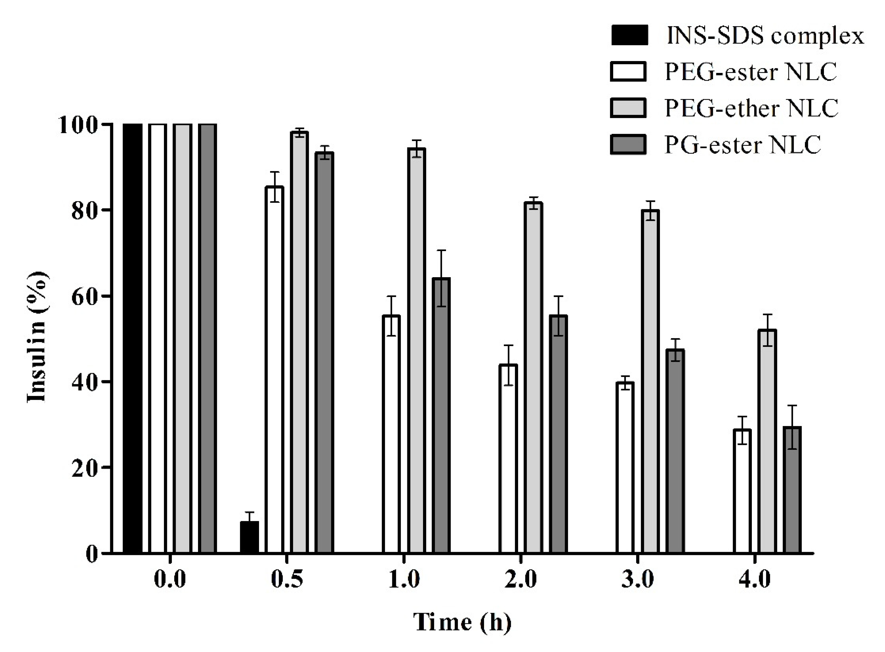
3.5. Proteolysis
4. Conclusions
Author Contributions
Funding
Acknowledgments
Conflicts of Interest
References
- Dumont, C.; Bourgeois, S.; Fessi, H.; Jannin, V. Lipid-based nanosuspensions for oral delivery of peptides, a critical review. Int. J. Pharm. 2018, 541, 117–135. [Google Scholar] [CrossRef] [PubMed]
- Li, P.; Nielsen, H.M.; Müllertz, A. Oral delivery of peptides and proteins using lipid-based drug delivery systems. Expert Opin. Drug Deliv. 2012, 9, 1289–1304. [Google Scholar] [CrossRef] [PubMed]
- Niu, Z.; Conejos-Sánchez, I.; Griffin, B.T.; O’Driscoll, C.M.; Alonso, M.J. Lipid-based nanocarriers for oral peptide delivery. Adv. Drug Deliv. Rev. 2016, 106, 337–354. [Google Scholar] [CrossRef]
- Bonengel, S.; Jelkmann, M.; Abdulkarim, M.; Gumbleton, M.; Reinstadler, V.; Oberacher, H.; Prüfert, F.; Bernkop-Schnürch, A. Impact of different hydrophobic ion pairs of octreotide on its oral bioavailability in pigs. J. Control Release 2018, 273, 21–29. [Google Scholar] [CrossRef]
- Zhang, N.; Ping, Q.N.; Huang, G.H.; Xu, W.F. Investigation of lectin-modified insulin liposomes as carriers for oral administration. Int. J. Pharm. 2005, 294, 247–259. [Google Scholar] [CrossRef]
- Matougui, N.; Boge, L.; Groo, A.-C.; Umerska, A.; Ringstad, L.; Bysell, H.; Saulnier, P. Lipid-based nanoformulations for peptide delivery. Int. J. Pharm. 2016, 502, 80–97. [Google Scholar] [CrossRef] [PubMed]
- Dumont, C.; Jannin, V.; Miolane, C.; Lelong, Q.; Valour, J.-P.; Urbaniak, S.; Fessi, H.; Bourgeois, S. A proof-of-concept for developing oral lipidized peptide nanostructured lipid carrier formulations. J. Drug Deliv. Sci. Technol. 2019, 54, 101394. [Google Scholar] [CrossRef]
- Bernkop-Schnürch, A.; Jalil, A. Do drug release studies from sedds make any sense? J. Control Release 2018, 271, 55–59. [Google Scholar] [CrossRef]
- Mehnert, W.; Mäder, K. Solid lipid nanoparticles: Production, characterization and applications. Adv. Drug Deliv. Rev. 2012, 64, 83–101. [Google Scholar] [CrossRef]
- Martins, S.; Sarmento, B.; Ferreira, D.C.; Souto, E.B. Lipid-based colloidal carriers for peptide and protein delivery—Liposomes versus lipid nanoparticles. Int. J. Nanomed. 2007, 2, 595–607. [Google Scholar]
- Muntoni, E.; Anfossi, L.; Milla, P.; Marini, E.; Ferraris, C.; Capucchio, M.T.; Colombino, E.; Segale, L.; Porta, M.; Battaglia, L. Glargine insulin loaded lipid nanoparticles: Oral delivery of liquid and solid oral dosage forms. Nutr. Metab. Cardiovasc. Dis. 2021, 31, 691–698. [Google Scholar] [CrossRef]
- Olbrich, C.; Müller, R. Enzymatic degradation of sln—Effect of surfactant and surfactant mixtures. Int. J. Pharm. 1999, 180, 31–39. [Google Scholar] [CrossRef]
- Nazir, I.; Asim, M.H.; Dizdarević, A.; Bernkop-Schnürch, A. Self-emulsifying drug delivery systems: Impact of stability of hydrophobic ion pairs on drug release. Int. J. Pharm. 2019, 561, 197–205. [Google Scholar] [CrossRef]
- Griesser, J.; Hetényi, G.; Moser, M.; Demarne, F.; Jannin, V.; Bernkop-Schnürch, A. Hydrophobic ion pairing: Key to highly payloaded self-emulsifying peptide drug delivery systems. Int. J. Pharm. 2017, 520, 267–274. [Google Scholar] [CrossRef]
- Hu, F.Q.; Jiang, S.P.; Du, Y.Z.; Yuan, H.; Ye, Y.Q.; Zeng, S. Preparation and characteristics of monostearin nanostructured lipid carriers. Int. J. Pharm. 2006, 314, 83–89. [Google Scholar] [CrossRef]
- Shahzadi, I.; Asim, M.H.; Dizdarević, A.; Wolf, J.D.; Kurpiers, M.; Matuszczak, B.; Bernkop-Schnürch, A. Arginine-based cationic surfactants: Biodegradable auxiliary agents for the formation of hydrophobic ion pairs with hydrophilic macromolecular drugs. J. Colloid Interface Sci. 2019, 552, 287–294. [Google Scholar] [CrossRef] [PubMed]
- Tan, A.; Simovic, S.; Davey, A.K.; Rades, T.; Boyd, B.J.; Prestidge, C.A. Silica nanoparticles to control the lipase-mediated digestion of lipid-based oral delivery systems. Mol. Pharm. 2010, 7, 522–532. [Google Scholar] [CrossRef] [PubMed]
- Wang, J.; Yadav, V.; Smart, A.L.; Tajiri, S.; Basit, A.W. Toward oral delivery of biopharmaceuticals: An assessment of the gastrointestinal stability of 17 peptide drugs. Mol. Pharm. 2015, 12, 966–973. [Google Scholar] [CrossRef] [PubMed] [Green Version]
- Shahzadi, I.; Nazir, I.; Nhu Quynh Phan, T.; Bernkop-Schnürch, A. About the impact of superassociation of hydrophobic ion pairs on membrane permeability. Eur. J. Pharm. Biopharm. 2020, 151, 1–8. [Google Scholar] [CrossRef]
- Shahzadi, I.; Dizdarević, A.; Efiana, N.A.; Matuszczak, B.; Bernkop-Schnürch, A. Trypsin decorated self-emulsifying drug delivery systems (sedds): Key to enhanced mucus permeation. J. Colloid Interface Sci. 2018, 531, 253–260. [Google Scholar] [CrossRef]
- Yoon, G.; Park, J.W.; Yoon, I.-S. Solid lipid nanoparticles (slns) and nanostructured lipid carriers (nlcs): Recent advances in drug delivery. J. Pharm. Investig. 2013, 43, 353–362. [Google Scholar] [CrossRef]
- Üner, M. Preparation, characterization and physico-chemical properties of solid lipid nanoparticles (sln) and nanostructured lipid carriers (nlc): Their benefits as colloidal drug carrier systems. Pharmazie 2006, 61, 375–386. [Google Scholar]
- Bekard, I.B.; Asimakis, P.; Bertolini, J.; Dunstan, D.E. The effects of shear flow on protein structure and function. Biopolymers 2011, 95, 733–745. [Google Scholar] [CrossRef]
- Bekard, I.B.; Dunstan, D.E. Shear-induced deformation of bovine insulin in couette flow. J. Phys. Chem. B 2009, 113, 8453–8457. [Google Scholar] [CrossRef]
- Beloqui, A.; Solinís, M.Á.; Rodríguez-Gascón, A.; Almeida, A.J.; Préat, V. Nanostructured lipid carriers: Promising drug delivery systems for future clinics. Nanomed. Nanotechnol. Biol. Med. 2016, 12, 143–161. [Google Scholar] [CrossRef] [PubMed]
- Shah, N.V.; Seth, A.K.; Balaraman, R.; Aundhia, C.J.; Maheshwari, R.A.; Parmar, G.R. Nanostructured lipid carriers for oral bioavailability enhancement of raloxifene: Design and in vivo study. J. Adv. Res. 2016, 7, 423–434. [Google Scholar] [CrossRef] [PubMed] [Green Version]
- Hu, F.-Q.; Jiang, S.-P.; Du, Y.-Z.; Yuan, H.; Ye, Y.-Q.; Zeng, S. Preparation and characterization of stearic acid nanostructured lipid carriers by solvent diffusion method in an aqueous system. Colloids Surf. B Biointerfaces 2005, 45, 167–173. [Google Scholar] [CrossRef]
- Leonaviciute, G.; Zupančič, O.; Prüfert, F.; Rohrer, J.; Bernkop-Schnürch, A. Impact of lipases on the protective effect of sedds for incorporated peptide drugs towards intestinal peptidases. Int. J. Pharm. 2016, 508, 102–108. [Google Scholar] [CrossRef] [PubMed]
- Simon-Assmann, P.; Turck, N.; Sidhoum-Jenny, M.; Gradwohl, G.; Kedinger, M. In vitro models of intestinal epithelial cell differentiation. Cell Biol. Toxicol. 2007, 23, 241–256. [Google Scholar] [CrossRef] [PubMed]
- Kauffman, A.L.; Gyurdieva, A.V.; Mabus, J.R.; Ferguson, C.; Yan, Z.; Hornby, P.J. Alternative functional in vitro models of human intestinal epithelia. Front. Pharmacol. 2013, 4, 79. [Google Scholar] [CrossRef] [PubMed] [Green Version]
- Shahzadi, I.; Jalil, A.; Asim, M.H.; Hupfauf, A.; Gust, R.; Nelles, P.A.; Knabl, L.; Bernkop-Schnürch, A. Lipophilic arginine esters: The gateway to preservatives without side effects. Mol. Pharm. 2020, 17, 3129–3139. [Google Scholar] [CrossRef]
- Zahir-Jouzdani, F.; Lupo, N.; Hermann, M.; Prüfert, F.; Atyabi, F.; Bernkop Schnürch, A. Glyceryl ester surfactants: Promising excipients to enhance the cell permeating properties of sedds. Eur. J. Pharm. Biopharm. 2018, 129, 154–161. [Google Scholar] [CrossRef]
- Boethling, R.; Sommer, E.; DiFiore, D. Designing small molecules for biodegradability. Chem. Rev. 2007, 107, 2207–2227. [Google Scholar] [CrossRef] [Green Version]
- Liu, J.; Hirschberg, C.; Fanø, M.; Mu, H.; Müllertz, A. Evaluation of self-emulsifying drug delivery systems for oral insulin delivery using an in vitro model simulating the intestinal proteolysis. Eur. J. Pharm. Sci. 2020, 147, 105272. [Google Scholar] [CrossRef]
- Li, P.; Ford, L.; Haque, S.; McInerney, M.P.; Williams, H.D.; Scammells, P.J.; Thompson, P.E.; Jannin, V.; Porter, C.J.; Benameur, H. Lipophilic salts and lipid-based formulations: Enhancing the oral delivery of octreotide. Pharm. Res. 2021, 38, 1125–1137. [Google Scholar] [CrossRef]
- Bakala-N’Goma, J.-C.; Williams, H.D.; Sassene, P.J.; Kleberg, K.; Calderone, M.; Jannin, V.; Igonin, A.; Partheil, A.; Marchaud, D.; Jule, E. Toward the establishment of standardized in vitro tests for lipid-based formulations. 5. Lipolysis of representative formulations by gastric lipase. Pharm. Res. 2015, 32, 1279–1287. [Google Scholar] [CrossRef]
- Dening, T.J.; Joyce, P.; Rao, S.; Thomas, N.; Prestidge, C.A. Nanostructured montmorillonite clay for controlling the lipase-mediated digestion of medium chain triglycerides. ACS Appl. Mater. Interfaces 2016, 8, 32732–32742. [Google Scholar] [CrossRef]
- Arnold, Y.E.; Imanidis, G.; Kuentz, M. In vitro digestion kinetics of excipients for lipid-based drug delivery and introduction of a relative lipolysis half life. Drug Dev. Ind. Pharm. 2012, 38, 1262–1269. [Google Scholar] [CrossRef] [PubMed]
- Malhaire, H.; Gimel, J.-C.; Roger, E.; Benoît, J.-P.; Lagarce, F. How to design the surface of peptide-loaded nanoparticles for efficient oral bioavailability? Adv. Drug Deliv. Rev. 2016, 106, 320–336. [Google Scholar] [CrossRef] [PubMed]
- Christiansen, A.; Backensfeld, T.; Weitschies, W. Effects of non-ionic surfactants on in vitro triglyceride digestion and their susceptibility to digestion by pancreatic enzymes. Eur. J. Pharm. Sci. 2010, 41, 376–382. [Google Scholar] [CrossRef] [PubMed]
- Ansari, M.J.; Anwer, M.K.; Jamil, S.; Al-Shdefat, R.; Ali, B.E.; Ahmad, M.M.; Ansari, M.N. Enhanced oral bioavailability of insulin-loaded solid lipid nanoparticles: Pharmacokinetic bioavailability of insulin-loaded solid lipid nanoparticles in diabetic rats. Drug Deliv. 2016, 23, 1972–1979. [Google Scholar] [CrossRef] [PubMed] [Green Version]
- Qi, J.; Zhuang, J.; Lu, Y.; Dong, X.; Zhao, W.; Wu, W. In vivo fate of lipid-based nanoparticles. Drug Discov. Today 2017, 22, 166–172. [Google Scholar] [CrossRef] [PubMed]


| NLCs | Constituents | S:L Ratio | Surfactant (% w/v) | |
|---|---|---|---|---|
| PEG-ester NLC | Solid lipid (S) | Stearic acid | 70:30 | 1.0 |
| Liquid lipid (L) | Glyceryl monooleate (Type 40) NF (PeceolTM) | |||
| Surfactant | Polyoxyl hydrogenated castor oil (Kolliphor® RH 40) | |||
| PEG-ether NLC | Solid lipid (S) | Stearic acid | 70:30 | 1.0 |
| Liquid lipid (L) | Oleic acid | |||
| Surfactant | Polyoxyethylene-23-laurylether (BrijTM L23) | |||
| PG-ester NLC | Solid lipid (S) | Stearic acid | 70:30 | 1.0 |
| Liquid lipid (L) | Glyceryl monooleate (Type 40) NF (PeceolTM) | |||
| Surfactants | Polyglyceryl-4 Caprate (Tegosoft® PC41) and Polyglyceryl-6 Caprylate (Tegosolve® 90MB) | |||
| NLCs | INS-SDS Having Been Added during Preparation Process (mg) | Entrapment Efficacy (%) |
|---|---|---|
| PEG-ester NLC | 10 | 61.5 ± 2.2 |
| PEG-ether NLC | 10 | 58.6 ± 3.0 |
| PG-ester NLC | 10 | 73.0 ± 6.3 |
| Formulations | Blank NLCs | INS-SDS Complex Loaded NLCs | ||||
|---|---|---|---|---|---|---|
| Parameters | Size (nm) | PDI | Zeta Potential (mV) | Size (nm) | PDI | Zeta Potential (mV) |
| PEG-ester NLC | 64.3 ± 2.6 | 0.5 | −11.3 | 99.3 ± 1.3 | 0.4 | −17.4 |
| PEG-ether NLC | 143.4 ± 17.0 | 0.5 | −20.8 | 134.4 ± 3.3 | 0.2 | −19.6 |
| PG-ester NLC | 121.8 ± 3.9 | 0.2 | −2.2 | 217.3 ± 8.4 | 0.3 | −18.3 |
Publisher’s Note: MDPI stays neutral with regard to jurisdictional claims in published maps and institutional affiliations. |
© 2021 by the authors. Licensee MDPI, Basel, Switzerland. This article is an open access article distributed under the terms and conditions of the Creative Commons Attribution (CC BY) license (https://creativecommons.org/licenses/by/4.0/).
Share and Cite
Shahzadi, I.; Fürst, A.; Knoll, P.; Bernkop-Schnürch, A. Nanostructured Lipid Carriers (NLCs) for Oral Peptide Drug Delivery: About the Impact of Surface Decoration. Pharmaceutics 2021, 13, 1312. https://doi.org/10.3390/pharmaceutics13081312
Shahzadi I, Fürst A, Knoll P, Bernkop-Schnürch A. Nanostructured Lipid Carriers (NLCs) for Oral Peptide Drug Delivery: About the Impact of Surface Decoration. Pharmaceutics. 2021; 13(8):1312. https://doi.org/10.3390/pharmaceutics13081312
Chicago/Turabian StyleShahzadi, Iram, Andrea Fürst, Patrick Knoll, and Andreas Bernkop-Schnürch. 2021. "Nanostructured Lipid Carriers (NLCs) for Oral Peptide Drug Delivery: About the Impact of Surface Decoration" Pharmaceutics 13, no. 8: 1312. https://doi.org/10.3390/pharmaceutics13081312