
Nanoemulsions: A Review on the Conceptualization of Treatment for Psoriasis Using a ‘Green’ Surfactant with Low-Energy Emulsification Method


Abstract
:1. Introduction
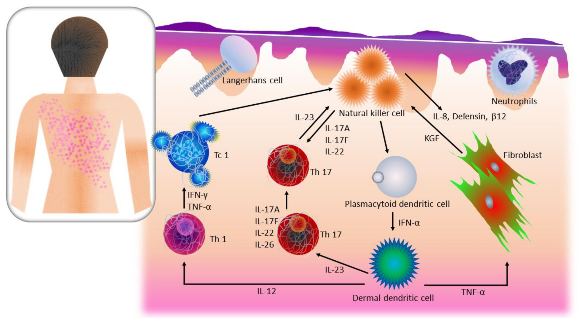
2. Pathogenic Pathways of Psoriasis
2.1. T Cell Activation, Cytokine Release, and Over-Proliferation of Keratinocytes in Psoriasis
2.2. Natural Killer Cells in Psoriasis
2.3. Nuclear Factor-Kappa B in Psoriasis
2.4. Langerhans’ Cells in Psoriasis
3. Current Psoriatic Treatments
4. Nanoemulsions as a Topical Drug Delivery System
4.1. Definition of Nanoemulsion
4.2. Components of Nanoemulsion
4.3. Physicochemical Characteristics of Nanoemulsion
4.4. Selection of Oil Phase
4.5. Selection of Biosurfactants
5. Formation of Low-Energy Nanoemulsions
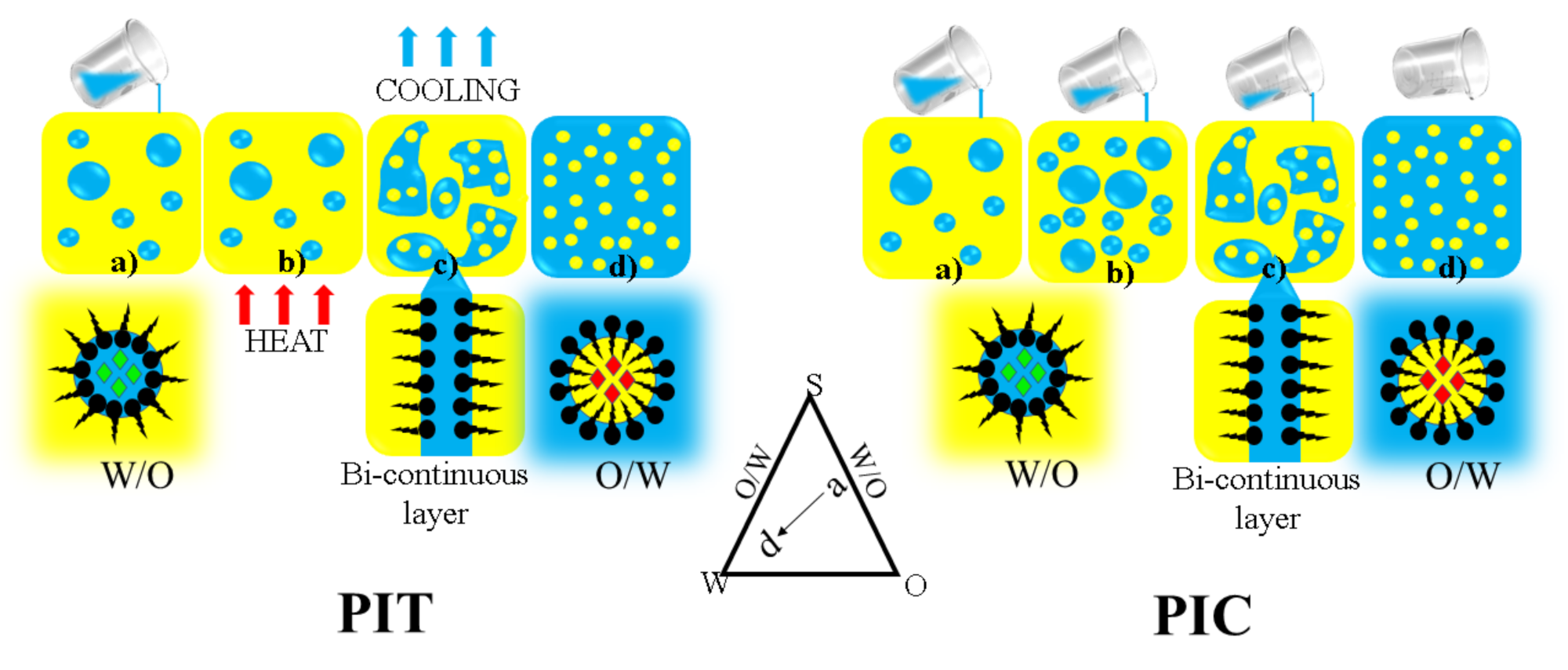
5.1. Phase Inversion Emulsification
5.2. Spontaneous Emulsification
5.3. Self-Nanoemulsifying Drug Delivery Systems
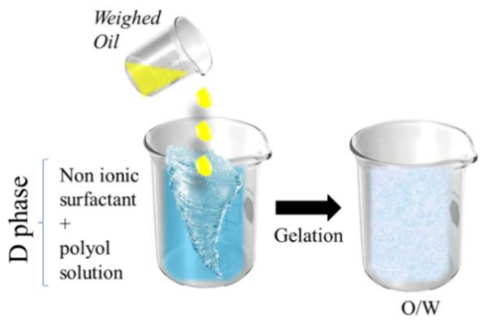
5.4. D-Phase Emulsification
6. The Recent Development of Nanoemulsions as a Treatment for Psoriasis
7. Conclusions
Author Contributions
Funding
Institutional Review Board Statement
Informed Consent Statement
Data Availability Statement
Conflicts of Interest
References
- Cowden, A.; Van Voorhees, A.S. Treatment of Psoriasis. In Introduction: History of Psoriasis and Psoriasis; Weinberg, J.M., Ed.; Birkhäuser: Basel, Swetzerland, 2008; pp. 1–9. [Google Scholar] [CrossRef]
- Rendon, A.; Schäkel, K. Psoriasis Pathogenesis and Treatment. Int. J. Mol. Sci. 2019, 20, 1475. [Google Scholar] [CrossRef] [PubMed] [Green Version]
- Affandi, A.M.; Khan, I.; Saaya, N.N. Epidemiology and Clinical Features of Adult Patients with Psoriasis in Malaysia: 10-Year Review from the Malaysian Psoriasis Registry (2007–2016). Dermatol. Res. Pract. 2018, 2018, 1–8. [Google Scholar] [CrossRef] [PubMed] [Green Version]
- Krueger, J.G.; Bowcock, A. Psoriasis pathophysiology: Current concepts of pathogenesis. Ann. Rheum. Dis. 2005, 64, ii30–ii36. [Google Scholar] [CrossRef] [PubMed]
- Salim, N.; Ahmad, N.; Musa, S.H.; Hashim, R.; Tadros, T.F.; Basri, M. Nanoemulsion as a topical delivery system of antipsoriatic drugs. RSC Adv. 2016, 6, 6234–6250. [Google Scholar] [CrossRef]
- Sarkar, S. A Treatise on Topical Corticosteroid in Dermatology. Indian J. Dermatol. 2018, 63, 530–531. [Google Scholar]
- Campalani, E.; Allen, M.H.; Fairhurst, D.; Young, H.; Mendonca, C.O.; Burden, A.D.; Griffiths, C.; Crook, M.A.; Barker, J.N.W.N.; Smith, C. Apolipoprotein E gene polymorphisms are associated with psoriasis but do not determine disease response to acitretin. Br. J. Dermatol. 2005, 154, 345–352. [Google Scholar] [CrossRef]
- Warren, R.B.; Griffiths, C.E. Systemic therapies for psoriasis: Methotrexate, retinoids, and cyclosporine. Clin. Dermatol. 2008, 26, 438–447. [Google Scholar] [CrossRef]
- Thami, G.P.; Sarkar, R. Coal tar: Past, present and future. Clin. Exp. Dermatol. 2002, 27, 99–103. [Google Scholar] [CrossRef]
- Raza, K.; Kumar, M.; Kumar, P.; Malik, R.; Sharma, G.; Kaur, M.; Katare, O.P. Topical Delivery of Aceclofenac: Challenges and Promises of Novel Drug Delivery Systems. BioMed Res. Int. 2014, 2014, 1–11. [Google Scholar] [CrossRef]
- Pradhan, M.; Singh, D.; Singh, M.R. Novel colloidal carriers for psoriasis: Current issues, mechanistic insight and novel delivery approaches. J. Control. Release 2013, 170, 380–395. [Google Scholar] [CrossRef]
- Chen, J.; Wan, Z. Controllable synthesis of lanthanide upconversion nanomaterials through impurity doping. In Fabrication and Self-Assembly of Nanobiomaterials; Grumezescu, A.M., Ed.; William Andrew Publishing: Norwich, NY, USA, 2016. [Google Scholar] [CrossRef]
- Solans, C.; Izquierdo, P.; Nolla, J.; Azemar, N.; García-Celma, M.-J. Nano-emulsions. Curr. Opin. Colloid Interface Sci. 2005, 10, 102–110. [Google Scholar] [CrossRef]
- Nafisi, S.; Maibach, H.I. Basic Physical Sciences for the Formulation of Cosmetic Products. In Cosmetic Science and Technology; Sakamoto, K., Lochhead, H., Maibach, H., Yamashita, Y., Eds.; Elsevier: Amsterdam, The Netherlands, 2017; pp. 337–369. ISBN 9780128020548. [Google Scholar]
- Mason, T.G.; Wilking, J.N.; Meleson, K.; Chang, C.B.; Graves, S.M. Nanoemulsions: Formation, structure, and physical properties. J. Phys. Condens. Matter 2006, 18, R635–R666. [Google Scholar] [CrossRef] [Green Version]
- Fakruddin. Biosurfactant: Production and Application. J. Pet. Environ. Biotechnol. 2012, 3, 1–6. [Google Scholar] [CrossRef] [Green Version]
- Das, R.P.; Jain, A.K.; Ramesh, V. Current concepts in the pathogenesis of psoriasis. Indian J. Dermatol. 2009, 54, 7–12. [Google Scholar] [CrossRef]
- Gahan, P.B. The Adaptive Immune System. In Molecular Biology of the Cell, 4th ed.; Alberts, B., Johnson, A., Lewis, J., Raff, M., Roberts, K., Walter, P., Eds.; Garland Science Inc.: New York, NY, USA, 2002; ISBN-10: 0-8153-4072-9. [Google Scholar]
- Sérézal, I.G.; Classon, C.; Cheuk, S.; Barrientos-Somarribas, M.; Wadman, E.; Martini, E.; Chang, D.; Landén, N.X.; Ehrström, M.; Nylén, S.; et al. Resident T Cells in Resolved Psoriasis Steer Tissue Responses that Stratify Clinical Outcome. J. Investig. Dermatol. 2018, 138, 1754–1763. [Google Scholar] [CrossRef] [Green Version]
- Cosmi, L.; Liotta, F.; Maggi, E.; Romagnani, S.; Annunziato, F. Th17 and Non-Classic Th1 Cells in Chronic Inflammatory Disorders: Two Sides of the Same Coin. Int. Arch. Allergy Immunol. 2014, 164, 171–177. [Google Scholar] [CrossRef]
- Zaba, L.C.; Fuentes-Duculan, J.; Eungdamrong, N.J.; Abello, M.V.; Novitskaya, I.; Pierson, K.C.; Gonzalez, J.; Krueger, J.G.; Lowes, M.A. Psoriasis Is Characterized by Accumulation of Immunostimulatory and Th1/Th17 Cell-Polarizing Myeloid Dendritic Cells. J. Investig. Dermatol. 2009, 129, 79–88. [Google Scholar] [CrossRef] [Green Version]
- Zaba, L.C.; Krueger, J.G.; Lowes, M.A. Resident and “Inflammatory” Dendritic Cells in Human Skin. J. Investig. Dermatol. 2009, 129, 302–308. [Google Scholar] [CrossRef] [Green Version]
- Mackay, L.K.; Kallies, A. Transcriptional Regulation of Tissue-Resident Lymphocytes. Trends Immunol. 2017, 38, 94–103. [Google Scholar] [CrossRef]
- Cheuk, S.; Wikén, M.; Blomqvist, L.; Nylén, S.; Talme, T.; Ståhle, M.; Eidsmo, L. Epidermal Th22 and Tc17 Cells Form a Localized Disease Memory in Clinically Healed Psoriasis. J. Immunol. 2014, 192, 3111–3120. [Google Scholar] [CrossRef] [Green Version]
- Gottlieb, A.B. Therapeutic options in the treatment of psoriasis and atopic dermatitis. J. Am. Acad. Dermatol. 2005, 53, S3–S16. [Google Scholar] [CrossRef] [PubMed]
- Lande, R.; Botti, E.; Jandus, C.; Dojcinovic, D.; Fanelli, G.; Conrad, C.; Chamilos, G.; Feldmeyer, L.; Marinari, B.; Chon, S.; et al. The antimicrobial peptide LL37 is a T-cell autoantigen in psoriasis. Nat. Commun. 2014, 5, 5621. [Google Scholar] [CrossRef] [PubMed]
- Lande, R.; Chamilos, G.; Ganguly, D.; Demaria, O.; Frasca, L.; Durr, S.; Conrad, C.; Schröder, J.; Gilliet, M. Cationic antimicrobial peptides in psoriatic skin cooperate to break innate tolerance to self-DNA. Eur. J. Immunol. 2014, 45, 203–213. [Google Scholar] [CrossRef] [PubMed]
- Conrad, C.; Gilliet, M. Psoriasis: From Pathogenesis to Targeted Therapies. Clin. Rev. Allergy Immunol. 2018, 54, 102–113. [Google Scholar] [CrossRef]
- Di Meglio, P.; Villanova, F.; Navarini, A.A.; Mylonas, A.; Tosi, I.; Nestle, F.O.; Conrad, C. Targeting CD8+ T cells prevents psoriasis development. J. Allergy Clin. Immunol. 2016, 138, 274–276.e6. [Google Scholar] [CrossRef] [Green Version]
- Brilot, F.; Strowig, T. NK cells interactions with dendritic cells shape innate and adaptive immunity. Front. Biosci. 2008, 13, 6443–6454. [Google Scholar] [CrossRef] [Green Version]
- Marshall, J.D.; Heeke, D.S.; Abbate, C.; Yee, P.; Van Nest, G. Induction of interferon-gamma from natural killer cells by immunostimulatory CpG DNA is mediated through plasmacytoid-dendritic-cell-produced interferon-alpha and tumour necrosis factor-alpha. Immunology 2006, 117, 38–46. [Google Scholar] [CrossRef]
- Kryczek, I.; Bruce, A.T.; Gudjonsson, J.E.; Johnston, A.; Aphale, A.; Vatan, L.; Szeliga, W.; Wang, Y.; Liu, Y.; Welling, T.H.; et al. Induction of IL-17+ T Cell Trafficking and Development by IFN-γ: Mechanism and Pathological Relevance in Psoriasis. J. Immunol. 2008, 181, 4733–4741. [Google Scholar] [CrossRef] [Green Version]
- Elewaut, D.; Kronenberg, M. Molecular biology of NK T cell specificity and development. Semin. Immunol. 2000, 12, 561–568. [Google Scholar] [CrossRef]
- Bonish, B.; Jullien, D.; Dutronc, Y.; Huang, B.B.; Modlin, R.; Spada, F.M.; Porcelli, S.A.; Nickoloff, B.J. Overexpression of CD1d by Keratinocytes in Psoriasis and CD1d-Dependent IFN-γ Production by NK-T Cells. J. Immunol. 2000, 165, 4076–4085. [Google Scholar] [CrossRef] [Green Version]
- Kirby, O.A.-J.B.; Al-Jiffri, O. Investigation of Cytomegalovirus and Human Herpes Viruses 6 and 7 as Possible Causative Antigens in Psoriasis. Acta Derm. Venereol. 2000, 80, 404–406. [Google Scholar] [CrossRef] [Green Version]
- Tsuruta, D. NF-κB Links Keratinocytes and Lymphocytes in the Pathogenesis of Psoriasis. Recent Patents Inflamm. Allergy Drug Discov. 2009, 3, 40–48. [Google Scholar] [CrossRef]
- Begon, E.; Michel, L.; Flageul, B.; Beaudoin, I.; Jean-Louis, F.; Bachelez, H.; Dubertret, L.; Musette, P. Expression, subcellular localization and cytokinic modulation of Toll-like receptors (TLRs) in normal human keratinocytes: TLR2 up-regulation in psoriatic skin. Eur. J. Dermatol. 2007, 17, 497–506. [Google Scholar] [CrossRef]
- Salskov-Iversen, M.L.; Johansen, C.; Kragballe, K.; Iversen, L. Caspase-5 Expression Is Upregulated in Lesional Psoriatic Skin. J. Investig. Dermatol. 2011, 131, 670–676. [Google Scholar] [CrossRef] [Green Version]
- Raj, D.; Brash, D.E.; Grossman, D. Keratinocyte Apoptosis in Epidermal Development and Disease. J. Investig. Dermatol. 2006, 126, 243–257. [Google Scholar] [CrossRef] [Green Version]
- Ghosh, S.; Hayden, M. Celebrating 25 years of NF-κB research. Immunol. Rev. 2012, 246, 5–13. [Google Scholar] [CrossRef]
- Abdou, A.G.; Hanout, H.M. Evaluation of survivin and NF-κB in psoriasis, an immunohistochemical study. J. Cutan. Pathol. 2008, 35, 445–451. [Google Scholar] [CrossRef]
- Romani, N.; Clausen, B.E.; Stoitzner, P. Langerhans cells and more: Langerin-expressing dendritic cell subsets in the skin. Immunol. Rev. 2010, 234, 120–141. [Google Scholar] [CrossRef] [Green Version]
- Fujita, H.; Nograles, K.E.; Kikuchi, T.; Gonzalez, J.; Carucci, J.; Krueger, J.G. Human Langerhans cells induce distinct IL-22-producing CD4+ T cells lacking IL-17 production. Proc. Natl. Acad. Sci. USA 2009, 106, 21795–21800. [Google Scholar] [CrossRef] [Green Version]
- Feldman, S.R.; Goffe, B. The Challenge of Managing Psoriasis: Unmet Medical Needs and Stakeholder Perspectives. Am. Health Drug Benefits 2016, 9, 504–513. [Google Scholar]
- Luger, T.A.; Cambazard, F.; Larsen, F.G.; Bourcier, M.; Gupta, G.; Clonier, F.; Kidson, P.; Shear, N.H. A Study of the Safety and Efficacy of Calcipotriol and Betamethasone Dipropionate Scalp Formulation in the Long-Term Management of Scalp Psoriasis. Dermatology 2008, 217, 321–328. [Google Scholar] [CrossRef] [Green Version]
- Weiss, S.C.; Nguyen, J.; Chon, S.; Kimball, A.B. A randomized controlled clinical trial assessing the effect of betamethasone valerate 0.12% foam on the short-term treatment of stasis dermatitis. J. Drugs. Dermatol. 2005, 4, 339–345. [Google Scholar] [PubMed]
- Aggarwal, P.; Aggarwal, K.; Jain, V.K. Tacalcitol: A useful adjunct to narrow band ultraviolet B phototherapy in psoriasis. J Dermatolog Treat. 2016, 27, 546–551. [Google Scholar] [CrossRef] [PubMed]
- Colombo, G.L.; Di Matteo, S.; Bruno, G.; Girolomoni, G.; Vena, G.A. Calcipotriol and betamethasone dipropionate in the treatment of mild-to-moderate psoriasis: A cost-effectiveness analysis of the ointment versus gel formulation. Clinicoecon Outcomes Res. 2012, 4, 261–268. [Google Scholar] [CrossRef] [PubMed] [Green Version]
- Mrowietz, U.; Mrowietz, M.; Christoph, O.; Christoph, E. Effective treatment and improvement of quality of life in patients with scalp psoriasis by topical use of calcipotriol/betamethasone (Xamiol® Gel). J. Ger. Soc. Dermatol. JDDG 2011, 9, 825–831. [Google Scholar] [CrossRef] [PubMed]
- Gooderham, M.; Debarre, J.; Keddy-Grant, J.; Xu, Z.; Kurvits, M.; Goodfield, M. Safety and efficacy of calcipotriol plus betamethasone dipropionate gel in the treatment of scalp psoriasis in adolescents 12–17 years of age. Br. J. Dermatol. 2014, 171, 1470–1477. [Google Scholar] [CrossRef]
- Langley, R.G.; Gupta, A.; Papp, K.; Wexler, D.; Østerdal, M.L.; Çurčić, D. Calcipotriol plus Betamethasone Dipropionate Gel Compared with Tacalcitol Ointment and the Gel Vehicle Alone in Patients with Psoriasis Vulgaris: A Randomized, Controlled Clinical Trial. Dermatology 2011, 222, 148–156. [Google Scholar] [CrossRef]
- Puig, L.; Ribera, M.; Hernanz, J.M.; Belinchón, I.; Santos-Juanes, J.; Linares, M.; Querol, I.; Colomé, E.; Caballé, G. Treatment of Scalp Psoriasis: Review of the Evidence and Delphi Consensus of the Psoriasis Group of the Spanish Academy of Dermatology and Venereology. Actas Dermo-Sifiliográficas (Engl. Ed.) 2010, 101, 827–846. [Google Scholar] [CrossRef]
- Sekhon, S.; Jeon, C.; Nakamura, M.; Afifi, L.; Yan, D.; Wu, J.J.; Liao, W.; Bhutani, T. Review of the mechanism of action of coal tar in psoriasis. J. Dermatolog. Treat. 2018; 29, 230–232. [Google Scholar] [CrossRef]
- Saraceno, R.; Camplone, G.; D’Agostino, M.; de Simone, C.; Di Cesare, A.; Filosa, G.; Frascione, P.; Gabellini, M.; Lunghi, F.; Mazzotta, A.; et al. Efficacy and maintenance strategies of two-compound formulation calcipotriol and betamethasone dipropionate gel (Xamiol® gel) in the treatment of scalp psoriasis: Results from a study in 885 patients. J. Dermatol. Treat. 2013, 25, 30–33. [Google Scholar] [CrossRef]
- Williams, H. Exorex for Psoriasis: The Importance of Randomized Controlled Trials in Testing "New" Products. Arch. Dermatol. 2002, 137, 1637–1638. [Google Scholar] [CrossRef]
- Mason, A.R.; Mason, J.; Cork, M.; Dooley, G.; Hancock, H. Topical treatments for chronic plaque psoriasis. Cochrane Database Syst. Rev. 2013, 3. [Google Scholar] [CrossRef] [Green Version]
- Basrai, S.; Wright, S.; Patel, G.K. Psoriasis. InnovAiT 2018, 11, 413–419. [Google Scholar] [CrossRef]
- Higgins, E. Psoriasis. Medicine 2017, 45, 368–378. [Google Scholar] [CrossRef]
- Van Onselen, J. Prescribing for mild-to-moderate psoriasis in adults. Nurse Prescr. 2012, 10, 582–589. [Google Scholar] [CrossRef]
- Aronson, J.K. (Ed.) Meyler’s Side Effects of Drug: The International Encyclopedia of Adverse Drug Reactions and Interactions, 16th ed.; Elsevier Science: Amsterdam, The Netherlands, 2015; ISBN 9780444537164. [Google Scholar]
- Trémezaygues, L.; Reichrath, J. Vitamin D analogs in the treatment of psoriasis: Where are we standing and where will we be going? Dermato-Endocrinology 2011, 3, 180–186. [Google Scholar] [CrossRef]
- Maul, J.-T.; Anzengruber, F.; Conrad, C.; Cozzio, A.; Häusermann, P.; Jalili, A.; Kolios, A.G.; Laffitte, E.; Lapointe, A.-K.; Mainetti, C.; et al. Topical Treatment of Psoriasis Vulgaris: The Swiss Treatment Pathway. Dermatology 2021, 237, 1–13. [Google Scholar] [CrossRef]
- Lambert, J.; Trompke, C. Tacalcitol ointment for long-term control of chronic plaque psoriasis in dermatological practice. Dermatology 2002, 204, 321–324. [Google Scholar] [CrossRef]
- Sadakane, K.; Ichinose, T. Effect of the Hand Antiseptic Agents Benzalkonium Chloride, Povidone-Iodine, Ethanol, and Chlorhexidine Gluconate on Atopic Dermatitis in NC/Nga Mice. Int. J. Med. Sci. 2015, 12, 116–125. [Google Scholar] [CrossRef] [Green Version]
- Pittelkow, M.R.; Genebriera, J. Psoriasis. In Pharmacology and Therapeutics; Waldman, S.A., Terzic, A., Egan, L.J., Elghozi, J.L., Jahangir, A., Kane, G.C., Kraft, W.K., Lewis, L.D., Morrow, J.D., Zingman, L.V., et al., Eds.; Elsevier: Amsterdam, The Netherlands, 2009; pp. 983–1005. [Google Scholar] [CrossRef]
- Hollywood, K.A.; Winder, C.L.; Dunn, W.B.; Xu, Y.; Broadhurst, D.; Griffiths, C.E.M.; Goodacre, R. Exploring the mode of action of dithranol therapy for psoriasis: A metabolomic analysis using HaCaT cells. Mol. Biosyst. 2015, 11, 2198–2209. [Google Scholar] [CrossRef]
- Singh, K.; Argáez, C. Cyclosporine for Moderate to Severe Plaque Psoriasis in Adults: A Review of Clinical Effectiveness and Safety; Canadian Agency for Drugs and Technologies in Health: Ottawa, ON, Canada, 2018. [Google Scholar]
- Sekhon, S.; Bhutani, T.; Koo, J.Y.M. Cyclosporine. In Comprehensive, Dermatologic Drug Therapy, 4th ed.; Wolverton, S.E., Ed.; Elsevier: Amsterdam, The Netherlands, 2021; pp. 187–198. [Google Scholar] [CrossRef]
- Balak, D.D. Fumaric acid esters in the management of psoriasis. Psoriasis Targets Ther. 2015, 5, 9–23. [Google Scholar] [CrossRef] [Green Version]
- Deeks, E.D. Apremilast: A Review in Psoriasis and Psoriatic Arthritis. Drugs 2015, 75, 1393–1403. [Google Scholar] [CrossRef]
- Chimenti, M.S.; Gramiccia, T.; Saraceno, R.; Bianchi, L.; Garofalo, V.; Buonomo, O.; Perricone, R.; Chimenti, S.; Chiricozzi, A. Apremilast for the treatment of psoriasis. Expert Opin Pharmacother. 2015, 16, 2083–2094. [Google Scholar] [CrossRef]
- Prinz, J.C.; Puig, L.; Girolomoni, G. Treatment of psoriasis with etanercept: The typical patient profile. J. Eur. Acad. Dermatol. Venereol. 2016, 30, 1092–1099. [Google Scholar] [CrossRef]
- Lim, H.; Lee, S.H.; Lee, H.T.; Lee, J.U.; Son, J.Y.; Shin, W.; Heo, Y.S. Structural Biology of the TNFα Antagonists Used in the Treatment of Rheumatoid Arthritis. Int. J. Mol. Sci. 2018, 19, 768. [Google Scholar] [CrossRef] [PubMed] [Green Version]
- Blauvelt, A.; Reich, K.; Lebwohl, M.; Burge, D.; Arendt, C.; Peterson, L.; Drew, J.; Rolleri, R.; Gottlieb, A.B. Certolizumab pegol for the treatment of patients with moderate-to-severe chronic plaque psoriasis: Pooled analysis of week 16 data from three randomized controlled trials. J. Eur. Acad. Dermatol. Venereol. 2018, 33, 546–552. [Google Scholar] [CrossRef] [PubMed]
- Savage, L.J.; Wittmann, M.; McGonagle, D.; Helliwell, P.S. Ustekinumab in the Treatment of Psoriasis and Psoriatic Arthritis. Rheumatol. Ther. 2015, 2, 1–16. [Google Scholar] [CrossRef] [PubMed] [Green Version]
- Pithadia, D.J.; Reynolds, K.A.; Lee, E.B.; Liao, W.; Wu, J.J. Tildrakizumab in the treatment of psoriasis: Latest evidence and place in therapy. Ther. Adv. Chronic Dis. 2019, 10. [Google Scholar] [CrossRef] [PubMed] [Green Version]
- Nogueira, M.; Torres, T. Guselkumab for the treatment of psoriasis—Evidence to date. Drugs Context 2019, 8, 1–11. [Google Scholar] [CrossRef]
- Banaszczyk, K. Risankizumab in the treatment of psoriasis—Literature review. Reumatologia 2019, 57, 158–162. [Google Scholar] [CrossRef]
- Yang, E.J.; Beck, K.M.; Liao, W. Secukinumab in the treatment of psoriasis: Patient selection and perspectives. Psoriasis Targets Ther. 2018, 8, 75–82. [Google Scholar] [CrossRef] [Green Version]
- Blegvad, C.; Skov, L.; Zachariae, C. Ixekizumab for the treatment of psoriasis: An update on new data since first approval. Expert Rev. Clin. Immunol. 2019, 15, 111–121. [Google Scholar] [CrossRef]
- Foulkes, A.C.; Warren, R.B. Brodalumab in psoriasis: Evidence to date and clinical potential. Drugs Context 2019, 8, 212570. [Google Scholar] [CrossRef]
- Hegazi, A.G.; Raboh, F.A.A. Bee venom and propolis as new treatment modality in patients with localized plaque psoriasis. Int. Res. J. Med. Med. Sci. 2013, 1, 27–33. [Google Scholar]
- Wahedi, H.M.; Jeong, M.; Chae, J.K.; Do, S.G.; Yoon, H.; Kim, S.Y. Aloesin from Aloe vera accelerates skin wound healing by modulating MAPK/Rho and Smad signaling pathways in vitro and in vivo. Phytomedicine 2017, 28, 19–26. [Google Scholar] [CrossRef]
- El-Gammal, A.; Di Nardo, V.; Daaboul, F.; Tchernev, G.; Wollina, U.; Lotti, J.; Lotti, T. Is There a Place for Local Natural Treatment of Psoriasis? Open Access Maced. J. Med. Sci. 2018, 6, 839–842. [Google Scholar] [CrossRef] [Green Version]
- Varma, S.R.; Sivaprakasam, T.O.; Mishra, A.; Prabhu, S.; Rafiq, M.; Rangesh, P. Imiquimod-induced psoriasis-like inflammation in differentiated Human keratinocytes: Its evaluation using curcumin. Eur. J. Pharmacol. 2017, 813, 33–41. [Google Scholar] [CrossRef]
- Janeczek, M.; Moy, L.; Lake, E.P.; Swan, J. Review of the Efficacy and Safety of Topical Mahonia aquifolium for the Treatment of Psoriasis and Atopic Dermatitis. J. Clin. Aesthetic Dermatol. 2018, 11, 42–47. [Google Scholar]
- Oršolić, N.; Skurić, J.; Đikić, D.; Stanić, G. Inhibitory effect of a propolis on Di-n-Propyl Disulfide or n-Hexyl salycilate-induced skin irritation, oxidative stress and inflammatory responses in mice. Fitoterapia 2014, 93, 18–30. [Google Scholar] [CrossRef]
- Bawa, R. Regulating Nanomedicine—Can the FDA Handle It? Curr. Drug Deliv. 2011, 8, 227–234. [Google Scholar] [CrossRef]
- Patra, J.K.; Das, G.; Fraceto, L.F.; Campos, E.V.R.; del Pilar Rodriguez-Torres, M.; Acosta-Torres, L.S.; Diaz-Torres, L.A.; Grillo, R.; Swamy, M.K.; Sharma, S.; et al. Nano based drug delivery systems: Recent developments and future prospects. J. Nanobiotechnol. 2018, 16, 71. [Google Scholar] [CrossRef] [Green Version]
- Senapati, S.; Mahanta, A.K.; Kumar, S.; Maiti, P. Controlled drug delivery vehicles for cancer treatment and their performance. Signal Transduct. Target. Ther. 2018, 3, 7. [Google Scholar] [CrossRef] [Green Version]
- Akçan, R.; Aydogan, H.C.; Yildirim, M.Ş.; Taştekin, B.; Sağlam, N. Nanotoxicity: A challenge for future medicine. Turk. J. Med. Sci. 2020, 50, 1180–1196. [Google Scholar] [CrossRef]
- Patel, M.M.; Patel, B.M. Crossing the Blood–Brain Barrier: Recent Advances in Drug Delivery to the Brain. CNS Drugs 2017, 31, 109–133. [Google Scholar] [CrossRef]
- Jaiswal, M.; Dudhe, R.; Sharma, P.K. Nanoemulsion: An advanced mode of drug delivery system. 3 Biotech 2015, 5, 123–127. [Google Scholar] [CrossRef] [Green Version]
- Panzarini, E.; Inguscio, V.; Tenuzzo, B.A.; Carata, E.; Dini, L. Nanomaterials and Autophagy: New Insights in Cancer Treatment. Cancers 2013, 5, 296–319. [Google Scholar] [CrossRef]
- Da Silva, A.C.F.; Costa, A.M.; Ascenso, A.; Ribeiro, H.M.; Marto, J.; Gonçalves, L.M.; Carvalheiro, M.; Simões, S. Nanoemulsions for cosmetic products. In Nanocosmetics; Nanda, A., Nanda, S., Nguyen, T.A., Rajendran, S., Slimani, Y., Eds.; Micro and Nano Technologies; Elsevier: Amsterdam, The Netherlands, 2020; pp. 59–77. [Google Scholar] [CrossRef]
- International Union of Pure and Applied Chemistry (IUPAC). Compendium of Chemical Terminology Version 2.3.3; Gold Book Blackwell Scientific Publications: Oxford, UK, 1997. [Google Scholar]
- Simonazzi, A.; Cid, A.G.; Villegas, M.; Romero, A.I.; Palma, S.D.; Bermúdez, J.M. Nanotechnology applications in drug controlled release. In Drug Targeting and Stimuli Sensitive Drug Delivery Systems; Grumezescu, A.M., Ed.; William Andrew Publishing: Norwich, NY, USA, 2018; pp. 81–116. [Google Scholar]
- Shah, P.; Bhalodia, D.; Shelat, P. Nanoemulsion: A pharmaceutical review. Syst. Rev. Pharm. 2010, 1, 24. [Google Scholar] [CrossRef]
- Shakeel, F.; Baboota, S.; Ahuja, A.; Ali, J.; Shafiq, S. Skin permeation mechanism and bioavailability enhancement of celecoxib from transdermally applied nanoemulsion. J. Nanobiotech. 2008, 6, 8. [Google Scholar] [CrossRef] [Green Version]
- Saxena, A.; Maity, T.; Paliwal, A.; Wadhwa, S. Technological Aspects of Nanoemulsions and Their Applications in the Food Sector. In Nanotechnology Applications in Food; Grumezescu, A.M., Opiera, A.E., Eds.; Academic Press: Cambridge, MA, USA, 2017; pp. 129–152. [Google Scholar]
- Tadros, T.; Izquierdo, P.; Esquena, J.; Solans, C. Formation and stability of nano-emulsions. Adv. Colloid Interface Sci. 2004, 108–109, 303–318. [Google Scholar] [CrossRef]
- Wooster, T.J.; Golding, M.; Sanguansri, P. Impact of Oil Type on Nanoemulsion Formation and Ostwald Ripening Stability. Langmuir 2008, 24, 12758–12765. [Google Scholar] [CrossRef]
- McClements, D.J.; Rao, J. Food-Grade Nanoemulsions: Formulation, Fabrication, Properties, Performance, Biological Fate, and Potential Toxicity. Crit. Rev. Food Sci. Nutr. 2011, 51, 285–330. [Google Scholar] [CrossRef]
- Kulkarni, V.S.; Shaw, C. (Eds.) Preparation and Stability Testing. In Essential Chemistry for Formulators of Semisolid and Liquid Dosages; Academic Press: Boston, MA, USA, 2016; pp. 99–135. ISBN 9780128010723. [Google Scholar]
- Gökçe, E.H.; Yapar, E.A.; Tanrıverdi, S.T.; Özer, Ö. Nanocarriers in cosmetology. In Nanobiomaterials in Galenic Formulations and Cosmetics; Grumezescu, A.M., Ed.; William Andrew Publishing: Norwich, NY, USA, 2016; pp. 363–393. [Google Scholar]
- Gurpret, K.; Singh, S.K. Review of Nanoemulsion Formulation and Characterization Techniques. Indian J. Pharm. Sci. 2018, 80, 781–789. [Google Scholar] [CrossRef]
- Benichou, A.; Garti, N. Recent Developments in Double Emulsions for Food Applications. Food Emuls. 2004, 132, 353–412. [Google Scholar] [CrossRef]
- Chen, J.; Jiang, Q.-D.; Wu, Y.-M.; Liu, P.; Yao, J.-H.; Lu, Q.; Zhang, H.; Duan, J.-A. Potential of Essential Oils as Penetration Enhancers for Transdermal Administration of Ibuprofen to Treat Dysmenorrhoea. Molecules 2015, 20, 18219–18236. [Google Scholar] [CrossRef] [PubMed] [Green Version]
- de Oca-Ávalos, J.M.M.; Candal, R.J.; Herrera, M.L. Nanoemulsions: Stability and physical properties. Curr. Opin. Food Sci. 2017, 16, 1–6. [Google Scholar] [CrossRef]
- Danaei, M.; Dehghankhold, M.; Ataei, S.; Davarani, F.H.; Javanmard, R.; Dokhani, A.; Khorasani, S.; Mozafari, M.R. Impact of Particle Size and Polydispersity Index on the Clinical Applications of Lipidic Nanocarrier Systems. Pharmaceutics 2018, 10, 57. [Google Scholar] [CrossRef] [Green Version]
- Joseph, E.; Singhvi, G. Multifunctional nanocrystals for cancer therapy: A potential nanocarrier. In Nanomaterials for Drug Delivery and Therapy; Grumezescu, A.M., Ed.; William Andrew Publishing: Norwich, NY, USA, 2019; pp. 91–116. [Google Scholar]
- Nastiti, C.M.R.R.; Ponto, T.; Abd, E.; Grice, J.E.; Benson, H.A.E.; Roberts, M. Topical Nano and Microemulsions for Skin Delivery. Pharmaceutics 2017, 9, 37. [Google Scholar] [CrossRef]
- Zheng, Y.; Zheng, M.; Ma, Z.; Xin, B.; Guo, R.; Xu, X. Sugar fatty acid esters. In Polar Lipids: Biology, Chemistry, and Technology; Ahmad, M.U., Xu, X., Eds.; AOCS Press: Urbana, IL, USA, 2015; pp. 215–243. [Google Scholar] [CrossRef]
- Pavoni, L.; Perinelli, D.R.; Bonacucina, G.; Cespi, M.; Palmieri, G.F. An Overview of Micro- and Nanoemulsions as Vehicles for Essential Oils: Formulation, Preparation and Stability. Nanomaterials 2020, 10, 135. [Google Scholar] [CrossRef] [Green Version]
- Esquerdo, V.M.; Silva, P.P.; Dotto, G.L.; Pinto, L.A. Nanoemulsions From Unsaturated Fatty Acids Concentrates of Carp Oil Using Chitosan, Gelatin, and Their Blends as Wall Materials. Eur. J. Lipid Sci. Technol. 2018, 120, 1700240. [Google Scholar] [CrossRef]
- Katzer, T.; Chaves, P.; Bernardi, A.; Pohlmann, A.R.; Guterres, S.S.; Beck, R.C.R. Castor oil and mineral oil nanoemulsion: Development and compatibility with a soft contact lens. Pharm. Dev. Technol. 2013, 19, 232–237. [Google Scholar] [CrossRef]
- Bhusal, P.; Subedi, K.S. Efficient emulsification method for corn oil. In Proceeding of the International Conference of the Korean Society of Pharmaceutical Science and Technology, Ramada Plaza, Jeju Island, South Korea, 2 December 2010. [Google Scholar]
- Sungpud, C.; Panpipat, W.; Chaijan, M.; Yoon, A.S. Techno-biofunctionality of mangostin extract-loaded virgin coconut oil nanoemulsion and nanoemulgel. PLoS ONE 2020, 15, e0227979. [Google Scholar] [CrossRef] [Green Version]
- Rodrigues, R.F.; Costa, I.C.; Almeida, F.; Cruz, R.; Ferreira, A.M.; Vilhena, J.C.; Florentino, A.C.; Carvalho, J.C.; Fernandes, C.P. Development and characterization of evening primrose (Oenothera biennis) oil nanoemulsions. Rev. Bras. Farm. 2015, 25, 422–425. [Google Scholar] [CrossRef] [Green Version]
- Han, B.; Yu, B.; Liu, L.; Xiu, Y.; Wang, H. Experimental investigation of the strong stability, antibacterial and anti-inflammatory effect and high bioabsorbability of a perilla oil or linseed oil nanoemulsion system. RSC Adv. 2019, 9, 25739–25749. [Google Scholar] [CrossRef] [Green Version]
- Ren, J.-N.; Dong, M.; Hou, Y.-Y.; Fan, G.; Pan, S.-Y. Effect of olive oil on the preparation of nanoemulsions and its effect on aroma release. J. Food Sci. Technol. 2018, 55, 4223–4231. [Google Scholar] [CrossRef]
- Raviadaran, R.; Ng, M.H.; Manickam, S.; Chandran, D. Ultrasound-assisted production of palm oil-based isotonic W/O/W multiple nanoemulsion encapsulating both hydrophobic tocotrienols and hydrophilic caffeic acid with enhanced stability using oil-based Sucragel. Ultrason. Sonochem. 2020, 64, 104995. [Google Scholar] [CrossRef]
- Arianto, A.; Cindy, C. Preparation and Evaluation of Sunflower Oil Nanoemulsion as a Sunscreen. Open Access Maced. J. Med. Sci. 2019, 7, 3757–3761. [Google Scholar] [CrossRef] [Green Version]
- Rinaldi, F.; Hanieh, P.N.; Longhi, C.; Carradori, S.; Secci, D.; Zengin, G.; Ammendolia, M.G.; Mattia, E.; Del Favero, E.; Marianecci, C.; et al. Neem oil nanoemulsions: Characterisation and antioxidant activity. J. Enzym. Inhib. Med. Chem. 2017, 32, 1265–1273. [Google Scholar] [CrossRef] [Green Version]
- Faiyazuddin, M.; Akhtar, N.; Akhter, J.; Suri, S.; Shakeel, F.; Shafiq, S.; Mustafa, G. Production, characterization, in vitro and ex vivo studies of babchi oil-encapsulated nanostructured solid lipid carriers produced by a hot aqueous titration method. Pharmazie 2010, 65, 348–355. [Google Scholar]
- Sumaiyah; Leisyah, B.M. The Effect of Antioxidant of Grapeseed Oil As Skin Anti-Aging in Nanoemulsion And Emulsion Preparations. Rasayan J. Chem. 2019, 12, 1185–1194. [Google Scholar] [CrossRef]
- Rajitha, P.; Shammika, P.; Aiswarya, S.; Gopikrishnan, A.; Jayakumar, R.; Sabitha, M. Chaulmoogra oil based methotrexate loaded topical nanoemulsion for the treatment of psoriasis. J. Drug Deliv. Sci. Technol. 2019, 49, 463–476. [Google Scholar] [CrossRef]
- Wulansari, A.; Jufri, M.; Budianti, A. Studies on the Formulation, Physical Stability, and in Vitro Antibacterial Activity of Tea Tree Oil (Melaleuca Alternifolia) Nanoemulsion Gel. Int. J. Appl. Pharm. 2017, 9, 135–139. [Google Scholar] [CrossRef] [Green Version]
- Lococo, D.; Mora-Huertas, C.E.; Fessi, H.; Zaanoun, I.; Elaissari, A. Argan oil nanoemulsions as new hydrophobic drug-loaded delivery system for transdermal application. J. Biomed. Nanotechnol. 2012, 8, 843–848. [Google Scholar] [CrossRef] [PubMed]
- El-Refai, A.A.; Rabie, M.M.; El-Gammal, R.E.; Al-Saban, W. Nanoemulsion of Sesame Seeds Oil: Preparation, Evaluation and Stability. Asian J. Chem. 2019, 31, 3004–3008. [Google Scholar] [CrossRef]
- Costa, I.; Rodrigues, R.F. Development of Jojoba Oil (Simmondsia chinensis (Link) C.K. Schneid.) Based Nanoemulsions. Lat. Am. J. Phar. 2014, 33, 459–463. [Google Scholar]
- Silva, P.D.C.E.; Pereira, L.A.S.; Carvalho, G.R.; Campelo, P.H.; Botrel, D.A.; Oliveira, J.E.; Marconcini, J.M. Production and Stability of Carnauba Wax Nanoemulsion. Adv. Sci. Eng. Med. 2017, 9, 977–985. [Google Scholar] [CrossRef]
- Jadhav, A.; Holkar, C.; Karekar, S.; Pinjari, D.; Pandit, A. Ultrasound assisted manufacturing of paraffin wax nanoemulsions: Process optimization. Ultrason. Sonochem. 2015, 23, 201–207. [Google Scholar] [CrossRef] [PubMed]
- Souza, C.; Freitas, L.; Campos, P.M.B.G.M. Topical Formulation Containing Beeswax-Based Nanoparticles Improved In Vivo Skin Barrier Function. AAPS PharmSciTech 2017, 18, 2505–2516. [Google Scholar] [CrossRef]
- Kim, E.-H.; Cho, W.-G. Candelilla Wax Nanoemulsions Prepared by Phase Inversion Composition (PIC) Method. J. Korean Oil Chem. Soc. 2014, 31, 203–209. [Google Scholar] [CrossRef]
- Ishaka, A.; Imam, M.U.; Mahamud, R.; Zakaria, Z.A.B.; Ismail, M. Characterization of rice bran wax policosanol and its nanoemulsion formulation. Int. J. Nanomed. 2014, 9, 2261–2269. [Google Scholar] [CrossRef] [Green Version]
- Cameotra, S.S.; Makkar, R.S.; Kaur, J.; Mehta, S.K. Synthesis of Biosurfactants and Their Advantages to Microorganisms and Mankind. In Biosurfactants: Advances in Experimental Medicine and Biology; Sen, R., Ed.; Springer: New York, NY, USA, 2010; Volume 672, pp. 261–280. [Google Scholar] [CrossRef]
- Ahmad, S.; Tripathy, D.B.; Mishra, A. Sustainable Nanomaterials. Encycl. Inorg. Bioinorg. Chem. 2016, 1–17. [Google Scholar] [CrossRef]
- Marchant, R.; Banat, I.M. Biosurfactants: A sustainable replacement for chemical surfactants? Biotechnol. Lett. 2012, 34, 1597–1605. [Google Scholar] [CrossRef]
- Long, X.; Sha, R.; Meng, Q.; Zhang, G. Mechanism Study on the Severe Foaming of Rhamnolipid in Fermentation. J. Surfactants Deterg. 2016, 19, 833–840. [Google Scholar] [CrossRef]
- Olasanmi, I.; Thring, R. The Role of Biosurfactants in the Continued Drive for Environmental Sustainability. Sustainability 2018, 10, 4817. [Google Scholar] [CrossRef] [Green Version]
- Randhawa, K.K.S.; Rahman, P.K.S.M. Rhamnolipid biosurfactants - past, present, and future scenario of global market. Front. Microbiol. 2014, 5, 454. [Google Scholar] [CrossRef] [Green Version]
- Arutchelvi, J.; Doble, M. Mannosylerythritol Lipids: Microbial Production and Their Applications. In Biosurfactants; Springer: Berlin/Heidelberg, Germany, 2010; Volume 20, pp. 145–177. [Google Scholar] [CrossRef]
- Franzetti, A.; Gandolfi, I.; Bestetti, G.; Smyth, T.J.P.; Banat, I.M. Production and applications of trehalose lipid biosurfactants. Eur. J. Lipid Sci. Technol. 2010, 112, 617–627. [Google Scholar] [CrossRef]
- Maeng, Y.; Kim, K.T.; Zhou, X.; Jin, L.; Kim, K.S.; Kim, Y.H.; Lee, S.; Park, J.H.; Chen, X.; Kong, M.; et al. A novel microbial technique for producing high-quality sophorolipids from horse oil suitable for cosmetic applications. Microb. Biotechnol. 2018, 11, 917–929. [Google Scholar] [CrossRef]
- Ahmadi-Ashtiani, H.-R.; Baldisserotto, A.; Cesa, E.; Manfredini, S.; Zadeh, H.S.; Gorab, M.G.; Khanahmadi, M.; Zakizadeh, S.; Buso, P.; Vertuani, S. Microbial Biosurfactants as Key Multifunctional Ingredients for Sustainable Cosmetics. Cosmetics 2020, 7, 46. [Google Scholar] [CrossRef]
- Amani, H.; Kariminezhad, H. Study on emulsification of crude oil in water using emulsan biosurfactant for pipeline transportation. Pet. Sci. Technol. 2016, 34, 216–222. [Google Scholar] [CrossRef]
- Sen, R. Surfactin: Biosynthesis, Genetics and Potential Applications. Adv. Exp. Med. Biol. 2010, 672, 316–323. [Google Scholar] [CrossRef]
- Hu, F.; Liu, Y.; Li, S. Rational strain improvement for surfactin production: Enhancing the yield and generating novel structures. Microb. Cell Factories 2019, 18, 1–13. [Google Scholar] [CrossRef] [Green Version]
- Martin, H.C.; Ibáñez, R.; Nothias, L.-F.; Boya, P.C.A.; Reinert, L.K.; Rollins-Smith, L.A.; Dorrestein, P.C.; Gutiérrez, M. Viscosin-like lipopeptides from frog skin bacteria inhibit Aspergillus fumigatus and Batrachochytrium dendrobatidis detected by imaging mass spectrometry and molecular networking. Sci. Rep. 2019, 9, 3019. [Google Scholar] [CrossRef]
- Saini, H.S.; Barragán-Huerta, B.E.; Lebrón-Paler, A.; Pemberton, J.E.; Vázquez, R.R.; Burns, A.M.; Marron, M.T.; Seliga, C.J.; Gunatilaka, A.A.L.; Maier, R. Efficient Purification of the Biosurfactant Viscosin from Pseudomonas libanensis Strain M9-3 and Its Physicochemical and Biological Properties. J. Nat. Prod. 2008, 71, 1011–1015. [Google Scholar] [CrossRef] [PubMed]
- Sørensen, D.; Nielsen, T.H.; Christophersen, C.; Gajhede, M. Cyclic lipoundecapeptide amphisin fromPseudomonassp. strain DSS73. Acta Crystallogr. Sect. C Cryst. Struct. Commun. 2001, 57, 1123–1124. [Google Scholar] [CrossRef] [PubMed] [Green Version]
- Bassarello, C.; Lazzaroni, S.; Bifulco, G.; Cantore, P.L.; Iacobellis, N.S.; Riccio, R.; Gomez-Paloma, L.; Evidente, A. Tolaasins A−E, Five New Lipodepsipeptides Produced by Pseudomonas tolaasii. J. Nat. Prod. 2004, 67, 811–816. [Google Scholar] [CrossRef] [PubMed]
- Bensaci, M.F. The Bioactive Properties of Syringomy Ties of Syringomycin E-Rhamnolipid Mixtures Syringopeptins. Ph.D. Thesis, Utah State University, Logan, Utah, 2009. [Google Scholar]
- Matsuyama, T.; Tanikawa, T.; Nakagawa, Y. Serrawettins and Other Surfactants Produced by Serratia. In Biosurfactants; Soberón-Chávez, G., Ed.; Springer: Berlin/Heidelberg, Germany, 2011; Volume 20, pp. 93–120. [Google Scholar] [CrossRef]
- Câmara, J.M.D.A.; Sousa, M.A.S.B.; Neto, E.L.B.; Oliveira, M.C.A. Application of rhamnolipid biosurfactant produced by Pseudomonas aeruginosa in microbial-enhanced oil recovery (MEOR). J. Pet. Explor. Prod. Technol. 2019, 9, 2333–2341. [Google Scholar] [CrossRef] [Green Version]
- Niu, Y.; Wu, J.; Wang, W.; Chen, Q. Production and characterization of a new glycolipid, mannosylerythritol lipid, from waste cooking oil biotransformation by Pseudozyma aphidis ZJUDM34. Food Sci. Nutr. 2019, 7, 937–948. [Google Scholar] [CrossRef] [Green Version]
- Janek, T.; Krasowska, A.; Czyżnikowska, Ż.; Łukaszewicz, M. Trehalose Lipid Biosurfactant Reduces Adhesion of Microbial Pathogens to Polystyrene and Silicone Surfaces: An Experimental and Computational Approach. Front. Microbiol. 2018, 9, 2441. [Google Scholar] [CrossRef] [Green Version]
- Kulakovskaya, E.; Kulakovskaya, T. Physicochemical Properties of Yeast Extracellular Glycolipids. In Extracellular Glycolipids of Yeasts; Academic Press: Cambridge, MA, USA, 2014; pp. 29–34. ISBN 9780124200968. [Google Scholar]
- Geissler, M.; Heravi, K.M.; Henkel, M.; Hausmann, R. Lipopeptide Biosurfactants From Bacillus Species. In Biobased surfactants; Academic Press: Stuttgart, Germany, 2019; pp. 205–240. [Google Scholar] [CrossRef]
- Groboillot, A.; Portet-Koltalo, F.; Le Derf, F.; Feuilloley, M.J.G.; Orange, N.; Poc, C.D. Novel Application of Cyclolipopeptide Amphisin: Feasibility Study as Additive to Remediate Polycyclic Aromatic Hydrocarbon (PAH) Contaminated Sediments. Int. J. Mol. Sci. 2011, 12, 1787–1806. [Google Scholar] [CrossRef] [Green Version]
- Renard, P.; Canet, I.; Sancelme, M.; Matulova, M.; Uhliarikova, I.; Eyheraguibel, B.; Nauton, L.; Devemy, J.; Traïkia, M.; Malfreyt, P.; et al. Cloud Microorganisms, an Interesting Source of Biosurfactants; IntechOpen: London, UK, 2019. [Google Scholar] [CrossRef] [Green Version]
- Otzen, D.E. Biosurfactants and surfactants interacting with membranes and proteins: Same but different? Biochim. Biophys. Acta Biomembr. 2017, 1859, 639–649. [Google Scholar] [CrossRef]
- Kaur, A.; Katiyar, S.S.; Kushwah, V.; Jain, S. Nanoemulsion loaded gel for topical co-delivery of clobitasol propionate and calcipotriol in psoriasis. Nanomedicine 2017, 13, 1473–1482. [Google Scholar] [CrossRef]
- Musa, S.H.; Basri, M.; Masoumi, H.R.F.; Shamsudin, N.; Salim, N. Enhancement of physicochemical properties of nanocolloidal carrier loaded with cyclosporine for topical treatment of psoriasis: In vitro diffusion and in vivo hydrating action. Int. J. Nanomed. 2017, 12, 2427–2441. [Google Scholar] [CrossRef] [Green Version]
- Sahu, S.; Katiyar, S.S.; Kushwah, V.; Jain, S. Active natural oil-based nanoemulsion containing tacrolimus for synergistic antipsoriatic efficacy. Nanomedicine 2018, 13, 1985–1998. [Google Scholar] [CrossRef] [PubMed]
- Ali, M.S.; Alam, M.S.; Imam, F.I.; Siddiqui, M.R. Topical nanoemulsion of turmeric oil for psoriasis: Characterization, ex-vivo and in-vivo assessment. Int. J. Drug Deliv. 2012, 4, 184–197. Available online: http://www.arjournals.org/index.php/ijdd/article/view/575 (accessed on 11 June 2021).
- Alam, M.S.; Ali, M.S. Design and Characterization of Nanostructure Topical Gel of Betamethasone Dipropionate for Psoriasis. J. App. Pharm. Sci. 2012, 2, 148–158. [Google Scholar] [CrossRef]
- Hussain, A.; Samad, A.; Singh, S.K.; Ahsan, M.N.; Haque, M.W.; Faruk, A.; Ahmed, F.J. Nanoemulsion gel-based topical delivery of an antifungal drug:in vitroactivity andin vivoevaluation. Drug Deliv. 2016, 23, 642–647. [Google Scholar] [CrossRef]
- Kaur, A.; Sharma, S.; Singh, M.; Kaushik, P.; Kutlehria, A. Formulation and Evaluation of Nano-Emulsion Gel of Amphotericin B for Treatment of Skin Infections. Int. Res. J. Pharm. 2018, 9, 99–102. [Google Scholar] [CrossRef]
- Eskandarii, E.F.; Monahdessi, M. Preparation and in-vitro evaluation of evening primerose-based nanoemulsion for the treatment of acne. Int. Pharm. Acta 2018, 1, 2. [Google Scholar] [CrossRef]
- Alam, S.; Ali, S.; Alam, N.; Siddiqui, M.R.; Shamim, M.; Safhi, M. In vivo study of clobetasol propionate loaded nanoemulsion for topical application in psoriasis and atopic dermatitis. Drug Invent. Today 2013, 5, 8–12. [Google Scholar] [CrossRef]
- Somagoni, J.; Boakye, C.H.A.; Godugu, C.; Patel, A.R.; Faria, H.A.M.; Zucolotto, V.; Singh, M. Nanomiemgel—A Novel Drug Delivery System for Topical Application—In Vitro and In Vivo Evaluation. PLoS ONE 2014, 9, e115952. [Google Scholar] [CrossRef] [Green Version]
- Ahmad, S.; Kumar, N. Omega 3—Fatty Acid (Epa and Dha) Rich Salmon Fish Oil Enhance AntiPsoriatic activity of Glucocorticoid (Betamethasone Dipropionate) in Nano Form. Int. J. Drug Dev. Res. 2014, 6, 61–76. [Google Scholar]
- Jaworska, M.; Sikora, E.; Zielina, M.; Ogonowski, J. Studies on the formation of O/W nano-emulsions, by low-energy emulsification method, suitable for cosmeceutical applications. Acta Biochim. Pol. 2013, 60, 779–782. [Google Scholar] [CrossRef]
- Ragelle, H.; Crauste-Manciet, S.; Seguin, J.; Brossard, D.; Scherman, D.; Arnaud, P.; Chabot, G.G. Nanoemulsion formulation of fisetin improves bioavailability and antitumour activity in mice. Int. J. Pharm. 2012, 427, 452–459. [Google Scholar] [CrossRef] [Green Version]
- Mashhadi, S.; Javadian, H.; Tyagi, I.; Agarwal, S.; Gupta, V.K. The effect of Na2SO4 concentration in aqueous phase on the phase inversion temperature of lemon oil in water nano-emulsions. J. Mol. Liq. 2016, 215, 454–460. [Google Scholar] [CrossRef]
- Chuesiang, P.; Siripatrawan, U.; Sanguandeekul, R.; McLandsborough, L.; McClements, D.J. Optimization of cinnamon oil nanoemulsions using phase inversion temperature method: Impact of oil phase composition and surfactant concentration. J. Colloid Interface Sci. 2018, 514, 208–216. [Google Scholar] [CrossRef]
- Ali, M.S.; Alam, M.S.; Alam, N.; Anwer, T.; Safhi, M.M.A. Accelerated Stability Testing of a Clobetasol Propionate Loaded Nanoemulsion as per ICH Guideline. Sci. Pharm. 2013, 81, 1089–1100. [Google Scholar] [CrossRef] [Green Version]
- Mohd, A.B.; Sanka, K.; Bandi, S.; Diwan, P.; Shastri, N. Solid self-nanoemulsifying drug delivery system (S-SNEDDS) for oral delivery of glimepiride: Development and antidiabetic activity in albino rabbits. Drug Deliv. 2013, 22, 499–508. [Google Scholar] [CrossRef]
- Zhao, T.; Maniglio, D.; Chen, J.; Chen, B.; Motta, A.; Migliaresi, C. Design and optimization of self-nanoemulsifying formulations for lipophilic drugs. Nanotechnology 2015, 26, 125102. [Google Scholar] [CrossRef]
- Bandyopadhyay, S.; Beg, S.; Katare, O.; Sharma, G.; Singh, B. QbD-Oriented Development of Self-Nanoemulsifying Drug Delivery Systems (SNEDDS) of Valsartan with Improved Biopharmaceutical Performance. Curr. Drug Deliv. 2015, 12, 544–563. [Google Scholar] [CrossRef]
- Shakeel, F.; Haq, N.; Alanazi, F.; Alsarra, I. Impact of Mixed Nonionic Surfactants on Self-Nanoemulsification Efficiency of Sefsol-218 in Glibenclamide Nanoemulsion. Curr. Nanosci. 2013, 9, 723–729. [Google Scholar] [CrossRef]
- Yukuyama, M.N.; Oseliero, P.L.F.; Kato, E.T.M.; Lobënberg, R.; Oliveira, C.; De Araujo, G.L.B.; Bou-Chacra, N.A. High internal vegetable oil nanoemulsion: D-phase emulsification as a unique low energy process. Colloids Surfaces A Physicochem. Eng. Asp. 2018, 554, 296–305. [Google Scholar] [CrossRef]
- Ishak, K.A.; Annuar, M.S.M. Phase inversion of medium-chain-length poly-3-hydroxyalkanoates (mcl-PHA)-incorporated nanoemulsion: Effects of mcl-PHA molecular weight and amount on its mechanism. Colloid Polym. Sci. 2016, 294, 1969–1981. [Google Scholar] [CrossRef]
- Perazzo, A.; Preziosi, V.; Guido, S. Phase inversion emulsification: Current understanding and applications. Adv. Colloid Interface Sci. 2015, 222, 581–599. [Google Scholar] [CrossRef]
- Perazzo, A.; Preziosi, V. Catastrophic Phase Inversion Techniques for Nanoemulsification. In Nanoemulsions; Jafari, S.M., McClements, D.J., Eds.; Academic Press: Cambridge, MA, USA, 2018; pp. 53–76. [Google Scholar] [CrossRef]
- Sokolov, Y.V. Nanoemulsion formation by low-energy methods: A review. News Pharm. 2014, 3, 16–19. [Google Scholar] [CrossRef]
- Maestro, A.; Solè, I.; González, C.; Solans, C.; Gutierrez, J.M. Influence of the phase behavior on the properties of ionic nanoemulsions prepared by the phase inversion composition method. J. Colloid Interface Sci. 2008, 327, 433–439. [Google Scholar] [CrossRef]
- Azmi, N.A.N.; Elgharbawy, A.A.M.; Motlagh, S.R.; Samsudin, N.; Salleh, H.M. Nanoemulsions: Factory for Food, Pharmaceutical and Cosmetics. Processes 2019, 7, 617. [Google Scholar] [CrossRef] [Green Version]
- A Date, A.; Desai, N.; Dixit, R.; Nagarsenker, M. Self-nanoemulsifying drug delivery systems: Formulation insights, applications and advances. Nanomedicine 2010, 5, 1595–1616. [Google Scholar] [CrossRef]
- Komaiko, J.S.; McClements, D.J. Formation of Food-Grade Nanoemulsions Using Low-Energy Preparation Methods: A Review of Available Methods. Compr. Rev. Food Sci. Food Saf. 2016, 15, 331–352. [Google Scholar] [CrossRef]
- Beg, S.; Kaur, R.; Khurana, R.K.; Rana, V.; Sharma, T.; Singh, B. QbD-Based Development of Cationic Self-nanoemulsifying Drug Delivery Systems of Paclitaxel with Improved Biopharmaceutical Attributes. AAPS PharmSciTech 2019, 20, 118. [Google Scholar] [CrossRef]
- Liu, Q.; Huang, H.; Chen, H.; Lin, J.; Wang, Q. Food-Grade Nanoemulsions: Preparation, Stability and Application in Encapsulation of Bioactive Compounds. Molecules 2019, 24, 4242. [Google Scholar] [CrossRef] [Green Version]
- Kawanami, T.; Togashi, K.; Fumoto, K.; Hirano, S.; Zhang, P.; Shirai, K.; Hirasawa, S. Thermophysical properties and thermal characteristics of phase change emulsion for thermal energy storage media. Energy 2016, 117, 562–568. [Google Scholar] [CrossRef]
- Zhang, W.; Qin, Y.; Chang, S.; Zhu, H.; Zhang, Q. Influence of oil types on the formation and stability of nano-emulsions by D phase emulsification. J. Dispers. Sci. Technol. 2021, 42, 1225–1232. [Google Scholar] [CrossRef]


| Anti-Psoriatic Drug/Material | Trade Name | Application | Mechanism | Reference | ||
|---|---|---|---|---|---|---|
| Conventional | Topical | Betamethasone/calcipotriol | Dovobet® gel | Gel | These drugs inhibit cell proliferation and initiate cell differentiation on the affected skin. They also serve as immunomarkers. | [48,49,50] |
| Betamethasone/calcipotriol | Xamiol® | Gel | ||||
| Calcipotriol | Calcipotriol® ointment | Ointment | ||||
| Calcipotriol | Dovonex® ointment | Ointment | [51] | |||
| Coal tar | Capasal® | Shampoo | These drugs inhibit cell proliferation and initiate cell differentiation on the affected skin. They also serve as immunomarkers. | [52,53] | ||
| Coal tar, coconut oil, salicylic acid, sulfur | Cocois® | Ointment | [52] | |||
| Coal tar | Carbo-Dome® | Cream | [54] | |||
| Coal tar | Exorex® | Lotion | [55] | |||
| Coal tar | Polytar® plus liquid | Solution | [56] | |||
| Coal tar | Psoriderm® cream | Cream | [57] | |||
| Coal tar, coconut oil, salicylic acid, sulfur | Sebco® | Ointment | [58] | |||
| Concentrate cetrimide, phenylethyl alcohol, undecenoic acid (antifungal and antibacterial) | Ceanel® | Shampoo | This drug demonstrates antibacterial activity. As the drug is hydrated, small inactive anions and complex cations are dissociated, activating its bactericidal properties. | [59,60] | ||
| Tacalcitol | Curatoderm® | Ointment | These drugs regulate cell apoptosis, differentiation, proliferation, and immunomodulation. | [59,61] | ||
| Tacalcitol | Curatoderm® lotion | Lotion | [62] | |||
| Benzalkonium chloride | Dermax® therapeutic shampoo | Shampoo | This drug exhibits antifungal and antibacterial activity. | [63,64] | ||
| Dithranol | Dithrocream® | Cream | These drugs inhibit hyperproliferation of keratinocyte and granulocyte function. They may also help to suppress the immunoresponse triggered by psoriasis. | [63] | ||
| 2% Dithranol | Dithrocream® | Cream | [63] | |||
| Dithranol | Micanol® | Cream | [65,66] | |||
| Systemic | Methotrexate | Otrexup™, Rasuvo®, Rheumatrex®, and Trexall™ | Subcutaneous injection or oral | This drug inhibits dihydrofolate reductase production, which offsets purine synthesis. This may also lead to lymphocyte apoptosis. | [66] | |
| Cyclosporine | Neoral, Sandimmune, and Gengraf | Oral | This drug inhibits production of calcineurin, leading to a reduction in IL-2 levels. | [67] | ||
| Acitretin | Soriatane and Neotigason | Oral | This drug helps normalize the proliferation of keratinocyte and differentiation by binding to the retinoid receptor. | [68] | ||
| Fumarate | Tecfidera | Oral | This drug regulates intracellular glutathione, Nrf2, HIF-1α, and NF-κB, and by doing so creates a shift from pro-inflammatory Th1/Th17 response to an anti-inflammatory/regulatory Th2 response. | [69] | ||
| Apremilast | Otezla, Aplex | Oral | This drug inhibits PDE4, thereby increasing the level of intracellular cAMP in immune and non-immune cells, reducing the inflammation. | [70] | ||
| Etanercept | Enbrel | Subcutaneous injection | This drug is a dimeric human fusion protein that is a TNF-α antagonist. TNF-α is the pro-inflammatory cytokine in pathogenesis. | [71] | ||
| Infliximab | Remicade | Intravenous | This drug is a chimeric IgG1κ monoclonal antibody that binds to soluble and transmembrane forms of TNF-α, another TNF-α antagonist. | [72] | ||
| Adalimumab | Humira, Mabura, Exemptia | Subcutaneous injection | This drug is a human monoclonal antibody that is yet another TNF-α antagonist. | [73] | ||
| Certolizumab | Cimzia | Subcutaneous injection | This drug is a humanized antigen-binding fragment (Fab) of monoclonal antibody TNF-α conjugated to polyethylene glycol. | [74] | ||
| Ustekinumab | Stelara | Subcutaneous injection | The drug is a human IgG1k monoclonal antibody that specifically binds to p40 protein subunit that is required for IL-12 and IL-23 cytokines IL-12/IL-23 p40 functionality. | [75] | ||
| Tildrakizumab | Ilumya, Ilumetri | Subcutaneous injection | The drug is a humanized IgG1κ, which inhibits IL-23 selectively by binding to its p19 subunit. | [76] | ||
| Guselkumab | Tremfya | Subcutaneous injection | The drug is a human immunoglobulin G1 lambda (IgG1λ) monoclonal antibody, inhibiting IL-23 selectively by binding to its p19 subunit. | [77] | ||
| Risankizumab | Skyrizi | Subcutaneous injection | The drug is a humanized IgG1 monoclonal antibody that inhibits IL-23 by binding to its p19 subunit. | [78] | ||
| Secukinumab | Cosentyx | Subcutaneous injection | This drug is a human IgG1κ monoclonal antibody that is a IL-17A antagonist. | [79] | ||
| Ixekizumab | Taltz | Subcutaneous injection | This drug is a humanized, immunoglobulin G4κ monoclonal antibody that binds selectively to IL-17A and thereby neutralizes it functionality. | [80] | ||
| Brodalumab | Siliq, Kyntheum | Subcutaneous injection | This drug is a human monoclonal IgG2 antibody that acts against IL-17RA. | [81] | ||
| Natural | Bee venom | - | Venom acupuncture | The venom has an analgesic and anti-inflammatory effect. | [82] | |
| Aloe vera (aloesin) | - | Gel | Aloesin initiates the upregulation of cytokines and growth factor (IL-1β, IL-6, TGF-β1, and TNF-α) release from macrophages. This enhances the angiogenesis in endothelial cells. It also accelerates wound healing in mice model by activating MAPK and Smad signalling proteins. | [83] | ||
| Turmeric (curcumin) | - | Powder | Curcumin inhibits the proliferation of IMQ-induced differentiated HaCaT cells (psoriatic-like cells) by downregulation of pro-inflammatory cytokines, IL-17, TNF-α, IFN-γ, and IL-6. | [84] | ||
| Oregon grape (berberine) | - | Ointment/cream | Berberine inhibits 5-lipoxygenase and lipid peroxidation in liposomes, as well as immune markers ICAM-1, CD-3, and HLA-DR, and keratins 6 and 16, resulting in an antiproliferative effect on human keratinocytes. | [85] | ||
| Propolis | - | Ethanolic extract | Propolis prevents the release of prostaglandins and leukotrienes, and it decreases neutrophil infiltration into the skin. | [86] | ||
| Neem (azadirachtin) | - | Soap | Azadirachtin modulates the activity of transcription factors NF-κB and cell apoptosis. | [87] | ||
| Oil Phase | Source | Matrix | Particle Size (nm) | Ref. | |
|---|---|---|---|---|---|
| Oils | Fish oil | Fishmeal and ensilage | Carp viscera fish oil, chitosan, gelatine | 96.5–166.5 | [115] |
| Castor oil | Seed of ricinus communis | Castor oil, mineral oil, sorbitan monostearate, polysorbate 80 | 234 | [116] | |
| Corn oil | Germ of corn | Corn oil, glycerine, Tween 80®, Span | 250 | [117] | |
| Coconut oil | Kernel of coconut | Tween 80® | 18–62 | [118] | |
| Evening primrose oil | Seeds of oenothera biennis | Virgin coconut oil, mangosteen peels, propylene glycol, Tween 20/Span 20 | 202- 215 | [119] | |
| Linseed oil | Seeds of linum usitatissimum | Evening primrose oil, sorbitan oleate, polysorbate 80 | 20.34–314.77 | [120] | |
| Olive oil | Olive fruit | Linseed oil, perilla oil, cremophor rh40, Span 80 | 39.22 | [121] | |
| Palm oil | Mesocarp of palm fruit | Olive oil, Tween 80 | 275.5 | [122] | |
| Sunflower oil | Seeds of sunflower | Palm oil, curcumin, Tween 80 | 100–500 | [123] | |
| Neem oil | Kernel of neem seed | Sunflower oil, Tween 80, sorbitol | 10 to 100 | [124] | |
| Babchi oil | Seed of Psoralea corylifolia | Neem oil, Tween 20 | 169–228 | [125] | |
| Grapeseed oil | Seeds of grapes | Babchi oil, cat, Tween 80, transcutol-p, distilled water | 137.49-163.82 | [126] | |
| Chaumogra oil | Seeds of hydnocarpus flacourtiaceae | Grapeseed oil, Tween 80, PEG 400 | 36.33-49.11 | [127] | |
| Tea tree oil | Leaves of Melaleuca alternifolia | Chaulmoogra oil, methotrexate, Tween 80, ethanol | 166.7–188.2 | [128] | |
| Argan oil | Kernel of argan fruit | Tea tree oil, Tween 80, propylene glycol | 129–231 | [129] | |
| Sesame seed oil | Sesame seeds | Argan oil, sorbitan monolaurate, water | 62.2 | [130] | |
| Jojoba oil | Seeds of simmondsia chinensis | Sesame seed oil, Tween 80, sorbitan monooleate | 30–300 | [131] | |
| Waxes | Carnauba wax | Leaves of Copernicia prunifera palm | Jojoba oil, sorbitan oleate, polysorbate 80 | 69 ± 0.63 | [132] |
| Paraffin wax | Petroleum | Carnauba wax, soybean lecithin, Tween 80 | 160.9 | [133] | |
| Bees wax | Bees | Paraffin wax, sodium | 95.72 ± 9.63 | [134] | |
| Candelilla | Leave of candelilla shrub | Lauryl sulphate, distilled water | 50 | [135] | |
| Rice bran wax | Bran oil of rice (Oryza sativa) | Beeswax, carnauba wax, Polaxamer 407, Tween 80 | 92.56–94.52 | [136] |
| Biosurfactant | Class | Source | Organism | Cosmetic Properties | Chemical Structure | CMC | Reference |
|---|---|---|---|---|---|---|---|
| Rhamnolipid | Glycolipid | Bacteria | Pseudomonas aeruginosa | This biosurfactant is a good emulsifying agent. It also has antimicrobial activity against Staphylococcus aureus, Bacillus cereus, Micrococcus luteus, and monocytogenes. It also is known for its skin-repairing abilities by reduced fibrosis. |  | 5–50 mg/L | [142] |
| Mannosylerthriol lipid | Glycolipid | Fungi | Pseudozyma and Ustilago | This biosurfactant has antiaging properties, particularly in the recovery of skin cells that have been damaged by UV radiation. It also exhibits antimicrobial activities. |  | 1.50–100 mg/L | [143] |
| Trehalose lipid | Glycolipid | Bacteria | Mycobacterium, Rhodococcus, Arthrobacter, Nocardia, and Gordonia | The biosurfactant can reduce the interfacial tension and increase pseudosolubility of hydrophobic compounds. It also exhibits antibacterial activity against Escherichia coli, Vibrio harveyi, Proteus vulgaris, and Candida albicans. The mode of biocidal action is by antiadhesion. It also has hydration and restructuring properties. |  | 37 mg/L | [144] |
| Sophorolipid | Glycolipid | Fungi | Rhodotorula bogoriensis | This biosurfactant has antiaging properties, specifically antiwrinkling. |  | 9.5 mg/L | [145] |
| Cellobioselipid | Glycolipid | Fungi | Pseudozymafusiformata, Cryptococcus humicola, Sclerotinia sclerotiorum, Phomopsis helianthi, Ustilago maydis | This biosurfactant can facilitate the dissolution and consumption of organic hydrophobic compounds. It is also known for its antifungal properties. |  | 2 × 10⁻⁵ M (pH 4.0), 3.3 × 10⁻⁵ M (pH 4.0) | [146] |
| Emulsan | Polymeric biosurfactant | Bacteria | Acinetobacter calcoaceticus | This biosurfactant is used in the removal of residual oil in oil tanks. |  | no information available | [147] |
| Surfactin | Lipopeptides and oligopeptides | Bacteria | Bacillus subtilis | This biosurfactant inhibits fibrin clot formation and has antibacterial properties. |  | 9.4 µM (pH 8.7) | [148,149] |
| Viscosin | Lipopeptides and oligopeptides | Bacteria | Pseudomonas sp. | This biosurfactant has antifungal activity against Aspergillus fumigatus and Batrachochytrium dendrobatidis. |  | 54 mg/L | [150,151] |
| Amphisin | Lipopeptides and oligopeptides | Bacteria | Pseudomonas sp. | This biosurfactant has antifungal activity. |  | 54 mg/L | [152] |
| Tolaasin | Lipopeptides and oligopeptides | Bacteria | Pseudomonas | This biosurfactant has antibacterial activity against Bacillus megaterium. |  | no information available | [153] |
| Syringomycin | Lipopeptides and oligopeptides | Bacteria | Pseudomonas sp. | This biosurfactant has fungicidal activity against various yeast, Aspergillus species, Candida, Cryptococcus, and Geotrichum. |  | 1.25 mg/mL | [154] |
| Serrawettin | Lipopeptides and oligopeptides | Bacteria | Serratia marcescens | This biosurfactant has slight or almost insignificant antibacterial activity. |  | no information available | [155] |
| Surfactant | Type | Chemical Grouping | CMC | Surface Tension (mN/m) | Reference |
|---|---|---|---|---|---|
| Biosurfactant | |||||
| Rhamnolipid | Anionic/nonionic | Glycolipid | 5 mg/L | 25–40 | [156] |
| Mannosylerthriol lipid | Anionic | Glycolipid | 1.50–100 mg/L | 30–33 | [157] |
| Trehalose lipid | N/A | Glycolipid | 1.04–37 mg/L | 34 | [158] |
| Sophorolipid | Anionic/nonionic | Glycolipid | 9.5 mg/L | 30–43 | [159] |
| Cellobioselipid | N/A | Glycolipid | 20 µM (pH 4.0), 33 µM (pH 4.0) | 37 in 0.1 M NaHCO3 23 °C | [159] |
| Surfactin | Zwitterionic | Lipopeptides and oligopeptides | 20–130 mg/L | 27.9–36 | [160] |
| Viscosin | N/A | Lipopeptides and oligopeptides | 54 mg/L | 28 | [151] |
| Amphisin | N/A | Lipopeptides and oligopeptides | 54 mg/L | 32 | [161] |
| Syringomycin | N/A | Lipopeptides and oligopeptides | 1.25 mg/mL | 33 | [162] |
| Synthetic | |||||
| Alkyl polyglucosides | Nonionic | Glycolipid | 67–87 mg/L | 28–29 | [163] p |
| Dodecyl maltoside | Nonionic | Glycolipid | 0.2 mM (102 mg/L) | 35.5 | |
| SDS | Anionic | Organic sodium salt | 7–8 mM (2–3 g/L) | 38 | |
| Hexa-ethyleneglycol mono n-dodecyl ether (C12E6) | Nonionic | Alcohol ethoxylate | 0.087 mM (39 mg/L) | 33 | |
| Tetraethylene glycol monodecyl ether (C10E4) | Nonionic | Glycol ether | 0.68 mM (227 mg/L) | 30 | |
| Triton X-100 | Nonionic | Poly (ethylene glycol) derivative | 140 mg/L | 30–31 | |
| Tween/polysorbate 80 | Nonionic | Polysorbates | 17 mg/L | 42 | |
| Tween/polysorbate 20 | Nonionic | Polysorbates | 70 mg/L | 30 | |
| Active Ingredient | Oil | Surfactant | Matrix | Particle Size (nm) | NE Method | Ref | |
|---|---|---|---|---|---|---|---|
| Psoriasis Treatment | Clobitasol propionate and calcipotriol | Capmul MCM C8 EP | Cremophor RH 40 | Capmul MCM C8 EP, Cremophor RH 40, Labrafil 1944 CS | 35.45 ± 2.68 | Spontaneous emulsification | [164] |
| Cyclosporin | Virgin coconut oil, nutmeg oil | Polysorbate 80 (Tween 80) | Virgin coconut oil, nutmeg oil, Tween 80, Xanthan gum, Phenonip | 100–200 | Standard sample-dilution | [165] | |
| Tacrolimus and Kalonji oil | Tacrolimus and Kalonji oil | Cremophor RH 40 | Tacrolimus and Kalonji oil, Cremophor RH 40, polyethylene glycol 400 | 93.37 ± 2.58 | Spontaneous emulsification | [166] | |
| Methotrexate | Chaulmoogra oil | Tween 80 | Chaulmoogra oil, Tween 80, ethanol | 34 (with negative surface charge) | Low-energy self-emulsification | [127] | |
| Turmeric oil | Turmeric oil | Tween 20/ Tween 80/ Labrasol/ Lecithin | Turmeric oil, Tween 20/ Tween 80/ Labrasol/ Lecithin, isopropyl alcohol | 20–200 | Spontaneous emulsification | [167] | |
| Betamethasone dipropionate (BD) | Eucalyptus oil and babchi oil | Labrasol, Tween 80, Tween 20, Brij35 | Eucalyptus oil and babchi oil, labrasol/ Tween 80/ Tween 20/ Brij35, Ethanol/ propanol/ PEG 200/ Glycol/ Capryol and Pleurol oleic | 155.08 | Spontaneous emulsification | [168] | |
| Amphotericin B (amb) | Sefsol-218 oil | Tween 80 | Sefsol-218 oil, Tween 80, Transcutol-P | 97.04 ± 7.4 | Slow and spontaneous emulsification titration | [169] | |
| Amphotericin B | Sesame oil or soya bean oil | Tween 80 | Sesame oil or soya bean oil, Tween 80, glycerol, α-tocopherol (antioxidant) | - | Hot homogenization | [170] | |
| Evening primrose oil | Evening primrose oil | Tween 80 | Evening primrose oil, Tween 80 | 164 | - | [171] | |
| Clobetasol propionate | Eucalyptus oil | Tween 20 | Eucalyptus oil, Tween 20, ethanol | 10–200 | Aqueous phase titration | [172] | |
| Aceclofenac and capsaicin | Olive oil and miglyol | Polysorbate 80 | Olive oil and miglyol, Tween 80, Transcutol | 200–220 | High-pressure homogenization | [173] | |
| Betamethasone Dipropionate (BD) | Salmon fish oil | Tween 80 | Salmon fish oil, Tween 80, Transcutol P, carbopol 971 (gelling agent) | 129.89 | Aqueous phase titration | [174] | |
| Other applications | Hexadecane and oleic acid mixture | Hexadecane | Oleyl ammonium (cationic surfactant) | Hexadecane, Oleylammonium (cationic surfactant) | Micelle size (µm range) | Phase inversion composition (PIC) | [175] |
| Fisetin | Miglyol 812N | Tween 80 | Miglyol 812N, Tween 80, Lipoid E80 | 153 ± 2 | Phase inversion temperature (PIT) | [176] | |
| Lemon oil | Lemon oil | Tween 40 | Lemon oil, Tween 40 | 9.6 and 11.1 | Phase inversion temperature (PIT) | [177] | |
| Cinnamon oil | Cinnamon oil and medium-chain triglyceride (MCT) | Tween 80 | Cinnamon oil, medium-chain triglyceride (MCT), Tween 80 | 101 | Phase inversion temperature (PIT) | [178] | |
| Celecoxib | Eucalyptus oil | Tween 20 | Eucalyptus oil, Tween 20, ethanol | 85.31–86.01 | Self-nanoemulsifying drug delivery systems (SNEDDS) | [179] | |
| Glimepiride | Miglyol 812 | Tween 80 | Miglyol 812, Tween 80, PEG 400 | <200 | SNEDDS | [180] | |
| Ibuprofen | Lemon essential oil | Cremophor RH40 | Lemon essential oil, Cremophor RH40, Transcutol HP | 40–99 | SNEDDS | [181] | |
| Valsartan | Lauroglycol FCC and Capmul MCM L8 | Tween 40 | Lauroglycol FCC and Capmul MCM L8, Tween 40, Tween 80 | - | SNEDDS | [182] | |
| Glibenclamide | Labrasol, HCO-60 | Tween-80 | Labrasol, HCO-60, Tween-80, Cremophor-EL | 39.1 | SNEDDS | [183] | |
| Olive oil | Olive oil | Polyethylene glycol monooleyl ether (Oleth-20) | Olive oil, polyethylene glycol monooleyl ether (Oleth-20) | 20–30 | D-phase emulsification | [184] |
Publisher’s Note: MDPI stays neutral with regard to jurisdictional claims in published maps and institutional affiliations. |
© 2021 by the authors. Licensee MDPI, Basel, Switzerland. This article is an open access article distributed under the terms and conditions of the Creative Commons Attribution (CC BY) license (https://creativecommons.org/licenses/by/4.0/).
Share and Cite
Dinshaw, I.J.; Ahmad, N.; Salim, N.; Leo, B.F. Nanoemulsions: A Review on the Conceptualization of Treatment for Psoriasis Using a ‘Green’ Surfactant with Low-Energy Emulsification Method. Pharmaceutics 2021, 13, 1024. https://doi.org/10.3390/pharmaceutics13071024
Dinshaw IJ, Ahmad N, Salim N, Leo BF. Nanoemulsions: A Review on the Conceptualization of Treatment for Psoriasis Using a ‘Green’ Surfactant with Low-Energy Emulsification Method. Pharmaceutics. 2021; 13(7):1024. https://doi.org/10.3390/pharmaceutics13071024
Chicago/Turabian StyleDinshaw, Ignatius Julian, Noraini Ahmad, Norazlinaliza Salim, and Bey Fen Leo. 2021. "Nanoemulsions: A Review on the Conceptualization of Treatment for Psoriasis Using a ‘Green’ Surfactant with Low-Energy Emulsification Method" Pharmaceutics 13, no. 7: 1024. https://doi.org/10.3390/pharmaceutics13071024