
Synthesis of Novel, Dual-Targeting 68Ga-NODAGA-LacN-E[c(RGDfK)]2 Glycopeptide as a PET Imaging Agent for Cancer Diagnosis
 ,
,
Abstract
:
1. Introduction
2. Materials and Methods
2.1. General
2.2. Chemical Synthesis
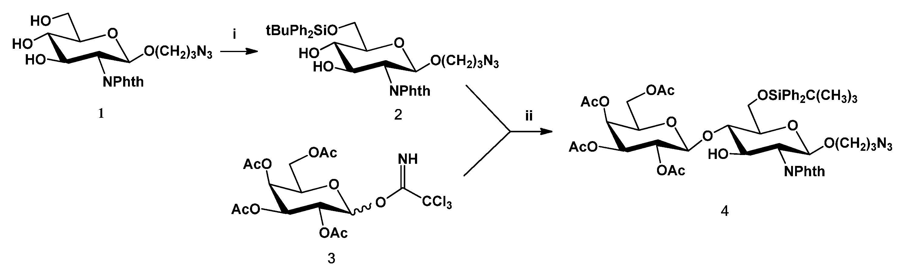
2.2.1. 3-Azidopropyl 6-O-tert-butyldiphenylsilyl-2-phtalimide-2-deoxy-β-D-glucopyranoside (2)
2.2.2. 3-Azidopropyl 2,3,4,6-tetra-O-acetyl-β-D-galactopyranosyl-(1→4)-6-O-tert-butyldiphenylsilyl-2-phtalimide-2-deoxy-β-D-glucopyranoside (4)
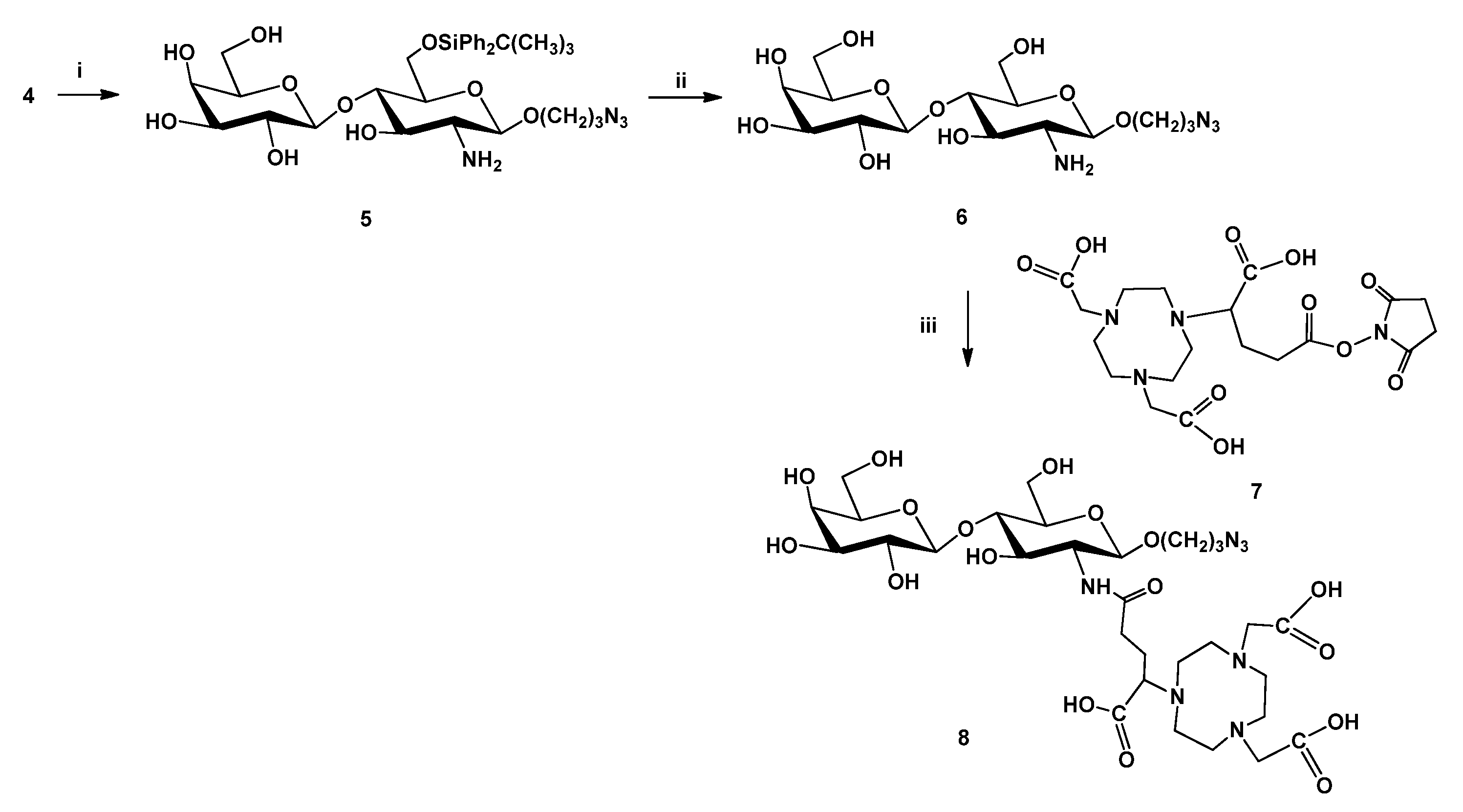
2.2.3. 3-Azidopropyl β-D-galactopyranosyl-(1→4)-6-O-tert-butyldiphenylsilyl-2-amino-2-deoxy-β-D-glucopyranoside (5)
2.2.4. 3-Azidopropyl β-D-galactopyranosyl-(1→4)-2-amino-2-deoxy-β-D-glucopyranoside (6)
2.2.5. NODAGA-LacN (8)
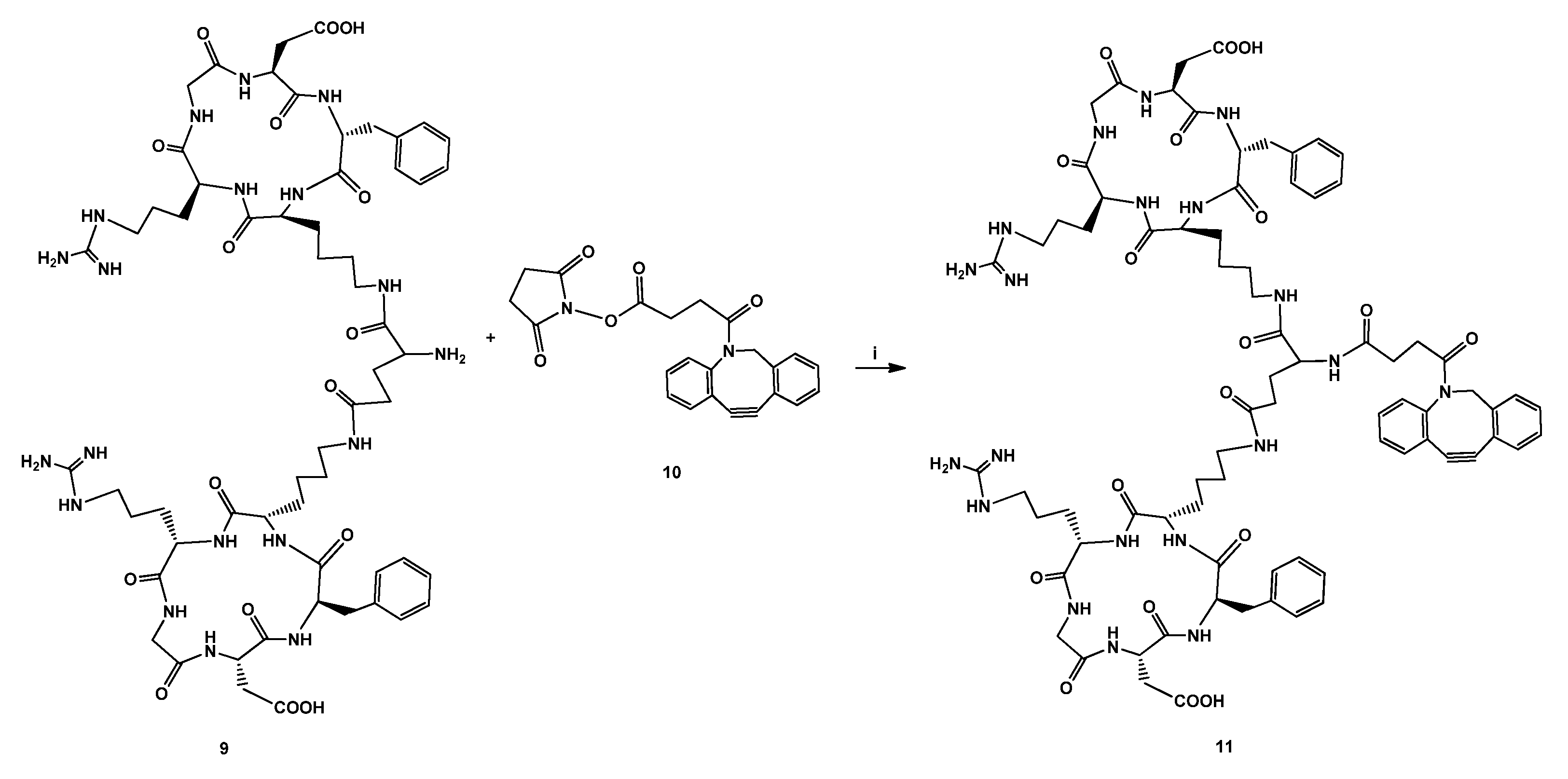
2.2.6. DBCO-E[c(RGDfK)]2 (11)
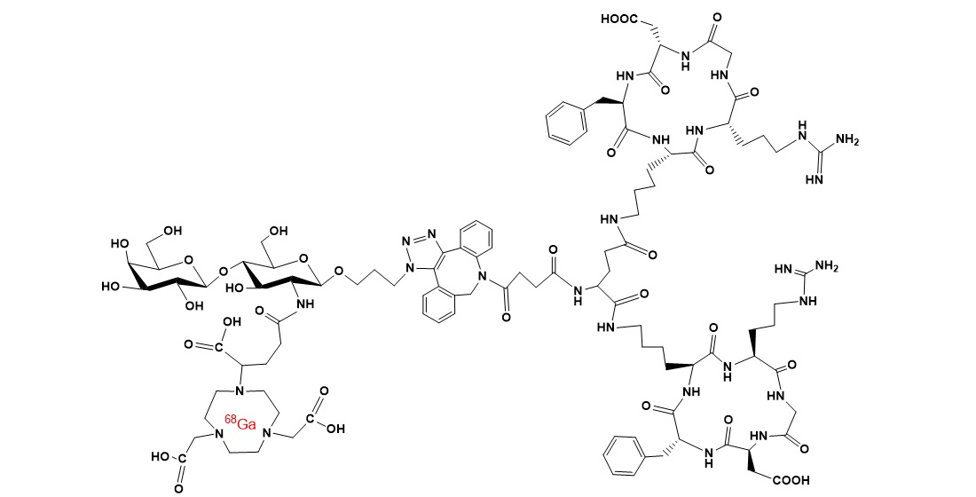
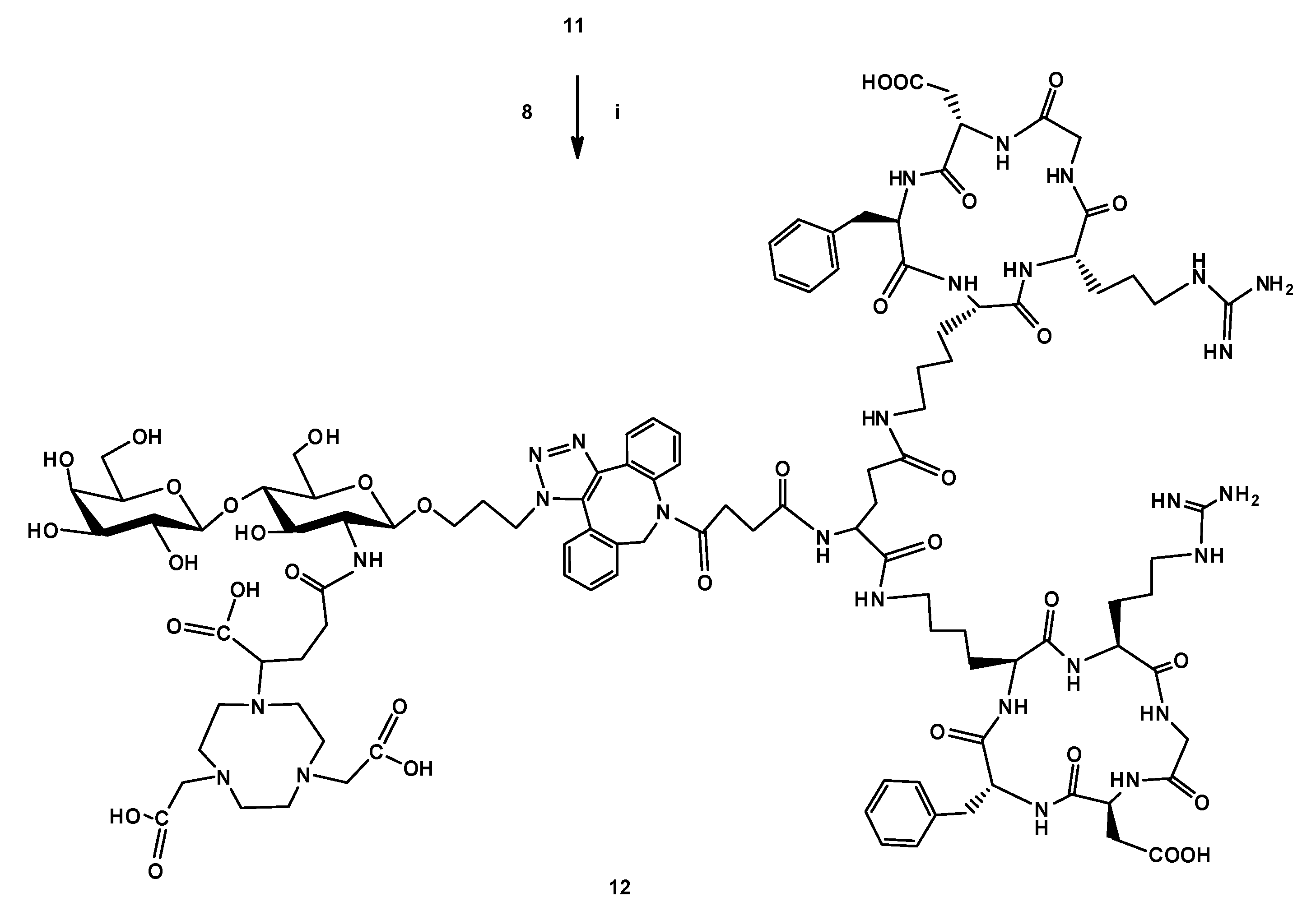
2.2.7. NODAGA-LacN-E[c(RGDfK)]2 (12)
2.3. Radiochemistry
2.3.1. Investigation of 68Ga Labeling of NODAGA-LacN-E[c(RGDfK)]2 Using Different Ligand Concentrations (10, 17, 23 and 32 µM)
2.3.2. Investigation of 68Ga Labeling of NODAGA-LacN-E[c(RGDfK)]2 Using Different Temperatures (Room Temperature, 37, 60, 80 and 95 °C)
2.3.3. Synthesis of 68Ga-NODAGA-LacN-E[c(RGDfK)]2 Radiotracer with Optimal Reaction Procedure
2.3.4. Determination of logP Value of 68Ga-NODAGA-LacN-E[c(RGDfK)]2
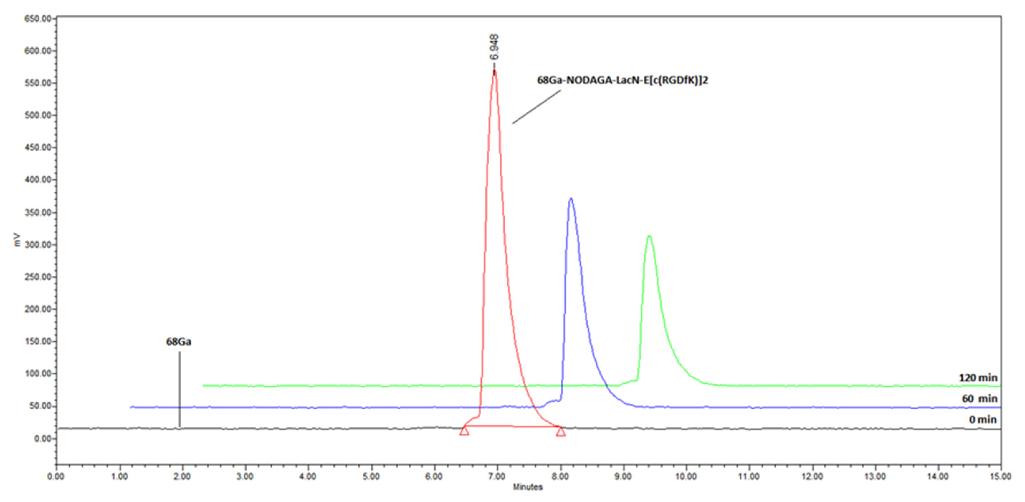
2.3.5. Determination of in Vitro Stability of 68Ga-NODAGA-LacN-E[c(RGDfK)]2 in Human Serum, Na2EDTA and Oxalic Acid
3. Results and Discussion
3.1. Chemistry
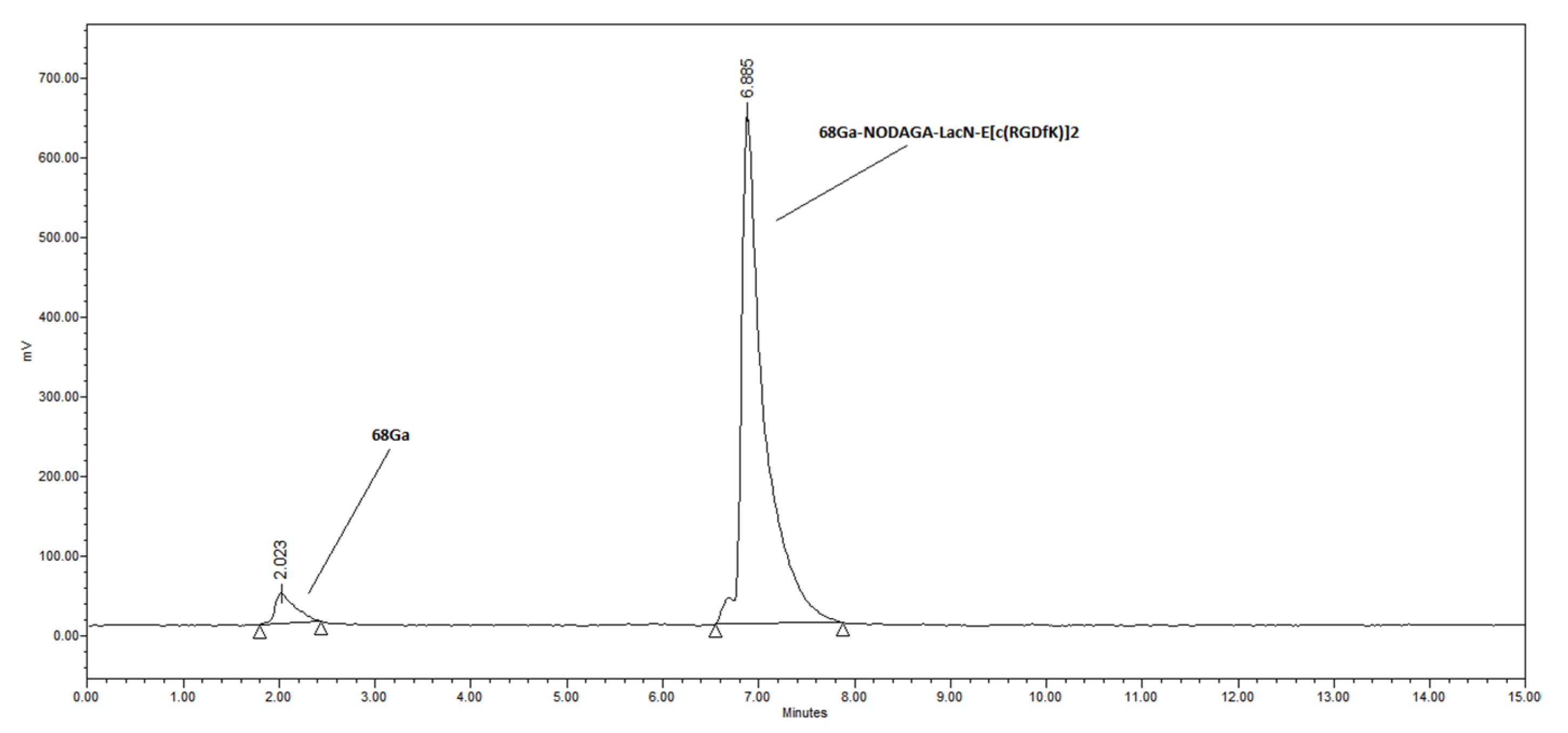
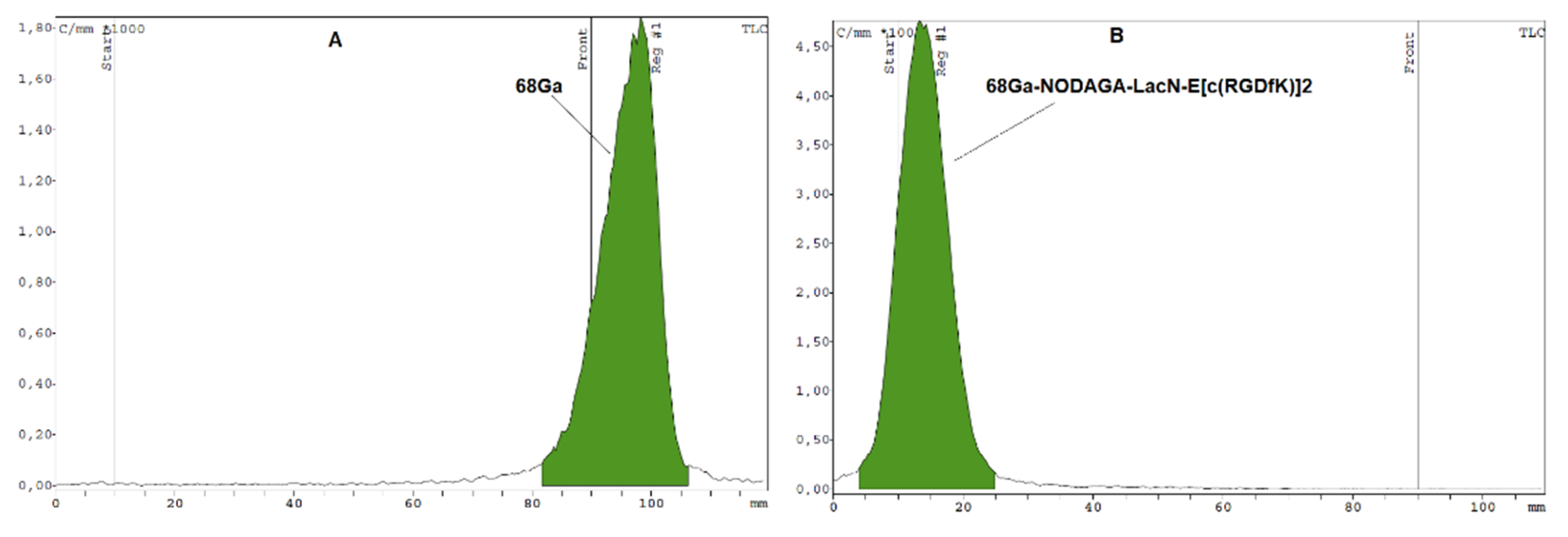
3.2. Radiochemistry
4. Conclusions
Supplementary Materials
Author Contributions
Funding
Institutional Review Board Statement
Informed Consent Statement
Data Availability Statement
Conflicts of Interest
References
- Ambrosini, V.; Fani, M.; Fanti, S.; Forrer, F.; Maecke, H.R. Radiopeptide imaging and therapy in Europe. J. Nucl. Med. 2011, 52, 42s–55s. [Google Scholar] [CrossRef] [PubMed] [Green Version]
- Liu, Z.; Wang, F.; Chen, X. Integrin αvβ3-targeted cancer therapy. Drug Dev. Res. 2008, 69, 329–339. [Google Scholar] [CrossRef] [Green Version]
- Simons, M. Angiogenesis, where do we stand now? Circulation 2005, 111, 1556–1566. [Google Scholar] [CrossRef] [PubMed] [Green Version]
- Chen, H.; Niu, G. Clinical application of radiolabeled RGD peptides for PET imaging of integrin αvβ3. Theranostics 2016, 6, 78–92. [Google Scholar] [CrossRef] [PubMed] [Green Version]
- Haubner, R.; Weber, W.A.; Beer, A.J.; Vabuliene, E.; Reim, D.; Sarbia, M.; Becker, K.-F.; Goebel, M.; Hein, R.; Wester, H.-J.; et al. Noninvasive visualization of the activated αvβ3 integrin in cancer patients by positron emission tomography and [18F]Galacto-RGD. PLoS Med. 2005, 2, e70. [Google Scholar] [CrossRef] [Green Version]
- Haubner, R.; Kuhnast, B.; Mang, C.; Weber, W.A.; Kessler, H.; Wester, H.-J.; Schwaiger, M. [18F]Galacto-RGD: Synthesis, radiolabeling, metabolic stability, and radiation dose estimates. Bioconjug. Chem. 2004, 15, 61–69. [Google Scholar] [CrossRef] [PubMed]
- Zhernosekov, K.P.; Filosofov, D.V.; Baum, R.P.; Aschoff, P.; Bihl, H.; Razbash, A.A.; Jahn, M.; Jennewein, M.; Rösch, F. Processing of generator-produced 68Ga for medical application. J. Nucl. Med. 2007, 48, 1741–1748. [Google Scholar] [CrossRef] [PubMed]
- Froidevaux, S.; Hintermann, E.; Török, M.; Macke, H.R.; Beglinger, C.; Eberle, A.N. Differential regulation of somatostatin receptor type 2 (sst 2) expression in AR4-2J tumour cells implanted into mice during octreotide treatment. Cancer Res. 1999, 59, 3652–3657. [Google Scholar]
- Abbasi Gharibkandi, N.; Conlon, J.M.; Hosseinimehr, S.J. Strategies for improving stability and pharmacokinetic characteristics of radiolabeled peptides for imaging and therapy. Peptides 2020, 133, 170385. [Google Scholar] [CrossRef]
- Werle, M.; Bernkop-Schnurch, A. Strategies to improve plasma halflife time of peptide and protein drugs. Amino Acids 2006, 30, 351–367. [Google Scholar] [CrossRef]
- Seetharaman, J.; Kaningsberg, A.; Slaaby, R.; Leffler, H.; Barondes, S.H.; Rini, J.M. X-ray crystal structure of the human galectin-3 carbohydrate recognition domain at 2.1-Å resolution. J. Biol. Chem. 1998, 273, 13047–13052. [Google Scholar] [CrossRef] [Green Version]
- Liu, F.T.; Rabinovich, G.A. Galectins as modulators of tumour progression. Nat. Rev. Cancer 2005, 5, 29–41. [Google Scholar] [CrossRef]
- Danguy, A.; Camby, I.; Kiss, R. Galectins and cancer. Biochim. Biophys. Acta 2002, 1572, 285–293. [Google Scholar] [CrossRef]
- Prieto, V.G.; Mourad-Zeidan, A.; Melnikova, V.; Johnson, M.M.; Lopez, A.; Diwan, A.H.; Lazar, A.J.F.; Shen, S.S.; Zhang, P.S.; Reed, J.A.; et al. Galectin-3 expression is associated with tumor progression and pattern of sun exposure in melanoma. Clin. Cancer Res. 2006, 12, 6709–6715. [Google Scholar] [CrossRef] [Green Version]
- Endo, K.; Kohnoe, S.; Tsujita, E.; Watanabe, A.; Nakashima, H.; Baba, H.; Maehara, Y. Galectin-3 expression is a potent prognostic marker in colorectal cancer. Anticancer Res. 2005, 25, 3117–3121. [Google Scholar]
- Povegliano, L.Z.; Oshima, C.T.F.; Lima, F.O.; Scherholz, P.L.A.; Forones, N.M. Immunoexpression of Galectin-3 in colorectal cancer and its relationship with survival. J. Gastrointest. Cancer 2010, 42, 217–221. [Google Scholar] [CrossRef] [PubMed]
- Ehlerding, E.B.; Sun, L.; Lan, X.; Zeng, D.; Cai, W. Dual-targeted molecular imaging of cancer. J. Nucl. Med. 2018, 59, 390–395. [Google Scholar] [CrossRef] [PubMed]
- Li, Z.B.; Wu, Z.; Chen, K.; Ryu, E.K.; Chen, X. 18F-labeled BBN-RGD heterodimer for prostate cancer imaging. J. Nucl. Med. 2008, 49, 453–461. [Google Scholar] [CrossRef] [PubMed] [Green Version]
- Zhang, J.; Niu, G.; Lang, L.; Li, F.; Fan, X.; Yan, X.; Yao, S.; Yan, W.; Huo, L.; Chen, L.; et al. Clinical translation of a dual integrin αvβ3– and gastrin-releasing peptide receptor–targeting PET radiotracer, 68Ga-BBN-RGD. J. Nucl. Med. 2017, 58, 228–234. [Google Scholar] [CrossRef] [Green Version]
- Zhang, J.; Mao, F.; Niu, G.; Peng, L.; Lang, L.; Li, F.; Ying, H.; Wu, H.; Pan, B.; Zhu, Z.; et al. 68Ga-BBN-RGD PET/CT for GRPR and Integrin αvβ3 imaging in patients with breast cancer. Theranostics 2018, 8, 1121–1130. [Google Scholar] [CrossRef]
- Kwon, L.Y.; Scollard, D.A.; Reilly, R.M. 64Cu-labeled trastuzumab fab-PEG24-EGF radioimmunoconjugates bispecific for HER2 and EGFR: Pharmacokinetics, biodistribution, and tumor imaging by PET in comparison to monospecific agents. Mol. Pharm. 2017, 14, 492–501. [Google Scholar] [CrossRef] [PubMed]
- Razumienko, E.; Dryden, L.; Scollard, D.; Reilly, R.M. MicroSPECT/CT imaging of co-expressed HER2 and EGFR on subcutaneous human tumor xenografts in athymic mice using 111In-labeled bispecific radioimmunoconjugates. Breast Cancer Res. Treat. 2013, 138, 709–718. [Google Scholar] [CrossRef]
- Shi, S.; Zhou, M.; Li, X.; Hu, M.; Li, C.; Li, M.; Sheng, F.; Li, Z.; Wu, G.; Luo, M.; et al. Synergistic active targeting of dually integrin alphavbeta3/CD44-targeted nanoparticles to B16F10 tumors located at different sites of mouse bodies. J. Control. Release 2016, 235, 1–13. [Google Scholar] [CrossRef] [PubMed]
- Van Hattum, H.; Branderhorst, H.M.; Moret, E.E.; Nilsson, U.J.; Leffler, H.; Pieters, R.J. Tuning the preference of thiodigalactoside- and lactosamine-based ligands to galectin-3 over galectin-1. J. Med. Chem. 2013, 56, 1350–1354. [Google Scholar] [CrossRef] [PubMed]
- Shi, J.; Wang, L.; Kim, Y.S.; Zhai, S.; Jia, B.; Wang, F.; Liu, S. 99mTcO(MAG2-3G3-dimer): A new integrin αvβ3-targeted SPECT radiotracer with high tumor uptake and favorable pharmacokinetics. Eur. J. Nucl. Med. Mol. Imaging 2009, 36, 1874–1884. [Google Scholar] [CrossRef] [PubMed]
- Janssen, M.; Oyen, W.J.; Massuger, L.F.; Frielink, C.; Dijkgraaf, I.; Edwards, D.S.; Radjopadhye, M.; Corstens, F.; Boermanl, O. Comparison of a monomeric and dimeric radiolabeled RGD-peptide for tumor targeting. Cancer Biother. Radiopharm. 2002, 17, 641–646. [Google Scholar] [CrossRef]
- Reichert, D.; Lewis, J.; Anderson, C. Metal complexes as diagnostic tools. Coord. Chem. Rev. 1999, 184, 3–66. [Google Scholar] [CrossRef]
- Koeman, F.A.W.; Meissner, J.W.G.; Ritter, H.R.P.; Kamerling, J.P.; Vliegenthart, J.F.G. Synthesis of structural elements of the capsular polysaccharide of Streptococcus Pneumoniae Type 14. J. Carbohydr. Chem. 1994, 13, 1–26. [Google Scholar] [CrossRef]
- Agard, N.J.; Baskin, J.M.; Prescher, J.A.; Lo, A.; Bertozzi, C.R. A Comparative study of bioorthogonal reactions with azides. ACS Chem. Biol. 2006, 1, 644–648. [Google Scholar] [CrossRef]
- Satpati, D.; Bauer, N.; Hausner, S.H.; Sutcliffe, J.L. Synthesis of [64Cu]DOTA-ADIBON3-Ala-PEG28-A20FMDV2 via copper-free click chemistry for PET imaging of integrin αvβ6. J. Radioanal. Nucl. Chem. 2014, 302, 765–771. [Google Scholar] [CrossRef]
- Jeon, J.; Kang, J.A.; Shim, H.E.; Nam, Y.R.; Yoon, S.; Kim, H.R.; Lee, D.E.; Park, S.H. Efficient method for iodine radioisotope labeling of cyclooctyne containing molecules using strain-promoted copper-free click reaction. Bioorg. Med. Chem. 2015, 23, 3303–3308. [Google Scholar] [CrossRef] [PubMed]


| Ligand Concentration (µM) | Radiochemical Purity (%) |
|---|---|
| 10 | 85.96 ± 5.02 |
| 17 | 90.80 ± 1.51 |
| 23 | 92.82 ± 1.13 |
| 32 | 95.02 ± 0.62 |
| Temperature (°C) | Radiochemical Purity (%) |
|---|---|
| room temperature | 0 |
| 37 | 8.53 ± 1.06 |
| 60 | 92.84 ± 0.96 |
| 80 | 94.26 ± 0.72 |
| 95 | 95.02 ± 0.62 |
Publisher’s Note: MDPI stays neutral with regard to jurisdictional claims in published maps and institutional affiliations. |
© 2021 by the authors. Licensee MDPI, Basel, Switzerland. This article is an open access article distributed under the terms and conditions of the Creative Commons Attribution (CC BY) license (https://creativecommons.org/licenses/by/4.0/).
Share and Cite
Gyuricza, B.; Szabó, J.P.; Arató, V.; Szücs, D.; Vágner, A.; Szikra, D.; Fekete, A. Synthesis of Novel, Dual-Targeting 68Ga-NODAGA-LacN-E[c(RGDfK)]2 Glycopeptide as a PET Imaging Agent for Cancer Diagnosis. Pharmaceutics 2021, 13, 796. https://doi.org/10.3390/pharmaceutics13060796
Gyuricza B, Szabó JP, Arató V, Szücs D, Vágner A, Szikra D, Fekete A. Synthesis of Novel, Dual-Targeting 68Ga-NODAGA-LacN-E[c(RGDfK)]2 Glycopeptide as a PET Imaging Agent for Cancer Diagnosis. Pharmaceutics. 2021; 13(6):796. https://doi.org/10.3390/pharmaceutics13060796
Chicago/Turabian StyleGyuricza, Barbara, Judit P. Szabó, Viktória Arató, Dániel Szücs, Adrienn Vágner, Dezső Szikra, and Anikó Fekete. 2021. "Synthesis of Novel, Dual-Targeting 68Ga-NODAGA-LacN-E[c(RGDfK)]2 Glycopeptide as a PET Imaging Agent for Cancer Diagnosis" Pharmaceutics 13, no. 6: 796. https://doi.org/10.3390/pharmaceutics13060796