
Biopharmaceutics of Topical Ophthalmic Suspensions: Importance of Viscosity and Particle Size in Ocular Absorption of Indomethacin

 , ,
, ,  , ,
, ,
Abstract
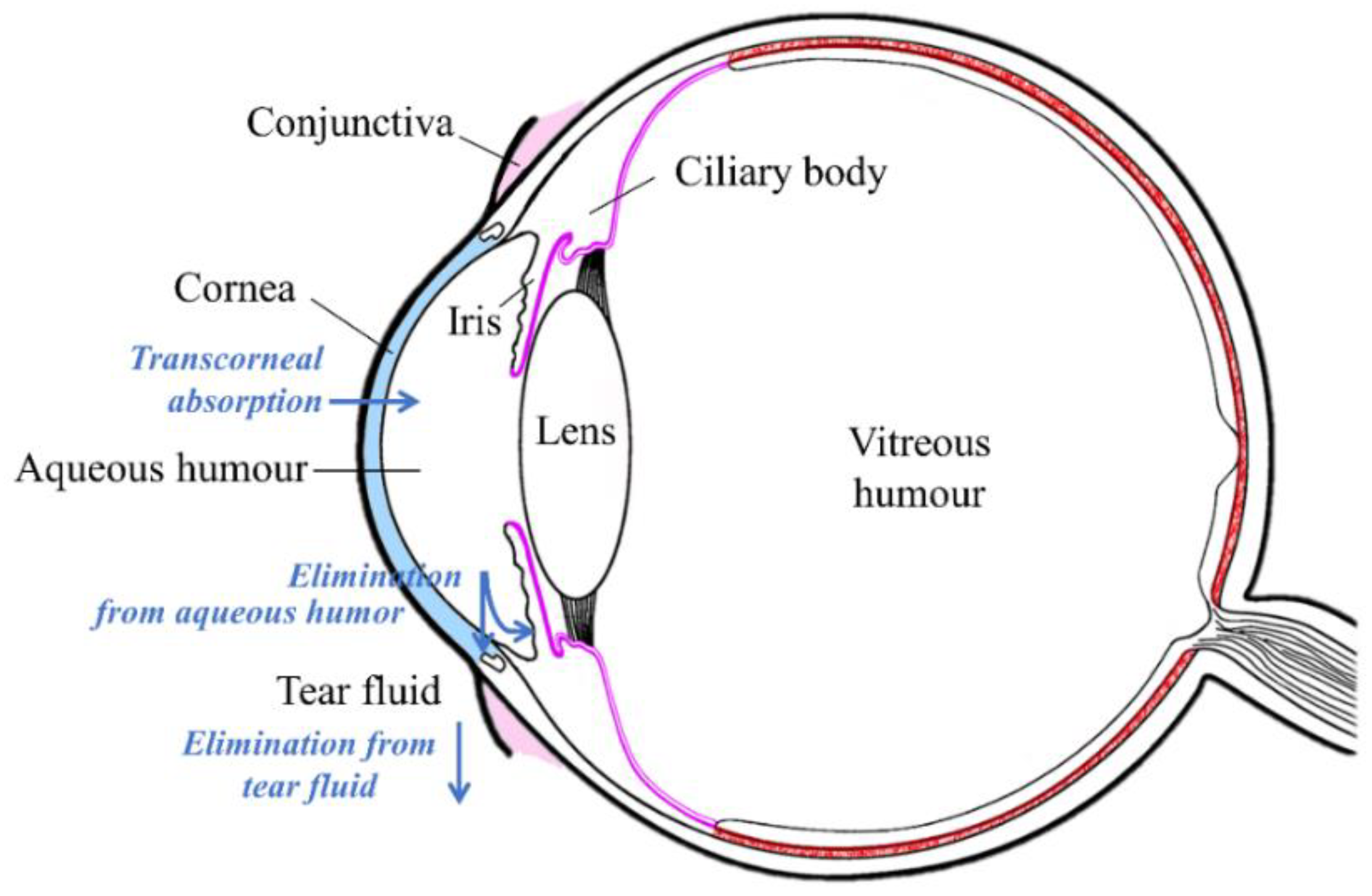
:1. Introduction
2. Materials and Methods
3. Results
3.1. Physical and Chemical Characterization of the Suspensions
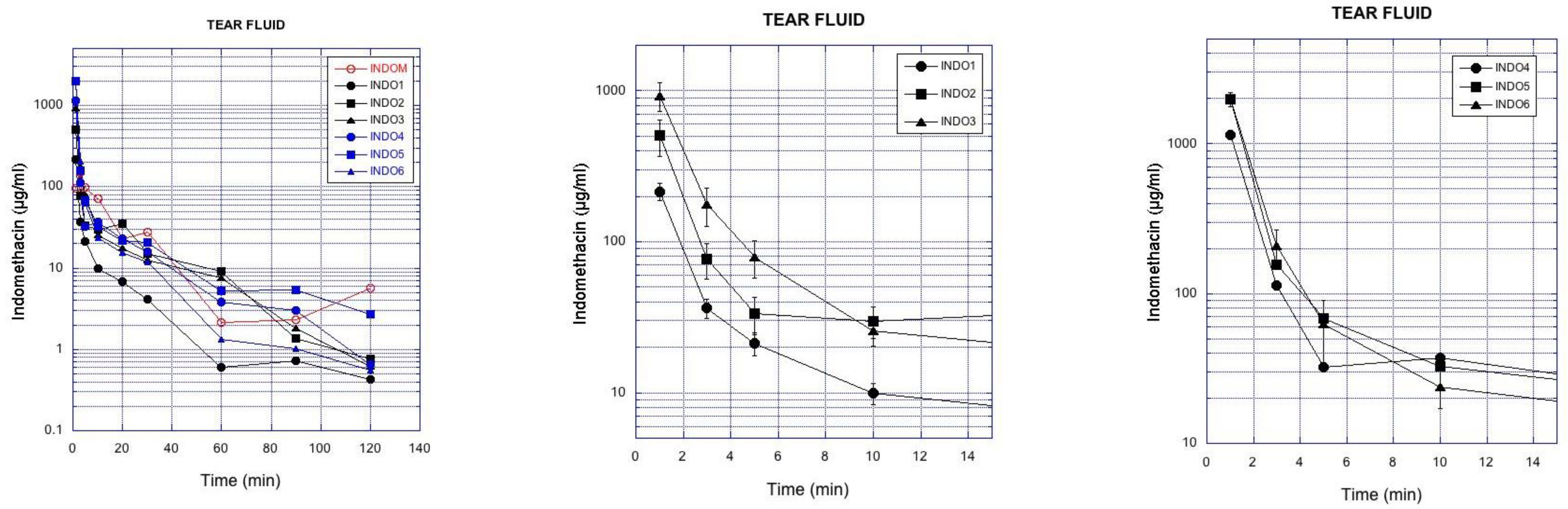
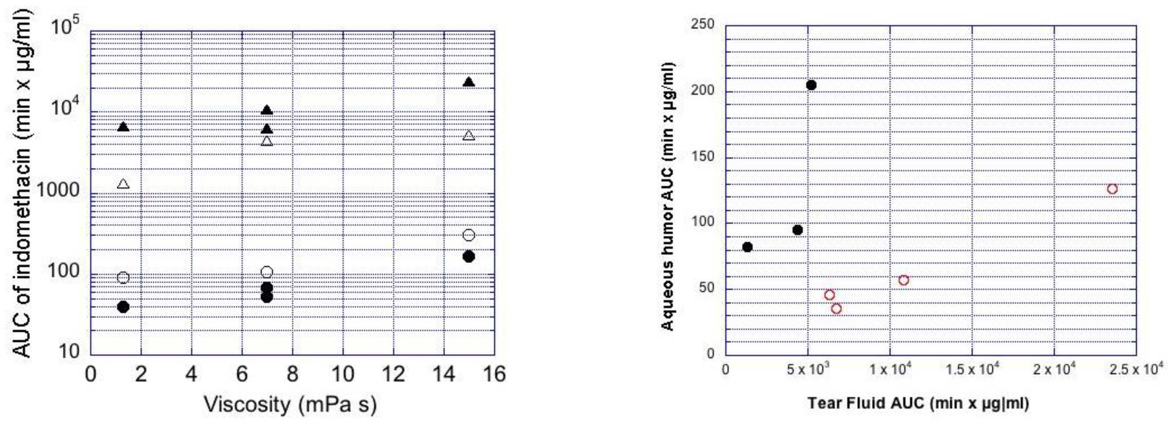
3.2. Indomethacin Absorption into Rabbit Eyes
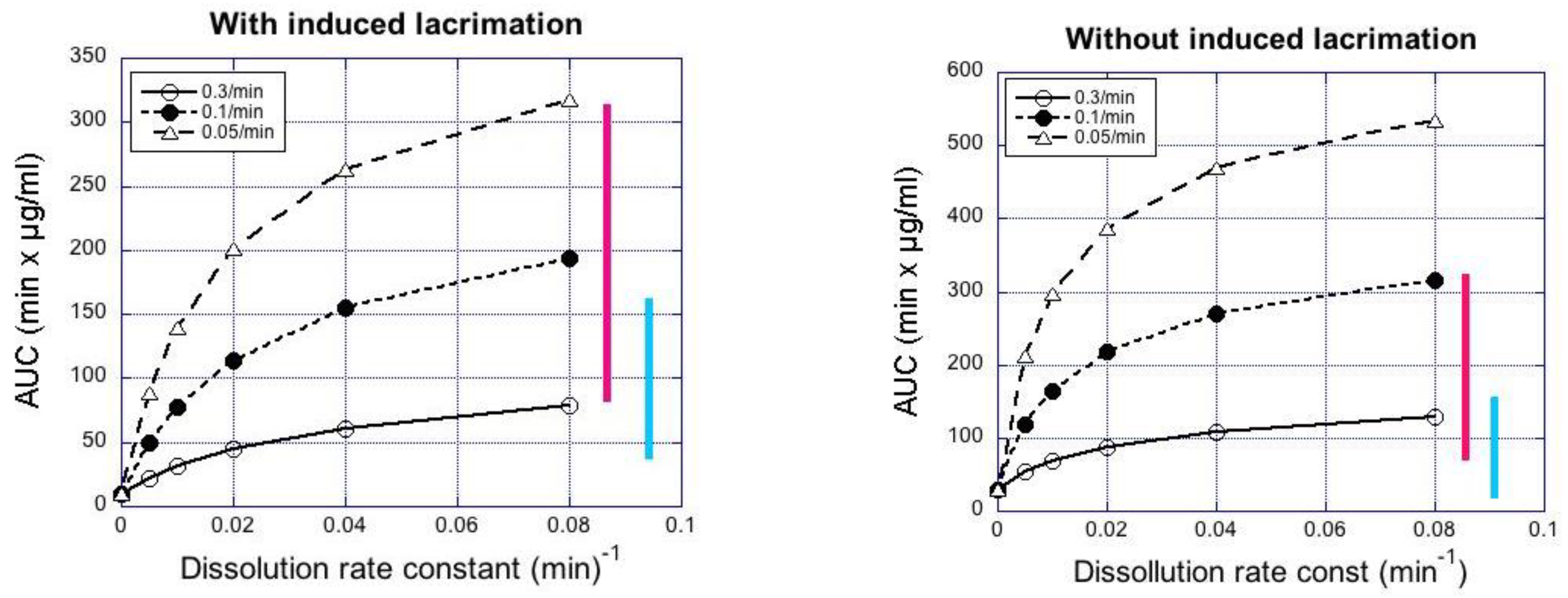
3.3. Pharmacokinetic Simulations
4. Discussion
5. Conclusions
Supplementary Materials
Author Contributions
Funding
Institutional Review Board Statement
Informed Consent Statement
Data Availability Statement
Acknowledgments
Conflicts of Interest
References
- Urtti, A.; Pipkin, J.D.; Rork, G.S.; Sendo, T.; Finne, U.; Repta, A.J. Controlled drug delivery devices for experimental ocular studies with timolol. 2. Ocular and systemic absorption in rabbits. Int. J. Pharm. 1990, 61, 241–249. [Google Scholar] [CrossRef]
- Huang, H.S.; Schoenwald, R.D.; Lach, J.L. Corneal permeation of beta-blocking agents II: Assessment of barrier contributions. J. Pharm. Sci. 1983, 72, 1272–1279. [Google Scholar]
- Lee, V.H.L.; Robinson, J.R. Mechanistic and quantitative evaluation of precorneal pilocarpine disposition in albino rabbits. J. Pharm. Sci. 1979, 68, 673–684. [Google Scholar] [CrossRef]
- Thombre, A.G.; Himmelstein, K.J. Quantitative evaluation of topically applied pilocarpine in precorneal area. J. Pharm. Sci. 1984, 73, 219–222. [Google Scholar] [CrossRef]
- Urtti, A.; Salminen, L.; Miinalainen, O. Systemic absorption of ocular pilocarpine is modified by polymer matrices. Int. J. Pharm. 1985, 23, 147–161. [Google Scholar] [CrossRef]
- Trueblood, J.H.; Rossomondo, R.M.; Carlton, W.H.; Wilson, L.A. Corneal contact times of ophthalmic vehicles. Arch. Ophthalmol. 1975, 93, 127–130. [Google Scholar] [CrossRef] [PubMed]
- Schoenwald, R.D.; Stewart, P. Effect of particle-size on ophthalmic bioavailability of dexamethasone suspensions in rabbits. J. Pharm. Sci. 1980, 69, 391–394. [Google Scholar] [CrossRef] [PubMed]
- Sieg, J.W.; Robinson, J.R. Mechanistic studies on transcorneal permeation of fluorometholone. J. Pharm. Sci. 1981, 70, 1026–1029. [Google Scholar] [CrossRef]
- Vooturi, S.; Bourne, D.; Panda, J.J.; Choi, S.; Kim, H.; Yandrapu, S.K.; Kompella, U.B. Effect of particle size and viscosity of suspensions on topical ocular bioavailability of budesonide, a corticosteroid. J. Ocul. Pharm. Ther. 2020, 36, 404–409. [Google Scholar] [CrossRef]
- Hui, H.W.; Robinson, J.R. Effect of particle dissolution rate on ocular drug bioavailability. J. Pharm. Sci. 1986, 75, 280–287. [Google Scholar] [CrossRef]
- Johansen, S.; Prause, J.U.; Rask-Pedersen, E. A bioavailability comparison in rabbits after a single topical ocular application of prednisolone acetate formulated as a high-viscosity gel and as an aqueous suspension. Acta Ophthalmol. Scand. 1996, 74, 253–258. [Google Scholar] [CrossRef] [PubMed]
- Calvo, P.; Alonso, M.J.; Vila-Jato, J.L.; Robinson, J.R. Improved ocular bioavailability of indomethacin by novel ocular drug carriers. J. Pharm. Pharm. 1996, 48, 1147–1152. [Google Scholar] [CrossRef] [PubMed]
- Begines, B.; Ortiz, T.; Perez-Aranda, M.; Martinez, G.; Merinero, M.; Arguelles-Arias, M.; Alcudia, A. Polymeric nanoparticles for drug delivery: Recent developments and future prospects. Nanomaterials 2020, 10, 1403. [Google Scholar] [CrossRef]
- Hany, S.M.; Ali, A.B.; York, P.; Ahmed, M.A.; Ali, C.; Blagden, N. Hydrocortisone nanosuspensions for ophthalmic delivery: A comparative study between microfluidic nanoprecipitation and wet milling. J. Control. Release 2011, 149, 175–181. [Google Scholar]
- Luschmanna, C.; Herrmann, W.; Strauß, O.; Luschmann, K.; Goepferich, A. Ocular delivery systems for poorly soluble drugs: An In-vivo evaluation. Int. J. Pharm. 2013, 455, 331–337. [Google Scholar] [CrossRef]
- Sheppard, J.D.; Cockrum, P.C.; Justice, A.; Jasek, M.C. In vivo pharmacokinetics of bromfenac ophthalmic solution 0.075%, Bromfenac ophthalmic solution 0.07%, and Nepafenac/Amfenac ophthalmic suspension 0.3% in rabbits. Ophthalmol. Ther. 2018, 7, 157–165. [Google Scholar] [CrossRef] [Green Version]
- Zhou, X.; Li, X.; Xu, J.; Cheng, Y.; Cao, F. Latanoprost-loaded cyclodextrin microaggregate suspension eye drops for enhanced bioavailability and stability. Eur. J. Pharm. Sci. 2021, 160, 105758. [Google Scholar] [CrossRef]
- Baba, K.; Hashida, N.; Tujikawa, M.; Quantock, A.J.; Nishida, K. The generation of fluorometholone nanocrystal eye drops, their metabolization to dihydrofluorometholone and penetration into rabbit eyes. Int. J. Pharm. 2021, 592, 120067. [Google Scholar] [CrossRef]
- Maurice, D.M.; Mishima, S. Ocular pharmacokinetics. In Pharmacology of the Eye: Handbook of Experimental Pharmacology; Sears, M.L., Ed.; Springer: Berlin/Heidelberg, Germany, 1984; Volume 69, pp. 19–116. [Google Scholar]
- Harigaya, Y.; Jiang, X.; Zhang, H.; Chandaroy, P.; Stier, E.M.; Pan, Y. Bioequivalence study methods with pharmacokinetic endpoints for topical ophthalmic corticosteroid suspensions and effects of subject demographics. Pharm. Res. 2018, 36, 13. [Google Scholar] [CrossRef]
- Ramsay, E.; del Amo, E.; Toropainen, E.; Tengvall-Unadike, U.; Ranta, V.P.; Urtti, A.; Ruponen, M. Corneal and conjunctival drug permeability: Systematic comparison and pharmacokinetic impact in the eye. Eur. J. Pharm. Sci. 2018, 119, 83–89. [Google Scholar] [CrossRef] [PubMed] [Green Version]
- Chrai, S.S.; Robinson, J.R. Ocular evaluation of methylcellulose vehicle in albino rabbits. J. Pharm. Sci. 1974, 63, 1218–1223. [Google Scholar] [CrossRef]
- Saettone, M.F.; Giannaccini, B.; Teneggi, A.; Savigni, P.; Tellini, G. Vehicle effects on ophthalmic bioavailability: The influence of different polymers on the acitivity of pilocarpine in rabbit and man. J. Pharm. Pharmacol. 1982, 34, 464–466. [Google Scholar] [CrossRef]
- Sinko, P.J. Martin’s Physical Pharmacy and Pharmaceutical Sciences: Physical Chemical and Biopharmaceutical Principles in the Pharmaceutical Sciences; Lippincott Williams & Wilkins: London, UK, 2016. [Google Scholar]
- Saettone, M.F.; Giannaccini, B.; Ravecca, S.; La Marca, F.; Tota, G. Polymer effects on ocular bioavailability—The influence of different liquid vehicles on the mydriatic response of tropicamide in humans and rabbits. Int. J. Pharm. 1984, 20, 187–202. [Google Scholar] [CrossRef]
- Le Merdy, M.; Fan, J.; Bolger, M.B.; Lukacova, V.; Spires, J.; Tsakalazou, E.; Patel, V.; Xu, L.; Stewart, S.; Chockalingam, A.; et al. Application of mechanistic ocular absorption modeling and simulation to understand the impact of formulation properties on ophthalmic bioavailability in rabbit: A case study using dexamethasone suspension. AAPS J. 2019, 21, 65. [Google Scholar] [CrossRef]
- Fayaaz, A.; Ranta, V.; Toropainen, E.; Vellonen, K.S.; D’Amico Ricci, G.; Reinisalo, M.; Heikkinen, E.M.; Gardner, I.; Urtti, A.; Jamei, M.; et al. Ocular intracameral pharmacokinetics for a cocktail of timolol, betaxolol and atenolol in rabbits. Mol. Pharm. 2020, 17, 588–594. [Google Scholar] [CrossRef] [Green Version]
- Deng, F.; Ranta, V.P.; Kidron, H.; Urtti, A. General pharmacokinetic model for topically administered ocular drug solutions and suspensions. Pharm. Res. 2016, 33, 2680–2690. [Google Scholar] [CrossRef]
- Chambers, W.A. Ophthalmic generics—Are they really the same? Ophthalmology 2012, 119, 1095–1096. [Google Scholar] [CrossRef] [PubMed]
- U.S. Department of Health and Human Services Food and Drug Administration. FDA’s Guidance for Industry. Statistical Approaches to Establishing Bioequivalence. Available online: https://www.fda.gov/media/70958/download (accessed on 28 February 2001).


| Sample | Particle Size | Milling Parameters * | Viscosity | Calculated Viscosity (mPa·s) |
|---|---|---|---|---|
| INDO1 | Small | Six cycles at 700 rpm with 1 mm pearls | Low | ≈1.3 (HPMC E5) |
| INDO2 | Small | Six cycles at 700 rpm with 1 mm pearls | Medium | ≈7 (HPMC 4000) |
| INDO3 | Small | Six cycles at 700 rpm with 1 mm pearls | High | ≈15 (HPMC K35M) |
| INDO4 | Large | One cycle at 1000 rpm with 5 mm pearls | Low | ≈1.3 (HPMC E5) |
| INDO5 | Large | One cycle at 1000 rpm with 5 mm pearls | Medium | ≈7 (HPMC 4000) |
| INDO6 | Large | One cycle at 1000 rpm with 5 mm pearls | High | ≈15 (HPMC K35M) |
| Formulation Component | Role | Concentration |
|---|---|---|
| Indomethacin (pre-milled to a certain particle size) | Active pharmaceutical ingredient | 0.5 wt.% |
| HPMC | Wetting and viscosity-increasing agent | 0.3 wt.% |
| Sodium dihydrogen phosphate, disodium phosphate | Buffering agents | 78.16 and 9.84 mM |
| Sodium chloride | Tonicity adjusting agent | 35 mM |
| EDTA | Chelating agent | 0.05% |
| Methylparaben | Preservative | 0.0205% |
| Propylparaben | Preservative | 0.026% |
| Deionized water | Aqueous vehicle | ad 50 mL |
| Compound | ESI | Precursor Ion | Product Ion 1 | CE (eV) | Product Ion 2 | CE (eV) | Internal Standard |
|---|---|---|---|---|---|---|---|
| Indomethacin | + | 358.01 | 138.9 | 17 | 110.9 | 49 | Indomethacin-d4 |
| Indomethacin-d4 | + | 364.01 | 143 | 16 | / |
| Sample | Particle Size | Calculated Viscosity (mPa·s) | Osmolality (mOsm/kg) | pH | |||||
|---|---|---|---|---|---|---|---|---|---|
| Laser Light Diffraction | SEM | ||||||||
| d10 (µm) | d20 (µm) | d50 (µm) | d80 (µm) | d90 (µm) | Visual Range (µm) | ||||
| INDO1 | 0.19 | 0.22 | 0.43 | 2.48 | 5.90 | 0.1–2 | ≈1.3 (HPMC E5) | 241 | 5.80 |
| INDO2 | 0.29 | 0.45 | 1.33 | 7.30 | 14.00 | 0.1–3 | ≈7 (HPMC 4000) | 239 | 5.90 |
| INDO3 | 0.18 | 0.22 | 0.37 | 3.28 | 12.81 | 0.1–4 | ≈15 (HPMC K35M) | 239 | 5.84 |
| INDO4 | 0.69 | 1.22 | 3.23 | 5.56 | 7.21 | 0.4–4 | ≈1.3 (HPMC E5) | 241 | 5.82 |
| INDO5 | 0.74 | 1.50 | 3.50 | 6.03 | 7.40 | 0.4–3 | ≈7 (HPMC 4000) | 242 | 5.89 |
| INDO6 | 0.80 | 1.32 | 3.12 | 6.40 | 9.42 | 0.4–4 | ≈15 (HPMC K35M) | 236 | 5.91 |
| Indom® | 0.90 | 2.75 | 5.48 | 9.06 | 11.40 | 0.5–5 (bulk), 20–50 (few) | ≈7 (measured) | 232 | 5.90 |
| Formulation | AUC (min × µg/mL) | CV% | C1min (µg/mL) | CV% |
|---|---|---|---|---|
| Indom® | 6312 | 25 | 216 | 33 |
| INDO1 | 1299 | 15 | 63 | 19 |
| INDO2 | 4379 | 17 | 238 | 20 |
| INDO3 | 5197 | 17 | 299 | 20 |
| INDO4 | 6718 | 20 | 400 | 24 |
| INDO5 | 10,849 | 19 | 445 | 24 |
| INDO6 | 23,517 | 11 | 1480 | 13 |
| Formulation | AUC0–4h (min × µg/mL) | AUC (min × µg/mL) | Cmax (µg/mL) | Tmax (min) | Terminal Half-Life (min) |
|---|---|---|---|---|---|
| Indom® | 45.9 | 52.1 | 0.012 | 78 | 402 |
| INDO1 | 82.0 | 89.7 | 0.008 | 59 | 636 |
| INDO2 | 94.9 | 104.7 | 0.009 | 63 | 654 |
| INDO3 | 204.9 | 301.6 | 0.032 | 127 | 13,826 |
| INDO4 | 35.5 | 39.2 | 0.010 | 59 | 256 |
| INDO5 | 57.5 | 67.8 | 0.015 | 76 | 422 |
| INDO6 | 126.3 | 166.2 | 0.024 | 107 | 744 |
Publisher’s Note: MDPI stays neutral with regard to jurisdictional claims in published maps and institutional affiliations. |
© 2021 by the authors. Licensee MDPI, Basel, Switzerland. This article is an open access article distributed under the terms and conditions of the Creative Commons Attribution (CC BY) license (http://creativecommons.org/licenses/by/4.0/).
Share and Cite
Toropainen, E.; Fraser-Miller, S.J.; Novakovic, D.; Del Amo, E.M.; Vellonen, K.-S.; Ruponen, M.; Viitala, T.; Korhonen, O.; Auriola, S.; Hellinen, L.; et al. Biopharmaceutics of Topical Ophthalmic Suspensions: Importance of Viscosity and Particle Size in Ocular Absorption of Indomethacin. Pharmaceutics 2021, 13, 452. https://doi.org/10.3390/pharmaceutics13040452
Toropainen E, Fraser-Miller SJ, Novakovic D, Del Amo EM, Vellonen K-S, Ruponen M, Viitala T, Korhonen O, Auriola S, Hellinen L, et al. Biopharmaceutics of Topical Ophthalmic Suspensions: Importance of Viscosity and Particle Size in Ocular Absorption of Indomethacin. Pharmaceutics. 2021; 13(4):452. https://doi.org/10.3390/pharmaceutics13040452
Chicago/Turabian StyleToropainen, Elisa, Sara J. Fraser-Miller, Dunja Novakovic, Eva M. Del Amo, Kati-Sisko Vellonen, Marika Ruponen, Tapani Viitala, Ossi Korhonen, Seppo Auriola, Laura Hellinen, and et al. 2021. "Biopharmaceutics of Topical Ophthalmic Suspensions: Importance of Viscosity and Particle Size in Ocular Absorption of Indomethacin" Pharmaceutics 13, no. 4: 452. https://doi.org/10.3390/pharmaceutics13040452