
Formulation Considerations for the Management of Dry Eye Disease


Abstract
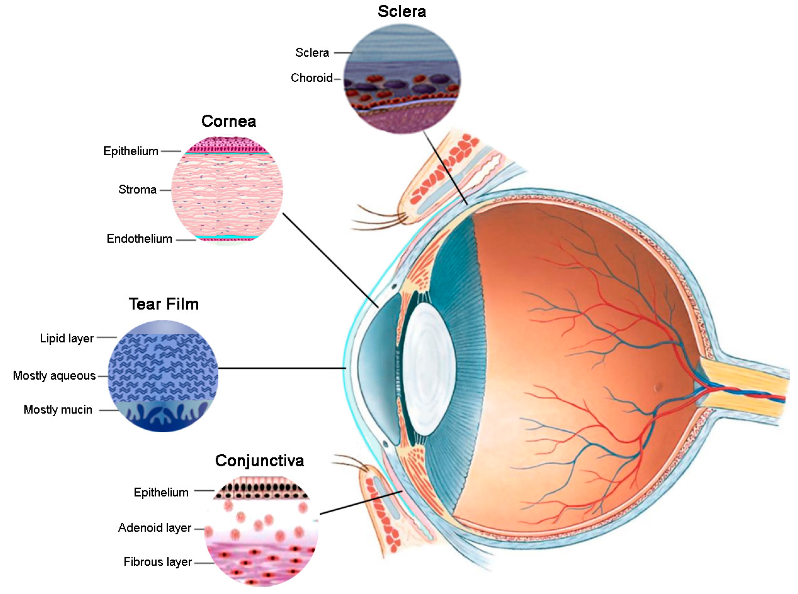
:1. Introduction
2. Formulation Challenges
2.1. Rapid Precorneal Clearance
2.2. Poor Drug Penetration
2.3. Dose Volume
2.4. Visual Disturbance
2.5. Preservative Toxicity
2.6. Poor Tolerability of Formulation Excipients
2.7. Poor Patient Compliance
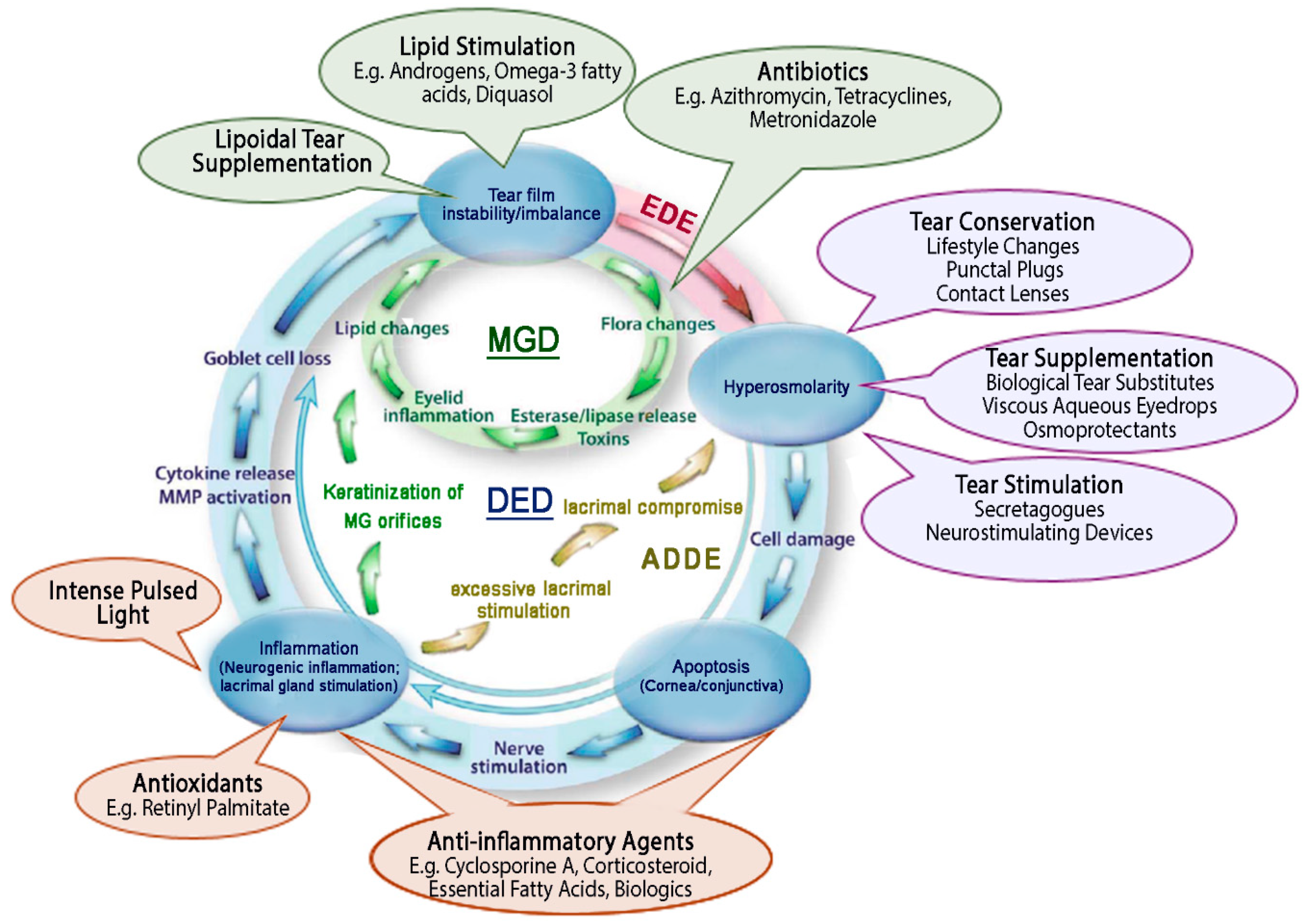
3. Management of Dry Eye Disease
3.1. Tear Supplementation
3.1.1. Aqueous Tear Supplementation
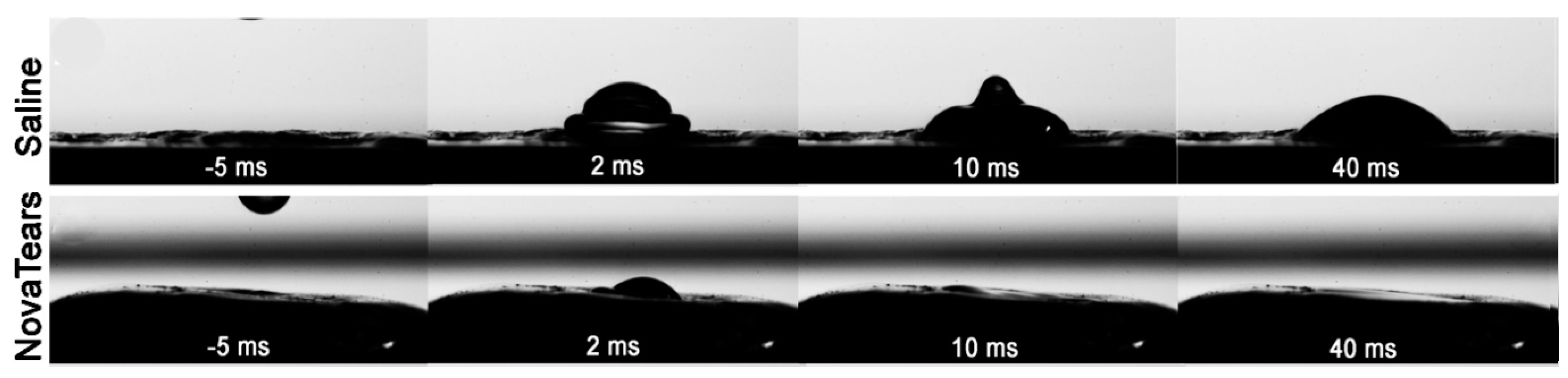
3.1.2. Lipoidal Tear Supplementation
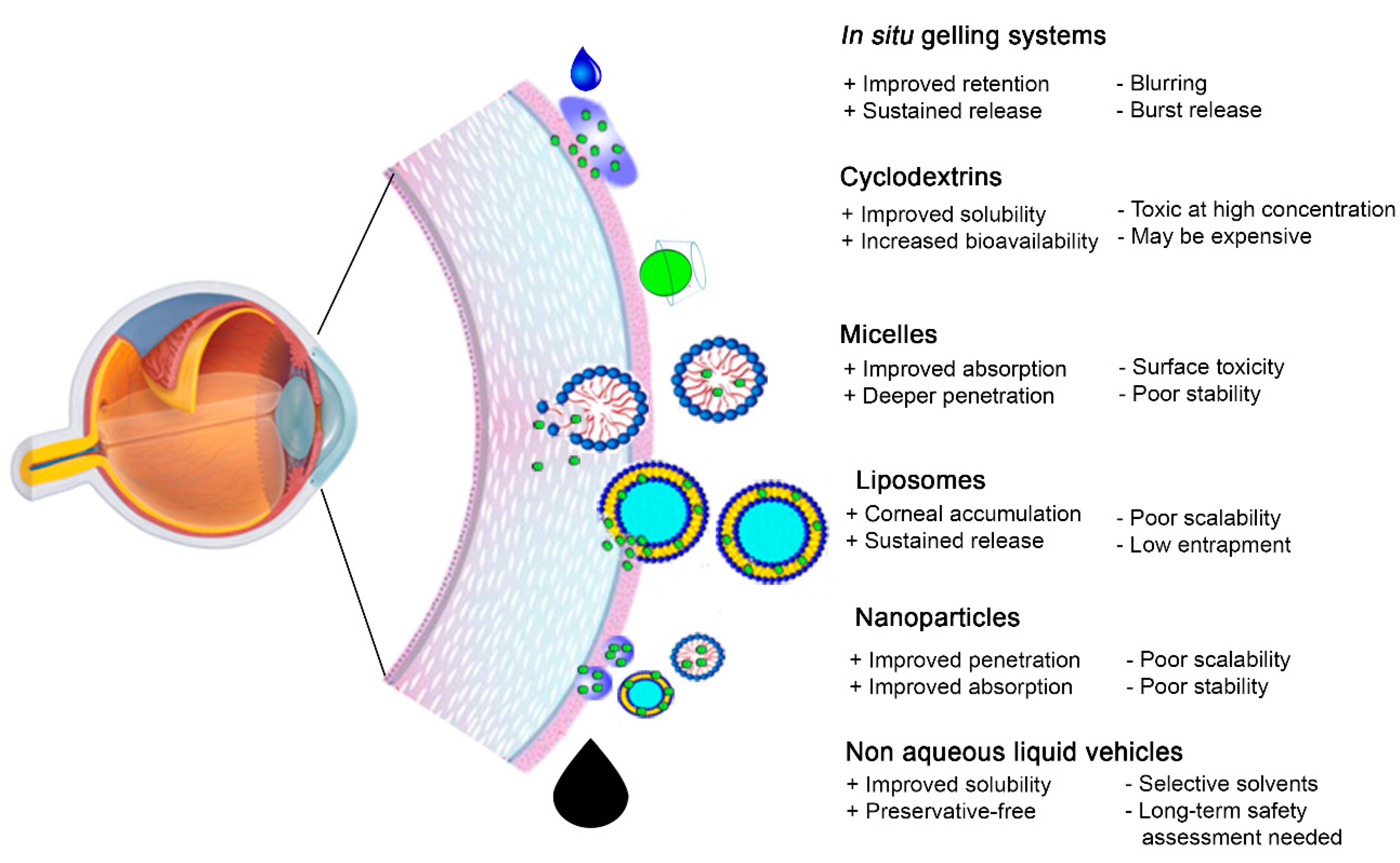
3.2. Medicated Topical Ophthalmic Formulations
3.2.1. Cyclodextrins
3.2.2. Micelles
3.2.3. Liposomes
3.2.4. Nanoformulations
3.2.5. Non-Aqueous Vehicles
4. Concluding Remarks
Author Contributions
Funding
Institutional Review Board Statement
Informed Consent Statement
Data Availability Statement
Conflicts of Interest
References
- Stapleton, F.; Alves, M.; Bunya, V.Y.; Jalbert, I.; Lekhanont, K.; Malet, F.; Na, K.-S.; Schaumberg, D.; Uchino, M.; Vehof, J.; et al. TFOS DEWS II Epidemiology Report. Ocul. Surf. 2017, 15, 334–365. [Google Scholar] [CrossRef] [PubMed]
- Craig, J.P.; Nichols, K.K.; Akpek, E.K.; Caffery, B.; Dua, H.S.; Joo, C.-K.; Liu, Z.; Nelson, J.D.; Nichols, J.J.; Tsubota, K.; et al. TFOS DEWS II Definition and Classification Report. Ocul. Surf. 2017, 15, 276–283. [Google Scholar] [CrossRef] [PubMed]
- Koh, S. Mechanisms of Visual Disturbance in Dry Eye. Cornea 2016, 35, S83–S88. [Google Scholar] [CrossRef] [PubMed]
- Puell, M.C.; Benítez-del-Castillo, J.M.; Martínez-de-la-Casa, J.; Sánchez-Ramos, C.; Vico, E.; Pérez-Carrasco, M.J.; Pedraza, C.; Del-Hierro, A. Contrast sensitivity and disability glare in patients with dry eye. Acta Ophthalmol. Scand. 2006, 84, 527–531. [Google Scholar] [CrossRef] [PubMed]
- Mathews, P.M.; Ramulu, P.Y.; Swenor, B.S.; Utine, C.A.; Rubin, G.S.; Akpek, E.K. Functional impairment of reading in patients with dry eye. Br. J. Ophthalmol. 2017, 101, 481–486. [Google Scholar] [CrossRef]
- Reddy, P.; Grad, O.; Rajagopalan, K. The Economic Burden of Dry Eye: A Conceptual Framework and Preliminary Assessment. Cornea 2004, 23, 751–761. [Google Scholar] [CrossRef]
- Mertzanis, P.; Abetz, L.; Rajagopalan, K.; Espindle, D.; Chalmers, R.; Snyder, C.; Caffery, B.; Edrington, T.; Simpson, T.; Nelson, J.D.; et al. The Relative Burden of Dry Eye in Patients’ Lives: Comparisons to a U.S. Normative Sample. Invest. Ophthalmol. Vis. Sci. 2005, 46, 46–50. [Google Scholar] [CrossRef]
- Uchino, M.; Schaumberg, D.A. Dry Eye Disease: Impact on Quality of Life and Vision. Curr. Ophthalmol. Rep. 2013, 1, 51–57. [Google Scholar] [CrossRef] [Green Version]
- Nichols, K.K.; Foulks, G.N.; Bron, A.J.; Glasgow, B.J.; Dogru, M.; Tsubota, K.; Lemp, M.A.; Sullivan, D.A. The International Workshop on Meibomian Gland Dysfunction: Executive Summary. Invest. Ophthalmol. Vis. Sci. 2011, 52, 1922–1929. [Google Scholar] [CrossRef] [Green Version]
- Baudouin, C.; Messmer, E.M.; Aragona, P.; Geerling, G.; Akova, Y.A.; Benítez-del-Castillo, J.; Boboridis, K.G.; Merayo-Lloves, J.; Rolando, M.; Labetoulle, M. Revisiting the vicious circle of dry eye disease: A focus on the pathophysiology of meibomian gland dysfunction. Br. J. Ophthalmol. 2016, 100, 300–306. [Google Scholar] [CrossRef]
- Tomlinson, A.; Bron, A.J.; Korb, D.R.; Amano, S.; Paugh, J.R.; Pearce, E.I.; Yee, R.; Yokoi, N.; Arita, R.; Dogru, M. The International Workshop on Meibomian Gland Dysfunction: Report of the Diagnosis Subcommittee. Investig. Ophthalmol. Vis. Sci. 2011, 52, 2006–2049. [Google Scholar] [CrossRef] [PubMed]
- Bron, A.J.; Yokoi, N.; Gaffney, E.; Tiffany, J.M. Predicted Phenotypes of Dry Eye: Proposed Consequences of Its Natural History. Ocul. Surf. 2009, 7, 78–92. [Google Scholar] [CrossRef]
- Bron, A.J.; de Paiva, C.S.; Chauhan, S.K.; Bonini, S.; Gabison, E.E.; Jain, S.; Knop, E.; Markoulli, M.; Ogawa, Y.; Perez, V.; et al. TFOS DEWS II pathophysiology report. Ocul. Surf. 2017, 15, 438–510. [Google Scholar] [CrossRef] [PubMed]
- Baudouin, C.; Aragona, P.; Messmer, E.M.; Tomlinson, A.; Calonge, M.; Boboridis, K.G.; Akova, Y.A.; Geerling, G.; Labetoulle, M.; Rolando, M. Role of Hyperosmolarity in the Pathogenesis and Management of Dry Eye Disease: Proceedings of the OCEAN Group Meeting. Ocul. Surf. 2013, 11, 246–258. [Google Scholar] [CrossRef] [PubMed] [Green Version]
- Davies, N.M. Biopharmaceutical considerations in topical ocular drug delivery. Clin. Exp. Pharmacol. Physiol. 2000, 27, 558–562. [Google Scholar] [CrossRef]
- Abdelkader, H.; Alany, R.G. Controlled and continuous release ocular drug delivery systems: Pros and cons. Curr. Drug Deliv. 2012, 9, 421–430. [Google Scholar] [CrossRef]
- Järvinen, K.; Järvinen, T.; Urtti, A. Ocular absorption following topical delivery. Adv. Drug Del. Rev. 1995, 16, 3–19. [Google Scholar] [CrossRef]
- Lederer, C.M.; Harold, R.E. Drop Size of Commercial Glaucoma Medications. Am. J. Ophthalmol. 1986, 101, 691–694. [Google Scholar] [CrossRef]
- Lee, V.H.; Robinson, J.R. Topical ocular drug delivery: Recent developments and future challenges. J. Ocul. Pharmacol. Ther. 1986, 2, 67–108. [Google Scholar] [CrossRef]
- Pearce, E.I.; Dorman, M.; Wilkinson, B.C.; Oliver, K.M. Effect of Blink Frequency on Tear Turnover Rate. Investig. Ophthalmol. Vis. Sci. 2011, 52, 3726. [Google Scholar]
- Saarinen-Savolainen, P.; Järvinen, T.; Araki-Sasaki, K.; Watanabe, H.; Urtti, A. Evaluation of Cytotoxicity of Various Ophthalmic Drugs, Eye Drop Excipients and Cyclodextrins in an Immortalized Human Corneal Epithelial Cell Line. Pharm. Res. 1998, 15, 1275–1280. [Google Scholar] [CrossRef] [PubMed]
- Leibowitz, H.M.; Kupferman, A. Drug interaction in the eye: Concurrent corticosteroid-antibiotic therapy for inflammatory keratitis. Arch. Ophthalmol. 1977, 95, 682–685. [Google Scholar] [CrossRef] [PubMed]
- Yamada, M.; Mochizuki, H.; Yamada, K.; Kawai, M.; Mashima, Y. Aqueous humor levels of topically applied levofloxacin, norfloxacin, and lomefloxacin in the same human eyes. J. Cataract Refract. Surg. 2003, 29, 1771–1775. [Google Scholar] [CrossRef]
- Duvvuri, S.; Majumdar, S.; Mitra, A.K. Role of Metabolism in Ocular Drug Delivery. Curr. Drug Metabol. 2004, 5, 507–515. [Google Scholar] [CrossRef] [PubMed]
- Ensign, L.M.; Schneider, C.; Suk, J.S.; Cone, R.; Hanes, J. Mucus penetrating nanoparticles: Biophysical tool and method of drug and gene delivery. Adv. Mater. 2012, 24, 3887–3894. [Google Scholar] [CrossRef] [PubMed]
- Schuster, B.S.; Suk, J.S.; Woodworth, G.F.; Hanes, J. Nanoparticle diffusion in respiratory mucus from humans without lung disease. Biomaterials 2013, 34, 3439–3446. [Google Scholar] [CrossRef] [Green Version]
- Ghate, D.; Edelhauser, H.F. Barriers to glaucoma drug delivery. J. Glaucoma 2008, 17, 147–156. [Google Scholar] [CrossRef]
- Prausnitz, M.R.; Noonan, J.S. Permeability of cornea, sclera, and conjunctiva: A literature analysis for drug delivery to the eye. J. Pharm. Sci. 1998, 87, 1479–1488. [Google Scholar] [CrossRef]
- Koevary, S.B. Pharmacokinetics of topical ocular drug delivery: Potential uses for the treatment of diseases of the posterior segment and beyond. Curr. Drug Metabol. 2003, 4, 213–222. [Google Scholar] [CrossRef]
- Geroski, D.H.; Edelhauser, H.F. Transscleral drug delivery for posterior segment disease. Adv. Drug Del. Rev. 2001, 52, 37–48. [Google Scholar] [CrossRef]
- Hamalainen, K.M.; Kananen, K.; Auriola, S.; Kontturi, K.; Urtti, A. Characterization of paracellular and aqueous penetration routes in cornea, conjunctiva, and sclera. Investig. Ophthalmol. Vis. Sci. 1997, 38, 627–634. [Google Scholar] [PubMed]
- Urtti, A. Challenges and obstacles of ocular pharmacokinetics and drug delivery. Adv. Drug Del. Rev. 2006, 58, 1131–1135. [Google Scholar] [CrossRef] [PubMed]
- Van Santvliet, L.; Ludwig, A. Determinants of eye drop size. Surv. Ophthalmol. 2004, 49, 197–213. [Google Scholar] [CrossRef] [PubMed]
- Sklubalova, Z.; Zatloukal, Z. Systematic study of factors affecting eye drop size and dosing variability. Pharmazie 2005, 60, 917–921. [Google Scholar]
- Ianchulev, T.; Chayet, A.; Kahook, M.; Packer, M.; Pasquale, L.; Weinreb, R.N. Pharmacodynamic profile of mydriatic agents delivered by ocular piezo-ejection microdosing compared with conventional eyedropper. Ther. Deliv. 2016, 7, 751–760. [Google Scholar] [CrossRef] [PubMed]
- Van Santvliet, L.; Caljon, K.; Pieters, L.; Ludwig, A. Physicochemical properties, NMR spectroscopy and tolerance of inclusion complexes of antazoline and tetracaine with hydroxypropyl-β-cyclodextrin. Int. J. Pharm. 1998, 171, 147–156. [Google Scholar] [CrossRef]
- Jho, C.; Carreras, M. The effect of viscosity on the drop weight technique for the measurement of dynamic surface tension. J. Colloid Interface Sci. 1984, 99, 543–548. [Google Scholar] [CrossRef]
- Agarwal, P.; Khun, D.; Krösser, S.; Eickhoff, K.; Wells, F.S.; Willmott, G.R.; Craig, J.P.; Rupenthal, I.D. Preclinical studies evaluating the effect of semifluorinated alkanes on ocular surface and tear fluid dynamics. Ocul. Surf. 2019, 17, 241–249. [Google Scholar] [CrossRef]
- Simmons, P.A.; Vehige, J.G. Clinical performance of a mid-viscosity artificial tear for dry eye treatment. Cornea 2007, 26, 294–302. [Google Scholar] [CrossRef]
- Sall, K.N.; Cohen, S.M.; Christensen, M.T.; Stein, J.M. An evaluation of the efficacy of a cyclosporine-based dry eye therapy when used with marketed artificial tears as supportive therapy in dry eye. Eye Contact Lens 2006, 32, 21–26. [Google Scholar] [CrossRef]
- Ludwig, A.; Reimann, H. Eye. In Practical Pharmaceutics: An International Guideline for the Preparation, Care and Use of Medicinal Products; Bouwman-Boer, Y., Fenton-May, V.I., Le Brun, P., Eds.; Springer International Publishing: Berlin, Germany, 2015; pp. 163–188. [Google Scholar]
- Scherer, D.; Alvarez-Gonzalez, E.; Pettigrew, A. EyeSol: A Novel Topical Ocular Drug Delivery System for Poorly Soluble Drugs. Drug Dev. Del. 2013, 13, 40–44. [Google Scholar]
- Baudouin, C. Short term comparative study of topical 2% carteolol with and without benzalkonium chloride in healthy volunteers. Br. J. Ophthalmol. 1998, 82, 39–42. [Google Scholar] [CrossRef] [Green Version]
- Ishibashi, T.; Yokoi, N.; Kinoshita, S. Comparison of the Short-Term Effects on the Human Corneal Surface of Topical Timolol Maleate with and without Benzalkonium Chloride. J. Glaucoma 2003, 12, 486–490. [Google Scholar] [CrossRef] [PubMed]
- Leung, E.W.; Medeiros, F.A.; Weinreb, R.N. Prevalence of Ocular Surface Disease in Glaucoma Patients. J. Glaucoma 2008, 17, 350–355. [Google Scholar] [CrossRef] [PubMed] [Green Version]
- Walsh, K.; Jones, L. The use of preservatives in dry eye drops. Clin. Ophthalmol. 2019, 13, 1409–1425. [Google Scholar] [CrossRef] [Green Version]
- Epstein, S.P.; Ahdoot, M.; Marcus, E.; Asbell, P.A. Comparative toxicity of preservatives on immortalized corneal and conjunctival epithelial cells. J. Ocul. Pharmacol. Ther. 2009, 25, 113–119. [Google Scholar] [CrossRef]
- Baudouin, C.; Labbé, A.; Liang, H.; Pauly, A.; Brignole-Baudouin, F. Preservatives in eyedrops: The good, the bad and the ugly. Prog. Retin. Eye Res. 2010, 29, 312–334. [Google Scholar] [CrossRef]
- Durand-Cavagna, G.; Delort, P.; Duprat, P.; Bailly, Y.; Plazonnet, B.; Gordon, L.R. Corneal toxicity studies in rabbits and dogs with hydroxyethyl cellulose and benzalkonium chloride. Fundam. Appl. Toxicol. 1989, 13, 500–508. [Google Scholar] [CrossRef]
- Furrer, P.; Mayer, J.M.; Gurny, R. Ocular tolerance of preservatives and alternatives. Eur. J. Pharm. Biopharm. 2002, 53, 263–280. [Google Scholar] [CrossRef]
- Gomes, J.A.P.; Azar, D.T.; Baudouin, C.; Efron, N.; Hirayama, M.; Horwath-Winter, J.; Kim, T.; Mehta, J.S.; Messmer, E.M.; Pepose, J.S.; et al. TFOS DEWS II iatrogenic report. Ocul. Surf. 2017, 15, 511–538. [Google Scholar] [CrossRef]
- Prow, T.W.; Bhutto, I.; Kim, S.Y.; Grebe, R.; Merges, C.; McLeod, D.S.; Uno, K.; Mennon, M.; Rodriguez, L.; Leong, K.; et al. Ocular nanoparticle toxicity and transfection of the retina and retinal pigment epithelium. Nanomedicine 2008, 4, 340–349. [Google Scholar] [CrossRef] [PubMed] [Green Version]
- Abdelkader, H.; Pierscionek, B.; Carew, M.; Wu, Z.; Alany, R.G. Critical appraisal of alternative irritation models: Three decades of testing ophthalmic pharmaceuticals. Br. Med. Bull. 2015, 113, 59–71. [Google Scholar] [CrossRef] [PubMed] [Green Version]
- Cho, P.; Brown, B. Disruption of the tear film by the application of small drops of saline and surfactant. Cont. Lens Anterior Eye 1998, 21, 73–80. [Google Scholar] [CrossRef]
- Sall, K.; Stevenson, O.D.; Mundorf, T.K.; Reis, B.L. Two multicenter randomized studies of the efficacy and safety of cyclosporine ophthalmic emulsion in moderate to severe dry eye disease. Ophthalmology 2000, 107, 631–639. [Google Scholar] [CrossRef]
- Swanson, M. Compliance with and typical usage of artificial tears in dry eye conditions. J. Am. Optom. Assoc. 1998, 69, 649–655. [Google Scholar]
- Yu, J.; Asche, C.V.; Fairchild, C.J. The economic burden of dry eye disease in the United States: A decision tree analysis. Cornea 2011, 30, 379–387. [Google Scholar] [CrossRef]
- Jampel, H.D.; Schwartz, G.F.; Robin, A.L.; Abrams, D.A.; Johnson, E.; Miller, R.B. Patient preferences for eye drop characteristics: A willingness-to-pay analysis. Arch. Ophthalmol. 2003, 121, 540–546. [Google Scholar] [CrossRef] [Green Version]
- Tsai, J.C. Medication adherence in glaucoma: Approaches for optimizing patient compliance. Curr. Opin. Ophthalmol. 2006, 17, 190–195. [Google Scholar]
- Robin, A.L.; Covert, D. Does Adjunctive Glaucoma Therapy Affect Adherence to the Initial Primary Therapy? Ophthalmology 2005, 112, 863–868. [Google Scholar] [CrossRef]
- Robin, A.L.; Novack, G.D.; Covert, D.W.; Crockett, R.S.; Marcic, T.S. Adherence in Glaucoma: Objective Measurements of Once-Daily and Adjunctive Medication Use. Am. J. Ophthalmol. 2007, 144, 533–540.e532. [Google Scholar] [CrossRef]
- Jones, L.; Downie, L.E.; Korb, D.; Benitez-del-Castillo, J.M.; Dana, R.; Deng, S.X.; Dong, P.N.; Geerling, G.; Hida, R.Y.; Liu, Y.; et al. TFOS DEWS II Management and Therapy Report. Ocul. Surf. 2017, 15, 575–628. [Google Scholar] [CrossRef]
- Geerling, G.; Tauber, J.; Baudouin, C.; Goto, E.; Matsumoto, Y.; O’Brien, T.; Rolando, M.; Tsubota, K.; Nichols, K.K. The International Workshop on Meibomian Gland Dysfunction: Report of the Subcommittee on Management and Treatment of Meibomian Gland Dysfunction. Investig. Ophthalmol. Vis. Sci. 2011, 52, 2050–2064. [Google Scholar] [CrossRef] [PubMed] [Green Version]
- Downie, L.E.; Rumney, N.; Gad, A.; Keller, P.R.; Purslow, C.; Vingrys, A.J. Comparing self-reported optometric dry eye clinical practices in Australia and the United Kingdom: Is there scope for practice improvement? Ophthalmic Physiol. Opt. 2016, 36, 140–151. [Google Scholar] [CrossRef] [PubMed]
- Essa, L.; Laughton, D.; Wolffsohn, J.S. Can the optimum artificial tear treatment for dry eye disease be predicted from presenting signs and symptoms? Cont. Lens Anterior Eye 2017, 41, 60–68. [Google Scholar] [CrossRef] [Green Version]
- Lopez Bernal, D.; Ubels, J.L. Artificial tear composition and promotion of recovery of the damaged corneal epithelium. Cornea 1993, 12, 115–120. [Google Scholar] [CrossRef] [PubMed]
- Kiss, H.J.; Németh, J. Isotonic Glycerol and Sodium Hyaluronate Containing Artificial Tear Decreases Conjunctivochalasis after One and Three Months: A Self-Controlled, Unmasked Study. PLoS ONE 2015, 10, e0132656. [Google Scholar] [CrossRef] [Green Version]
- Troiano, P.; Monaco, G. Effect of hypotonic 0.4% hyaluronic acid drops in dry eye patients: A cross-over study. Cornea 2008, 27, 1126–1130. [Google Scholar] [CrossRef]
- Holly, F.J.; Lamberts, D.W. Effect of nonisotonic solutions on tear film osmolality. Investig. Ophthalmol. Vis. Sci. 1981, 20, 236–245. [Google Scholar]
- Papa, V.; Aragona, P.; Russo, S.; Di Bella, A.; Russo, P.; Milazzo, G. Comparison of Hypotonic and Isotonic Solutions Containing Sodium Hyaluronate on the Symptomatic Treatment of Dry Eye Patients. Ophthalmologica 2001, 215, 124–127. [Google Scholar] [CrossRef]
- Aragona, P.; Di Stefano, G.; Ferreri, F.; Spinella, R.; Stilo, A. Sodium hyaluronate eye drops of different osmolarity for the treatment of dry eye in Sjögren’s syndrome patients. Br. J. Ophthalmol. 2002, 86, 879–884. [Google Scholar] [CrossRef] [Green Version]
- Vogel, R.; Crockett, R.S.; Oden, N.; Laliberte, T.W.; Molina, L. Demonstration of Efficacy in the Treatment of Dry Eye Disease with 0.18% Sodium Hyaluronate Ophthalmic Solution (Vismed, Rejena). Am. J. Ophthalmol. 2010, 149, 594–601. [Google Scholar] [CrossRef] [PubMed]
- Yancey, P.H. Organic osmolytes as compatible, metabolic and counteracting cytoprotectants in high osmolarity and other stresses. J. Exp. Biol. 2005, 208, 2819–2830. [Google Scholar] [CrossRef] [PubMed] [Green Version]
- Deng, R.; Su, Z.; Hua, X.; Zhang, Z.; Li, D.-Q.; Pflugfelder, S.C. Osmoprotectants suppress the production and activity of matrix metalloproteinases induced by hyperosmolarity in primary human corneal epithelial cells. Mol. Vis. 2014, 20, 1243–1252. [Google Scholar] [PubMed]
- Chen, W.; Zhang, X.; Li, J.; Wang, Y.; Chen, Q.; Hou, C.; Garrett, Q. Efficacy of osmoprotectants on prevention and treatment of murine dry eye. Investig. Ophthalmol. Vis. Sci. 2013, 54, 6287–6297. [Google Scholar] [CrossRef] [Green Version]
- Giannaccare, G.; Fresina, M.; Versura, P. A Novel Osmoprotectant Tear Substitute for the Treatment of Dry Eye Disease. Int. J. Ophthalmol. Clin. Res. 2016, 3, 058. [Google Scholar] [CrossRef]
- Simmons, P.A.; Carlisle-Wilcox, C.; Chen, R.; Liu, H.; Vehige, J.G. Efficacy, Safety, and Acceptability of a Lipid-Based Artificial Tear Formulation: A Randomized, Controlled, Multicenter Clinical Trial. Clin. Ther. 2015, 37, 858–868. [Google Scholar] [CrossRef] [Green Version]
- Kaercher, T.; Buchholz, P.; Kimmich, F. Treatment of patients with keratoconjunctivitis sicca with Optive™: Results of a multicenter, open-label observational study in Germany. Clin. Ophthalmol. 2009, 3, 33–39. [Google Scholar]
- Matsuo, T.; Tsuchida, Y.; Morimoto, N. Trehalose eye drops in the treatment of dry eye syndrome. Ophthalmology 2002, 109, 2024–2029. [Google Scholar] [CrossRef]
- Mateo Orobia, A.J.; Cristobal, J.; Casas Pascual, P.; Lafuente Ojeda, N.; Perez Garcia, D.; Peiro Embid, C.; Korobko Kulikova, V.; Del Buey, M.; Lavilla, L. The effect of trehalose 3% as adjuvant therapy on Lasik procedure. Acta Ophthalmol. 2015, 93, ABS15-0490. [Google Scholar] [CrossRef]
- Ubels, J.; Clousing, D.; Van Haitsma, T.; Hong, B.-S.; Stauffer, P.; Asgharian, B.; Meadows, D. Pre-clinical investigation of the efficacy of an artificial tear solution containing hydroxypropyl-guar as a gelling agent. Curr. Eye Res. 2004, 28, 437–444. [Google Scholar] [CrossRef] [PubMed]
- Koffler, B.H.; McDonald, M.; Nelinson, D.S.; LAC-07-01 Study Group. Improved Signs, Symptoms, and Quality of Life Associated With Dry Eye Syndrome: Hydroxypropyl Cellulose Ophthalmic Insert Patient Registry. Eye Contact Lens 2010, 36, 170–176. [Google Scholar] [CrossRef] [PubMed]
- Luchs, J.I.; Nelinson, D.S.; Macy, J.I.; LAC-07-01 Study Group. Efficacy of Hydroxypropyl Cellulose Ophthalmic Inserts (LACRISERT) in Subsets of Patients with Dry Eye Syndrome: Findings from a Patient Registry. Cornea 2010, 29, 1417–1427. [Google Scholar] [CrossRef] [PubMed]
- Cohen, S.; Martin, A.; Sall, K. Evaluation of clinical outcomes in patients with dry eye disease using lubricant eye drops containing polyethylene glycol or carboxymethylcellulose. Clin. Ophthalmol. 2014, 8, 157–164. [Google Scholar] [CrossRef] [Green Version]
- Christensen, M.T.; Cohen, S.; Rinehart, J.; Akers, F.; Pemberton, B.; Bloomenstein, M.; Lesher, M.; Kaplan, D.; Meadows, D.; Meuse, P.; et al. Clinical evaluation of an HP-guar gellable lubricant eye drop for the relief of dryness of the eye. Curr. Eye Res. 2004, 28, 55–62. [Google Scholar] [CrossRef] [PubMed]
- Craig, J.P.; Tomlinson, A. Importance of the lipid layer in human tear film stability and evaporation. Optom. Vis. Sci. 1997, 74, 8–13. [Google Scholar] [CrossRef]
- Craig, J.P.; Muntz, A.; Wang, M.T.; Luensmann, D.; Tan, J.; Huarte, S.T.; Xue, A.L.; Jones, L.; Dp Willcox, M.; Wolffsohn, J.S. Developing evidence-based guidance for the treatment of dry eye disease with artificial tear supplements: A six-month multicentre, double-masked randomised controlled trial. Ocul. Surf. 2021, 20, 62–69. [Google Scholar] [CrossRef]
- McCulley, J.P.; Shine, W.E. Meibomian gland function and the tear lipid layer. Ocul. Surf. 2003, 1, 97–106. [Google Scholar] [CrossRef]
- Tomlinson, A.; Madden, L.C.; Simmons, P.A. Effectiveness of Dry Eye Therapy under Conditions of Environmental Stress. Curr. Eye Res. 2013, 38, 229–236. [Google Scholar] [CrossRef] [Green Version]
- Wang, M.T.M.; Cho, I.S.H.; Jung, S.H.; Craig, J.P. Effect of lipid-based dry eye supplements on the tear film in wearers of eye cosmetics. Cont. Lens Anterior Eye 2017, 40, 236–241. [Google Scholar] [CrossRef]
- Benelli, U. Systane® lubricant eye drops in the management of ocular dryness. Clin. Ophthalmol. 2011, 5, 783–790. [Google Scholar] [CrossRef] [Green Version]
- Pucker, A.D.; Haworth, K.M. The Presence and Significance of Polar Meibum and Tear Lipids. Ocul. Surf. 2015, 13, 26–42. [Google Scholar] [CrossRef] [PubMed]
- Gokul, A.; Wang, M.T.M.; Craig, J.P. Tear lipid supplement prophylaxis against dry eye in adverse environments. Cont. Lens Anterior Eye 2017, 41, 97–100. [Google Scholar] [CrossRef]
- Muntz, A.; Marasini, S.; Wang, M.T.M.; Craig, J.P. Prophylactic action of lipid and non-lipid tear supplements in adverse environmental conditions: A randomised crossover trial. Ocul. Surf. 2020, 18, 920–925. [Google Scholar] [CrossRef] [PubMed]
- Pult, H.; Riede-Pult, B.H.; Purslow, C. A comparison of an eyelid-warming device to traditional compress therapy. Optom. Vis. Sci. 2012, 89, E1035–E1041. [Google Scholar] [CrossRef] [PubMed]
- Craig, J.P.; Purslow, C.; Murphy, P.J.; Wolffsohn, J.S.W. Effect of a liposomal spray on the pre-ocular tear film. Cont. Lens Anterior Eye 2010, 33, 83–87. [Google Scholar] [CrossRef] [PubMed] [Green Version]
- Acar, D.; Molina-Martínez, I.T.; Gómez-Ballesteros, M.; Guzmán-Navarro, M.; Benítez-Del-Castillo, J.M.; Herrero-Vanrell, R. Novel liposome-based and in situ gelling artificial tear formulation for dry eye disease treatment. Cont. Lens Anterior Eye 2018, 41, 93–96. [Google Scholar] [CrossRef] [PubMed]
- Steven, P.; Augustin, A.J.; Geerling, G.; Kaercher, T.; Kretz, F.; Kunert, K.; Menzel-Severing, J.; Schrage, N.; Schrems, W.; Krösser, S.; et al. Semifluorinated Alkane Eye Drops for Treatment of Dry Eye Disease Due to Meibomian Gland Disease. J. Ocul. Pharmacol. Ther. 2017, 33, 678–685. [Google Scholar] [CrossRef]
- Steven, P.; Scherer, D.; Krösser, S.; Beckert, M.; Cursiefen, C.; Kaercher, T. Semifluorinated Alkane Eye Drops for Treatment of Dry Eye Disease--A Prospective, Multicenter Noninterventional Study. J. Ocul. Pharmacol. Ther. 2015, 31, 498–503. [Google Scholar] [CrossRef] [Green Version]
- Scifo, C.; Barabino, S.; De Pasquale, G.; Blanco, A.R.; Mazzone, M.G.; Rolando, M. Effects of a New Lipid Tear Substitute in a Mouse Model of Dry Eye. Cornea 2010, 29, 802–806. [Google Scholar] [CrossRef]
- Lallemand, F.; Schmitt, M.; Bourges, J.-L.; Gurny, R.; Benita, S.; Garrigue, J.-S. Cyclosporine A delivery to the eye: A comprehensive review of academic and industrial efforts. Eur. J. Pharm. Biopharm. 2017, 117, 14–28. [Google Scholar] [CrossRef]
- Agarwal, P.; Rupenthal, I.D. Modern approaches to the ocular delivery of cyclosporine A. Drug Discov. Today 2016, 21, 977–988. [Google Scholar] [CrossRef] [PubMed]
- Mahmoudi, A.; Malaekeh-Nikouei, B.; Hanafi-Bojd, M.Y.; Toloei, M.; Hosseini, M.; Nikandish, M. Preliminary In Vivo Safety Evaluation of a Tacrolimus Eye Drop Formulation Using Hydroxypropyl Beta Cyclodextrin After Ocular Administration in NZW Rabbits. Clin. Ophthalmol. 2020, 14, 947–953. [Google Scholar] [CrossRef] [PubMed] [Green Version]
- Soliman, O.A.E.-A.; Mohamed, E.A.M.; El-Dahan, M.S.; Khatera, N.A.A. Potential Use of Cyclodextrin Complexes for Enhanced Stability, Anti-inflammatory Efficacy, and Ocular Bioavailability of Loteprednol Etabonate. AAPS PharmSciTech 2017, 18, 1228–1241. [Google Scholar] [CrossRef] [PubMed]
- Messner, M.; Kurkov, S.V.; Jansook, P.; Loftsson, T. Self-assembled cyclodextrin aggregates and nanoparticles. Int. J. Pharm. 2010, 387, 199–208. [Google Scholar] [CrossRef]
- Oculis. OCS-02: The Potential of Topical Biologic for Ophthalmic Use. Available online: https://oculis.com/ocs-02-dry-eye/ (accessed on 20 December 2020).
- Di Tommaso, C.; Bourges, J.L.; Valamanesh, F.; Trubitsyn, G.; Torriglia, A.; Jeanny, J.C.; Behar-Cohen, F.; Gurny, R.; Moller, M. Novel micelle carriers for cyclosporin A topical ocular delivery: In vivo cornea penetration, ocular distribution and efficacy studies. Eur. J. Pharm. Biopharm. 2012, 81, 257–264. [Google Scholar] [CrossRef]
- Quintana-Hau, J.D.; Cruz-Olmos, E.; López-Sánchez, M.I.; Sánchez-Castellanos, V.; Baiza-Durán, L.; González, J.R.; Mondragón-Flores, R.; Hernández-Santoyo, A.; Tornero-Montaño, R. Characterization of the Novel Ophthalmic Drug Carrier Sophisen in Two of Its Derivatives: 3A Ofteno™ and Modusik-A Ofteno™. Drug Dev. Ind. Pharm. 2005, 31, 263–269. [Google Scholar] [CrossRef]
- Mandal, A.; Gote, V.; Pal, D.; Ogundele, A.; Mitra, A.K. Ocular Pharmacokinetics of a Topical Ophthalmic Nanomicellar Solution of Cyclosporine (Cequa®) for Dry Eye Disease. Pharm. Res. 2019, 36, 36. [Google Scholar] [CrossRef]
- Li, N.; Zhuang, C.-Y.; Wang, M.; Sui, C.-G.; Pan, W.-S. Low molecular weight chitosan-coated liposomes for ocular drug delivery: In vitro and in vivo studies. Drug Deliv. 2011, 19, 28–35. [Google Scholar] [CrossRef]
- Soriano-Romaní, L.; Vicario-de-la-Torre, M.; Crespo-Moral, M.; López-García, A.; Herrero-Vanrell, R.; Molina-Martínez, I.T.; Diebold, Y. Novel anti-inflammatory liposomal formulation for the pre-ocular tear film: In vitro and ex vivo functionality studies in corneal epithelial cells. Exp. Eye Res. 2017, 154, 79–87. [Google Scholar] [CrossRef]
- Linares-Alba, M.A.; Gómez-Guajardo, M.B.; Fonzar, J.F.; Brooks, D.E.; García-Sánchez, G.A.; Bernad-Bernad, M.J. Preformulation studies of a liposomal formulation containing sirolimus for the treatment of dry eye disease. J. Ocul. Pharmacol. Ther. 2016, 32, 11–22. [Google Scholar] [CrossRef] [Green Version]
- Nikoofal-Sahlabadi, S.; Mohajeri, S.A.; Banaee, T.; Abedini, E.; Malaekeh-Nikouei, B. Evaluation of cyclosporine A eye penetration after administration of liposomal or conventional forms in animal model. Nanomed. J. 2012, 1, 48–54. [Google Scholar]
- Karn, P.R.; Jin, S.-E.; Lee, B.J.; Sun, B.K.; Kim, M.-S.; Sung, J.-H.; Hwang, S.-J. Preparation and evaluation of cyclosporin A-containing proliposomes: A comparison of the supercritical antisolvent process with the conventional film method. Int. J. Nanomed. 2014, 9, 5079–5091. [Google Scholar]
- Begines, B.; Ortiz, T.; Pérez-Aranda, M.; Martínez, G.; Merinero, M.; Argüelles-Arias, F.; Alcudia, A. Polymeric Nanoparticles for Drug Delivery: Recent Developments and Future Prospects. Nanomaterials 2020, 10, 1403. [Google Scholar] [CrossRef] [PubMed]
- Agarwal, P.; Huang, D.; Thakur, S.S.; Rupenthal, I.D. Nanotechnology for ocular drug delivery. In Design of Nanostructures for Versatile Therapeutic Applications; Grumezescu, A.M., Ed.; William Andrew Publishing: Norwich, NY, USA, 2018; Chapter 4; pp. 137–188. [Google Scholar]
- Lallemand, F.; Daull, P.; Benita, S.; Buggage, R.; Garrigue, J.S. Successfully improving ocular drug delivery using the cationic nanoemulsion, novasorb. J. Drug Deliv. 2012, 2012, 604204. [Google Scholar] [CrossRef]
- Daull, P.; Lallemand, F.; Philips, B.; Lambert, G.; Buggage, R.; Garrigue, J.S. Distribution of cyclosporine a in ocular tissues after topical administration of cyclosporine a cationic emulsions to pigmented rabbits. Cornea 2013, 32, 345–354. [Google Scholar] [CrossRef]
- Liu, S.; Chang, C.N.; Verma, M.S.; Hileeto, D.; Muntz, A.; Stahl, U.; Woods, J.; Jones, L.W.; Gu, F.X. Phenylboronic acid modified mucoadhesive nanoparticle drug carriers facilitate weekly treatment of experimentallyinduced dry eye syndrome. Nano Res. 2015, 8, 621–635. [Google Scholar] [CrossRef]
- Lai, S.K.; Wang, Y.Y.; Hanes, J. Mucus-penetrating nanoparticles for drug and gene delivery to mucosal tissues. Adv. Drug Del. Rev. 2009, 61, 158–171. [Google Scholar] [CrossRef] [Green Version]
- Schopf, L.R.; Popov, A.M.; Enlow, E.M.; Bourassa, J.L.; Ong, W.Z.; Nowak, P.; Chen, H. Topical ocular drug delivery to the back of the eye by mucus-penetrating particles. Transl. Vis. Sci. Technol. 2015, 4, 11. [Google Scholar] [CrossRef] [Green Version]
- Luschmann, C.; Tessmar, J.; Schoeberl, S.; Strauss, O.; Framme, C.; Luschmann, K.; Goepferich, A. Developing an in situ nanosuspension: A novel approach towards the efficient administration of poorly soluble drugs at the anterior eye. Eur. J. Pharm. Sci. 2013, 50, 385–392. [Google Scholar] [CrossRef]
- Gökçe, E.H.; Sandri, G.; Eǧrilmez, S.; Bonferoni, M.C.; Güneri, T.; Caramella, C. Cyclosporine a-loaded solid lipid nanoparticles: Ocular tolerance and in vivo drug release in rabbit eyes. Curr. Eye Res. 2009, 34, 996–1003. [Google Scholar] [CrossRef]
- Leonardi, A.; Bucolo, C.; Romano, G.L.; Platania, C.B.; Drago, F.; Puglisi, G.; Pignatello, R. Influence of different surfactants on the technological properties and in vivo ocular tolerability of lipid nanoparticles. Int. J. Pharm. 2014, 470, 133–140. [Google Scholar] [CrossRef] [PubMed]
- BenEzra, D.; Maftzir, G.; De Courten, C.; Timonen, P. Ocular penetration of cyclosporin A. III: The human eye. Br. J. Ophthalmol. 1990, 74, 350–352. [Google Scholar] [CrossRef] [PubMed] [Green Version]
- BenEzra, D.; Pe’er, J.; Brodsky, M.; Cohen, E. Cyclosporine eyedrops for the treatment of severe vernal keratoconjunctivitis. Am. J. Ophthalmol. 1986, 101, 278–282. [Google Scholar] [CrossRef]
- Liang, Y.; Kociok, N.; Leszczuk, M.; Hiebl, W.; Theisinger, B.; Lux, A.; Joussen, A.M. A cleaning solution for silicone intraocular lenses: "sticky silicone oil". Br. J. Ophthalmol. 2008, 92, 1522–1527. [Google Scholar] [CrossRef] [PubMed]
- Herbert, E.N.; Groenewald, C.; Wong, D. Treatment of retinal folds using a modified macula relocation technique with perfluoro-hexyloctane tamponade. Br. J. Ophthalmol. 2003, 87, 921–922. [Google Scholar] [CrossRef] [PubMed]
- Jonas, J.B.; Jager, M. Perfluorohexyloctane endotamponade for treatment of subfoveal hemorrhage. Eur. J. Ophthalmol. 2002, 12, 534–536. [Google Scholar] [CrossRef] [PubMed]
- Agarwal, P.; Scherer, D.; Günther, B.; Rupenthal, I.D. Semifluorinated alkane based systems for enhanced corneal penetration of poorly soluble drugs. Int. J. Pharm. 2018, 538, 119–129. [Google Scholar] [CrossRef]
- Wirta, D.L.; Torkildsen, G.L.; Moreira, H.R.; Lonsdale, J.D.; Ciolino, J.B.; Jentsch, G.; Beckert, M.; Ousler, G.W.; Steven, P.; Krösser, S. A Clinical Phase II Study to Assess Efficacy, Safety, and Tolerability of Waterfree Cyclosporine Formulation for Treatment of Dry Eye Disease. Ophthalmology 2019, 126, 792–800. [Google Scholar] [CrossRef] [Green Version]
- Agarwal, P.; Craig, J.P.; Krösser, S.; Eickhoff, K.; Swift, S.; Rupenthal, I.D. Topical semifluorinated alkane-based azithromycin suspension for the management of ocular infections. Eur. J. Pharm. Biopharm. 2019, 142, 83–91. [Google Scholar] [CrossRef]
- Garnock-Jones, K.P. Azithromycin 1.5% Ophthalmic Solution. Drugs 2012, 72, 361–373. [Google Scholar] [CrossRef]
- Doan, S.; Gabison, E.; Chiambaretta, F.; Touati, M.; Cochereau, I. Efficacy of azithromycin 1.5% eye drops in childhood ocular rosacea with phlyctenular blepharokeratoconjunctivitis. J. Ophthalmic Inflamm. Infect. 2013, 3, 38. [Google Scholar] [CrossRef] [PubMed] [Green Version]


| Stage 1 | |
| Education | Provide information on the condition, its management, treatment and prognosis |
| Environmental modification | Minimize exposure to high temperature/low-humidity environments and air conditioning/forced hot-air systems; use humidifiers and air filters indoors; avoid exposure to pollutants, volatile organic compounds and wind drafts |
| Lifestyle modification | Lower video display terminals to below the eye level; take periodic breaks; increase blink frequency (blinking exercises); avoid smoking and alcohol consumption |
| Dietary changes | Increase dietary intake of omega-3 essential fatty acids |
| Medication review | Identify and potentially modify/eliminate any offending systemic and topical medications (e.g., antihistamines, antidepressants, anxiolytics, oestrogen-containing hormone replacement therapy) |
| Lid hygiene | Apply warm compresses and lid massage |
| Tear supplementation | Apply low viscosity eyedrops; consider lipid-containing eyedrops for patience with evidence of MGD |
| Stage 2 (If Above Options Are Inadequate or Insufficient): | |
| Tear supplementation | Apply low to moderate viscosity preservative-free eyedrops to minimise preservative-induced toxicity |
| Tear conservation | Use moisture chamber spectacles/goggles and/or punctal occlusion (collagen plugs-short term/silicone plugs-long term) |
| In-office treatments (for MGD) | Apply heat and express meibomian glands (including device-assisted therapies, such as LipiFlow); use intense pulsed light |
| Prescription medicine | Consider one or more of the following:
|
| Stage 3 (If Above Options Are Insufficient or Inadequate): | |
| Tear stimulation | Take oral secretagogues |
| Biological tear substitutes | Apply autologous/allogeneic serum eyedrops |
| Therapeutic contact lenses | Use soft bandage lenses; rigid mini-scleral and scleral contact lenses |
| Stage 4 (If Above Options Are Insufficient or Inadequate): | |
| Prescription medicine | Use topical corticosteroid (for longer duration) and systemic anti-inflammatory agents |
| Surgical intervention | Consider punctal cautery; amniotic membrane grafts; tarsorrhaphy; salivary gland transplantation |
Publisher’s Note: MDPI stays neutral with regard to jurisdictional claims in published maps and institutional affiliations. |
© 2021 by the authors. Licensee MDPI, Basel, Switzerland. This article is an open access article distributed under the terms and conditions of the Creative Commons Attribution (CC BY) license (http://creativecommons.org/licenses/by/4.0/).
Share and Cite
Agarwal, P.; Craig, J.P.; Rupenthal, I.D. Formulation Considerations for the Management of Dry Eye Disease. Pharmaceutics 2021, 13, 207. https://doi.org/10.3390/pharmaceutics13020207
Agarwal P, Craig JP, Rupenthal ID. Formulation Considerations for the Management of Dry Eye Disease. Pharmaceutics. 2021; 13(2):207. https://doi.org/10.3390/pharmaceutics13020207
Chicago/Turabian StyleAgarwal, Priyanka, Jennifer P. Craig, and Ilva D. Rupenthal. 2021. "Formulation Considerations for the Management of Dry Eye Disease" Pharmaceutics 13, no. 2: 207. https://doi.org/10.3390/pharmaceutics13020207