1. Introduction
Dermal drug delivery is an important administration route and can be used for the local and systemic administration of active ingredients (AI). In most cases, topical products that allow for the effective dermal or transdermal penetration of the AI are desired. The penetration efficacy is known to be influenced by various parameters, i.e., the type of AI (polarity, molecular weight, etc.), the type of vehicle, type of skin, and type of application (for example, with or without massage) [
1,
2,
3,
4]. Today, different in vitro, ex vivo, and in vivo methods are available for the testing of the dermal penetration efficacy, where the most often used method is in vitro testing with Franz diffusion cells [
5]. Testing the dermal penetration with Franz diffusion cells is tedious work and requires experienced and careful handling. Recent results, however, now demonstrated that results obtained by this technique can even be misleading if poorly soluble and non-dissolved active compounds are tested [
6]. The reason for this is that the recommended test setup of Franz diffusion cells requires an acceptor medium that provides sink conditions for the test compound. For a correct test setup, the acceptor medium needs to be in close contact with the test membrane to allow for a non-hindered diffusion of the active compounds through the membrane into the acceptor medium. Recent studies have now demonstrated that the acceptor medium can also penetrate into the test membrane [
6]. In these cases, the acceptor medium is soaked in the test membrane, and thus the formulation that is applied onto the test membrane or skin will also come into direct contact with the acceptor medium. If non-dissolved active compounds come into contact with this acceptor medium-soaked membrane, they will dissolve and quickly penetrate through the test membrane/skin, leading to results that suggest good permeability of the test compound [
6]. In a physiological setup, i.e., skin in its original state, the skin is not soaked with solvents and thus non-dissolved active compounds will possess different dissolution behaviors than in the Franz diffusion setup. Recent tests showed opposite results in penetration efficacy for suspensions that contained microparticles or nanocrystals when compared to a patch in which the active compound was loaded in amorphous state [
6]. When tested in vitro with Franz diffusion cells, the suspension that contained the AI in a non-dissolved state as microcrystals led to the highest penetration efficacy, and the patch resulted in the lowest penetration. In contrast, the lowest penetration efficacy was found for the microparticle suspension, and the best penetration for the patch when the formulations were tested with the ex vivo porcine ear model with subsequent digital image analysis. Based on these findings, it was concluded that the use of the ex vivo porcine ear model with subsequent digital image analysis is a more suitable model for testing the dermal penetration efficacy of active compounds from topical formulations [
6].
A further advantage of the ex vivo penetration model is its ability to determine changes in the stratum corneum thickness and other skin conditions (for example, transepidermal water loss, water content of the skin, skin friction, skin pH, etc.). This means that the influence of the formulation on the skin properties can also be determined. With this, the ex vivo porcine ear model represents a highly sensitive model that allows for the determination of the penetration efficacy of chemical compounds and the detection of changes in skin conditions that occur upon treatment of the skin with the topical formulation. This combination therefore enables a more holistic view of the performance of topical formulations and should allow for the development of more effective topical products, i.e., products that provide excellent penetration for active compounds and skin-caring properties at the same time [
7,
8,
9].
As the ex vivo porcine ear model is able to detect changes in skin hydration, it was hypothesized that it might also be useful to investigate to what extent skin hydration influences the dermal penetration efficacy of active compounds. Skin hydration can be achieved by a direct hydration of the skin, i.e., by wetting the skin with water and/or by the application of hydrogels or other water-rich vehicles. In addition, skin hydration can also be increased by indirect hydration, i.e., by the application of occlusive formulations that moist the skin by preventing the evaporation of water from the skin. As high skin hydration is considered to cause better dermal penetration, it was speculated that the combination of direct hydration and indirect hydration by occlusion might lead to a “super hydration” of the skin, which might result in a super-additive penetration efficacy. Moreover, it was speculated that massage (rubbing of the skin), which is often considered to foster penetration efficacy [
10], might be able to further improve the penetration efficacy. The aim of this study was to prove or to disprove these theories.
Vaseline was used as vehicle in this study. It is an isogel that consists of a complex mixture of liquid and solid hydrocarbons. The solid hydrocarbons are considered to form a three-dimensional crystalline network in which the liquid hydrocarbons are incorporated. Vaseline is also known as petroleum jelly and is an often-used vehicle for the formulation of dermal drug products. It can be used as a vehicle for active pharmaceutical compounds without further excipients and/or as a base for the formulation of various creams and ointments. Vaseline has now been available for 150 years, and many studies have been performed to understand the special properties and manifold applications and mechanisms of action after the topical application of this important pharmaceutical excipient [
11,
12]. Vaseline is a very cost-efficient excipient, and thus it is an attractive ingredient for topical products [
13]. The major properties of Vaseline include its excellent spreading properties on skin and its highly occlusive properties, which are associated with improved dermal penetration efficacy of active ingredients (AI). Nile red (NR) is a lipophilic fluorescent dye and was used as surrogate for an active pharmaceutical or cosmetic ingredient.
4. Discussion
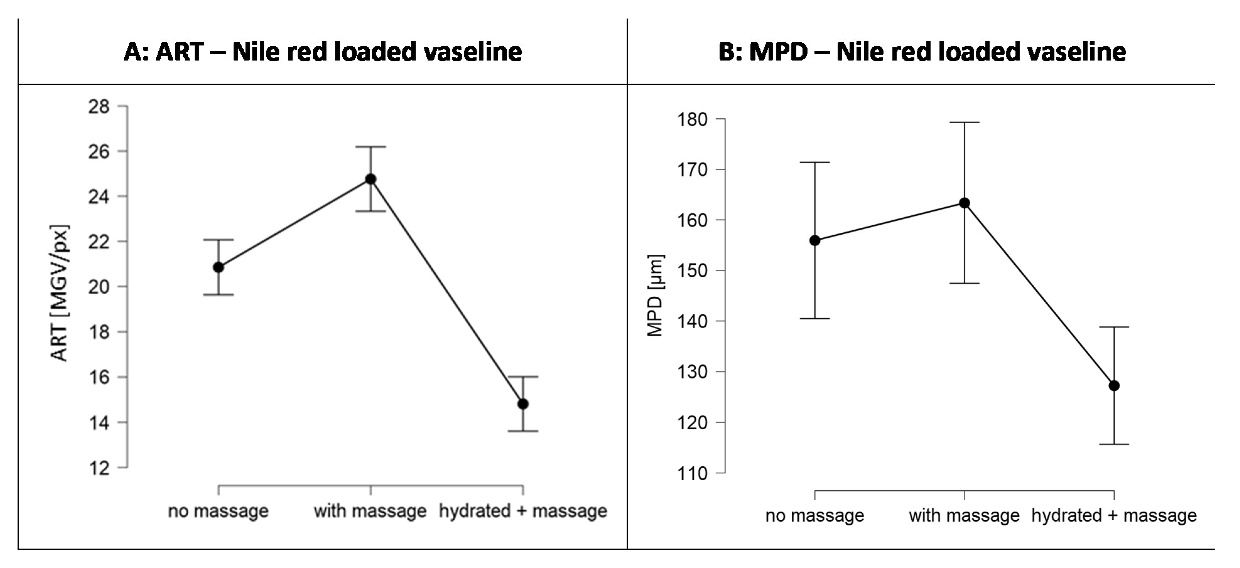
Based on the data obtained it can be concluded that the expected super-hydration effect by applying vaseline on hydrated skin with massage to improve the dermal penetration efficacy was not observed. Instead, it was found that penetration of the lipophilic AI surrogate was hampered by this procedure. Hence, improved dermal drug delivery cannot be achieved by applying drug loaded vaseline on hydrated skin with massage. Instead, it can be concluded that drug loaded Vaseline should be applied on rather dry skin, where massage can be used to further improve the penetration results.
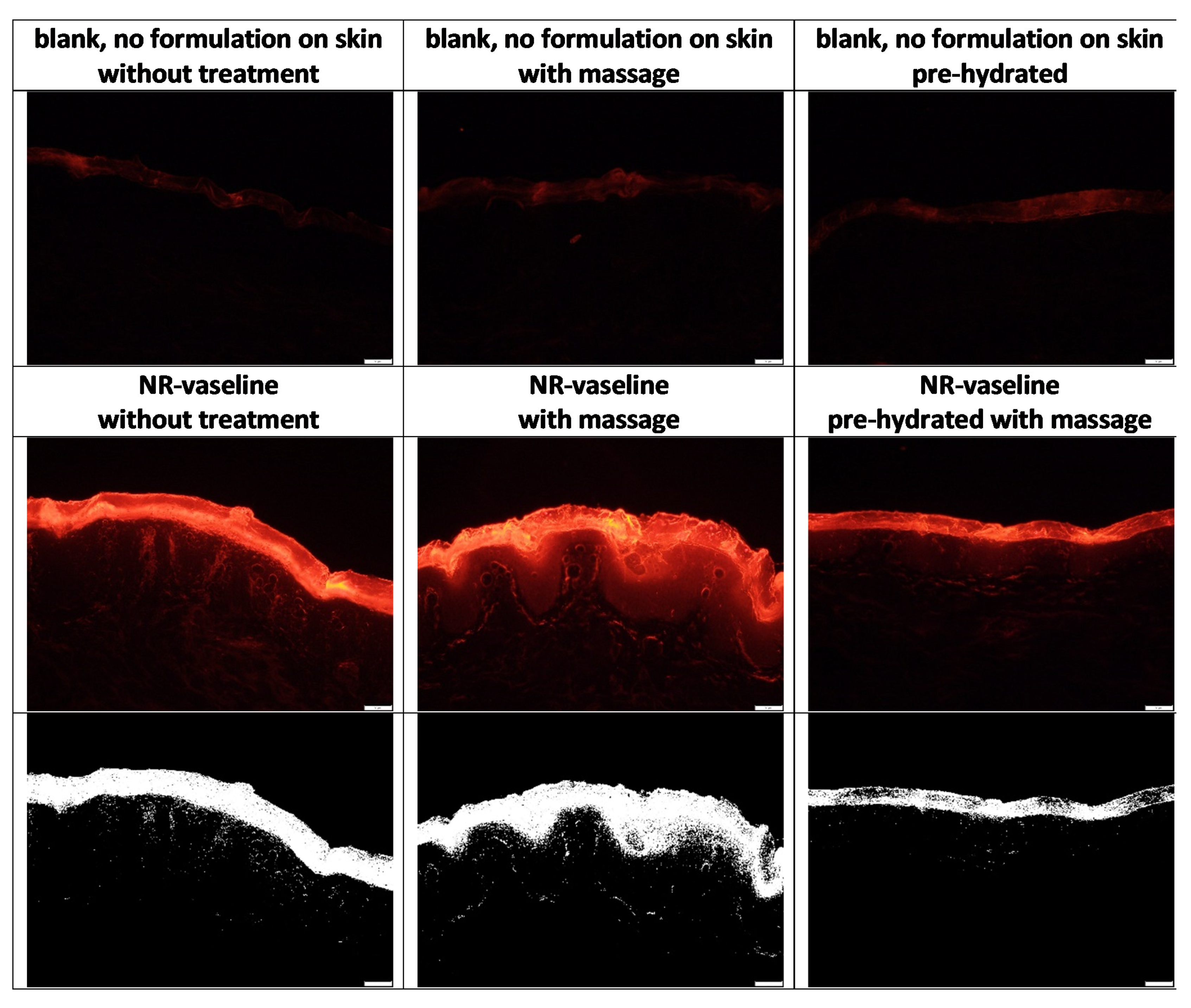
Despite these findings with important practical relevance, of course it is also interesting to understand the underlying mechanisms for the observed effects. After reconsideration of the results obtained it was hypothesized that the observed reduced penetration of Nile red from the Vaseline when applied on wet skin with massage (cf.
Figure 6) is caused by the formation of an aqueous layer on top of the skin. The aqueous layer acts as a water barrier not only for the paraffin but also for lipophilic AI surrogate Nile red. Hence, the hydrophilic barrier formed, hampers the penetration of the lipophilic dye.
The increased penetration efficacy, and especially the increased penetration depth when applied on dry skin with massage (cf.
Figure 6), cannot be explained by this theory, because the squeezed in water from deeper layers should at least cause a reduced penetration depth of Nile red when compared to the unmassaged, dry skin. Therefore, as an alternative, possible explanation for the increased penetration efficacy of Nile red after massage on dry skin, the penetration of the mineral oil after syneresis into the skin needs to be considered. The penetration of paraffin into the SC was confirmed by the data obtained from the biophysical skin properties (cf.
Section 3.3.2) and also previous studies already confirmed the penetration of paraffin into the skin [
11,
12,
22,
23]. Massage was shown to foster local syneresis of Vaseline and to increase the penetration of the expulsed mineral oil into the skin. According to Neubert and Wepf, active compounds might penetrate via the hydrophilic pathway of the skin and via the hydrophobic pathway [
21]. Therefore, in case of the penetrated non-polar mineral oil and/or mineral waxes, it can be assumed that they penetrate via the hydrophobic pathway (
Figure 9D).
Based on the images obtained from fluorescence microscopy (
Figure 4), it can be seen that the mineral oil acts as a solvent for the Nile red. Therefore, it appears reasonable that the AI-surrogate being dissolved in the mineral oil penetrates the skin together with the hydrocarbons (paraffin) and will not be left behind on top of the skin. Hence, it can be hypothesized that the Nile red enters the skin not only via passive diffusion but also via convection, i.e., via a solvent drag mechanism. Drug uptake via convection and solvent drag mechanisms are well described effects for oral application but were so far very rarely proposed, e.g., only for ethanolic solvents, for dermal application [
24,
25,
26].
The assumption that Nile red enters the skin via a solvent drag mechanism together with the mineral oil in which it is dissolved is underlined by the different penetration patterns observed from the skin biopsies treated with Nile red loaded Vaseline with and without massage (
Figure 5—lower,
Figure 10). When the Nile red loaded Vaseline was applied on wet skin with massage, the Nile red is mainly located in the SC and almost no penetration into the epidermis and deeper layers of the skin is visible. This can be explained by the formation of a “waterfront” on top of the skin that is created during the massage. The waterfront hampers the penetration of the lipophilic dye. If a deeper penetration is detected (
Figure 9B,C), the penetration pattern is characterized by single spots that penetrate individually into the skin. In contrast, “reddish waves” are clearly visible in the skin when the Nile red was applied on dry skin with massage. The waves seem to form a rim at the basal layer of the epidermis. After the rim (corresponding to the basal layer of the epidermis), the penetration of Nile red appears similar to the penetration pattern seen for the hydrated skin after the Nile red passed the stratum corneum. Hence, for the dry and massaged skin, it seems as the Nile red uses a “lift” until the end of the epidermis. The lift seems to be not present in the hydrated skin and only to a small extend in the dry skin were the Nile red loaded Vaseline was applied without massage (
Figure 10). The “lift” can be considered to be the penetrated mineral oil, which’s penetration seems to stop at the end of the basal layer.
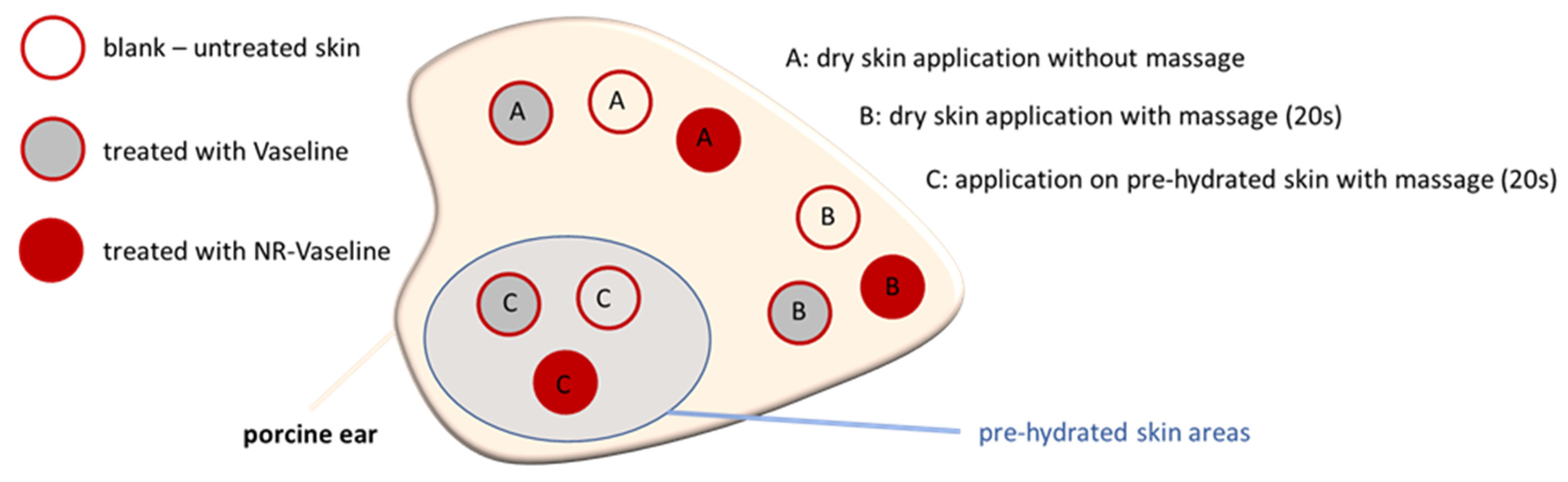
The proposed penetration of paraffin into the skin and the solvent drag and waterfront hypotheses were proven in the next steps of the study. First, the penetration of paraffin and also a possible penetration of water into the skin was investigated by applying pure paraffin and pure water without any dye on skin and by comparing the changes in the skin autofluorescence between untreated and treated skin. Secondly, the solvent drag and waterfront hypotheses were proven by adding 0.003% (
w/
w) of a water-soluble, fluorescent dye (sodium-fluorescein) as a surrogate for a hydrophilic AI to Vaseline. Skin penetration experiments were performed similarly to those performed for the Nile red loaded Vaseline, i.e., on skin with and without massage and on pre-hydrated skin with massage (
Figure 1,
Figure 10,
Figure 11 and
Figure 12).
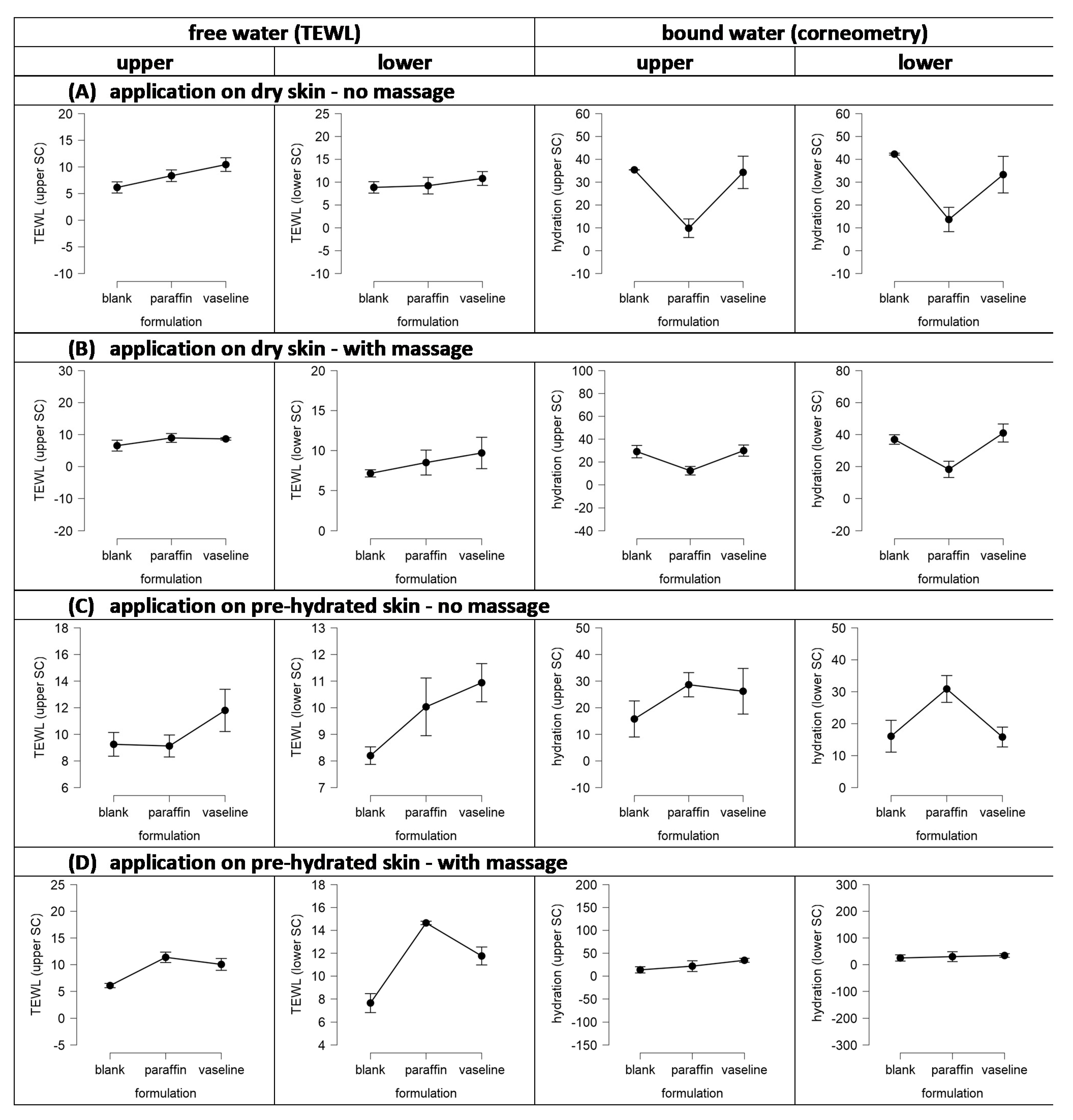
Both, the application of pure water and the application of pure paraffin resulted in significant changes in the autofluorescence of the skin (
Figure 11). Water reduced the autofluorescence of the skin and paraffin increased it (
Figure 11A). A more detailed analysis of the data revealed that the changes in the autofluorescence of the skin upon skin treatment with water were more pronounced in the deeper layers of the skin (
Figure 11B) and less pronounced in the stratum corneum (
Figure 11C). In contrast, changes in skin autofluorescence upon treatment with paraffin were less pronounced in the deeper layers of the dermis and most pronounced in the stratum corneum. Data therefore indicate that both, water and paraffin, penetrate into the skin. Water has no fluorescent properties and thus—once it penetrates into the skin tissue—decreases the autofluorescence of the skin tissue. Paraffin was found to possess a weak autofluorescence and thus increases the autofluorescence of the skin tissue [
27]. The strong increase in the autofluorescence of the stratum corneum and the very limited increase of the autofluorescence in the lower dermis upon the skin treatment with paraffin indicates, that the paraffin enters the stratum corneum but cannot penetrate into deeper layers of the skin. The data therefore substantiate the findings of previous studies, that could already show a penetration of paraffin and other liquids into the skin [
11,
12,
22,
23,
24,
25,
26,
28]. Further, data of this study also demonstrate that water can enter the skin if applied on skin. The differences in autofluorescence between untreated skin and skin treated with water in the lower dermis indicate that water can penetrate into deeper layers of the skin. The limited differences between untreated and water treated SC indicate that the hydration of the stratum corneum is only limited. This finding is in line with the data obtained from the analysis of the biophysical skin parameters (cf.
Section 3.3).
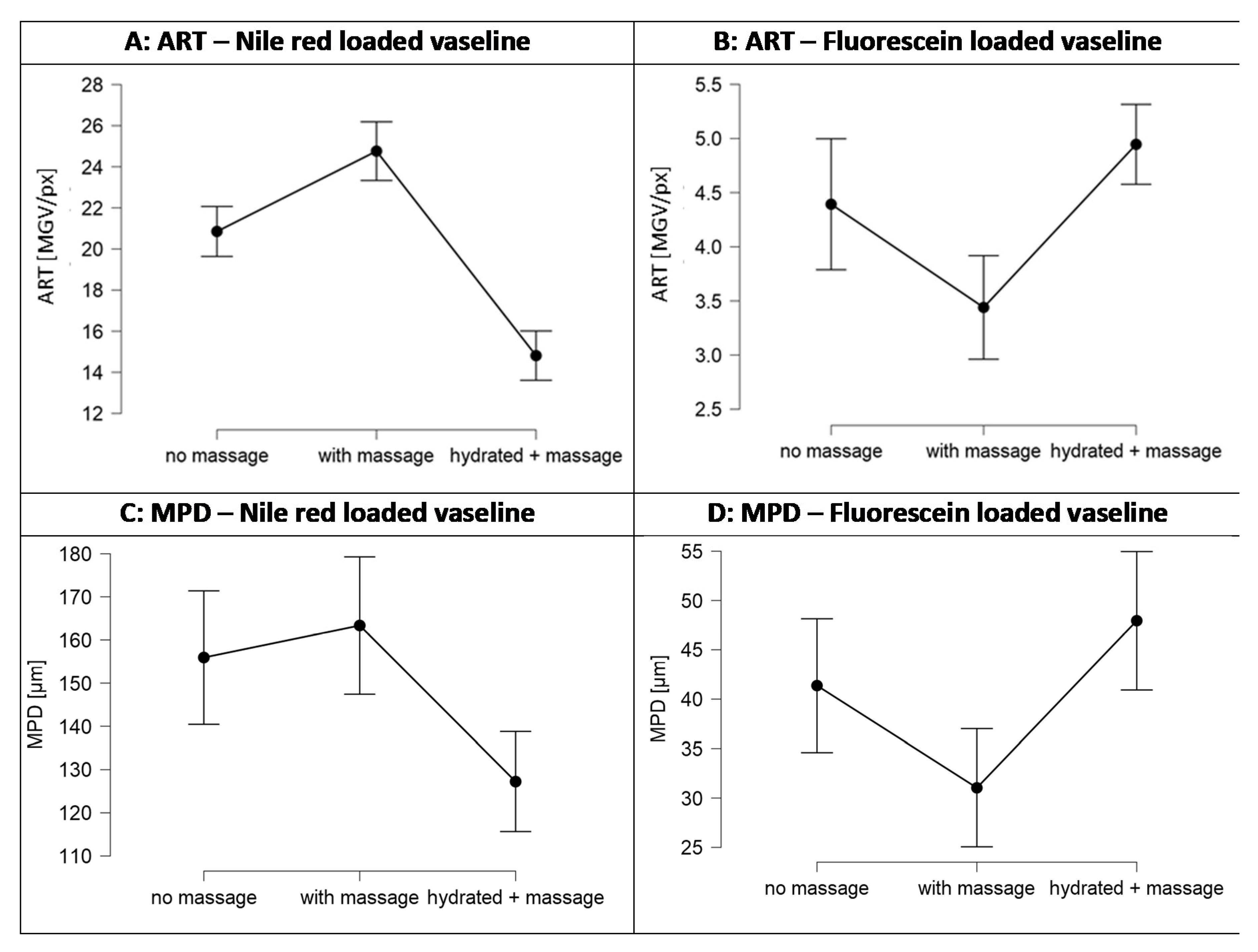
The solvent drag and waterfront hypotheses could also be confirmed, i.e., results obtained from the penetration studies with fluorescein loaded Vaseline prove the theory and confirm that the penetration of a hydrophilic AI is oppositely affected by skin hydration and syneresis of Vaseline due to massage. In fact, massage decreased the penetration of fluorescein and hydration increased it (
Figure 12B,D). The decrease in penetration depth due to massage was about 25% and the increase due to application on pre-hydrated skin was about 15% for fluorescein. In contrast, massage increased the penetration of Nile red by about 15% and application on pre-hydrated skin decreased the penetration by about 25% (
Figure 6 and
Figure 12A,C).
Data therefore demonstrate that both, skin condition and type of application, can strongly affect the penetration efficacy of active compounds into the skin and confirm that an “all fits one” approach, i.e., massage and application on moist skin, is not appropriate, because—depending on the type of AI and the skin condition—it might have penetration enhancing or penetration reducing effects. The mechanism of an aqueous barrier between skin and topical formulation, which was found in this study, can be conclusively linked to the observations by H. Maibach and its co-workers, which stated: “occlusion is widely utilized to enhance the penetration of applied drugs in clinical practice; however, occlusion does not increase the percutaneous absorption of all chemicals” [
29].
The results of this study fully confirm the observed effects by Maibach and co-workers and shed new light on the use of occlusive vehicles and skin massage for the improved dermal delivery of active compounds. In addition, data provide first evidence, that active compounds must not necessarily leave the vehicle to penetrate into the skin as they can directly penetrate along with the vehicle or parts of the vehicle in which they are dissolved. Based on the results obtained in this study it can be expected that many liquid ingredients of topical formulations might be able to penetrate into the skin. Despite paraffin, which was shown to penetrate the skin in this study, also other lipids (for example oils with low viscosity, e.g., medium chain triglycerides) can penetrate the skin [
27,
28] and thus allow for an enhanced penetration of lipophilic compounds which can be dissolved in these oils. The study could also demonstrate that water can penetrate into the skin. Thus, in theory, compounds being dissolved in the water might directly penetrate into the skin. These theories should be evaluated in more detail in future studies.
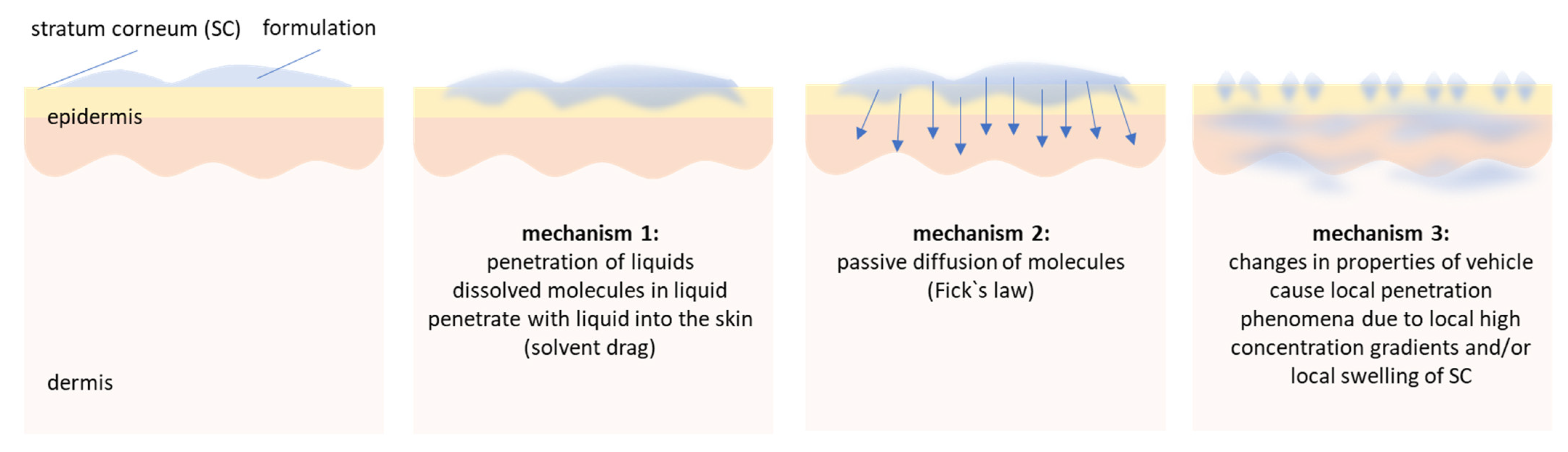
Nevertheless, data of this study already allow to conclude that penetration of active compounds from vehicles is not a static process that can be simply explained by Fick’s law of diffusion and by a simple consideration of distribution coefficients of the active compound between vehicle and skin. Dermal penetration of active compounds is rather a highly dynamic process that is composed of a complex interplay between skin, vehicle, type of application and changes in skin, vehicle and drug distribution after topical application. Based on the data so far it can be assumed that dermal penetration of active compounds from vehicles that are applied on the skin can be considered to be composed of at least three different mechanisms.
The first mechanism involves the expulsion of low viscous liquids, e.g., water or oils, from the vehicle and their subsequent penetration into the skin. Active compounds that are dissolved in these liquids can enter the skin via convection, i.e., a solvent drag mechanism either via the polar or non-polar route. The second mechanism is the classical penetration pathway, i.e., passive diffusion of the AI from the vehicle into the skin. The third mechanism involves changes in skin properties and/or the vehicle that can influence and modify the dermal penetration of active compounds over time. For example, the penetration of the liquids into the skin, the non-penetrated formulation on top of the skin and also mechanical skin treatments (e.g., massage) can modify the skin properties and thus can alter the passive diffusion of the AI. Finally, when the formulation on top of the skin dries out, other phenomena might start to modulate the penetration efficacy. Possible changes include coalescence of oil droplets in emulsions and/or—in case the formulation contains particulate materials—the formation of an aqueous meniscus [
6]. The aqueous meniscus connects particles to the skin, causes a local swelling of the SC and fosters the penetration of active compounds that are dissolved in the liquid of the meniscus (
Figure 13).
It can be assumed that a detailed and mechanistic understanding of these complex processes will allow for the development of topical products with tailor-made penetration profiles and optimal skin-caring properties at the same time. More research needs to be conducted to understand all these mechanisms, effects and possible interdependencies that orchestrate together the dermal penetration of active compounds in more detail.


 and
and 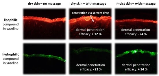


