
Polymeric Nanomaterials for Efficient Delivery of Antimicrobial Agents


1
School of Public Health and Management, Ningxia Medical University, Yinchuan 750004, China
2
State Key Laboratory of High-Efficiency Coal Utilization and Green Chemical Engineering, Ningxia University, Yinchuan 750021, China
*
Author to whom correspondence should be addressed.
Pharmaceutics 2021, 13(12), 2108; https://doi.org/10.3390/pharmaceutics13122108
Submission received: 5 November 2021
/
Revised: 29 November 2021
/
Accepted: 3 December 2021
/
Published: 7 December 2021
(This article belongs to the Special Issue Functional Polymers as Innovative Tools in the Delivery of Antimicrobial Agents)
Abstract
:Bacterial infections have threatened the lives of human beings for thousands of years either as major diseases or complications. The elimination of bacterial infections has always occupied a pivotal position in our history. For a long period of time, people were devoted to finding natural antimicrobial agents such as antimicrobial peptides (AMPs), antibiotics and silver ions or synthetic active antimicrobial substances including antimicrobial peptoids, metal oxides and polymers to combat bacterial infections. However, with the emergence of multidrug resistance (MDR), bacterial infection has become one of the most urgent problems worldwide. The efficient delivery of antimicrobial agents to the site of infection precisely is a promising strategy for reducing bacterial resistance. Polymeric nanomaterials have been widely studied as carriers for constructing antimicrobial agent delivery systems and have shown advantages including high biocompatibility, sustained release, targeting and improved bioavailability. In this review, we will highlight recent advances in highly efficient delivery of antimicrobial agents by polymeric nanomaterials such as micelles, vesicles, dendrimers, nanogels, nanofibers and so forth. The biomedical applications of polymeric nanomaterial-based delivery systems in combating MDR bacteria, anti-biofilms, wound healing, tissue engineering and anticancer are demonstrated. Moreover, conclusions and future perspectives are also proposed.
1. Introduction
Infectious diseases induced by bacteria, virus and fungi have been considered as one of the biggest enemies that threatened the lives of human beings for a long time [1]. Since the discovery of penicillin in 1928, antibiotics have played an unprecedented role in saving lives of human beings and caused revolutionary changes in medicine. However, with overuse and improper use of antibiotics, the emergence of bacterial drug resistance is becoming a severe problem. In particular, combating MDR bacteria such as methicillin-resistant Staphylococcus aureus (S. aureus) (MRSA) has drawn wide attention and efforts [2,3]. Non-antibiotic antimicrobial agents such as AMPs [4,5,6], silver nanoparticles (AgNPs) [7,8,9], metal oxides [10,11,12], antimicrobial peptoids [13,14] and polymers [15,16,17,18] are alternatives for treating infectious diseases that kill bacteria in a physical manner and avoid the generation of drug resistance. For instance, cationic compounds including AMPs, antimicrobial peptoids and polymers, as well as their corresponding nanostructures, strongly interacted with the negatively charged cell membrane of bacteria, resulting in the disruption of the cell membrane and outflow of the content of bacteria [19,20]. Metal (oxide) nanoparticles such as widely studied AgNPs kill bacteria via heavy metal ions induced by the denaturation of proteins or genetic materials, while ZnO and TiO2 nanoparticles eliminate bacteria by reacting with reactive oxygen species (ROS) generated from photocatalytic process [12,21]. Moreover, emerging antimicrobial agents including gases, photothermal sensitizers and carbon materials were also developed to combat bacterial infections [17,22,23,24].
The efficient delivery of antimicrobial agents to the target preventing the defense system of bacteria including efflux pump, degrading enzymes and resistance genes is critical for reducing the emergence of drug resistance [25,26,27]. Polymeric nanomaterials are promising vehicles for the efficient delivery of antimicrobial agents due to their tailorable chemical compositions, microstructures and biological properties for a wide range of biomedical applications [28,29,30]. For instance, low dimensional nanostructures including dendrimers [31,32], polymeric nanoparticles [33,34], micelles [35,36], vesicles [37,38] and nanogels [39,40] have shown superiorities in the delivery of antimicrobial agents to the areas of infections and on-demand release. Polymeric nanofibers and hydrogels are beneficial for the long-term release of antimicrobial agents and wound coverage [41,42]. Very recently, metal organic frameworks (MOFs) have attracted attention as emerging carriers for the efficient delivery of metal ions, metal nanoparticles, antibiotics and enzymes due to their highly porous structures [43,44,45,46]. There are several advantages of using polymeric nanomaterials as carriers to accomplish the on-demand delivery of antimicrobial agents: (i) reduced dosage and drug resistance; (ii) increased in vivo circulation stability; (iii) enhanced penetration ability; (iv) prolonged antimicrobial performance; and (v) improved bioavailability. Therefore, apart from the wide attentional broad spectrum antimicrobial properties, biomedical applications including combating MDR bacteria, anti-biofilm, anticancer, wound healing and tissue engineering based on the polymeric antimicrobial agent delivery systems have been rapidly developed [47,48,49,50].
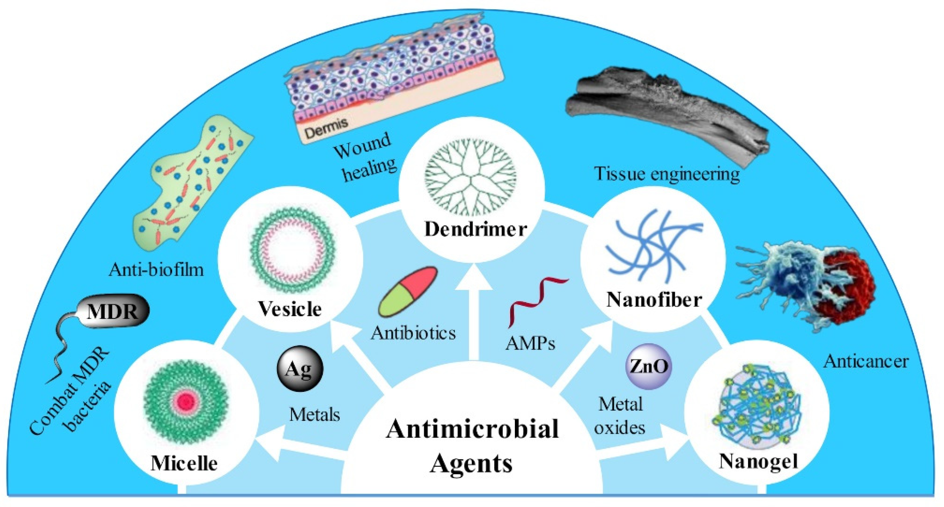
In this review, we aim to present the state of the art of polymeric nanomaterials as carriers for the efficient delivery of antimicrobial agents from the following aspects: (1) classification of polymeric nanoparticles based on their nanostructures; (2) the structural features and corresponding advantages in delivery of antimicrobial agents; and (3) biomedical applications benefiting from the constructed delivery systems, as illustrated in Figure 1.
2. Efficient Delivery of Antimicrobial Agents by Diverse Polymeric Nanostructures
2.1. Self-Assembled Polymeric Nanoparticles
Polymer self-assembly has been recognized as one of the most versatile strategies for preparing soft nanomaterials with various morphologies and functionalities from small building blocks [51,52,53,54,55,56]. Typically, polymer micelles and vesicles are the most easily obtained and widely studied nano-objects due to their well-organized structures [57,58,59,60]. Polymer micelles are formed by the regular arrangement of building blocks with hydrophobic components forming the cores and hydrophilic polymer chains covering the surface. Moreover, the hydrophobic cores facilitated the efficient encapsulation of hydrophobic drugs [61,62], while polymer vesicles are hollow bilayer nanostructures with hydrophobic membranes, hydrophilic coronas and interior cavities, endowing them with superiorities in loading and delivering hydrophobic, hydrophilic and large-sized cargoes [63,64,65,66]. The design of polymer vesicles for meeting the requirements of various applications mainly focuses on the chemical composition and structure of coronas and membranes, such as permeability and homogeneity of the membrane, symmetricity of the corona and so forth [67,68,69]. Despite the wide applications of polymer micelles and vesicles in cancer therapy, gene delivery and cell imaging, they also exhibit considerable potentials in the efficient delivery of antimicrobial agents [36,70,71].
Polymer micelles, core-shell nanostructures that usually self-assembled from amphiphilic block copolymers, are regarded as one of the most extensively studied nanostructures for antimicrobial agent delivery [35,72]. Typically, hydrophobic antibiotics, AMPs and AgNPs can be loaded in the hydrophobic core, and amphiphilic antimicrobial molecules are usually decorated on the surface of polymer micelles by covalent bonding or electrostatic interaction [73,74,75,76]. For instance, a poor water-soluble anti-fungi agent, amphotericin B, could be encapsulated in the core of micelles and showed ultra-long sustained release for 150 h, resulting in reduced hemotoxicity and comparable anti-fungi activity compared with free amphotericin B [77]. Xiong and coworkers [78] functionalized terpyridine on the surface of polymer micelles; after chelating with Fe(II), the micelles displayed excellent biofilm inhibition activity up to 99.9% at a concentration of 128 μM. Recently, Lee et al. [79] prepared AMP-covered micelles by the co-assembly of chimeric antimicrobial lipopeptide and a biodegradable amphiphilic polymer (poly-(lactic-co-glycolic acid)-b-poly(ethylene glycol), (PLGA-b-PEG)). The chimeric peptide HnMc and PEG formed the shell of the micelles in which PEG protected HnMc from proteolytic degradation. Moreover, HnMc on the surface could help micelles in preferentially binding and killing bacteria. Due to the synergy between HnMc and PEG, the micelles targeted a wide range of bacteria preferentially including Escherichia coli (E. coli), Listeria monocytogenes, Pseudomonas aeruginosa (P. aeruginosa) and S. aureus instead of mammalian cells. Moreover, in vivo experiments also demonstrated superior anti-inflammatory effects of the micelles in a mouse model of drug-resistant P. aeruginosa lung infection with highly targeted abilities, as shown in Figure 2.
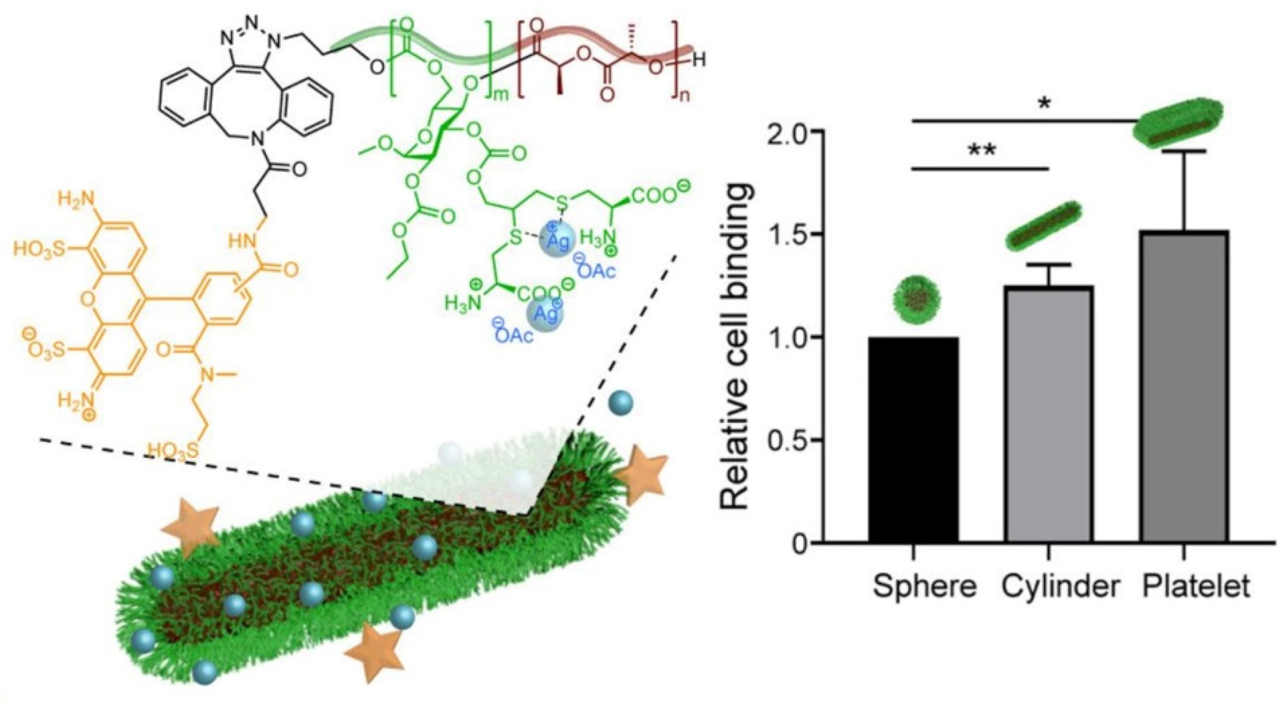
Very recently, Wooley and coworkers [80] fabricated spherical micelles, cylinders and nanoplates derived from the crystallization-driven self-assembly (CDSA) of an amphiphilic block copolymer composed of zwitterionic poly(ᴅ-glucose carbonate) and semicrystalline poly(ʟ-lactide) segments (PDGC-b-PLLA). As illustrated in Figure 3, fluorescent molecule and cysteine were modified on the polymer in order to afford tracing ability and to chelate with silver ions, respectively. The morphology of the nanostructures could be well controlled by the hydrophilic-to-hydrophobic ratios, which exhibited negligible cytotoxicity, immunotoxicity and cytokine adsorption. However, the nanostructures offered substantial silver ion loading capacity, extended release and in vitro antimicrobial activity. Compared with spherical micelles, the cylinders and nanoplates exhibited enhanced association with uroepithelial cells due to their high aspect ratio, resulting in improved inhibition of the growth of E. coli in recurrent urinary tract infections.
Compared with polymer micelles, polymer vesicles are closed hollow spheres with more complicated structures usually acting as simple mimics of biological cells [81]. There are three compartmentalized regions that should be considered for realizing different functions, namely the inner hydrophilic cavity, hydrophobic membrane and hydrophilic corona in contact with external environments [82]. Therefore, both hydrophilic and hydrophobic compounds and even nanoparticles could be encapsulated in the interior cavity or membrane of vesicles, respectively. Moreover, hydrophilic molecules could also be linked onto the coronas of polymer vesicles by covalent bonding. Considering the structural feasibility of polymer vesicles, a large variety of antimicrobial agents could be loaded and delivered to combat bacteria with high loading efficiency, controlled release manner, targeting capability and improved bioavailability [83,84]. For example, Du and coworkers [35] deposited ultrafine AgNPs with a diameter of 1.9 ± 0.4 nm on the membrane of polymer vesicles by in situ reduction of silver ions to inhibit the growth of Gram-negative and Gram-positive bacteria. Battaglia et al. [71] reported the intracellular delivery of metronidazole or doxycycline to P. gingivalis-infected oral epithelial cells by polymer vesicles, which were disassembled in early endosomes due to the acidic condition, resulting in the release of loaded cargoes.
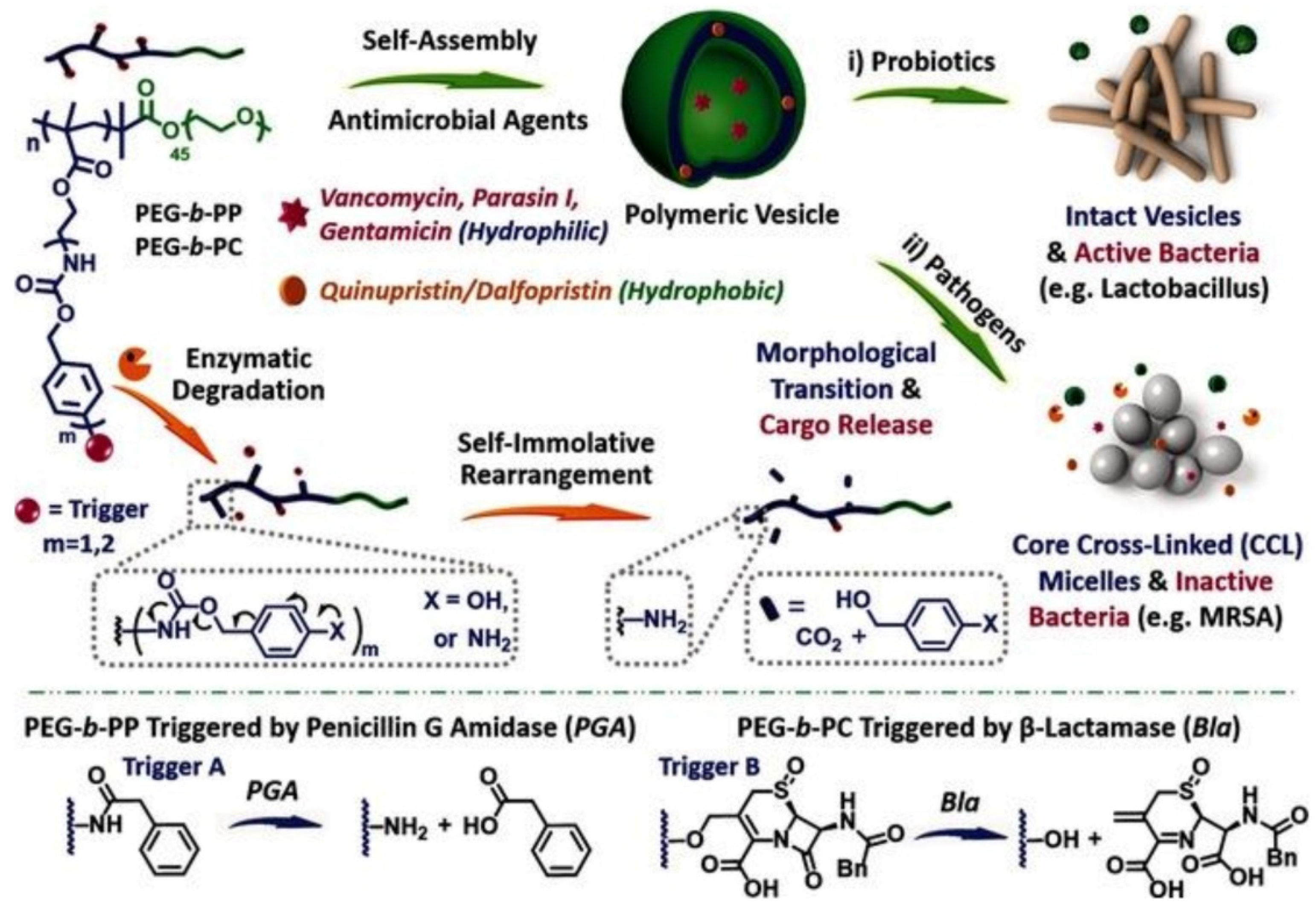
Recently, Liu and coworkers [85] designed enzyme-responsive polymer vesicles for bacterial strain-selective delivery of antimicrobials, as shown in Figure 4. Both hydrophilic and hydrophobic antimicrobials including vancomycin, gentamicin, quinupristin and dalfopristin could be encapsulated either in the interior cavity or membrane of the polymer vesicles with high efficiency. The PEG chains covered on the surface of the vesicle could reduce cytotoxicity and improve biocompatibility, while the self-immolative side chains could be degraded by penicillin Gamidase (PGA) and β-lactamase (Bla), which are overexpressed by drug resistant bacterial strains. Without the trigger by PGA and Bla, the encapsulated antimicrobials were well protected by vesicles. Upon being exposed to drug-resistant bacteria, the membrane of the vesicle was degraded, resulting in the sustained release of antimicrobials, as well as the elimination of bacteria. Considering that Bla is the main cause of bacterial resistance to β-lactam antibiotic drugs that are secreted by MRSA, selective antimicrobial activity of the antimicrobials-loaded vesicles was achieved.
Loading bioactive enzymes by polymer vesicles to generate antimicrobial active species triggered by external stimuli is another effective method for combating bacteria. For example, Blackman et al. [86] prepared glucose oxidase-loaded semipermeable polymer vesicles by polymerization-induced self-assembly inspired by honey. Hydrogen peroxide, an effective antimicrobial agent, could be generated in response to glucose to switch on antimicrobial activity of the vesicles. In the absence of glucose, the vesicles were completely nontoxic to bacteria, while the vesicles showed seven-log reduction in bacterial growth at high glucose concentrations against a range of Gram-negative and Gram-positive bacterial pathogens including S. aureus, S. epidermidis, E. coli and Klebsiella pneumoniae (K. pneumonia), even the MRSA clinical isolate. More importantly, the toxicity of the vesicle toward human fibroblasts at different dosage and glucose concentrations was also evaluated, demonstrating that the optimal concentration of the vesicle was 0.69 mg mL−1 at physiological blood glucose level to effectively eliminate bacteria while preserving good compatibility to mammalian cells.
2.2. Dendrimers
Dendrimers are highly branched, globular macromolecules with many arms emanating from a central core, which have shown unique structural properties such as high degree of branching, multivalency, globular architecture and well-defined molecular weight, rendering them promising scaffolds for drug delivery [87,88]. Many commercial drugs with anticancer and antimicrobial activity have been successfully loaded within dendrimers including poly(amidoamine) (PAMAM), poly(propylene imine) (PPI) and poly(etherhydroxylamine) (PEHAM), either via physical interactions or by chemical bonding to improve their water solubility [89]. Dendrimers themselves could be used as effective antimicrobial agents [90]. For instance, those with positively charged surfaces usually have strong interaction with negatively charged bacterial cell membranes, while those with metal cores can release active antimicrobial agents such as metal ions and ROS, resulting in the death of bacteria [91].
Moreover, antimicrobial agents including antibiotics, AMPs, AgNPs and metal oxide nanoparticles could be also effectively loaded by dendrimers [89,92]. For example, Tang et al. [93] prepared silver-dendrimer nanocomposites by loading AgNPs in low generation poly(amido amine) dendrimers. The AgNPs were formed by an in situ reduction of silver ions enriched by the amine groups of dendrimers. The factors that influenced the size of AgNPs were discussed, and the average diameter of the AgNPs could be controlled from 7.6 to 16.2 nm. The synthesized silver-dendrimer nanocomposite was used as antimicrobial agent in the fabrication of cotton fabrics, which exhibited excellent antimicrobial activity against both of E. coli and S. aureus. Recently, Huang and coworkers [94] reported PLGA nanoparticles and PAMAM dendrimers in order to effectively encapsulate and deliver platensimycin, a potent inhibitor for the synthesis of bacterial fatty acid, respectively, to combat MDR bacteria. Benefiting from the improved pharmacokinetics, both the platensimycin-loaded PLGA nanoparticles and PAMAM dendrimers showed enhanced antimicrobial activity and reduced cytotoxicity compared with free platensimycin, resulting in an efficient inhibition of S. aureus biofilm formation and the full survival of MRSA-infected mice.
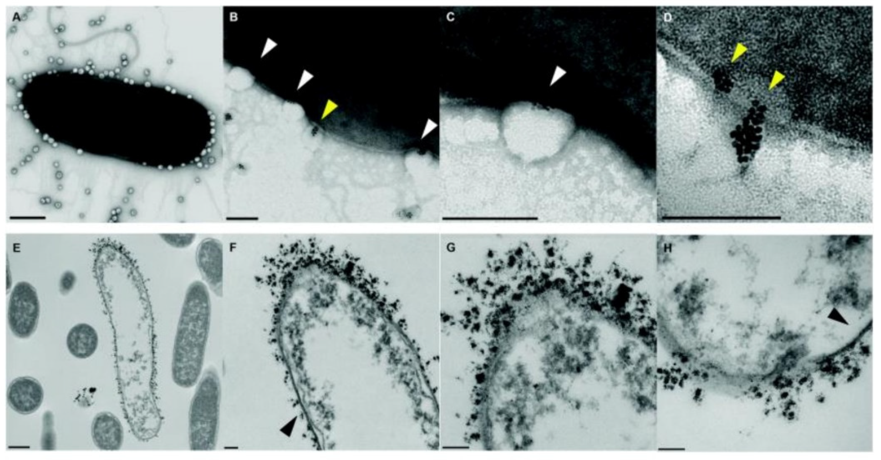
Dendrimers are ideal platforms for compacting and delivering deoxyribonucleic acids (DNAs) and ribonucleic acids (RNAs) for gene therapy due to their hyperbranched structure and strong positive charges, especially PAMAM [95,96]. Recently, antisense therapy strategy has been developed to treat bacterial infections facilitated by the dendrimers-based antisense delivery system [97]. For example, the G3 PAMAM dendrimer has good antimicrobial activity, as shown in Figure 5. However, the cytotoxicity of the G3 PAMAM dendrimer toward mammalian cells is also high. Luo et al. [98] conjugated LED209, a specific inhibitor of quorum sensor QseC of Gram-negative bacteria, onto the surface of G3 PAMAM to generate PAMAM-LED209 in order to reduce cytotoxicity to mammalian cells while retaining the excellent antibacterial activity of the G3 PAMAM dendrimer. In addition, PAMAM-LED209 also inhibited the virulence gene expression of Gram-negative bacteria and prevented the generation of drug resistance. As shown in Figure 5, compared with the control group (Figure 5A), entero-hemorrhagic E. coli (EHEC) were severely damaged after being treated with G3 PAMAM and G3 PAMAM-LED209 for 300 min (Figure 5B,C), demonstrating that G3 PAMAM-LED209 retained strong antibacterial activity toward resistant Gram-negative bacteria after functionalization of LED209. The induction of the resistance of G3 PAMAM-LED209 was also evaluated after 15 reproductions of bacteria, as illustrated in Figure 5D. The minimal inhibition concentration (MIC) of G3 PAMAM-LED209 barely changed, while the MIC values of classical antimicrobials, including ceftazidime, ampicillin and levofloxacin, increased by 8-fold to 64-fold. The cytotoxicity and antibacterial activity of terminally modified PAMAM are related to the conjugated ligand and degree of modification, as shown in Figure 5E. With an increase in modification ratio, the cytotoxicity of G3 PAMAM-PEG and G3 PAMAM-LED209 decreased dramatically to being almost nontoxic and then increased, while the antimicrobial activity of the G3 PAMAM-PEG and G3 PAMAM-LED209 decreased with an increase in modification ratio due to the shielding of positive charges. Therefore, there is an optimal modification ratio range for balancing cytotoxicity and antimicrobial activity, as pointed out by the arrow in Figure 5E. Moreover, the antibacterial potency of G3 PAMAM-LED209 is also higher than that of G3 PAMAM-PEG, which is indicated by area A and B in Figure 5E, demonstrating better biocompatibility and higher antibacterial potency than compared to G3 PAMAM-PEG.
2.3. Polymer Nanofibers
Polymer nanofibers are one dimensional nanostructures with large aspect ratio and high surface area and have shown significant potential for delivering antimicrobial agents locally into an infected area, especially in wound healing [42,99]. Typically, there are several methods for preparing nanofibers including self-assembly [100], template synthesis [101], phase separation [102] and electrospinning [103], among which electrospinning is a superior technique for preparing nanofibers with desired chemical compositions and diameters due to its simplicity and versatility [104,105,106]. Antimicrobial agents including antibiotics, AMPs, AgNPs and metal oxide nanoparticles could be incorporated into nanofibers by mixing with polymer precursors followed by electrospinning or attaching onto the surface of the nanofibers by noncovalent interactions or chemical bonds [107]. For instance, Schiffman et al. [108] immobilized zeolites nanoparticles with high silver ion change capability onto the surface of chitosan nanofibers. After ion exchange, silver ions were loaded in the zeolites to function as molecular delivery vehicles, and their ion release profiles and ability to inhibit E. coli were evaluated as a function of time. Interestingly, the zeolites immobilized on the nanofibers showed significantly enhanced antibacterial activity 11-times greater than that of the pure zeolites due to high porosity and hydrophilicity of the nanofibers.
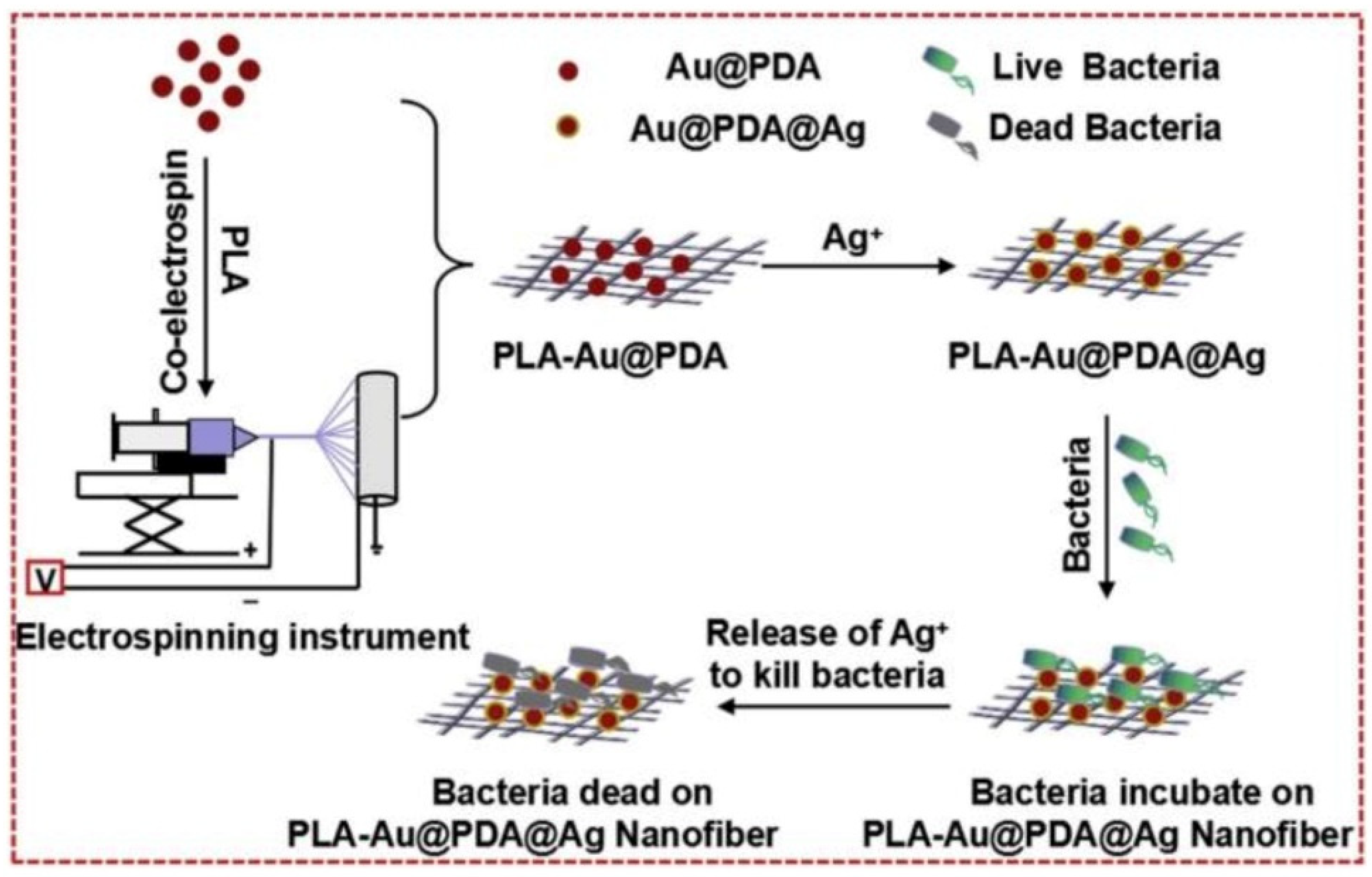
Recently, Tu and coworkers [109] reported the in situ deposition of AgNPs on gold/polydopamine core-shell nanoparticles encapsulated by poly(lactic acid) (PLA) nanofibers (PLA-Au@PDA@Ag), which could be applied to biological coatings for bacteriostatic functionality. The schematic illustration of the preparation and antimicrobial capability of the PLA-Au@PDA@Ag is presented in Figure 6. Chloroauric acid was reduced by ascorbic acid to afford gold nanoparticles. Following the polymerization of dopamine on the surface, Au@PDA core-shell nanoparticles formed, which were then mixed with PLA solution to produce PLA-Au@PDA hybrid nanofibers by electrospinning. Later, PLA-Au@PDA hybrid nanofibers were immersed in silver nitrate solution for in situ reduction of adsorbed silver ions into AgNPs to yield PLA-Au@PDA@Ag nanofibers. The hydrophilicity of the PLA-Au@PDA@Ag nanofibers significantly improved compared to that of PLA nanofibers, resulting in the promoted release of silver ions. Benefiting from the synergy between AuNPs, PDA and AgNPs, including AuNPs providing effective contact with microorganisms, PDA as binder was used to immobilize AgNPs and facilitated the release of silver ions; the PLA-Au@PDA@Ag nanofibers showed significant antibacterial ability against both of E. coli and S. aureus.
Due to their large exposed surface area and nanoporosity, polymer nanofiber meshes have shown distinct advantages in wound healing compared with hydrogels, films and foams [110]. The extracellular matrix (ECM) mimicking the structure of nanofibers facilitated the interaction with cells in the wound bed. Moreover, small molecules such as water, oxygen, nutrients and metabolic wastes could be efficiently exchanged due to the highly porous structure of nanofibers [111]. In order to promote the healing rate and elimination of bacteria, functional agents including enzymes, drugs and antimicrobial agents have been incorporated in polymer nanofibers. Rath et al. [112] loaded ZnO nanoparticles and cefazolin in the gelatin nanofibers to accelerate wound healing and prevented infection concurrently. Cefazolin was used to inhibit bacterial reproduction, while zinc cations could be released from ZnO nanoparticles to raise re-epithelialization, reduce inflammation and inhibit bacterial growth. Moreover, ROS was also produced by ZnO nanoparticles, thereby optimizing cell adhesion, proliferation and migration via growth factor mediated pathways, promoting the regeneration of the ECM.
2.4. Polymer Nanogels
Polymer nanogels are a class of nanoparticles composed of nanosized physically or chemically cross-linked hydrophilic or amphiphilic polymer networks [113]. They are of wide interest in various fields including drug delivery due to their flexible nanosize, good stability and high loading capacity, etc. [114]. As their analogues, polymer hydrogels have been widely used in antimicrobial applications due to their high water content, three-dimensional structure and stimuli-responsive sol-gel transition behavior [115]. There are several reviews summarizing the recent advances of antimicrobial polymer hydrogels [116,117,118]. Therefore, we will not discuss this part and focus on the nanogels as carriers for antimicrobial agent delivery in this section.
The stimuli-responsive swelling and collapsing of nanogels triggered by external stimuli including pH, temperature, enzymes or ionic strength render them ideal candidates in on-demand delivery and release of antimicrobial agents [119]. For instance, AMPs could be encapsulated in nanogels with high loading content via strong electrostatic interaction with negatively charged polymer chains, and they can be released when triggered by salt ions in physiological conditions [120,121]. El-Feky et al. [122] loaded silver sulfadiazine in alginate coated chitosan nanogels to heal burn wounds, and the nanogels showed a release profile of an initial burst followed by a slow and continuous release, resulting in excellent in vivo therapeutic efficacy.
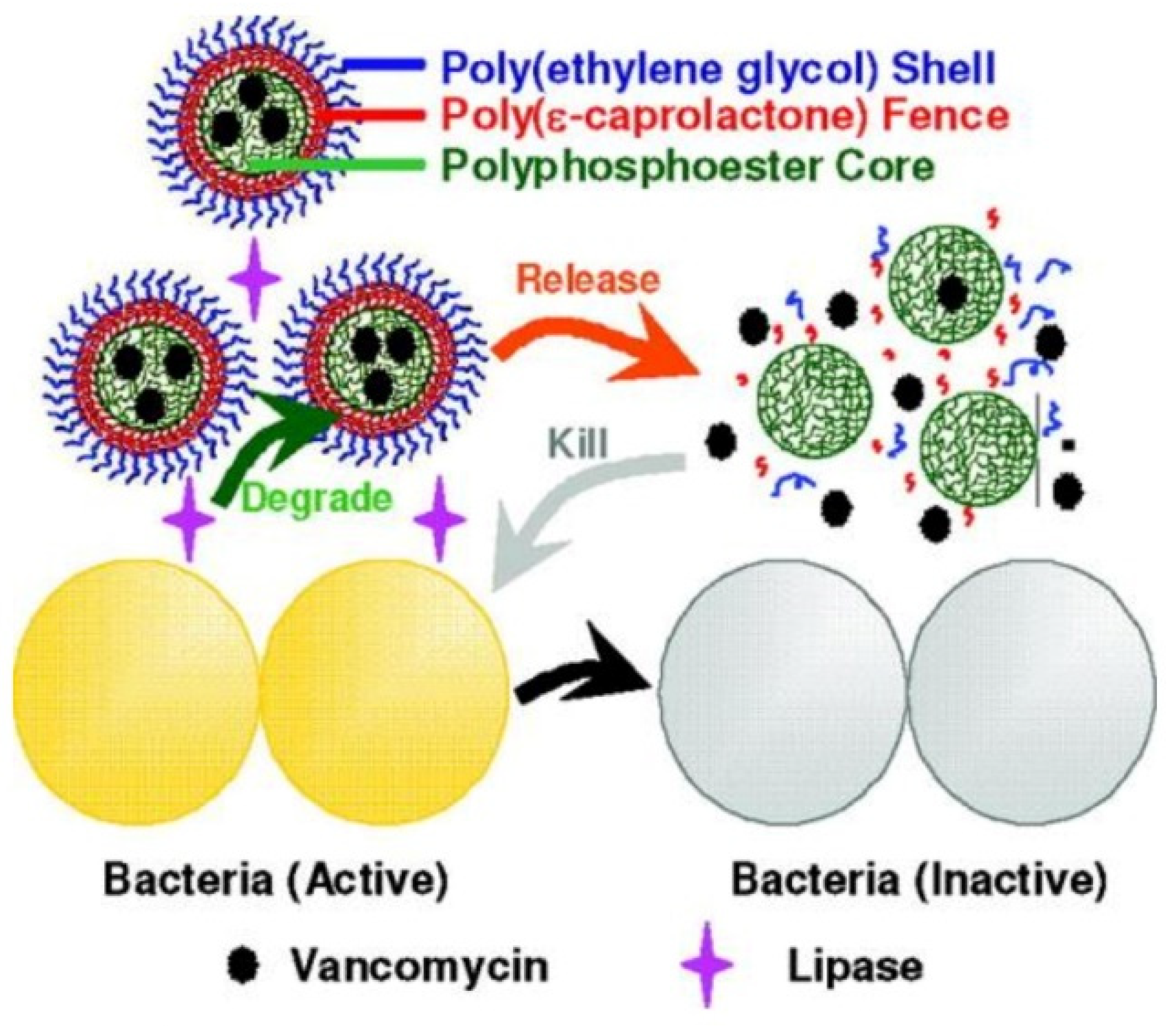
In addition, loading and delivery of antimicrobials including berberine, cyclodextrin, tetracycline hydrochloride and lincomycin hydrochloride by nanogels to combat bacteria and MDR bacteria were widely studied by Paunov, Schaefer and so forth [123,124,125,126]. Wang and coworkers [127] designed a lipase-sensitive polymeric triple-layered nanogel (TLN) formed by a cross-linked polyphosphoester core, poly(ε-caprolactone) (PCL) fence and PEG shell to encapsulate and deliver vancomycin, as illustrated in Figure 7. In aqueous solutions, hydrophobic PCL segments collapsed and covered the core to form a densely packed molecular fence to prevent the leakage of vancomycin. Once TLN was exposed to lipase secreting bacteria, the PCL chains were degraded to trigger the release of vancomycin, resulting in the inhibition of bacterial growth. They found that all encapsulated vancomycins were released within 24 h in the presence of S. aureus. Moreover, lipase secreting bacteria inside the cells could also be inhibited by TLN, demonstrating the versatility of the strategy of lipase-induced on-demand delivery and release of antimicrobials.
Recently, Knowles et al. [128] synthesized hybrid organic/inorganic AgNPs loaded nanofibrillar silk microgels to effectively eradicate bacteria by a two-step mechanism including bacterial adherence and consequent eradication. Compared with conventional AgNPs and silver ions, the hemolysis and cytotoxicity of hybrid microgels toward mammalian cell lines were significantly reduced due to the protection of the silk matrix. van Rijn and coworkers [129] prepared injectable nanogels loaded with hydrophobic triclosan in hydrophobic domains inside the nanogel networks through intraparticle self-assembly of aliphatic chains, which enhanced antimicrobial efficiency of triclosan up to 1000 times. As shown in Figure 8, a three-stage antimicrobial mechanism of the nanogels was proposed. Firstly, the nanogels attached onto the surfaces of the bacteria via electrostatic interaction to disturb the balance of charge density of the cell membranes. Secondly, bacterial cell membranes were destroyed by the insertion of hydrophobic aliphatic chains. Thirdly, loaded triclosan was released from the hydrophobic domains inside the nanogels and injected into the bacterial cell membranes, resulting in the death of bacteria. This approach dramatically increases the effective concentration of triclosan inside the bacteria. Moreover, both the MIC and minimal bactericidal concentration (MBC) against Gram-positive S. aureus and S. epidermidis decreased by three orders of magnitude compared with free triclosan, resulting in a decrease in the dosage of triclosan and reduction in drug resistance.
2.5. Hybrid Delivery Systems
Incorporating polymer nanoparticles including dendrimers, micelles and vesicles with high dimensional polymeric nanomaterials such as nanofibers, hydrogels and coatings as hybrid delivery systems could combine the advantages of both and achieve the hierarchical release of antimicrobial agents [130,131,132,133]. For example, Zhang and coworkers [130] developed a bioadhesive nanoparticle-hydrogel hybrid in order to enhance localized antimicrobial drug delivery. The antimicrobials ciprofloxacin was loaded in polymer nanoparticles that were embedded in hydrogels adhering to biological surfaces. Hydrogel network properties could be tailored independently for adhesion, which maintained controlled and prolonged ciprofloxacin release profiles from nanoparticles. Imae et al. [131] immobilized AgNPs-loaded amine-terminated fourth generation poly(amido amine) dendrimers onto the viscose rayon cellulose fibers, which exhibited excellent biocidal activity against E. coli with low weight percentage of silver of 0.2%. Du and coworkers [132] embedded penicillin encapsulated polypeptide polymersomes in the hydrogels to achieve quick and long-term antibacterial capability in which penicillin could be released from the hydrogel networks for quick bacteria elimination while the intrinsic antibacterial property of the polymersomes ensured long-term antibacterial activity. However, despite the advantages of hybrid delivery systems, the development of incorporation of different polymeric nanostructures as hybrid delivery platforms is still in its infancy, which may bring new opportunities in efficient loading and delivery of antimicrobial agents.
3. Biomedical Applications of Polymeric Nanomaterials Based Antimicrobial Agent Delivery Systems
3.1. Combating MDR Bacteria
The generation of drug resistance of pathogens is typically caused by the accumulation of drug resistant genes through mutation with the long-term use, especially overuse and improper use of antibiotics [25]. Therefore, the exploration of highly efficient delivery system to reduce dosage and improve bioavailability of antibiotics, as well as the delivery of non-antibiotic antimicrobial agents including AMPs, AgNPs, metal oxides, gases and so forth, is a promising strategy for reducing drug resistance [134,135]. Polymeric nanomaterial-based antimicrobial agent delivery systems have widely been used in combating MDR bacteria [136,137]. For instance, Liu et al. [138] conjugated quercetin and acetylcholine on the surface of selenium nanoparticles to combat MDR bacteria, which could effectively eliminate MRSA by destroying the membrane due to the synergy between quercetin, acetylcholine and selenium nanoparticles. Cationic polymeric star-shaped nanoparticles or dendrimers have also shown excellent antimicrobial activity against MDR bacteria even without loading antimicrobial agents [139,140,141], demonstrating the great potentials of polymeric nanomaterials in combating MDR bacteria.
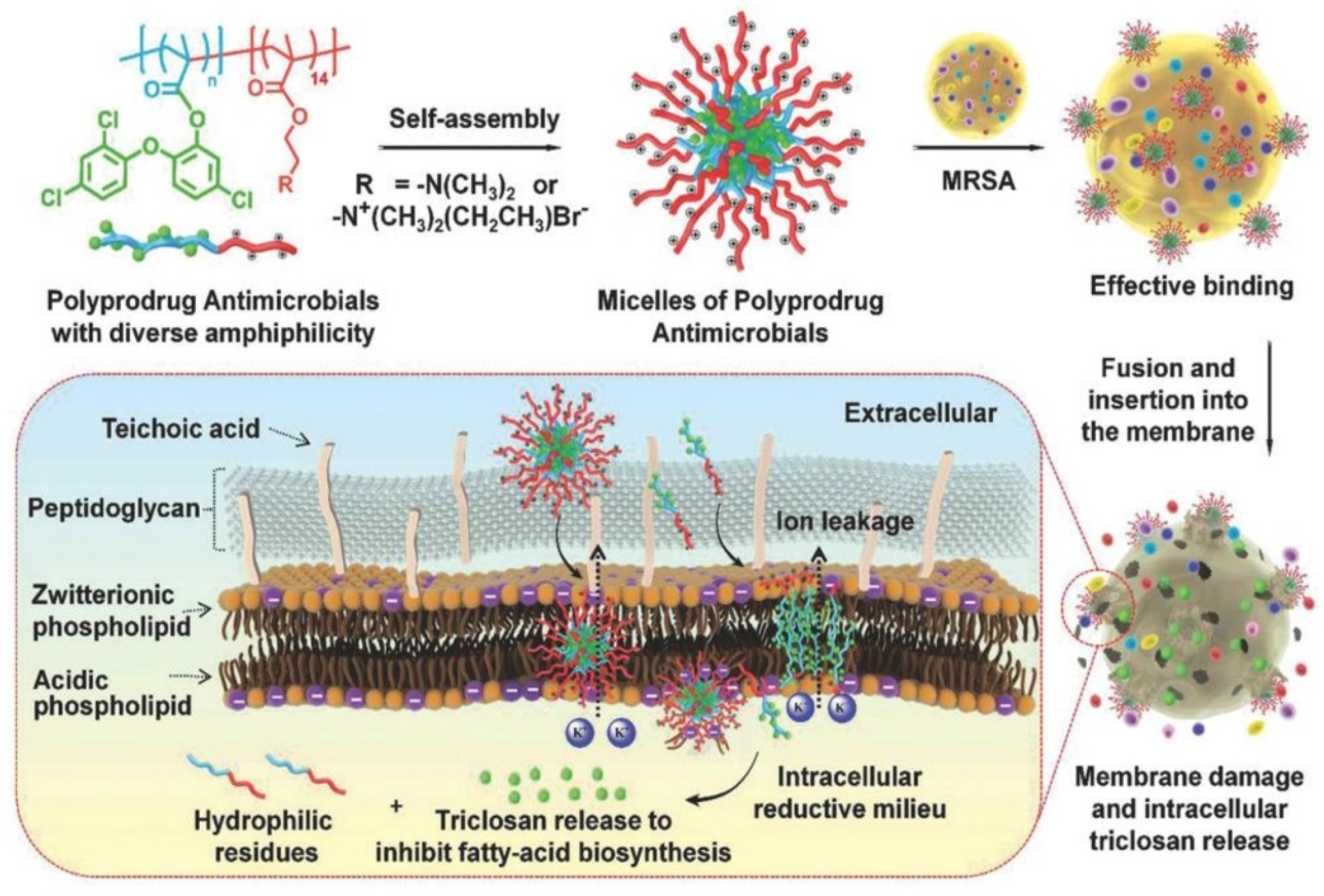
Hu et al. [142] prepared polyprodrug antimicrobials to combat MRSA by membrane damage and concurrent drug release, as shown in Figure 9. Triclosan was covalently linked with acrylic acid to produce a triclosan prodrug monomer (TMA). Then, TMA was copolymerized with quaternized N,N-dimethylaminoethyl methacrylate (QDMA), affording PQDMA-b-PTMA, which could self-assemble into prodrug micelles with positively charged surfaces. The hydrophilic–hydrophobic balance of the prodrug micelles was optimized to enhance interaction with bacterial cell membranes, resulting in improved antimicrobial activity. They proposed that the antimicrobial mechanism was as follows: (1) the prodrug micelles attached onto the surface of MRSA due to strong electrostatic interaction; (2) the prodrug micelles fused with and inserted into the cell membrane of MRSA; (3) the cell membrane of MRSA was damaged due to charge disorder, and prodrug micelles were encapsulated into the cell; (4) prodrug micelles were disassembled, and the linkage between triclosan and acrylic acid was broken due to the reductive milieu environment, resulting in the in situ release of triclosan and death of MRSA. It was noteworthy that no detectable resistance was observed due to the synergistic antibacterial mechanism, and prodrug micelles exhibited remarkable bacterial inhibition and low hemolysis toward red blood cells compared with commercial triclosan and vancomycin.
The combination of different classes of antimicrobial agents such as antimicrobials and AgNPs could afford synergistic effects, resulting in the efficient inhibition of MDR bacteria that is far better than its individual components [143,144]. Webster and coworkers [145] prepared polymer vesicles to co-deliver ampicillin and AgNPs simultaneously in the hydrophilic cavity and hydrophobic membrane, respectively. The AgNPs-embedded polymersomes exhibited potent antibacterial activity against E. coli transformed with a gene for ampicillin resistance in a dose-dependent fashion, while the free ampicillin, AgNPs decorated polymersomes without ampicillin and ampicillin loaded polymersomes without AgNPs had no effect on bacterial growth. TEM images in Figure 10 revealed that the interactions between vesicles, AgNPs and bacterial cells might result in the deformation and disruption of bacterial envelopes and consequently result in the death of bacteria. Later, the same group [146] functionalized proline-rich AMP PR-39 on the corona of polymer vesicles with AgNPs embedded in the membrane to combat MRSA with a AMP/AgNPs ratio-dependent behavior. A ratio of AgNPs-to-AMP of 1:5.8 corresponding to 11.6 μg mL−1 of AgNPs and 14.3 × 10⁻⁶ M of AMP exhibited the best MRSA inhibition activity, demonstrating the potentials of binary or ternary antimicrobial agent co-delivery systems in combating MDR bacteria.
3.2. Anti-Biofilm
Biofilms are matrix-enclosed communities of bacteria that show increased drug resistance and capability to evade the immune system [47]. It has been widely recognized that bacteria exist in the form of biofilms in many instances, which is hard to eliminate due to the protection of extracellular polymeric substances (EPS), a complex matrix composed of proteins, nucleic acids, phospholipids, polysaccharides, blood components and humic substances produced by bacteria [147]. Therefore, it is difficult for antimicrobials to penetrate the EPS to kill bacteria, resulting in the occurrence of drug resistance. The efficient delivery of antimicrobial agents by polymeric nanomaterials is considered a promising strategy for penetrating the biofilm and delivering antimicrobial agents to the deep end of the matrix to kill pathogens [148,149,150]. For example, Deoxyribonuclease I functionalized ciprofloxacin-loaded PLGA nanoparticles were prepared to target and disassemble the P. aeruginosa biofilm by degrading extracellular DNA that stabilizes the biofilm matrix and released ciprofloxacin inside the biofilm to effectively eliminate P. aeruginosa, as reported by Torrents and coworkers [151].
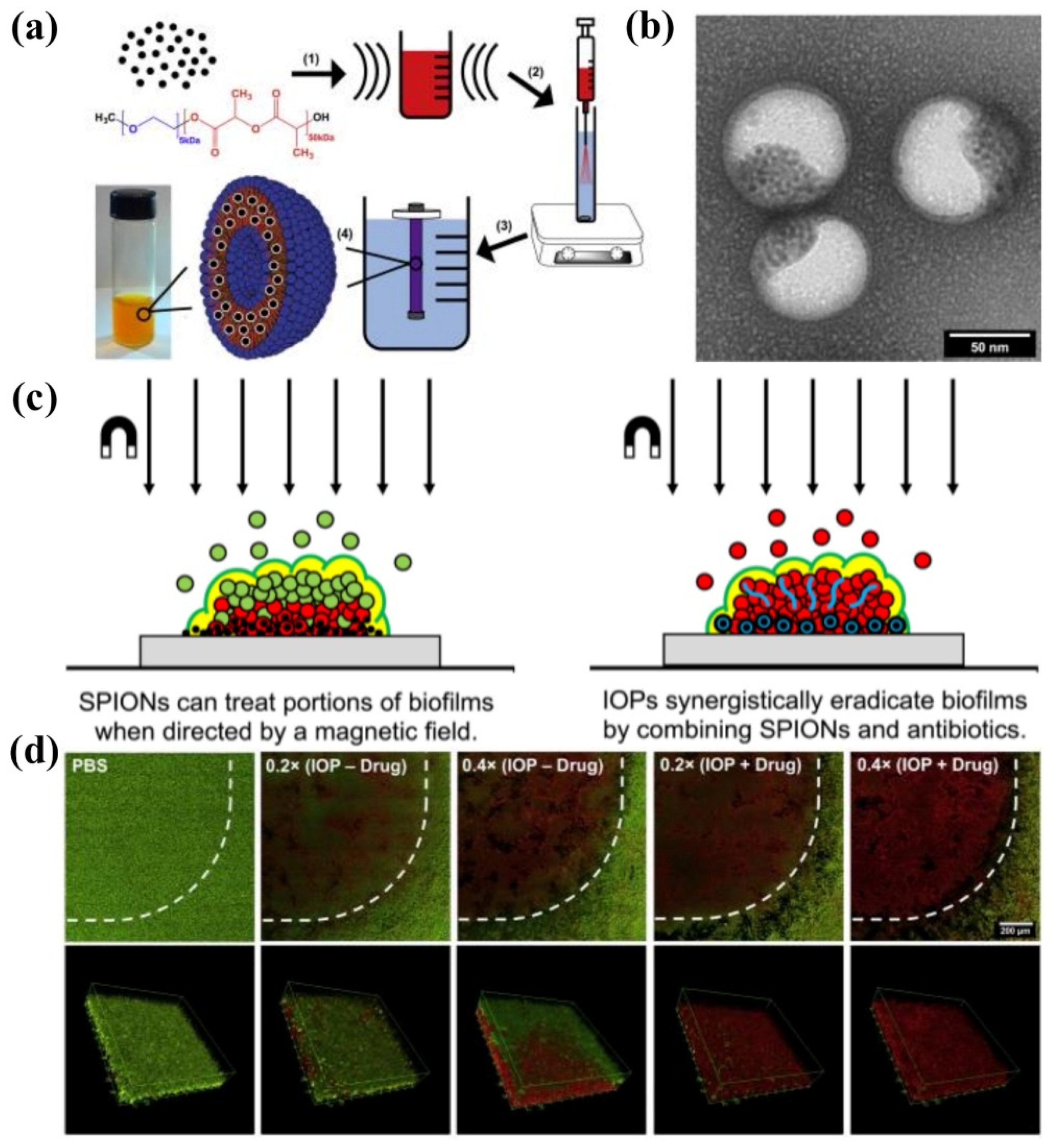
Webster et al. [152] prepared bifunctional polymersomes with methicillin encapsulated in the hydrophilic cavity and superparamagnetic iron oxide nanoparticles (SPIONs) embedded in the membrane, as illustrated in Figure 11. The iron oxide-encapsulated polymersomes (IOPs) penetrated into the S. epidermidis biofilm with high efficiency, promoted by external magnetic field. Comparing with individual SPIONs, methicillin and SPION co-encapsulated polymersomes showed enhanced penetration capability up to 20 μm due to the improved relaxivity and magneticity (Figure 11c). Thus, methicillin could be released into the deep end of the biofilm, resulting in the effective eradication of pathogens. The confocal microscopy images and the 3D reconstructions of z-stacks of the bacterial biofilm revealed the capability of IOPs to eradicate biofilms with and without methicillin, as shown in Figure 11d. When there was no methicillin, only bacteria in the bottom layer of the biofilm were killed. On the contrary, all bacteria throughout the biofilm were eliminated by the methicillin loaded IOPs. These organic/inorganic hybrid nanocarriers showed great promise as new weapons for eradicating persistent biofilm or drug-resistant bacteria.
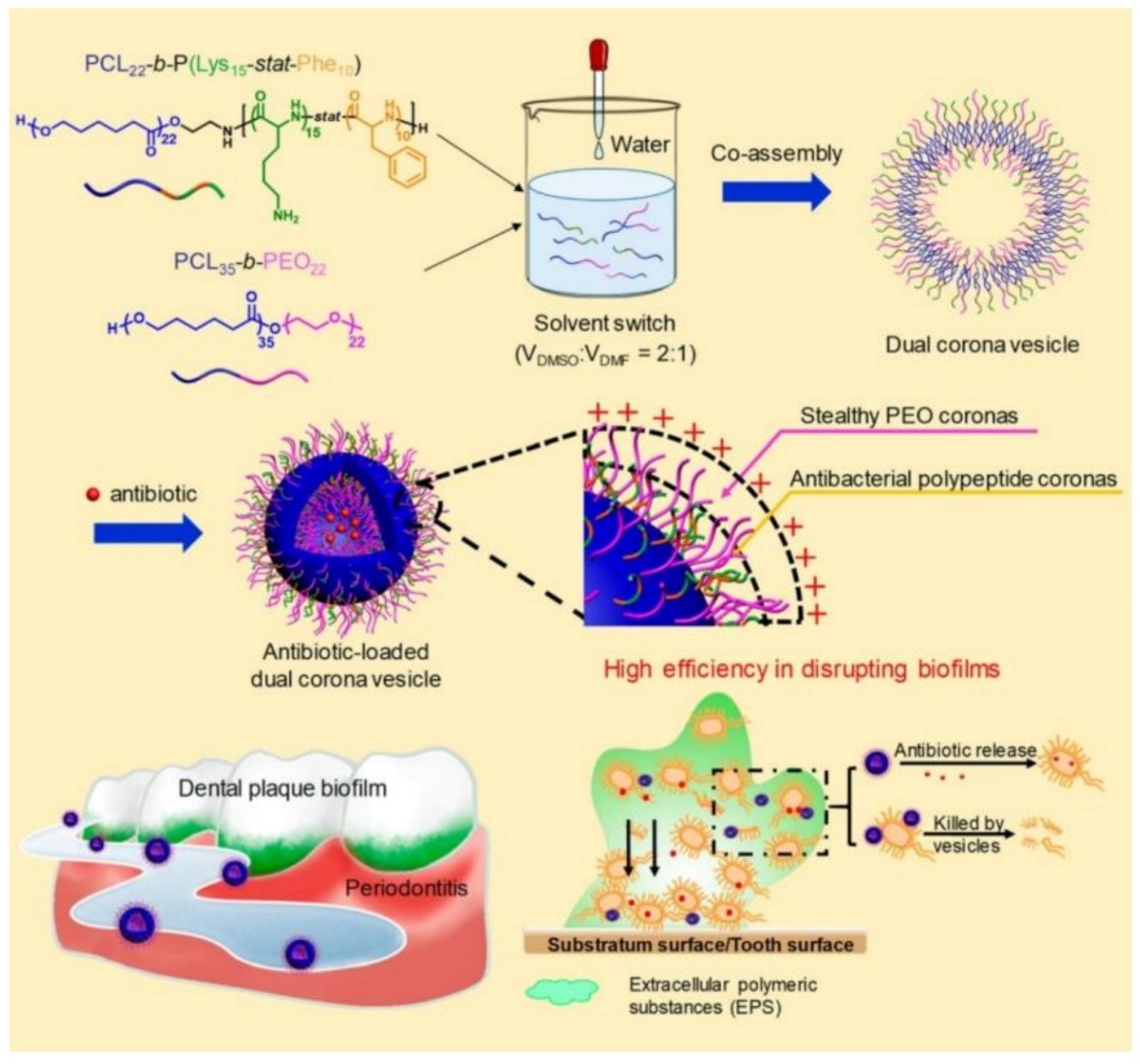
Recently, Du and coworkers [153] reported the treatment of periodontitis by efficiently disrupting biofilms using a dual corona antimicrobials-loaded polymer vesicle with stealthy poly(ethylene oxide) (PEO) corona to penetrate the biofilm and antibacterial polypeptide corona to provide intrinsic antimicrobial activity, as shown in Figure 12. The dual corona polymer vesicles were prepared by the co-assembly of two polymers PCL-b-poly(lysine-stat-phenylalanine) [PCL-b-P(Lys-stat-Phe)] and PEO-b-PCL with the same hydrophobic biodegradable PCL segment and different hydrophilic chains. Ciprofloxacin could be efficiently encapsulated in the cavity of the vesicles. Due to the protein-repelling ability of PEO, dual corona polymer vesicles penetrated the EPS of the biofilms with high efficiency, while the positive charged P(Lys-stat-Phe) allowed the vesicle to target and kill bacteria via electrostatic interaction. In addition, the encapsulated ciprofloxacin could be released as the polymer vesicle reached the deep end of the biofilm, resulting in a reduced dosage of the antimicrobials up to 50% to eradicate E. coli or S. aureus biofilms. In vivo experiment results demonstrated excellent performance of the dual corona vesicles in reducing dental plaque and alleviating inflammation using a rat periodontitis model.
Despite the strategy of delivering antibiotics to the deep end of biofilms by polymeric nanocarriers in order to reduce dosage and enhance antimicrobial activity, the efficient delivery of non-antibiotic antimicrobial agents including AMPs, AgNPs, photosensitizers and so forth for eliminating biofilms was also widely studied [154,155,156]. For instance, Haldar et al. [157] fabricated biodegradable polymer-coated AgNPs nanocomposite to eradicate biofilms, which reduced MRSA burden both on the catheter (>99.99% reduction) and in tissues surrounding the catheter (>99.999% reduction) in a mice model. Ji and coworkers [158] developed targeted photodynamic therapy strategies by using a supramolecular delivery system for the treatment of biofilms. The photosensitizer Chlorin e6 was grafted onto α-cyclodextrin, and the targeting group AMP Magainin I was covalently bound with PEG. Taking advantage of supramolecular recognition between α-cyclodextrin and PEG, targeting supramolecular micelles loaded with Chlorin e6 were formed, which exhibited excellent bacterial targeting effects and enhanced biofilm eradication ability against P. aeruginosa biofilm and MRSA biofilm. These results proved the versatility and great potential of polymeric nanomaterial-based antimicrobial agent delivery systems for eradicating biofilms.
3.3. Wound Healing
Wound infections induced by pathogens have become one of the main problems in wound care management systems, which impede the healing process and may result in life threatening complications. One of the approaches for treating wound infection is the use of wound dressings with antibacterial agents possessing broad-spectrum antimicrobial activity [159]. Typically, the moisture environment provided by the dressing has been shown to promote ulcer healing and to reduce pain experienced by patients [160]. Moreover, there are other requirements for wound dressings such as separating the wound with external environments and providing good breathability to promote wound healing. Polymeric nanomaterial-based delivery systems have shown considerable potentials in wound healing, especially polymer nanofibers and hydrogels [99,161]. For example, Lakshminarayanan et al. [162] prepared polydopamine crosslinked polyhydroxy antimicrobials loaded gelatin nanofiber mats for advanced wound dressings with long-term antimicrobial activity up to 20 days. The morphology of the nanofiber mats was retained for 1 month in an aqueous environment and showed comparable wound closure compared to commercially available silver-based dressings. Cai and coworkers [163] prepared composite hydrogels embedded with copper nanoparticles that could effectively convert NIR laser irradiation energy into localized heat for photothermal therapy. The synergistic effect of photothermal performance and rapid release of copper ions upon laser irradiation were responsible for excellent antimicrobial activity, reduced inflammatory response and promoted angiogenesis ability.
Antimicrobial agents including AMPs [49], antibiotics [164], AgNPs [165], metal oxide such as ZnO [166], photothermal sensitizers including porphyrin [167] and heavy metal ions [163] are usually used to improve the antimicrobial activity of polymeric wound dressings by covalent linkage, physical interaction or encapsulation. For example, Liu et al. [168] decorated chloramine on the surface of chitosan films by electrostatic interaction to heal MRSA infected wounds. Zhou and coworkers [167] prepared porphyrin containing alternating copolymer vesicles for the disinfection of drug-resistant bacteria infected wounds via photothermal effect. Fahimirad et al. [169] loaded recombinant LL37 AMP into chitosan nanoparticles for the elimination of MRSA infection during wound healing process with ultrahigh encapsulation efficiency of 78.52% and improved the activity and stability of LL37 AMP under thermal, salts and acidic pH treatments. Guo et al. [170] prepared injectable antimicrobial conductive quaternized chitosan hydrogels by loading graphene oxide via covalent bond for drug resistant bacterial disinfection and infectious wound healing, and the hybrid hydrogels showed excellent performance in the treatment of MRSA infected full-thickness defect mouse model.
Very recently, polymer vesicles loaded with antimicrobials have been explored as dressings in promoting wound healing by spraying onto wounds [167,171,172,173]. Du and coworkers [173] reported bifunctional polymer vesicles loaded with antimicrobials and antioxidant for healing infected diabetic wounds, as presented in Figure 13. As one of the chronically infected wounds, the diabetic wounds are difficult to heal due to high ROS concentration and recurrent infections, resulting in the occurrence of diabetic ulcers and chronic diabetic complications with very high mortality rate. Therefore, scavenging ROS is very important in the treatment of diabetic wounds. In this study, well-dispersed ceria nanoparticles were deposited on the membrane of ciprofloxacin-loaded polymer vesicles (CIP-Ceria-PVs). The CIP-Ceria-PVs could inhibit peroxide free radicals up to 50% at extremely low cerium concentrations of 1.25 μg mL−1, protecting normal L02 cells from the damage of peroxide free radicals. Moreover, CIP-Ceria-PVs exhibited enhanced antimicrobial activity compared with free ciprofloxacin due to scavenging ROS. In vivo studies in Figure 13b demonstrated the excellent wound healing capability of CIP-Ceria-PVs, and the diabetic wound was completely healed within 14 days. At the same time, they developed a H2S delivery polymer vesicle, which was capable of long-term H2S generation to promote the proliferation, migration of epidermal and endothelial cells and angiogenesis, accelerating the complete healing of diabetic wounds [172].
3.4. Tissue Engineering
The regeneration of adult tissue following an injury or degeneration is quite a limited process. Usually, the injury site is vulnerable to bacterial infections, which causes complications and delay of the regeneration of tissues [174]. Therefore, the prerequisite of tissue regeneration is to eliminate localized bacterial infections, followed by the delivery of bioactive molecules such as growth factor to the defected tissues. Antimicrobial polymer coatings on the surface of implants can provide appropriate biointerfaces to promote the regeneration of tissues. For instance, ZnO nanoparticles embedded PLA was dip coated on magnesium alloy, which helped to control the degradation and increase antibacterial activity [175]. Suteewong et al. [176] deposited polymethylmethacrylate (PMMA)/chitosan-silver hybrid nanoparticles on rubber substrate, which exhibited enhanced antibacterial activity toward E. coli and S. aureus and reduced cytotoxicity to L-929 fibroblast cells, demonstrating the potential of this hybrid nanoparticle coating at soft substrates. In addition, antimicrobial agents loaded with polymer nanomaterials can be used as bioadhesives to repair damaged soft tissues. Gu and coworkers [177] developed fast and high strength bioadhesives based on polysaccharides and peptide dendrimers with inherent hemostatic ability and antibacterial properties. Moreover, the bioadhesive showed a remarkable 5-fold increase in adhesion strength comparing with commercial bioadhesive Coseal.
Biocompatible polymeric nanoparticles have been investigated as delivery vehicles for various tissue engineering applications [178]. For example, Du and coworkers [179] prepared antibacterial peptide-mimetic alternating copolymers (PMACs) vesicles loaded with growth factor for bone regeneration. They designed a series of PMACs with different repeating units, and the PMAC with a repeating unit of 14 exhibited the best antibacterial activity against both E. coli and S. aureus with ultralow MICs of 8.0 μg mL−1. After self-assembling into vesicles in pure water, the antimicrobial activity of the vesicles was well-preserved. Growth factor could be encapsulated in antimicrobial vesicles and released during the long-term antibacterial process to promote the regeneration of bone with a 20 mm defect model in rabbits. Micro-CT, bone mineral content and BMD were used to evaluate the repair of bone defects with scaffolds at 4 weeks and 6 weeks after implantation. After 6 weeks, the defect in the rabbit bone was completely repaired, demonstrating the excellent bone repair capability of antimicrobial growth factor-loaded vesicles.
3.5. Anticancer
The anticancer application of antimicrobial agents is an attracting field since cancers are often accompanied by inflammation, and the drug resistance of cancer cells is becoming increasingly concerning [180]. Theoretically, antimicrobial agents that kill bacteria via non-selective behaviors such as damage of the cell membrane [181], elevating temperature [182] and induced degeneration of proteins and genetic materials [183] can also kill cancer cells. For instance, Shim et al. [183] prepared AgNPs loaded chitosan-alginate composite, exhibiting broad-spectrum antimicrobial activity and high toxicity toward breast cancer cell line MDA-MB-231; Jothivenkatachalam and coworkers [184] fabricated chitosan-copper nanocomposite for the inhibition of various microorganisms and A549 cancer cells by photocatalytic effect. In addition, AMPs with specific sequences and proper positive charge densities have shown anticancer and antiviral activities, such as cecropin A and B, magainins, melittin, defensins, lactoferricin and so forth, as summarized by Hoskin’s and Franco’s group, respectively [181,185]. However, the AMPs are vulnerable to enzymes and can easily cause immune responses; thus, the delivery system is critical for in vivo applications of AMPs. Hazekawa et al. [186] conjugated antimicrobial human peptide, LL-37 peptide fragment analog, with a PLGA copolymer. The formed micellar system significantly improved the permeability of the peptide to cancer cells, and the proliferation, migration and invasion in various cancer cell lines were effectively exhibited. The intracellular delivery of peptides by polymer carriers in oncology applications has been summarized by Pun et al. very recently [187].
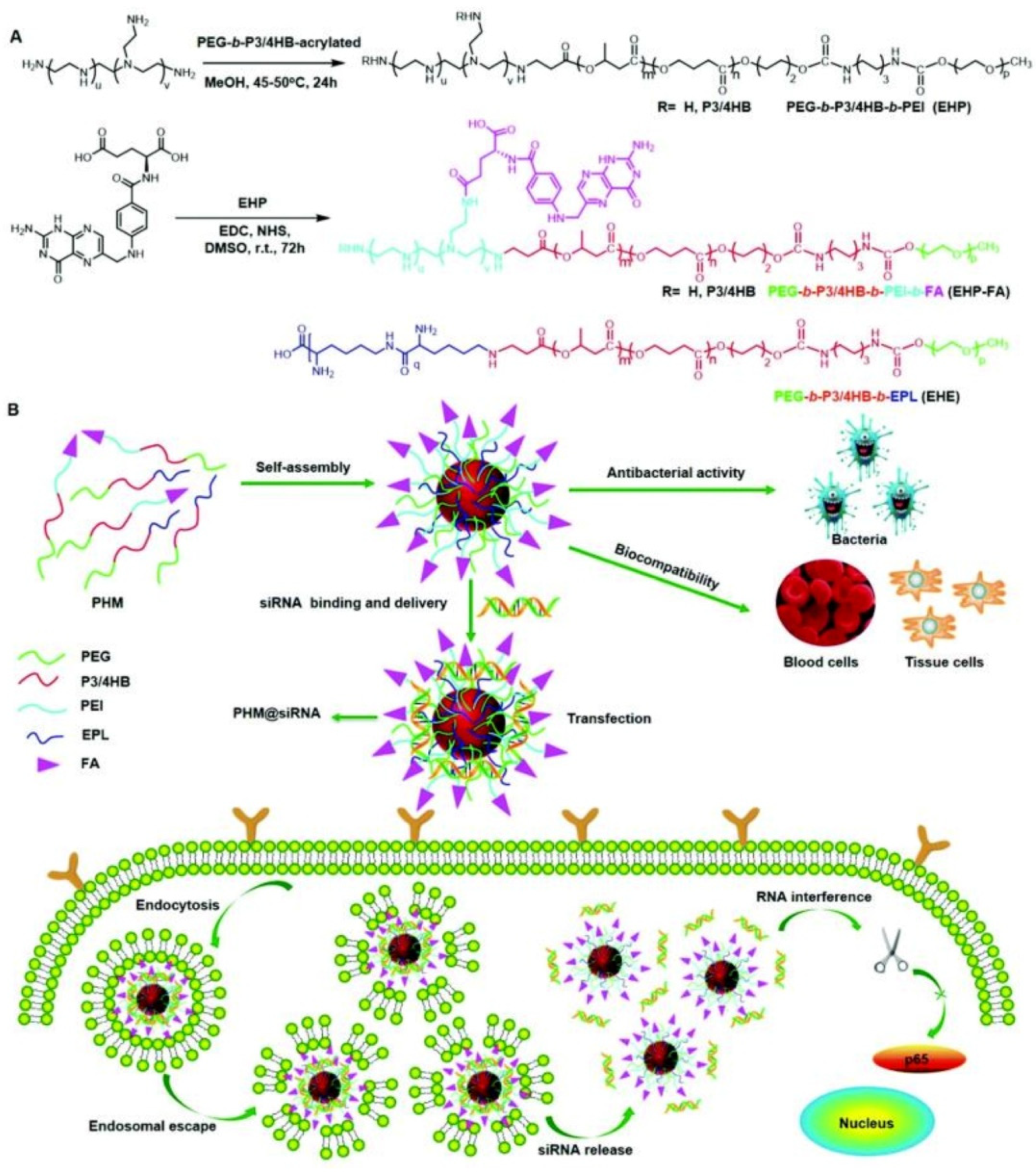
Another strategy for eliminating cancer cells using antimicrobial delivery systems is the co-delivery of antimicrobial and anticancer agents simultaneously or loading anticancer drugs with antimicrobial carriers [188,189]. For instance, Du and coworkers [190] proposed the concept of “armed” carrier to co-deliver anticancer and antiepileptic drugs with antibacterial polypeptide-grafted chitosan-based nanocapsules. Mahkam et al. [191] designed pH-responsive antibacterial clay/polymer nanocomposite as a carrier to deliver anticancer drug methotrexate and antibacterial agent ciprofloxacin with an ultrahigh efficiency of >90%, which showed enhanced antimicrobial and anticancer activity compared with free methotrexate and ciprofloxacin, demonstrating the potential of antibacterial nanocarriers in cancer therapy. Lei and coworkers [192] developed a class of multifunctional polymeric hybrid micelles (PHM) with high antibacterial activity for the efficient delivery of siRNA to cancer cells, as illustrated in Figure 14. The PHM was prepared by the co-assembly of EHP-FA and EHE, for which their structures were presented in Figure 14A. Due to the existence of positively charged poly(ethylene imine) (PEI) and poly-ε-l-lysine (EPL), the PHM showed high antibacterial activity against S. aureus in vitro and in vivo. On the contrary, PHM exhibited good hemocompatibility and lower cytotoxicity toward A549, HeLa, HepG2 and C2C12 cells benefiting from the shield effect of PEG. siRNA could be complexed onto PHM by electrostatic interaction, and PHM with folic acid decorated on the surface could effectively target FA receptor overexpressed HeLa cells and other low-expressed cancer cells, resulting in the targeted delivery of siRNA. In vitro experiments revealed that the PHM showed a high p65 gene silencing efficiency above 90% in various cancer cells, which is significantly higher than EHP-FA and EHE, demonstrating the potential of PHM as a safe and effective siRNA vector with high antibacterial activity for multifunctional gene therapy.
4. Conclusions and Future Perspectives
In summary, the recent progress of efficient loading, delivery and controlled release of antimicrobial agents in vivo or in vitro by polymeric nanomaterial-based delivery systems have been concluded. A large diversity of antimicrobial agents including antibiotics, AMPs, AgNPs, metal nanoparticles, metal oxides, gases, photosensitizers and so forth could be loaded and delivered by polymeric nanomaterials either by physical interactions or covalent bonding while maintaining the intrinsic antimicrobial activity of these antimicrobial agents. In order to fit the physiochemical properties of different kinds of antimicrobial agents to construct highly efficient delivery systems with superiorities such as high loading content and efficiency, good stability and on-demand release, polymeric nanomaterials with different chemical compositions and nanostructures including micelles, vesicles, dendrimers, nanofibers and nanogels etc. are developed. Benefiting from the versatility of polymeric nanomaterials, the antimicrobial agent delivery systems have shown significant potentials in a wide variety of biomedical applications, such as combating MDR bacteria, anti-biofilm, wound healing, tissue engineering and anticancer. Despite the rapid development of this field, the in vivo and intracellular delivery of antimicrobial agents is still in its early stage, and there are numerous challenges that should be considered in the future, which may bring new opportunities in the biomedical applications of antimicrobial agent-based delivery systems.
Non-covalent interactions such as hydrogen bonding, π-π stacking and coordination should be introduced to enhance the interactions between antimicrobial agents and the polymeric nanocarriers to increase loading content and efficiency. The strong interactions could also prevent the leakage of cargoes before reaching the target and enhance the stability of the delivery system. Modulation of the properties of different kinds of antimicrobial agents and the structural features of carriers may maximize the efficiency of the loaded antimicrobial agents. Targeting the infected area and high selectivity toward bacteria rather than mammalian cells should always be considered, which is very important for the reduction in side effects and drug resistance. Moreover, external stimuli, especially non-invasive stimuli-triggered release of loaded antimicrobial agents (in other words, the switchable antimicrobial activity of the delivery system), are also helpful for the reduction in side effects and drug resistance. However, the spatial and temporal sensitivity of the stimuli-triggered response still needs to be improved to meet practical applications. Furthermore, the generations of antimicrobial active species such as ROS or change of the micro-circumstance including elevating temperature triggered by stimuli or chemicals secreted by bacteria are also effective methods for eliminating bacteria without the generation of drug resistance. Regardless of the generation of drug resistance, taking advantage of the synergistic effect of multiple antibacterial agents is an effective strategy for eradicating MDR bacteria. In addition, the combination of antibacteria and anticancer simultaneously will be of great significance in cancer therapy.
The biosafety of polymeric nanomaterial-based delivery systems has always been selectively ignored in previous studies. Although many biodegradable polymers have been used, the cytotoxicity and hemolytic activity of the polymeric carriers, especially those with positively charged surfaces, should be evaluated systematically. In addition, the word “biocompatibility” is a comprehensive evaluation of in vivo delivery systems. If we claim that the carrier is biocompatible, numerous parameters should be evaluated more than cytotoxicity and hemolytic activity. The in vivo delivery of antimicrobial agents has been reported in many studies. However, very few investigated the stability of the delivery system in physiological conditions and the interactions between the carriers and proteins, salts, glucose, fatty acids, antigens and so forth. Moreover, the immune response of the delivery systems is also hardly investigated. Considering the complexity of the physiological condition, it is necessary to reveal the stability and true circulation behavior of the delivery systems in vivo and not only borrowing the results of in vitro experiments. Furthermore, the full life-cycle assessment of polymeric carriers should be conducted to explore blood circulation behavior, biodistribution, metabolism and organic accumulations, etc., which will be very valuable for the instructive design of polymer carriers to promote the clinical applications of polymeric nanomaterials-based antimicrobial delivery systems.
Author Contributions
Conceptualization, supervision and writing—review and editing, H.S.; resources and visualization, Y.W.; writing—original draft preparation, H.S. and Y.W. All authors have read and agreed to the published version of the manuscript.
Funding
This research was funded by Natural Science Foundation of Ningxia, grant number 2020AAC03003 and 2021AAC03026.
Institutional Review Board Statement
Not applicable.
Informed Consent Statement
Not applicable.
Acknowledgments
This work was supported by the Natural Science Foundation of Ningxia (2020AAC03003 and 2021AAC03026). H.S. thanks the Ningxia Youth Talent Support Project of Science and Technology and Young Scholars of Western China of CAS.
Conflicts of Interest
The authors declare no conflict of interest.
References
- Fournier, P.-E.; Drancourt, M.; Colson, P.; Rolain, J.-M.; La Scola, B.; Raoult, D. Modern clinical microbiology: New challenges and solutions. Nat. Rev. Microbiol. 2013, 11, 574–585. [Google Scholar] [CrossRef]
- Ghosh, C.; Sarkar, P.; Issa, R.; Haldar, J. Alternatives to Conventional Antibiotics in the Era of Antimicrobial Resistance. Trends Microbiol. 2019, 27, 323–338. [Google Scholar] [CrossRef]
- Monserrat-Martinez, A.; Gambin, Y.; Sierecki, E. Thinking Outside the Bug: Molecular Targets and Strategies to Overcome Antibiotic Resistance. Int. J. Mol. Sci. 2019, 20, 1255. [Google Scholar] [CrossRef] [Green Version]
- Faya, M.; Kalhapure, R.S.; Kumalo, H.M.; Waddad, A.Y.; Omolo, C.; Govender, T. Conjugates and nano-delivery of antimicrobial peptides for enhancing therapeutic activity. J. Drug Deliv. Sci. Technol. 2018, 44, 153–171. [Google Scholar] [CrossRef]
- Martin-Serrano, A.; Gomez, R.; Ortega, P.; Javier de la Mata, F. Nanosystems as Vehicles for the Delivery of Antimicrobial Peptides (AMPs). Pharmaceutics 2019, 11, 448. [Google Scholar] [CrossRef] [Green Version]
- Cui, Z.; Luo, Q.; Bannon, M.S.; Gray, V.P.; Bloom, T.G.; Clore, M.F.; Hughes, M.A.; Crawford, M.A.; Letteri, R.A. Molecular engineering of antimicrobial peptide (AMP)-polymer conjugates. Biomater. Sci. 2021, 9, 5069–5091. [Google Scholar] [CrossRef]
- Chung, I.-M.; Park, I.; Seung-Hyun, K.; Thiruvengadam, M.; Rajakumar, G. Plant-Mediated Synthesis of Silver Nanoparticles: Their Characteristic Properties and Therapeutic Applications. Nanoscale Res. Lett. 2016, 11, 40. [Google Scholar] [CrossRef] [Green Version]
- Tang, S.; Zheng, J. Antibacterial Activity of Silver Nanoparticles: Structural Effects. Adv. Healthc. Mater. 2018, 7, 1701503. [Google Scholar] [CrossRef] [PubMed]
- Yin, I.X.; Zhang, J.; Zhao, I.S.; Mei, M.L.; Li, Q.; Chu, C.H. The Antibacterial Mechanism of Silver Nanoparticles and Its Application in Dentistry. Int. J. Nanomed. 2020, 15, 2555–2562. [Google Scholar] [CrossRef] [Green Version]
- Chen, H.; Wang, R.; Zhang, J.; Hua, H.; Zhu, M. Synthesis of core-shell structured ZnO@m-SiO2 with excellent reinforcing effect and antimicrobial activity for dental resin composites. Dent. Mater. 2018, 34, 1846–1855. [Google Scholar] [CrossRef]
- Rodrigues, G.R.; Lopez-Abarrategui, C.; de la Serna Gomez, I.; Dias, S.C.; Otero-Gonzalez, A.J.; Franco, O.L. Antimicrobial magnetic nanoparticles based-therapies for controlling infectious diseases. Int. J. Pharm. 2019, 555, 356–367. [Google Scholar] [CrossRef] [PubMed]
- Ziental, D.; Czarczynska-Goslinska, B.; Mlynarczyk, D.T.; Glowacka-Sobotta, A.; Stanisz, B.; Goslinski, T.; Sobotta, L. Titanium Dioxide Nanoparticles: Prospects and Applications in Medicine. Nanomaterials 2020, 10, 387. [Google Scholar] [CrossRef] [PubMed] [Green Version]
- Zhen, J.-B.; Kang, P.-W.; Zhao, M.-H.; Yang, K.-W. Silver Nanoparticle Conjugated Star PCL-b-AMPs Copolymer as Nanocomposite Exhibits Efficient Antibacterial Properties. Bioconjugate Chem. 2020, 31, 51–63. [Google Scholar] [CrossRef] [PubMed]
- Chen, H.; Battalapalli, D.; Draz, M.S.; Zhang, P.; Ruan, Z. The application of cell-penetrating-peptides in antibacterial agents. Curr. Med. Chem. 2021, 28, 5896–5925. [Google Scholar] [CrossRef]
- Sun, H.; Hong, Y.; Xi, Y.; Zou, Y.; Gao, J.; Du, J. Synthesis, Self-Assembly, and Biomedical Applications of Antimicrobial Peptide-Polymer Conjugates. Biomacromolecules 2018, 19, 1701–1720. [Google Scholar] [CrossRef]
- Sun, H.; Wang, Y.; Song, J. Polymer Vesicles for Antimicrobial Applications. Polymers 2021, 13, 2903. [Google Scholar] [CrossRef] [PubMed]
- Duncan, M.J.; Wheatley, P.S.; Coghill, E.M.; Vornholt, S.M.; Warrender, S.J.; Megson, I.L.; Morris, R.E. Antibacterial efficacy from NO-releasing MOF-polymer films. Mater. Adv. 2020, 1, 2509–2519. [Google Scholar] [CrossRef]
- Jin, G.; Gao, Z.; Liu, Y.; Zhao, J.; Ou, H.; Xu, F.; Ding, D. Polymeric Nitric Oxide Delivery Nanoplatforms for Treating Cancer, Cardiovascular Diseases, and Infection. Adv. Healthc. Mater. 2021, 10, 2001550. [Google Scholar] [CrossRef] [PubMed]
- Simonson, A.W.; Aronson, M.R.; Medina, S.H. Supramolecular Peptide Assemblies as Antimicrobial Scaffolds. Molecules 2020, 25, 2751. [Google Scholar] [CrossRef] [PubMed]
- Mahata, D.; Mandal, S.M. Molecular self-assembly of copolymer from renewable phenols: New class of antimicrobial ointment base. J. Biomater. Sci. Polym. Ed. 2018, 29, 2187–2200. [Google Scholar] [CrossRef] [PubMed]
- Krol, A.; Pomastowski, P.; Rafinska, K.; Railean-Plugaru, V.; Buszewski, B. Zinc oxide nanoparticles: Synthesis, antiseptic activity and toxicity mechanism. Adv. Colloid Interface Sci. 2017, 249, 37–52. [Google Scholar] [CrossRef]
- Shi, L.; Chen, J.; Teng, L.; Wang, L.; Zhu, G.; Liu, S.; Luo, Z.; Shi, X.; Wang, Y.; Ren, L. The Antibacterial Applications of Graphene and Its Derivatives. Small 2016, 12, 4165–4184. [Google Scholar] [CrossRef]
- Han, W.; Wu, Z.; Li, Y.; Wang, Y. Graphene family nanomaterials (GFNs)—Promising materials for antimicrobial coating and film: A review. Chem. Eng. J. 2019, 358, 1022–1037. [Google Scholar] [CrossRef]
- Jiang, L.; Gan, C.R.R.; Gao, J.; Loh, X.J. A Perspective on the Trends and Challenges Facing Porphyrin-Based Anti-Microbial Materials. Small 2016, 12, 3609–3644. [Google Scholar] [CrossRef] [PubMed]
- Engler, A.C.; Wiradharma, N.; Ong, Z.Y.; Coady, D.J.; Hedrick, J.L.; Yang, Y.-Y. Emerging trends in macromolecular antimicrobials to fight multi-drug-resistant infections. Nano Today 2012, 7, 201–222. [Google Scholar] [CrossRef]
- Richter, A.P.; Brown, J.S.; Bharti, B.; Wang, A.; Gangwal, S.; Houck, K.; Hubal, E.A.C.; Paunov, V.N.; Stoyanov, S.D.; Velev, O.D. An environmentally benign antimicrobial nanoparticle based on a silver-infused lignin core. Nat. Nanotechnol. 2015, 10, 817–823. [Google Scholar] [CrossRef]
- Boge, L.; Hallstensson, K.; Ringstad, L.; Johansson, J.; Andersson, T.; Davoudi, M.; Larsson, P.T.; Mahlapuu, M.; Hakansson, J.; Andersson, M. Cubosomes for topical delivery of the antimicrobial peptide LL-37. Eur. J. Pharm. Biopharm. 2019, 134, 60–67. [Google Scholar] [CrossRef] [PubMed]
- Zhang, L.; Pornpattananangkul, D.; Hu, C.M.J.; Huang, C.M. Development of Nanoparticles for Antimicrobial Drug Delivery. Curr. Med. Chem. 2010, 17, 585–594. [Google Scholar] [CrossRef] [Green Version]
- Halbus, A.F.; Horozov, T.S.; Paunov, V.N. Colloid particle formulations for antimicrobial applications. Adv. Colloid Interface Sci. 2017, 249, 134–148. [Google Scholar] [CrossRef]
- Gao, W.; Chen, Y.; Zhang, Y.; Zhang, Q.; Zhang, L. Nanoparticle-based local antimicrobial drug delivery. Adv. Drug Deliv. Rev. 2018, 127, 46–57. [Google Scholar] [CrossRef]
- Kalhapure, R.S.; Suleman, N.; Mocktar, C.; Seedat, N.; Govender, T. Nanoengineered Drug Delivery Systems for Enhancing Antibiotic Therapy. J. Pharm. Sci. 2015, 104, 872–905. [Google Scholar] [CrossRef] [PubMed]
- Yan, Y.; Zhang, J.; Ren, L.; Tang, C. Metal-containing and related polymers for biomedical applications. Chem. Soc. Rev. 2016, 45, 5232–5263. [Google Scholar] [CrossRef] [PubMed] [Green Version]
- Chan, C.-F.; Huang, K.-S.; Lee, M.-Y.; Yang, C.-H.; Wang, C.-Y.; Lin, Y.-S. Applications of Nanoparticles for Antimicrobial Activity and Drug Delivery. Curr. Org. Chem. 2014, 18, 204–215. [Google Scholar] [CrossRef]
- Zazo, H.; Colino, C.I.; Lanao, J.M. Current applications of nanoparticles in infectious diseases. J. Control. Release 2016, 224, 86–102. [Google Scholar] [CrossRef]
- Lu, H.; Fan, L.; Liu, Q.; Wei, J.; Ren, T.; Du, J. Preparation of water-dispersible silver-decorated polymer vesicles and micelles with excellent antibacterial efficacy. Polym. Chem. 2012, 3, 2217–2227. [Google Scholar] [CrossRef]
- Basnet, P.; Skalko-Basnet, N. Nanodelivery Systems for Improved Topical Antimicrobial Therapy. Curr. Pharm. Des. 2013, 19, 7237–7243. [Google Scholar] [CrossRef]
- Sun, H.; Fan, L.; Zou, K.; Zhu, H.; Du, J. Decoration of homopolymer vesicles by antibacterial ultrafine silver nanoparticles. RSC Adv. 2014, 4, 41331–41335. [Google Scholar] [CrossRef]
- Walvekar, P.; Gannimani, R.; Salih, M.; Makhathini, S.; Mocktar, C.; Govender, T. Self-assembled oleylamine grafted hyaluronic acid polymersomes for delivery of vancomycin against methicillin resistant Staphylococcus aureus (MRSA). Colloids Surf. B 2019, 182, 110388. [Google Scholar] [CrossRef] [PubMed]
- Lim, Y.H.; Tiemann, K.M.; Heo, G.S.; Wagers, P.O.; Rezenom, Y.H.; Zhang, S.; Zhang, F.; Youngs, W.J.; Hunstad, D.A.; Wooley, K.L. Preparation and In Vitro Antimicrobial Activity of Silver-Bearing Degradable Polymeric Nanoparticles of Polyphosphoester-block-Poly(L-lactide). ACS Nano 2015, 9, 1995–2008. [Google Scholar] [CrossRef] [PubMed] [Green Version]
- Zare, E.N.; Makvandi, P.; Ashtari, B.; Rossi, F.; Motahari, A.; Perale, G. Progress in Conductive Polyaniline-Based Nanocomposites for Biomedical Applications: A Review. J. Med. Chem. 2020, 63, 1–22. [Google Scholar] [CrossRef] [PubMed]
- Mikhalevich, V.; Craciun, I.; Kyropoulou, M.; Palivan, C.G.; Meier, W. Amphiphilic Peptide Self-Assembly: Expansion to Hybrid Materials. Biomacromolecules 2017, 18, 3471–3480. [Google Scholar] [CrossRef]
- Kong, H.; Jang, J. Antibacterial properties of novel poly(methyl methacrylate) nanofiber containing silver nanoparticles. Langmuir 2008, 24, 2051–2056. [Google Scholar] [CrossRef]
- Quiros, J.; Boltes, K.; Aguado, S.; Guzman de Villoria, R.; Jose Vilatela, J.; Rosal, R. Antimicrobial metal-organic frameworks incorporated into electrospun fibers. Chem. Eng. J. 2015, 262, 189–197. [Google Scholar] [CrossRef]
- Gallis, D.F.S.; Butler, K.S.; Agola, J.O.; Pearce, C.J.; McBride, A.A. Antibacterial Countermeasures via Metal-Organic Framework Supported Sustained Therapeutic Release. ACS Appl. Mater. Interfaces 2019, 11, 7782–7791. [Google Scholar] [CrossRef] [PubMed]
- Kaur, N.; Tiwari, P.; Kapoor, K.S.; Saini, A.K.; Sharma, V.; Mobin, S.M. Metal-organic framework based antibiotic release and antimicrobial response: An overview. Crystengcomm 2020, 22, 7513–7527. [Google Scholar] [CrossRef]
- Yang, J.; Yang, Y.-W. Metal-Organic Frameworks for Biomedical Applications. Small 2020, 16, 1906846. [Google Scholar] [CrossRef] [PubMed]
- Kasimanickam, R.K.; Ranjan, A.; Asokan, G.V.; Kasimanickam, V.R.; Kastelic, J.P. Prevention and treatment of biofilms by hybrid- and nanotechnologies. Int. J. Nanomed. 2013, 8, 2809–2819. [Google Scholar] [CrossRef]
- Bai, M.; Li, C.; Cui, H.; Lin, L. Preparation of self-assembling Litsea cubeba essential oil/ diphenylalanine peptide micro/nanotubes with enhanced antibacterial properties against Staphylococcus aureus biofilm. LWT—Food Sci. Technol. 2021, 146, 111394. [Google Scholar] [CrossRef]
- Patrulea, V.; Borchard, G.; Jordan, O. An Update on Antimicrobial Peptides (AMPs) and Their Delivery Strategies for Wound Infections. Pharmaceutics 2020, 12, 840. [Google Scholar] [CrossRef]
- Drayton, M.; Kizhakkedathu, J.N.; Straus, S.K. Towards Robust Delivery of Antimicrobial Peptides to Combat Bacterial Resistance. Molecules 2020, 25, 3048. [Google Scholar] [CrossRef] [PubMed]
- Du, J.; Sun, H. Polymer/TiO2 hybrid vesicles for excellent UV screening and effective encapsulation of antioxidant agents. ACS Appl. Mater. Interfaces 2014, 6, 13535–13541. [Google Scholar] [CrossRef] [PubMed]
- Xiao, Y.; Sun, H.; Du, J. Sugar-Breathing Glycopolymersomes for Regulating Glucose Level. J. Am. Chem. Soc. 2017, 139, 7640–7647. [Google Scholar] [CrossRef] [PubMed]
- Sun, H.; Jiang, J.; Xiao, Y.; Du, J. Efficient Removal of Polycyclic Aromatic Hydrocarbons, Dyes, and Heavy Metal Ions by a Homopolymer Vesicle. ACS Appl. Mater. Interfaces 2018, 10, 713–722. [Google Scholar] [CrossRef] [PubMed]
- Warren, N.J.; Armes, S.P. Polymerization-Induced Self-Assembly of Block Copolymer Nano-objects via RAFT Aqueous Dispersion Polymerization. J. Am. Chem. Soc. 2014, 136, 10174–10185. [Google Scholar] [CrossRef]
- Qiu, H.; Hudson, Z.M.; Winnik, M.A.; Manners, I. Multidimensional hierarchical self-assembly of amphiphilic cylindrical block comicelles. Science 2015, 347, 1329–1332. [Google Scholar] [CrossRef] [PubMed] [Green Version]
- Sun, H.; Du, J. Intramolecular Cyclization-Induced Crystallization-Driven Self-Assembly of an Amorphous Poly(amic acid). Macromolecules 2020, 53, 11033–11039. [Google Scholar] [CrossRef]
- Shah, A.; Shahzad, S.; Munir, A.; Nadagouda, M.N.; Khan, G.S.; Shams, D.F.; Dionysiou, D.D.; Rana, U.A. Micelles as Soil and Water Decontamination Agents. Chem. Rev. 2016, 6042–6074. [Google Scholar] [CrossRef]
- Sun, H.; Du, J. Plasmonic vesicles with tailored collective properties. Nanoscale 2018, 10, 17354–17361. [Google Scholar] [CrossRef]
- Sun, H.; Zhu, Y.; Yang, B.; Wang, Y.; Wu, Y.; Du, J. Template-free fabrication of nitrogen-doped hollow carbon spheres for high-performance supercapacitors based on a scalable homopolymer vesicle. J. Mater. Chem. A 2016, 4, 12088–12097. [Google Scholar] [CrossRef]
- Wang, F.; Xiao, J.; Chen, S.; Sun, H.; Yang, B.; Jiang, J.; Zhou, X.; Du, J. Polymer Vesicles: Modular Platforms for Cancer Theranostics. Adv. Mater. 2018, 30, e1705674. [Google Scholar] [CrossRef] [PubMed]
- Owen, S.C.; Chan, D.P.Y.; Shoichet, M.S. Polymeric micelle stability. Nano Today 2012, 7, 53–65. [Google Scholar] [CrossRef]
- Cabral, H.; Miyata, K.; Osada, K.; Kataoka, K. Block Copolymer Micelles in Nanomedicine Applications. Chem. Rev. 2018, 118, 6844–6892. [Google Scholar] [CrossRef] [PubMed] [Green Version]
- Zhu, Y.; Yang, B.; Chen, S.; Du, J. Polymer vesicles: Mechanism, preparation, application, and responsive behavior. Prog. Polym. Sci. 2017, 64, 1–22. [Google Scholar] [CrossRef]
- Otrin, L.; Witkowska, A.; Marusic, N.; Zhao, Z.; Lira, R.B.; Kyrilis, F.L.; Hamdi, F.; Ivanov, I.; Lipowsky, R.; Kastritis, P.L.; et al. En route to dynamic life processes by SNARE-mediated fusion of polymer and hybrid membranes. Nat. Commun. 2021, 12, 4972. [Google Scholar] [CrossRef] [PubMed]
- Zhou, X.; Cornel, E.J.; Fan, Z.; He, S.; Du, J. Bone-Targeting Polymer Vesicles for Effective Therapy of Osteoporosis. Nano Lett. 2021, 21, 7998–8007. [Google Scholar] [CrossRef] [PubMed]
- Xi, D.; Xiao, M.; Cao, J.; Zhao, L.; Xu, N.; Long, S.; Fan, J.; Shao, K.; Sun, W.; Yan, X.; et al. NIR Light-Driving Barrier-Free Group Rotation in Nanoparticles with an 88.3% Photothermal Conversion Efficiency for Photothermal Therapy. Adv. Mater. 2020, 32, 1907855. [Google Scholar] [CrossRef]
- Sun, H.; Wang, F.; Du, J. Preparation, application and perspective in polymer vesicles with an inhomogeneous membrane. Sci. Sin. Chim. 2019, 49, 877–890. [Google Scholar] [CrossRef] [Green Version]
- Liu, D.; Sun, H.; Xiao, Y.; Chen, S.; Cornel, E.J.; Zhu, Y.; Du, J. Design principles, synthesis and biomedical applications of polymer vesicles with inhomogeneous membranes. J. Control. Release 2020, 326, 365–386. [Google Scholar] [CrossRef]
- Wei, P.; Cornel, E.J.; Du, J. Breaking the Corona Symmetry of Vesicles. Macromolecules 2021, 54, 7603–7611. [Google Scholar] [CrossRef]
- Chen, J.; Wang, F.; Liu, Q.; Du, J. Antibacterial polymeric nanostructures for biomedical applications. Chem. Commun. 2014, 50, 14482–14493. [Google Scholar] [CrossRef] [PubMed]
- Wayakanon, K.; Thornhill, M.H.; Douglas, C.W.I.; Lewis, A.L.; Warren, N.J.; Pinnock, A.; Armes, S.P.; Battaglia, G.; Murdoch, C. Polymersome-mediated intracellular delivery of antibiotics to treat Porphyromonas gingivalis-infected oral epithelial cells. FASEB J. 2013, 27, 4455–4465. [Google Scholar] [CrossRef] [PubMed]
- Glisoni, R.J.; Sosnik, A. Encapsulation of the Antimicrobial and Immunomodulator Agent Nitazoxanide within Polymeric Micelles. J. Nanosci. Nanotechnol. 2014, 14, 4670–4682. [Google Scholar] [CrossRef] [PubMed]
- Lin, W.; Huang, K.; Li, Y.; Qin, Y.; Xiong, D.; Ling, J.; Yi, G.; Tang, Z.; Lin, J.; Huang, Y.; et al. Facile In Situ Preparation and In Vitro Antibacterial Activity of PDMAEMA-Based Silver-Bearing Copolymer Micelles. Nanoscale Res. Lett. 2019, 14, 256. [Google Scholar] [CrossRef] [PubMed] [Green Version]
- Morteza, M.; Roya, S.; Hamed, H.; Amir, Z.; Abolfazl, A. Synthesis and evaluation of polymeric micelle containing piperacillin/tazobactam for enhanced antibacterial activity. Drug Deliv. 2019, 26, 1292–1299. [Google Scholar] [CrossRef] [Green Version]
- Chen, M.; Wei, J.; Xie, S.; Tao, X.; Zhang, Z.; Ran, P.; Li, X. Bacterial biofilm destruction by size/surface charge-adaptive micelles. Nanoscale 2019, 11, 1410–1422. [Google Scholar] [CrossRef] [PubMed]
- Rigo, S.; Huerlimann, D.; Marot, L.; Malmsten, M.; Meier, W.; Palivan, C.G. Decorating Nanostructured Surfaces with Antimicrobial Peptides to Efficiently Fight Bacteria. ACS Appl. Bio Mater. 2020, 3, 1533–1543. [Google Scholar] [CrossRef]
- Diaz, I.L.; Parra, C.; Linarez, M.; Perez, L.D. Design of Micelle Nanocontainers Based on PDMAEMA-b-PCL-b-PDMAEMA Triblock Copolymers for the Encapsulation of Amphotericin B. AAPS PharmSciTech 2015, 16, 1069–1078. [Google Scholar] [CrossRef] [PubMed] [Green Version]
- Qao, J.; Purro, M.; Liu, Z.; Xiong, M.P. Terpyridine-Micelles for Inhibiting Bacterial Biofilm Development. ACS Infect. Dis. 2018, 4, 1346–1354. [Google Scholar] [CrossRef]
- Park, S.-C.; Ko, C.; Hyeon, H.; Jang, M.-K.; Lee, D. Imaging and Targeted Antibacterial Therapy Using Chimeric Antimicrobial Peptide Micelles. ACS Appl. Mater. Interfaces 2020, 12, 54306–54315. [Google Scholar] [CrossRef]
- Song, Y.; Elsabahy, M.; Collins, C.A.; Khan, S.; Li, R.; Hreha, T.N.; Shen, Y.; Lin, Y.-N.; Letteri, R.A.; Su, L.; et al. Morphologic Design of Silver-Bearing Sugar-Based Polymer Nanoparticles for Uroepithelial Cell Binding and Antimicrobial Delivery. Nano Lett. 2021, 21, 4990–4998. [Google Scholar] [CrossRef]
- Song, J.; Zhou, J.; Duan, H. Self-Assembled Plasmonic Vesicles of SERS-Encoded Amphiphilic Gold Nanoparticles for Cancer Cell Targeting and Traceable Intracellular Drug Delivery. J. Am. Chem. Soc. 2012, 134, 13458–13469. [Google Scholar] [CrossRef] [PubMed]
- Blanazs, A.; Armes, S.P.; Ryan, A.J. Self-Assembled Block Copolymer Aggregates: From Micelles to Vesicles and their Biological Applications. Macromol. Rapid Commun. 2009, 30, 267–277. [Google Scholar] [CrossRef]
- Deng, Y.; Li, J.; Yu, J.; Zhao, J.; Tang, J. Silver nanoparticles well-dispersed in amine-functionalized, one-pot made vesicles as an effective antibacterial agent. Mater. Sci. Eng. C 2016, 60, 92–99. [Google Scholar] [CrossRef] [PubMed]
- Li, Y.-M.; Liu, S.-Y. Enzyme-triggered Transition from Polymeric Vesicles to Core Cross-linked Micelles for Selective Release of Antimicrobial Agents. Acta Polym. Sin. 2017, 7, 1178–1190. [Google Scholar] [CrossRef]
- Li, Y.; Liu, G.; Wang, X.; Hu, J.; Liu, S. Enzyme-Responsive Polymeric Vesicles for Bacterial-Strain-Selective Delivery of Antimicrobial Agents. Angew. Chem. Int. Ed. 2016, 55, 1760–1764. [Google Scholar] [CrossRef] [PubMed]
- Blackman, L.D.; Oo, Z.Y.; Qu, Y.; Gunatillake, P.A.; Cass, P.; Locock, K.E.S. Antimicrobial Honey-Inspired Glucose-Responsive Nanoreactors by Polymerization-Induced Self-Assembly. ACS Appl. Mater. Interfaces 2020, 12, 11353–11362. [Google Scholar] [CrossRef] [PubMed]
- Cheng, Y.; Zhao, L.; Li, Y.; Xu, T. Design of biocompatible dendrimers for cancer diagnosis and therapy: Current status and future perspectives. Chem. Soc. Rev. 2011, 40, 2673–2703. [Google Scholar] [CrossRef] [PubMed]
- Kesharwani, P.; Jain, K.; Jain, N.K. Dendrimer as nanocarrier for drug delivery. Prog. Polym. Sci. 2014, 39, 268–307. [Google Scholar] [CrossRef]
- Svenson, S. Dendrimers as versatile platform in drug delivery applications. Eur. J. Pharm. Biopharm. 2009, 71, 445–462. [Google Scholar] [CrossRef]
- Alfei, S.; Schito, A.M. From Nanobiotechnology, Positively Charged Biomimetic Dendrimers as Novel Antibacterial Agents: A Review. Nanomaterials 2020, 10, 2022. [Google Scholar] [CrossRef]
- Abd-El-Aziz, A.S.; Agatemor, C. Emerging Opportunities in the Biomedical Applications of Dendrimers. J. Inorg. Organomet. Polym. Mater. 2018, 28, 369–382. [Google Scholar] [CrossRef]
- Chen, S.; Huang, S.; Li, Y.; Zhou, C. Recent Advances in Epsilon-Poly-L-Lysine and L-Lysine-Based Dendrimer Synthesis, Modification, and Biomedical Applications. Front. Chem. 2021, 9, 659304. [Google Scholar] [CrossRef]
- Tang, J.; Chen, W.; Su, W.; Li, W.; Deng, J. Dendrimer-Encapsulated Silver Nanoparticles and Antibacterial Activity on Cotton Fabric. J. Nanosci. Nanotechnol. 2013, 13, 2128–2135. [Google Scholar] [CrossRef]
- Liu, X.; Wang, Z.; Feng, X.; Bai, E.; Xiong, Y.; Zhu, X.; Shen, B.; Duan, Y.; Huang, Y. Platensimycin-Encapsulated Poly(lactic-co-glycolic acid) and Poly(amidoamine) Dendrimers Nanoparticles with Enhanced Anti-Staphylococcal Activity In Vivo. Bioconjugate Chem. 2020, 31, 1425–1437. [Google Scholar] [CrossRef] [PubMed]
- Li, J.; Liang, H.; Liu, J.; Wang, Z. Poly (amidoamine) (PAMAM) dendrimer mediated delivery of drug and pDNA/siRNA for cancer therapy. Int. J. Pharm. 2018, 546, 215–225. [Google Scholar] [CrossRef] [PubMed]
- Lim, D.G.; Rajasekaran, N.; Lee, D.; Kim, N.A.; Jung, H.S.; Hong, S.; Shin, Y.K.; Kang, E.; Jeong, S.H. Polyamidoamine-Decorated Nanodiamonds as a Hybrid Gene Delivery Vector and siRNA Structural Characterization at the Charged Interfaces. ACS Appl. Mater. Interfaces 2017, 9, 31543–31556. [Google Scholar] [CrossRef] [PubMed]
- Pashaei-Asl, R.; Khodadadi, K.; Pashaei-Asl, F.; Haqshenas, G.; Ahmadian, N.; Pashaiasl, M.; Baghdadabadi, R.H. Legionella Pneumophila and Dendrimers-Mediated Antisense Therapy. Adv. Pharm. Bull. 2017, 7, 179–187. [Google Scholar] [CrossRef] [Green Version]
- Xue, X.Y.; Mao, X.G.; Li, Z.; Chen, Z.; Zhou, Y.; Hou, Z.; Li, M.K.; Meng, J.R.; Luo, X.X. A potent and selective antimicrobial poly(amidoamine) dendrimer conjugate with LED209 targeting QseC receptor to inhibit the virulence genes of gram negative bacteria. Nanomedicine 2015, 11, 329–339. [Google Scholar] [CrossRef]
- Homaeigohar, S.; Boccaccini, A.R. Antibacterial biohybrid nanofibers for wound dressings. Acta Biomater. 2020, 107, 25–49. [Google Scholar] [CrossRef] [PubMed]
- Schnaider, L.; Brahmachari, S.; Schmidt, N.W.; Mensa, B.; Shaham-Niv, S.; Bychenko, D.; Adler-Abramovich, L.; Shimon, L.J.W.; Kolusheva, S.; DeGrado, W.F.; et al. Self-assembling dipeptide antibacterial nanostructures with membrane disrupting activity. Nat. Commun. 2017, 8, 1365. [Google Scholar] [CrossRef]
- Feng, L.; Li, S.; Li, H.; Zhai, J.; Song, Y.; Jiang, L.; Zhu, D. Super-Hydrophobic Surface of Aligned Polyacrylonitrile Nanofibers. Angew. Chem. Int. Ed. 2002, 41, 1221–1223. [Google Scholar] [CrossRef]
- Ma, P.X.; Zhang, R.Y. Synthetic nano-scale fibrous extracellular matrix. J. Biomed. Mater. Res. 1999, 46, 60–72. [Google Scholar] [CrossRef] [Green Version]
- Elbahri, M.; Homaeigohar, S.; Abdelaziz, R.; Dai, T.; Khalil, R.; Zillohu, A.U. Smart Metal–Polymer Bionanocomposites as Omnidirectional Plasmonic Black Absorber Formed by Nanofluid Filtration. Adv. Funct. Mater. 2012, 22, 4771–4777. [Google Scholar] [CrossRef] [Green Version]
- Pant, B.; Park, M.; Park, S.-J. Drug Delivery Applications of Core-Sheath Nanofibers Prepared by Coaxial Electrospinning: A Review. Pharmaceutics 2019, 11, 305. [Google Scholar] [CrossRef] [PubMed] [Green Version]
- Xue, J.; Wu, T.; Dai, Y.; Xia, Y. Electrospinning and Electrospun Nanofibers: Methods, Materials, and Applications. Chem. Rev. 2019, 119, 5298–5415. [Google Scholar] [CrossRef] [PubMed]
- Wang, M.; Wang, K.; Yang, Y.; Liu, Y.; Yu, D.-G. Electrospun Environment Remediation Nanofibers Using Unspinnable Liquids as the Sheath Fluids: A Review. Polymers 2020, 12, 103. [Google Scholar] [CrossRef] [Green Version]
- Agarwal, S.; Greiner, A.; Wendorff, J.H. Functional materials by electrospinning of polymers. Prog. Polym. Sci. 2013, 38, 963–991. [Google Scholar] [CrossRef]
- Rieger, K.A.; Cho, H.J.; Yeung, H.F.; Fan, W.; Schiffman, J.D. Antimicrobial Activity of Silver Ions Released from Zeolites Immobilized on Cellulose Nanofiber Mats. ACS Appl. Mater. Interfaces 2016, 8, 3032–3040. [Google Scholar] [CrossRef] [PubMed]
- Zhang, Q.; Wang, Y.; Zhang, W.; Hickey, M.E.; Lin, Z.; Tu, Q.; Wang, J. In situ assembly of well-dispersed Ag nanoparticles on the surface of polylactic acid-Au@polydopamine nanofibers for antimicrobial applications. Colloids Surf. B 2019, 184, 110506. [Google Scholar] [CrossRef]
- Abrigo, M.; McArthur, S.L.; Kingshott, P. Electrospun Nanofibers as Dressings for Chronic Wound Care: Advances, Challenges, and Future Prospects. Macromol. Biosci. 2014, 14, 772–792. [Google Scholar] [CrossRef] [PubMed]
- Czaja, W.K.; Young, D.J.; Kawecki, M.; Brown, R.M. The Future Prospects of Microbial Cellulose in Biomedical Applications. Biomacromolecules 2007, 8, 1–12. [Google Scholar] [CrossRef] [PubMed]
- Rath, G.; Hussain, T.; Chauhan, G.; Garg, T.; Goyal, A.K. Development and characterization of cefazolin loaded zinc oxide nanoparticles composite gelatin nanofiber mats for postoperative surgical wounds. Mater. Sci. Eng. C 2016, 58, 242–253. [Google Scholar] [CrossRef]
- Oh, J.K.; Drumright, R.; Siegwart, D.J.; Matyjaszewski, K. The development of microgels/nanogels for drug delivery applications. Prog. Polym. Sci. 2008, 33, 448–477. [Google Scholar] [CrossRef]
- Karimi, M.; Ghasemi, A.; Zangabad, P.S.; Rahighi, R.; Basri, S.M.M.; Mirshekari, H.; Amiri, M.; Pishabad, Z.S.; Aslani, A.; Bozorgomid, M.; et al. Smart micro/nanoparticles in stimulus-responsive drug/gene delivery systems. Chem. Soc. Rev. 2016, 45, 1457–1501. [Google Scholar] [CrossRef] [Green Version]
- Veiga, A.S.; Schneider, J.P. Antimicrobial Hydrogels for the Treatment of Infection. Biopolymers 2013, 100, 637–644. [Google Scholar] [CrossRef] [Green Version]
- Ng, V.W.L.; Chan, J.M.W.; Sardon, H.; Ono, R.J.; Garcia, J.M.; Yang, Y.Y.; Hedrick, J.L. Antimicrobial hydrogels: A new weapon in the arsenal against multidrug-resistant infections. Adv. Drug Deliv. Rev. 2014, 78, 46–62. [Google Scholar] [CrossRef] [PubMed]
- Yang, K.; Han, Q.; Chen, B.; Zheng, Y.; Zhang, K.; Li, Q.; Wang, J. Antimicrobial hydrogels: Promising materials for medical application. Int. J. Nanomed. 2018, 13, 2217–2263. [Google Scholar] [CrossRef] [PubMed] [Green Version]
- Kundu, R.; Payal, P. Antimicrobial Hydrogels: Promising Soft Biomaterials. Chemistryselect 2020, 5, 14800–14810. [Google Scholar] [CrossRef]
- Sivaram, A.J.; Rajitha, P.; Maya, S.; Jayakumar, R.; Sabitha, M. Nanogels for delivery, imaging and therapy. Wiley Interdiscip. Rev. Nanomed. Nanobiotechnol. 2015, 7, 509–533. [Google Scholar] [CrossRef]
- Nordstrom, R.; Nystrom, L.; Andren, O.C.J.; Malkoch, M.; Umerska, A.; Davoudi, M.; Schmidtchen, A.; Malmsten, M. Membrane interactions of microgels as carriers of antimicrobial peptides. J. Colloid Interface Sci. 2018, 513, 141–150. [Google Scholar] [CrossRef]
- Nordstrom, R.; Andren, O.C.J.; Singh, S.; Malkoch, M.; Davoudi, M.; Schmidtchen, A.; Malmsten, M. Degradable dendritic nanogels as carriers for antimicrobial peptides. J. Colloid Interface Sci. 2019, 554, 592–602. [Google Scholar] [CrossRef]
- El-Feky, G.S.; El-Banna, S.T.; El-Bahy, G.S.; Abdelrazek, E.M.; Kamal, M. Alginate coated chitosan nanogel for the controlled topical delivery of Silver sulfadiazine. Carbohydr. Polym. 2017, 177, 194–202. [Google Scholar] [CrossRef] [PubMed]
- Al-Awady, M.J.; Fauchet, A.; Greenway, G.M.; Paunov, V.N. Enhanced antimicrobial effect of berberine in nanogel carriers with cationic surface functionality. J. Mater. Chem. B 2017, 5, 7885–7897. [Google Scholar] [CrossRef]
- Kettel, M.J.; Heine, E.; Schaefer, K.; Moeller, M. Chlorhexidine Loaded Cyclodextrin Containing PMMA Nanogels as Antimicrobial Coating and Delivery Systems. Macromol. Biosci. 2017, 17, 1600230. [Google Scholar] [CrossRef]
- Al-Awady, M.J.; Weldrick, P.J.; Hardman, M.J.; Greenway, G.M.; Paunov, V.N. Amplified antimicrobial action of chlorhexidine encapsulated in PDAC-functionalized acrylate copolymer nanogel carriers. Mater. Chem. Front. 2018, 2, 2032–2044. [Google Scholar] [CrossRef]
- Weldrick, P.J.; Iveson, S.; Hardman, M.J.; Paunov, V.N. Breathing new life into old antibiotics: Overcoming antibacterial resistance by antibiotic-loaded nanogel carriers with cationic surface functionality. Nanoscale 2019, 11, 10472–10485. [Google Scholar] [CrossRef]
- Xiong, M.-H.; Bao, Y.; Yang, X.-Z.; Wang, Y.-C.; Sun, B.; Wang, J. Lipase-Sensitive Polymeric Triple-Layered Nanogel for “On-Demand” Drug Delivery. J. Am. Chem. Soc. 2012, 134, 4355–4362. [Google Scholar] [CrossRef] [PubMed]
- Schnaider, L.; Toprakcioglu, Z.; Ezra, A.; Liu, X.; Bychenko, D.; Levin, A.; Gazit, E.; Knowles, T.P.J. Biocompatible Hybrid Organic/Inorganic Microhydrogels Promote Bacterial Adherence and Eradication In Vitro and In Vivo. Nano Lett. 2020, 20, 1590–1597. [Google Scholar] [CrossRef] [PubMed]
- Zu, G.; Steinmueller, M.; Keskin, D.; van der Mei, H.C.; Mergel, O.; van Rijn, P. Antimicrobial Nanogels with Nanoinjection Capabilities for Delivery of the Hydrophobic Antibacterial Agent Triclosan. ACS Appl. Polym. Mater. 2020, 2, 5779–5789. [Google Scholar] [CrossRef]
- Zhang, Y.; Zhang, J.; Chen, M.; Gong, H.; Tharnphiwatana, S.; Eckmann, L.; Gao, W.; Zhang, L. A Bioadhesive Nanoparticle-Hydrogel Hybrid System for Localized Antimicrobial Drug Delivery. ACS Appl. Mater. Interfaces 2016, 8, 18367–18374. [Google Scholar] [CrossRef] [PubMed] [Green Version]
- Kebede, M.A.; Imae, T.; Wu, C.-M.; Cheng, K.-B. Cellulose fibers functionalized by metal nanoparticles stabilized in dendrimer for formaldehyde decomposition and antimicrobial activity. Chem. Eng. J. 2017, 311, 340–347. [Google Scholar] [CrossRef]
- Hong, Y.; Xi, Y.; Zhang, J.; Wang, D.; Zhang, H.; Yan, N.; He, S.; Du, J. Polymersome-hydrogel composites with combined quick and long-term antibacterial activities. J. Mater. Chem. B 2018, 6, 6311–6321. [Google Scholar] [CrossRef] [PubMed]
- Zhou, W.; Jia, Z.; Xiong, P.; Yan, J.; Li, M.; Cheng, Y.; Zheng, Y. Novel pH-responsive tobramycin-embedded micelles in nanostructured multilayer-coatings of chitosan/heparin with efficient and sustained antibacterial properties. Mater. Sci. Eng. C 2018, 90, 693–705. [Google Scholar] [CrossRef] [PubMed]
- Brooks, B.D.; Brooks, A.E. Therapeutic strategies to combat antibiotic resistance. Adv. Drug Deliv. Rev. 2014, 78, 14–27. [Google Scholar] [CrossRef] [PubMed]
- Liu, S.Q.; Venkataraman, S.; Ong, Z.Y.; Chan, J.M.W.; Yang, C.; Hedrick, J.L.; Yang, Y.Y. Overcoming Multidrug Resistance in Microbials Using Nanostructures Self-Assembled from Cationic Bent-Core Oligomers. Small 2014, 10, 4130–4135. [Google Scholar] [CrossRef]
- Lakshminarayanan, R.; Ye, E.; Young, D.J.; Li, Z.; Loh, X.J. Recent Advances in the Development of Antimicrobial Nanoparticles for Combating Resistant Pathogens. Adv. Healthc. Mater. 2018, 7, 1701400. [Google Scholar] [CrossRef] [PubMed] [Green Version]
- Gupta, A.; Mumtaz, S.; Li, C.-H.; Hussain, I.; Rotello, V.M. Combatting antibiotic-resistant bacteria using nanomaterials. Chem. Soc. Rev. 2019, 48, 415–427. [Google Scholar] [CrossRef] [PubMed]
- Huang, X.; Chen, X.; Chen, Q.; Yu, Q.; Sun, D.; Liu, J. Investigation of functional selenium nanoparticles as potent antimicrobial agents against superbugs. Acta Biomater. 2016, 30, 397–407. [Google Scholar] [CrossRef]
- Lam, S.J.; O’Brien-Simpson, N.M.; Pantarat, N.; Sulistio, A.; Wong, E.H.; Chen, Y.Y.; Lenzo, J.C.; Holden, J.A.; Blencowe, A.; Reynolds, E.C.; et al. Combating multidrug-resistant Gram-negative bacteria with structurally nanoengineered antimicrobial peptide polymers. Nat. Microbiol. 2016, 1, 16162. [Google Scholar] [CrossRef] [PubMed]
- Lam, S.J.; Wong, E.H.; O’Brien-Simpson, N.M.; Pantarat, N.; Blencowe, A.; Reynolds, E.C.; Qiao, G.G. Bionano Interaction Study on Antimicrobial Star-Shaped Peptide Polymer Nanoparticles. ACS Appl. Mater. Interfaces 2016, 8, 33446–33456. [Google Scholar] [CrossRef]
- Siriwardena, T.N.; Stach, M.; He, R.; Gan, B.-H.; Javor, S.; Heitz, M.; Ma, L.; Cai, X.; Chen, P.; Wei, D.; et al. Lipidated Peptide Dendrimers Killing Multidrug-Resistant Bacteria. J. Am. Chem. Soc. 2017, 140, 423–432. [Google Scholar] [CrossRef] [Green Version]
- Cao, B.; Xiao, F.; Xing, D.; Hu, X. Polyprodrug Antimicrobials: Remarkable Membrane Damage and Concurrent Drug Release to Combat Antibiotic Resistance of Methicillin-Resistant Staphylococcus aureus. Small 2018, 14, 1802008. [Google Scholar] [CrossRef] [PubMed]
- Lee, N.-Y.; Ko, W.-C.; Hsueh, P.-R. Nanoparticles in the Treatment of Infections Caused by Multidrug-Resistant Organisms. Front. Pharmacol. 2019, 10, 1153. [Google Scholar] [CrossRef] [PubMed] [Green Version]
- Fatima, F.; Siddiqui, S.; Khan, W.A. Nanoparticles as Novel Emerging Therapeutic Antibacterial Agents in the Antibiotics Resistant Era. Biol. Trace Elem. Res. 2021, 199, 2552–2564. [Google Scholar] [CrossRef]
- Geilich, B.M.; van de Ven, A.L.; Singleton, G.L.; Sepúlveda, L.J.; Sridhar, S.; Webster, T.J. Silver nanoparticle-embedded polymersome nanocarriers for the treatment of antibiotic-resistant infections. Nanoscale 2015, 7, 3511–3519. [Google Scholar] [CrossRef] [PubMed]
- Bassous, N.J.; Webster, T.J. The Binary Effect on Methicillin-Resistant Staphylococcus aureus of Polymeric Nanovesicles Appended by Proline-Rich Amino Acid Sequences and Inorganic Nanoparticles. Small 2019, 15, 1804247. [Google Scholar] [CrossRef]
- Forier, K.; Raemdonck, K.; De Smedt, S.C.; Demeester, J.; Coenye, T.; Braeckmans, K. Lipid and polymer nanoparticles for drug delivery to bacterial biofilms. J. Control. Release 2014, 190, 607–623. [Google Scholar] [CrossRef] [PubMed] [Green Version]
- Martin, C.; Low, W.L.; Gupta, A.; Amin, M.C.I.M.; Radecka, I.; Britland, S.T.; Raj, P.; Kenward, K. Strategies for Antimicrobial Drug Delivery to Biofilm. Curr. Pharm. Des. 2015, 21, 43–66. [Google Scholar] [CrossRef] [PubMed]
- Mu, H.; Tang, J.; Liu, Q.; Sun, C.; Wang, T.; Duan, J. Potent Antibacterial Nanoparticles against Biofilm and Intracellular Bacteria. Sci. Rep. 2016, 6, 18877. [Google Scholar] [CrossRef] [Green Version]
- Li, C.; Cornel, E.J.; Du, J. Advances and Prospects of Polymeric Particles for the Treatment of Bacterial Biofilms. ACS Appl. Polym. Mater. 2021, 3, 2218–2232. [Google Scholar] [CrossRef]
- Baelo, A.; Levato, R.; Julian, E.; Crespo, A.; Astola, J.; Gavalda, J.; Engel, E.; Angel Mateos-Timoneda, M.; Torrents, E. Disassembling bacterial extracellular matrix with DNase-coated nanoparticles to enhance antibiotic delivery in biofilm infections. J. Control. Release 2015, 209, 150–158. [Google Scholar] [CrossRef]
- Geilich, B.M.; Gelfat, I.; Sridhar, S.; van de Ven, A.L.; Webster, T.J. Superparamagnetic iron oxide-encapsulating polymersome nanocarriers for biofilm eradication. Biomaterials 2017, 119, 78–85. [Google Scholar] [CrossRef] [PubMed] [Green Version]
- Xi, Y.; Wang, Y.; Gao, J.; Xiao, Y.; Du, J. Dual Corona Vesicles with Intrinsic Antibacterial and Enhanced Antibiotic Delivery Capabilities for Effective Treatment of Biofilm-Induced Periodontitis. ACS Nano 2019, 13, 13645–13657. [Google Scholar] [CrossRef]
- Pircalabioru, G.G.; Chifiriuc, M.-C. Nanoparticulate drug-delivery systems for fighting microbial biofilms: From bench to bedside. Future Microbiol. 2020, 15, 679–698. [Google Scholar] [CrossRef]
- Birk, S.E.; Boisen, A.; Nielsen, L.H. Polymeric nano- and microparticulate drug delivery systems for treatment of biofilms. Adv. Drug Deliv. Rev. 2021, 174, 30–52. [Google Scholar] [CrossRef] [PubMed]
- Porter, G.C.; Schwass, D.R.; Tompkins, G.R.; Bobbala, S.K.R.; Medlicott, N.J.; Meledandri, C.J. AgNP/Alginate Nanocomposite hydrogel for antimicrobial and antibiofilm applications. Carbohydr. Polym. 2021, 251, 117017. [Google Scholar] [CrossRef] [PubMed]
- Hoque, J.; Yadav, V.; Prakash, R.G.; Sanyal, K.; Haldar, J. Dual-Function Polymer-Silver Nanocomposites for Rapid Killing of Microbes and Inhibiting Biofilms. ACS Biomater. Sci. Eng. 2019, 5, 81–91. [Google Scholar] [CrossRef]
- Gao, Y.; Wang, J.; Hu, D.; Deng, Y.; Chen, T.; Jin, Q.; Ji, J. Bacteria-Targeted Supramolecular Photosensitizer Delivery Vehicles for Photodynamic Ablation Against Biofilms. Macromol. Rapid Commun. 2019, 40, 1800763. [Google Scholar] [CrossRef] [PubMed]
- Anjum, S.; Gupta, A.; Sharma, D.; Gautam, D.; Bhan, S.; Sharma, A.; Kapil, A.; Gupta, B. Development of novel wound care systems based on nanosilver nanohydrogels of polymethacrylic acid with Aloe vera and curcumin. Mater. Sci. Eng. C 2016, 64, 157–166. [Google Scholar] [CrossRef] [PubMed]
- Wu, J.; Zheng, Y.; Wen, X.; Lin, Q.; Chen, X.; Wu, Z. Silver nanoparticle/bacterial cellulose gel membranes for antibacterial wound dressing: Investigation in vitro and in vivo. Biomed. Mater. 2014, 9, 035005. [Google Scholar] [CrossRef]
- Gupta, A.; Briffa, S.M.; Swingler, S.; Gibson, H.; Kannappan, V.; Adamus, G.; Kowalczuk, M.; Martin, C.; Radecka, I. Synthesis of Silver Nanoparticles Using Curcumin-Cyclodextrins Loaded into Bacterial Cellulose-Based Hydrogels for Wound Dressing Applications. Biomacromolecules 2020, 21, 1802–1811. [Google Scholar] [CrossRef]
- Dhand, C.; Venkatesh, M.; Barathi, V.A.; Harini, S.; Bairagi, S.; Leng, E.G.T.; Muruganandham, N.; Low, K.Z.W.; Fazil, M.H.U.T.; Loh, X.J.; et al. Bio-inspired crosslinking and matrix-drug interactions for advanced wound dressings with long-term antimicrobial activity. Biomaterials 2017, 138, 153–168. [Google Scholar] [CrossRef]
- Tao, B.; Lin, C.; Deng, Y.; Yuan, Z.; Shen, X.; Chen, M.; He, Y.; Peng, Z.; Hu, Y.; Cai, K. Copper-nanoparticle-embedded hydrogel for killing bacteria and promoting wound healing with photothermal therapy. J. Mater. Chem. B 2019, 7, 2534–2548. [Google Scholar] [CrossRef]
- Hu, S.; Cai, X.; Qu, X.; Yu, B.; Yan, C.; Yang, J.; Li, F.; Zheng, Y.; Shi, X. Preparation of biocompatible wound dressings with long-term antimicrobial activity through covalent bonding of antibiotic agents to natural polymers. Int. J. Biol. Macromol. 2019, 123, 1320–1330. [Google Scholar] [CrossRef]
- Kalantari, K.; Mostafavi, E.; Afifi, A.M.; Izadiyan, Z.; Jahangirian, H.; Rafiee-Moghaddam, R.; Webster, T.J. Wound dressings functionalized with silver nanoparticles: Promises and pitfalls. Nanoscale 2020, 12, 2268–2291. [Google Scholar] [CrossRef] [PubMed]
- Alavi, M.; Nokhodchi, A. An overview on antimicrobial and wound healing properties of ZnO nanobiofilms, hydrogels, and bionanocomposites based on cellulose, chitosan, and alginate polymers. Carbohydr. Polym. 2020, 227, 115349. [Google Scholar] [CrossRef]
- Chen, C.; Chu, G.; Qi, M.; Liu, Y.; Huang, P.; Pan, H.; Wang, Y.; Chen, Y.; Zhou, Y. Porphyrin Alternating Copolymer Vesicles for Photothermal Drug-Resistant Bacterial Ablation and Wound Disinfection. ACS Appl. Bio Mater. 2020, 3, 9117–9125. [Google Scholar] [CrossRef]
- Qu, X.; Liu, H.; Zhang, C.; Lei, Y.; Lei, M.; Xu, M.; Jin, D.; Li, P.; Yin, M.; Payne, G.F.; et al. Electrofabrication of functional materials: Chloramine-based antimicrobial film for infectious wound treatment. Acta Biomater. 2018, 73, 190–203. [Google Scholar] [CrossRef]
- Fahimirad, S.; Ghaznavi-Rad, E.; Abtahi, H.; Sarlak, N. Antimicrobial Activity, Stability and Wound Healing Performances of Chitosan Nanoparticles Loaded Recombinant LL37 Antimicrobial Peptide. Int. J. Pept. Res. Ther. 2021, 27, 2505–2515. [Google Scholar] [CrossRef]
- Liang, Y.; Chen, B.; Li, M.; He, J.; Yin, Z.; Guo, B. Injectable Antimicrobial Conductive Hydrogels for Wound Disinfection and Infectious Wound Healing. Biomacromolecules 2020, 21, 1841–1852. [Google Scholar] [CrossRef]
- Zhou, J.; Yao, D.; Qian, Z.; Hou, S.; Li, L.; Jenkins, A.T.A.; Fan, Y. Bacteria-responsive intelligent wound dressing: Simultaneous In situ detection and inhibition of bacterial infection for accelerated wound healing. Biomaterials 2018, 161, 11–23. [Google Scholar] [CrossRef]
- Liu, D.; Liao, Y.; Cornel, E.J.; Lv, M.; Wu, T.; Zhang, X.; Fan, L.; Sun, M.; Zhu, Y.; Fan, Z.; et al. Polymersome Wound Dressing Spray Capable of Bacterial Inhibition and H2S Generation for Complete Diabetic Wound Healing. Chem. Mater. 2021, 33, 7972–7985. [Google Scholar] [CrossRef]
- Wang, T.; Li, Y.; Cornel, E.J.; Li, C.; Du, J. Combined Antioxidant-Antibiotic Treatment for Effectively Healing Infected Diabetic Wounds Based on Polymer Vesicles. ACS Nano 2021, 15, 9027–9038. [Google Scholar] [CrossRef]
- Nathanael, A.J.; Oh, T.H. Biopolymer Coatings for Biomedical Applications. Polymers 2020, 12, 3061. [Google Scholar] [CrossRef]
- Mousa, H.M.; Abdal-hay, A.; Bartnikowski, M.; Mohamed, I.M.A.; Yasin, A.S.; Ivanovski, S.; Park, C.H.; Kim, C.S. A Multifunctional Zinc Oxide/Poly(Lactic Acid) Nanocomposite Layer Coated on Magnesium Alloys for Controlled Degradation and Antibacterial Function. ACS Biomater. Sci. Eng. 2018, 4, 2169–2180. [Google Scholar] [CrossRef] [PubMed]
- Suteewong, T.; Wongpreecha, J.; Polpanich, D.; Jangpatarapongsa, K.; Kaewsaneha, C.; Tangboriboonrat, P. PMMA particles coated with chitosan-silver nanoparticles as a dual antibacterial modifier for natural rubber latex films. Colloids Surf. B 2019, 174, 544–552. [Google Scholar] [CrossRef]
- Zhu, H.; Mei, X.; He, Y.; Mao, H.; Tang, W.; Liu, R.; Yang, J.; Luo, K.; Gu, Z.; Zhou, L. Fast and High Strength Soft Tissue Bioadhesives Based on a Peptide Dendrimer with Antimicrobial Properties and Hemostatic Ability. ACS Appl. Mater. Interfaces 2020, 12, 4241–4253. [Google Scholar] [CrossRef]
- Balakrishnan, B. Role of Nanoscale Delivery Systems in Tissue Engineering. Curr. Pathobiol. Rep. 2021. [Google Scholar] [CrossRef]
- Zhou, C.; Yuan, Y.; Zhou, P.; Wang, F.; Hong, Y.; Wang, N.; Xu, S.; Du, J. Highly Effective Antibacterial Vesicles Based on Peptide-Mimetic Alternating Copolymers for Bone Repair. Biomacromolecules 2017, 18, 4154–4162. [Google Scholar] [CrossRef]
- Cirillo, S.; Tomeh, M.A.; Wilkinson, R.N.; Hill, C.; Brown, S.; Zhao, X. Designed Antitumor Peptide for Targeted siRNA Delivery into Cancer Spheroids. ACS Appl. Mater. Interfaces 2021, 3, 49713–49728. [Google Scholar] [CrossRef] [PubMed]
- Hoskin, D.W.; Ramamoorthy, A. Studies on anticancer activities of antimicrobial peptides. Biochim. Biophys. Acta Biomembr. 2008, 1778, 357–375. [Google Scholar] [CrossRef] [PubMed] [Green Version]
- Kirar, S.; Chaudhari, D.; Thakur, N.S.; Jain, S.; Bhaumik, J.; Laha, J.K.; Banerjee, U.C. Light-assisted anticancer photodynamic therapy using porphyrin-doped nanoencapsulates. J. Photochem. Photobiol. B 2021, 220, 112209. [Google Scholar] [CrossRef] [PubMed]
- Venkatesan, J.; Lee, J.-Y.; Kang, D.S.; Anil, S.; Kim, S.-K.; Shim, M.S.; Kim, D.G. Antimicrobial and anticancer activities of porous chitosan-alginate biosynthesized silver nanoparticles. Int. J. Biol. Macromol. 2017, 98, 515–525. [Google Scholar] [CrossRef] [PubMed]
- Nithya, A.; Mohan, S.C.; Jeganathan, K.; Jothivenkatachalam, K. A potential photocatalytic, antimicrobial and anticancer activity of chitosan-copper nanocomposite. Int. J. Biol. Macromol. 2017, 104, 1774–1782. [Google Scholar] [CrossRef]
- Felicio, M.R.; Silva, O.N.; Goncalves, S.; Santos, N.C.; Franco, O.L. Peptides with Dual Antimicrobial and Anticancer Activities. Front. Chem. 2017, 5, 5. [Google Scholar] [CrossRef] [Green Version]
- Mori, T.; Hazekawa, M.; Yoshida, M.; Nishinakagawa, T.; Uchida, T.; Ishibashi, D. Enhancing the anticancer efficacy of a LL-37 peptide fragment analog using peptide-linked PLGA conjugate micelles in tumor cells. Int. J. Pharm. 2021, 606, 120891. [Google Scholar] [CrossRef]
- Lv, S.; Sylvestre, M.; Prossnitz, A.N.; Yang, L.F.; Pun, S.H. Design of Polymeric Carriers for Intracellular Peptide Delivery in Oncology Applications. Chem. Rev. 2021, 121, 11653–11698. [Google Scholar] [CrossRef] [PubMed]
- Cortese, B.; D’Amone, S.; Testini, M.; Ratano, P.; Palama, I.E. Hybrid Clustered Nanoparticles for Chemo-Antibacterial Combinatorial Cancer Therapy. Cancers 2019, 11, 1338. [Google Scholar] [CrossRef] [PubMed] [Green Version]
- Zha, J.; Mao, X.; Hu, S.; Shang, K.; Yin, J. Acid- and Thiol-Cleavable Multifunctional Codelivery Hydrogel: Fabrication and Investigation of Antimicrobial and Anticancer Properties. ACS Appl. Bio Mater. 2021, 4, 1515–1523. [Google Scholar] [CrossRef]
- Zhou, C.C.; Wang, M.Z.; Zou, K.D.; Chen, J.; Zhu, Y.Q.; Du, J.Z. Antibacterial Polypeptide-Grafted Chitosan-Based Nanocapsules As an “Armed” Carrier of Anticancer and Antiepileptic Drugs. ACS Macro Lett. 2013, 2, 1021–1025. [Google Scholar] [CrossRef]
- Zeynabad, F.B.; Salehi, R.; Mahkam, M. Design of pH-responsive antimicrobial nanocomposite as dual drug delivery system for tumor therapy. Appl. Clay Sci. 2017, 141, 23–35. [Google Scholar] [CrossRef]
- Zhou, L.; Xi, Y.; Chen, M.; Niu, W.; Wang, M.; Ma, P.X.; Lei, B. A highly antibacterial polymeric hybrid micelle with efficiently targeted anticancer siRNA delivery and anti-infection in vitro/in vivo. Nanoscale 2018, 10, 17304–17317. [Google Scholar] [CrossRef] [Green Version]
Figure 1.
Schematic illustration of polymeric nanomaterials for efficient delivery of antimicrobial agents and their biomedical applications.
Figure 1.
Schematic illustration of polymeric nanomaterials for efficient delivery of antimicrobial agents and their biomedical applications.

Figure 2.
Targeted antibacterial activity of HnMc micelles in the mouse model of drug resistant P. aeruginosa lung infection. (a) Fluorescent imaging of infected mice after intravenous injection of fluorescent IR820-loaded HnMc micelles. (b) Survival rate of infected mice after administration of HnMc micelles. (c) Number of remaining cells in the infected lungs after administration of HnMc micelles. Inhibitory effects of HnMc micelles on the expression of TNF-α (d) and nitric oxide (e) in the blood of infected mice. (f) H&E-stained lung tissues. (i): control; (ii): P. aeruginosa + PBS; (iii): P. aeruginosa + gentamicin (5 mg kg−1); (iv): P. aeruginosa + scrambled HnMc micelle (5 mg kg−1); (v): P. aeruginosa + HnMc micelle (2.5 mg kg−1); and (vi): P. aeruginosa + HnMc micelle (5 mg kg−1) (reproduced with permission from Park et al. [79], ACS Applied Materials & Interfaces; published by American Chemical Society, 2020).

Figure 3.
Polymer micelles, cylinders and nanoplates derived from the CDSA of PDGC-b-PLLA and their antimicrobial activity (* p < 0.05 and ** p < 0.01 by t test) (reproduced with permission from Song et al. [80], Nano Letters; published by American Chemical Society, 2021).
Figure 3.
Polymer micelles, cylinders and nanoplates derived from the CDSA of PDGC-b-PLLA and their antimicrobial activity (* p < 0.05 and ** p < 0.01 by t test) (reproduced with permission from Song et al. [80], Nano Letters; published by American Chemical Society, 2021).

Figure 4.
Enzyme-responsive polymer vesicles for bacterial strain-selective delivery of antimicrobials (reproduced with permission from Li et al. [85], Angewandte Chemie International Edition; published by Wiley, 2015).
Figure 4.
Enzyme-responsive polymer vesicles for bacterial strain-selective delivery of antimicrobials (reproduced with permission from Li et al. [85], Angewandte Chemie International Edition; published by Wiley, 2015).

Figure 5.
The morphology of EHEC was investigated by transmission electron microscopy (TEM) after different treatments for 300 min. (A) Control; (B) 75 μg mL−1 of G3 PAMAM; and (C) 150 μg mL−1 of G3 PAMAM-LED209. (D) Induction of resistance to G3 PAMAM-LED209. (E) The influence of the conjugated ligand and degree of modification on cytotoxicity and antibacterial activity of terminally modified G3 PAMAM dendrimer. Area A and B showed increased antibacterial activity and reduced cytotoxicity with respect to G3 PAMAM-LED209 and G3 PAMAM-PEG, respectively (reproduced with permission from Xue et al. [98], Nanomedicine: Nanotechnology, Biology and Medicine; published by Elsevier, 2015).

Figure 6.
Schematic diagram illustrating the preparation of PLA-Au@PDA@Ag nanofibers and their antibacterial capacity (reproduced with permission from Zhang et al. [109], Colloids and Surfaces B: Biointerfaces; published by Elsevier, 2019).
Figure 6.
Schematic diagram illustrating the preparation of PLA-Au@PDA@Ag nanofibers and their antibacterial capacity (reproduced with permission from Zhang et al. [109], Colloids and Surfaces B: Biointerfaces; published by Elsevier, 2019).

Figure 7.
Schematic illustration of the on-demand delivery of vancomycin triggered by bacterial lipase to treat the bacterial infections using TLNs, which contains a bacterial lipase-sensitive PCL interlayer between the cross-linked polyphosphoester core and the shell of the PEG (reproduced with permission from Xiong et al. [127], Journal of the American Chemical Society; published by American Chemical Society, 2012).

Figure 8.
Schematic illustration of the bactericidal mechanism of Triclosan-loaded nanogels (adapted from Zu et al. [129], ACS Applied Polymer Materials; published by American Chemical Society, 2020).
Figure 8.
Schematic illustration of the bactericidal mechanism of Triclosan-loaded nanogels (adapted from Zu et al. [129], ACS Applied Polymer Materials; published by American Chemical Society, 2020).

Figure 9.
The polyprodrug antimicrobials with optimized hydrophilic–hydrophobic balance for efficiently eradicating MRSA with remarkable membrane damage and concurrent drug release profiles (reproduced with permission from Cao et al. [142], Small; published by Wiley, 2018).
Figure 9.
The polyprodrug antimicrobials with optimized hydrophilic–hydrophobic balance for efficiently eradicating MRSA with remarkable membrane damage and concurrent drug release profiles (reproduced with permission from Cao et al. [142], Small; published by Wiley, 2018).

Figure 10.
TEM images of bacteria-polymer vesicles interactions. Scale bars are 100 nm for (A–D,F–H) and 500 nm for (E), respectively. White arrows pointed out the indentation of bacterial cell membrane in regions of AgNPs loaded polymer vesicles; yellow arrows implied the polarization indicative of hydrophobic interactions of AgNPs inside the vesicles; black arrows revealed that the regions of the outer membrane with little AgNPs loaded polymer vesicles contact appeared morphologically normal (reproduced with permission from Geilich et al. [145], Nanoscale; published by Royal Society of Chemistry, 2015).

Figure 11.
(a) Synthesis of IOPs loaded with SPION and methicillin. (1) 5 nm monodisperse hydrophobic SPIONs are combined with mPEG-PDLLA co-polymer in organic solvent and ultrasonicated to create a uniform suspension. (2) This organic phase is injected through an atomizer into an actively stirring aqueous phase containing PBS and methicillin. (3) The mixture is dialyzed against pure PBS to remove the organic solvent and unencapsulated drug to yield (4) highly stable polymersome solution. (b) TEM image of SPIONs loaded polymersomes. (c) Magnetic field induced treatment of biofilm using SPIONs and/or antimicrobials. (d) Confocal microscopy images of LIVE/DEAD staining of S. epidermidis biofilms treated with IOPs with an external magnetic field (reproduced with permission from Geilich et al. [152], Biomaterials; published by Elsevier, 2017).

Figure 12.
Schematic model showing the treatment of periodontitis by efficiently disrupting biofilms via antimicrobials -loaded multifunctional dual corona vesicles (reproduced with permission from Xi et al. [153], ACS Nano; published by American Chemical Society, 2019).
Figure 12.
Schematic model showing the treatment of periodontitis by efficiently disrupting biofilms via antimicrobials -loaded multifunctional dual corona vesicles (reproduced with permission from Xi et al. [153], ACS Nano; published by American Chemical Society, 2019).

Figure 13.
(a) Illustration of the preparation of CIP-Ceria-PVs and the combined antioxidant-antimicrobials treatment of infected diabetic wounds. (b) Digital images of infected diabetic wounds at different time intervals under treatment. Scale bar: 2 cm (reproduced with permission from Wang et al. [173], ACS Nano; published by American Chemical Society, 2021).

Figure 14.
Scheme showing the synthesis and potential application of the PHM copolymer in siRNA delivery. (A) EHP and EHP-FA were synthesized by Michael addition and esterification reaction, respectively; PHM micelles were prepared by mixing EHP-FA and EHE copolymer; (B) schematic illustration of the application of PHM in siRNA delivery (reproduced with permission from Zhou et al. [192], Nanoscale; published by Royal Society of Chemistry, 2018).

Publisher’s Note: MDPI stays neutral with regard to jurisdictional claims in published maps and institutional affiliations. |
© 2021 by the authors. Licensee MDPI, Basel, Switzerland. This article is an open access article distributed under the terms and conditions of the Creative Commons Attribution (CC BY) license (https://creativecommons.org/licenses/by/4.0/).
Share and Cite
MDPI and ACS Style
Wang, Y.; Sun, H. Polymeric Nanomaterials for Efficient Delivery of Antimicrobial Agents. Pharmaceutics 2021, 13, 2108. https://doi.org/10.3390/pharmaceutics13122108
AMA Style
Wang Y, Sun H. Polymeric Nanomaterials for Efficient Delivery of Antimicrobial Agents. Pharmaceutics. 2021; 13(12):2108. https://doi.org/10.3390/pharmaceutics13122108
Chicago/Turabian StyleWang, Yin, and Hui Sun. 2021. "Polymeric Nanomaterials for Efficient Delivery of Antimicrobial Agents" Pharmaceutics 13, no. 12: 2108. https://doi.org/10.3390/pharmaceutics13122108
Note that from the first issue of 2016, this journal uses article numbers instead of page numbers. See further details here.