1. Introduction
Approximately 20% of the world’s population has been diagnosed with some type of cancer, making this one of the most fatal diseases worldwide. Further, the number of cancer patients in 2040 is predicted to increase by almost twice compared with the current estimates [
1,
2].
Cancer patients are commonly treated by surgery, chemotherapy, immunotherapy, and radiation therapy, as well as targeted therapy, a recently developed therapeutic strategy. Although current therapeutic approaches have been used to successfully treat patients, some of these methods have been linked to various serious side effects and patients often exhibit a cancer recurrence due to metastasis, as well as damage to normal organs [
2].
Among the current treatment methods, chemotherapy has been proven to be a highly efficient anticancer therapy. Nevertheless, chemotherapeutic drugs are often non-specific to tumors and can thus affect normal tissues, organs, and the nervous system. Additionally, the bioavailability of these compounds is generally poor [
3].
Given the aforementioned challenges and limitations, several studies have sought to improve these side effects and maximize the treatment efficiency of chemotherapeutic drugs. Targeted treatment methods using drug-loaded nanoparticles have recently begun to be actively studied and used in clinical trials [
4]. However, certain therapeutic drugs in nanoparticles can accumulate in healthy tissues or organs and cause side effects. Additionally, once nanoparticles are administered in vivo, they can temporarily or permanently remain in normal tissues, and organs and are therefore not suitable for targeted therapy [
5,
6,
7,
8]. Therefore, recent studies have developed precise and tumor-specific drug release technologies to minimize the toxicity to normal tissues and organs [
9,
10,
11].
Hydrogels can absorb a large amount of biological media and form a three-dimensional network structure. Therefore, these materials are applicable in a variety of biomedical fields [
12]. Hydrogels prepared using biocompatible materials can exhibit excellent biocompatibility and low cytotoxicity. Additionally, these hydrogels can be loaded with various anticancer drugs.
Several studies have recently evaluated injectable in situ-forming hydrogels that exist in liquid form at room temperature but undergo a phase transition when injected into a body [
13]. These hydrogels could thus be mixed with anticancer drugs and applied as intratumoral injections that solidify once they enter the body [
14,
15,
16]. Injectable in situ-forming hydrogels could thus eliminate the need for surgical procedures, as they can be intratumorally injected. Thus, injectable in situ-forming hydrogels could provide a far less invasive means to treat cancer patients. For intratumoral injection, injectable anticancer-loaded hydrogel formulations can be easily prepared without anticancer drugs loss and then delivered directly to specific tumor sites, thus minimizing drug accumulation in other organs [
17].
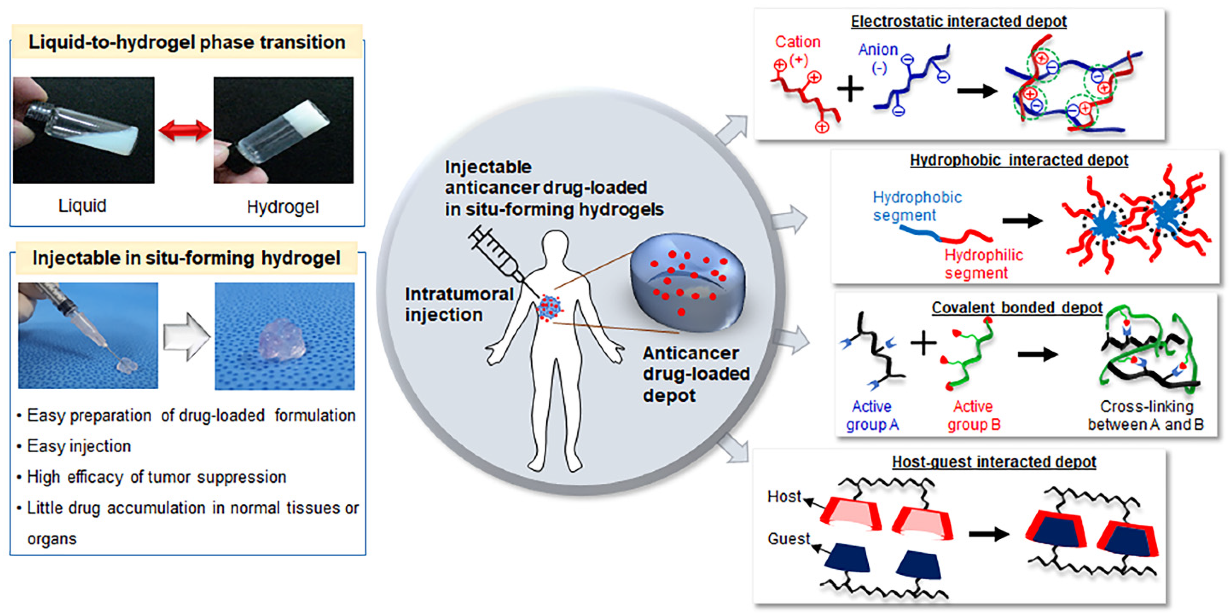
There are several types of injectable in situ-forming hydrogels, which are classified based on the mechanisms that mediate their liquid-to-hydrogel transition. These hydrogels can be fabricated via physical bonding (e.g., electrostatic interaction, hydrogen interaction, or hydrophobic interaction) and chemical bonding via covalent bond formation through light, enzymes, and click crosslinking agents [
18,
19,
20].
This review presents a comprehensive and detailed overview of the most recent advances in fabrication strategies for using injectable in situ-forming hydrogels for the intratumoral injection of anticancer drugs (
Figure 1). First, this review discusses the preparation of injectable in situ-forming hydrogel formulations via chemical and physical interaction and intratumoral injection in cancer therapy. Afterward, prospective future uses of injectable anticancer drug-loaded hydrogel formulations are proposed.
2. Injectable In Situ-Forming Hydrogels
Over the past decades, several studies have described the design and synthesis of several hydrogels for applications in tissue engineering, drug delivery, and bio-nanotechnology. Hydrogels consist of solvent-swelled polymer structures, and water is typically used as the solvent in biological applications. In situ-forming hydrogels can form by swelling in water through different driving forces including the covalent and ionic bonds of hydrophilic polymers [
21].
In situ-forming hydrogels offer several advantages in biomedical applications. Specifically, these materials can be prepared in liquid form at room temperature (i.e., outside the human body) and in the absence of trigger materials or mechanisms, after which they can be quickly injected into the body. Furthermore, different hydrogel formulations containing various types of drugs (e.g., anticancer drugs) can be prepared via simple physical mixing. Therefore, in situ-forming hydrogels are promising carrier candidates that could allow for the easy and convenient delivery of anticancer drugs.
For intratumoral injection, the in situ-forming hydrogels not only have to be stable, cost-efficient, and easy to manufacture, but must also be compatible with the drugs to be injected, in addition to promoting a suitable shape formation, uniform and consistent drug loading, and drug release [
22,
23,
24,
25]. Several fabrication methods of injectable in situ-forming hydrogels have been reported. This review will focus on injectable hydrogels formed via physical and chemical methods.
2.1. Injectable In Situ-Forming Hydrogels Prepared via Physical Interactions
The structure of certain biomaterials relies primarily on physical interactions such as ionic, hydrogen, and hydrophobic bonding, as well as inter- and intra-physical interactions. These physical interactions are relatively weak compared to chemical bonds, thus allowing for the formation of reversible hydrogel matrices according to variations in various biologic conditions [
26,
27]. The preparation of injectable in situ-forming hydrogels via physical interactions does not require the use of crosslinking agents or chemical modification. Therefore, various biomaterials can be used relatively easily as injectable in situ-forming hydrogels via physical interaction [
28,
29].
However, given that physical interactions depend on the physical properties of the biomaterial itself, some of the characteristics of these materials are inflexible, including their gelation time, in vitro and in vivo maintenance, and the mechanical properties of the formed hydrogel. Therefore, it may be difficult to accurately control the in vivo performance of these hydrogels [
27,
28,
29]. Nevertheless, these shortcomings can be overcome by using biomaterials with different molecular weights and concentrations, as well as different blends of biomaterials with other components. These procedures can result in the formation of hydrogels with different stiffnesses, viscosities, rheological behaviors, swelling and disintegration behaviors, and biocompatibility. Thus, injectable in situ-forming hydrogels prepared via physical interaction have been widely used for intratumoral injections due to the ease of biomaterial selection and applicability. The different types of injectable in situ-forming hydrogels prepared via physical interaction are described below.
2.1.1. Injectable In Situ-Forming Hydrogels Prepared via Electrostatic Interactions
Electrostatic interaction is one of the most common physical bonds. This type of bond consists of the interaction between two opposite charges such as positive and negative electrolyte groups. These electrostatic interactions can be caused by (poly)electrolyte groups with ionizing or protonating properties, as well as pH changes. These (poly)electrolyte groups can also be affected by external chemical, thermal, and mechanical factors.
Several typical biomaterials contain anionic (poly)electrolytes derived from natural materials such as alginate, chondroitin sulfate, hyaluronate, heparin, sodium carboxymethylcellulose, pectin, dextran sulfate, and xanthan, as well as synthetic materials such as polyacrylic acid derivatives. Cationic (poly)electrolytes include chitosan, polydiallydimethylammonium chloride, spermine, spermidine, polyethylenimine, and polylysine.
Electrostatic interactions can easily be formed between anionic (poly)electrolytes and cationic (poly)electrolytes by instantaneous physical crosslinking, but these interactions can also easily lead to a decreased cross-linking capability with several other (poly)electrolytes in physiological environments [
30,
31,
32]. Materials with anionic (poly)electrolytes (e.g., carboxylates, phosphates, and sulfates) and cationic (poly)electrolytes (e.g., protonated amines) can be individually prepared as injectable (i.e., liquid) formulations before mixing. The (poly)electrolytes in the injectable formulations can induce electrostatic interactions after mixing, resulting in hydrogel formation [
33,
34,
35,
36].
However, given the wide variety of electrostatic materials in living organisms, the performance of electrostatic-interaction-based hydrogels may vary depending on the surrounding environments (e.g., in the presence of biological materials and anionic and cationic (poly)electrolytes), as well as the body’s temperature. Resolving these limitations is thus necessary for the further development and widespread adoption of injectable intratumoral hydrogels prepared via electrostatic interactions [
29].
2.1.2. Injectable In Situ-Forming Hydrogels Prepared via Hydrophobic Interaction
Under physiological conditions, hydrophobic materials with non-polar groups repel water and aggregate among themselves or with other non-polar materials. In contrast, amphiphilic materials containing both hydrophilic and hydrophobic groups can either dissolve or precipitate in water depending on the environmental conditions. Certain environmental conditions favor the dissolution of hydrophilic structures in water, thus inducing the dissolution of amphiphilic materials in aqueous solution. In other conditions, precipitation can occur via the dehydration of the aqueous solution by the hydrophobic segments [
34,
37].
Given the difference between body temperature and room temperature, the solubility of amphiphilic materials in water may vary under physiological conditions. This phenomenon can cause amphiphilic materials to undergo a phase transition from their dissolved to their precipitate states.
Various amphiphilic materials have been developed and utilized as injectable in situ-forming hydrogels via hydrophobic interactions. Poly(ethylene glycol) (PEG) is among the most widely studied hydrophilic structures in the biomedical field. Various amphiphilic materials have been developed by incorporating hydrophobic structures into PEG blocks to be used as injectable in situ-forming hydrogels [
38]. The developed hydrophobic structures include poly(propylene oxide) (PPO), poly(lactide-co-glycolide) (PLGA), polylactic acid (PLA), poly(ε-caprolactone-co-D,L-lactic acid) (PCLA), polycaprolactone (PCL), poly(trimethylene carbonate) (PTMC), poly(δ-valerolactone), poly (1,4-dioxan-2-one) PDO, PCL-co-PTMC, PCL-co-PDO, polysebacic acid, polyphosphazenes, and poly(N-isopropylacrylamide). A variety of amphiphilic materials composed of hydrophobic and hydrophobic structures can be prepared as liner blocks, dendrimers, and network structures.
These materials possess varying advantages depending on their hydrophilic and hydrophobic group composition, including their adjustable in vivo mechanical properties and in vivo biodegradation [
38]. However, amphiphilic materials must be soluble in biological media to serve as carriers for therapeutic agents. Given that many of the developed amphiphilic materials are synthetic, potential challenges such as immune reactions should be comprehensively evaluated prior to their in vivo application [
39].
2.2. Injectable In Situ-Forming Hydrogels Prepared via Covalent Bonding
A covalent bond is the irreversible linking of one molecule (e.g., a biomaterial chain) to another. The formation of covalent bonds between intra- and inter-biomaterial chains can lead to permanent biomaterial fixing or promote changes in the physical properties of the original biomaterials [
40,
41]. If the biomaterial is water-soluble, it can form a water-swellable biomaterial network via the covalent bonding between intra- and inter-biomaterial chains, resulting in the formation of injectable in situ-forming hydrogels [
42]. Covalent bonding can improve the mechanical properties of injectable in situ-forming hydrogels, thus increasing their resistance to dissolution in aqueous solutions.
Covalent bonds between intra- and inter-biomaterial chains can be formed by chemical reactions that are initiated by heat, pressure, pH changes, or irradiation [
43], among which heat application is the most common strategy. Nevertheless, applying heat above body temperature would not be feasible in vivo, and this greatly limits the clinical applicability of injectable in situ-forming hydrogels prepared using heat.
Injectable in situ-forming hydrogels prepared via covalent bonding require functional or crosslinking materials with which to chemically react. Although most functional or crosslinking materials can cause toxicity, injectable hydrogels prepared via covalent bonding have been widely used in intratumoral injections. The sections below describe the types of injectable in situ-forming hydrogels prepared via photo-irradiation, click reaction, and enzyme activity to form covalent bonds.
2.2.1. Injectable In Situ-Forming Hydrogels Prepared via Photo-Irradiation
The photo-reaction method for hydrogel synthesis is generally regarded as fast and practical [
26,
44]. Additionally, various photosensitive materials have been developed [
45] and have been widely studied in a variety of biological fields [
46,
47,
48,
49,
50]. Among them, azobenzene, spiropyran, nitrobenzyl, galactose, 7-diethylamino-4-thiocoumarinylmethyl, cumarin, and cinnamic acid contain anthracene or acrylate groups (photosensitive chromophores), which can be activated within a few seconds of light irradiation.
Nevertheless, unreacted photosensitive chromophores can cause inflammatory reactions. Additionally, light irradiation (particularly UV light) can lead to cytotoxicity and potential genetic mutations. Furthermore, very few compounds could serve as suitable solvents and non-cytotoxic activators (i.e., photoinitiators) in vivo [
51,
52]. Instead, biomaterials containing these photosensitive chromophores or acrylate groups can also be easily activated by light irradiation. This can enable facile and rapid hydrogel formation via light irradiation when performing in vivo injections, thus enabling injection position control [
53].
2.2.2. Injectable In Situ-Forming Hydrogels Prepared via Click Reaction
Covalently click-cross-linked biomaterials prepared as a solution could be used for the development of injectable in situ-forming hydrogels. Click reactions, formed by the mixing of click-group-modified biomaterials, can rapidly form hydrogels with tunable mechanical properties for in vivo applications [
54,
55]. In recent years, click reaction materials (e.g., formed via the copper-catalyzed azide–alkyne cycloaddition or alkyne–azide reactions, Diels–Alder reaction, Schiff base formation, Michael addition, or thioenol addition, among others) have been introduced into biomaterials with various structures (e.g., linear, dendrimer, and network).
Click reactions between click reaction materials rapidly form bioorthogonally cross-linked hydrogels without catalysts or external energy in aqueous media, as well as under physiological environments [
54,
55,
56,
57]. Individual solutions of click-group-modified biomaterials can easily allow for the covalent formation of injectable in situ-forming hydrogels within a few seconds. However, click reactions are potentially cytotoxic, as they involve the use of copper. Additionally, a click reaction’s time may depend on the regiospecificity of the click reagents [
58,
59].
2.3. Injectable In Situ-Forming Hydrogels Prepared via Enzyme Activity
Enzyme-mediated crosslinking can enable the formation of hydrogels in physiological conditions, and injectable in situ-forming hydrogels prepared via enzymatic reactions are increasingly being used as alternatives to metal catalysts and photo-irradiation [
60].
Active enzymes for injectable in situ-forming hydrogels include laccases, horseradish peroxidase (HRP), transglutaminases (TGases, protein-glutamine gamma-glutamyltransferase), tyrosinase (Tyr), and lysyl oxidase coupled with hydrogen peroxide (H
2O
2) to support the reaction [
61,
62,
63,
64,
65,
66,
67,
68,
69]. Enzymatic-mediated crosslinking using HRP induces the binding of aniline, phenol, and its derivative tyramine in the presence of H
2O
2 [
70,
71,
72,
73]. Enzymatic-mediated crosslinking forms strong covalent bonding, with reactions occurring in less than 10 min. Additionally, the kinetics of in situ-forming hydrogel synthesis can be manipulated by controlling the enzyme concentration. Further, the products of these reactions tend to be highly biocompatible and are especially well suited to the preparation of injectable hydrogels. Nevertheless, several physicochemical factors such as the pH, temperature, and steric hindrance caused by the substrate structure can significantly affect the catalytic activity of enzymes [
74].
3. Intratumoral Injection Using Anticancer Drug-Loaded Injectable Hydrogels
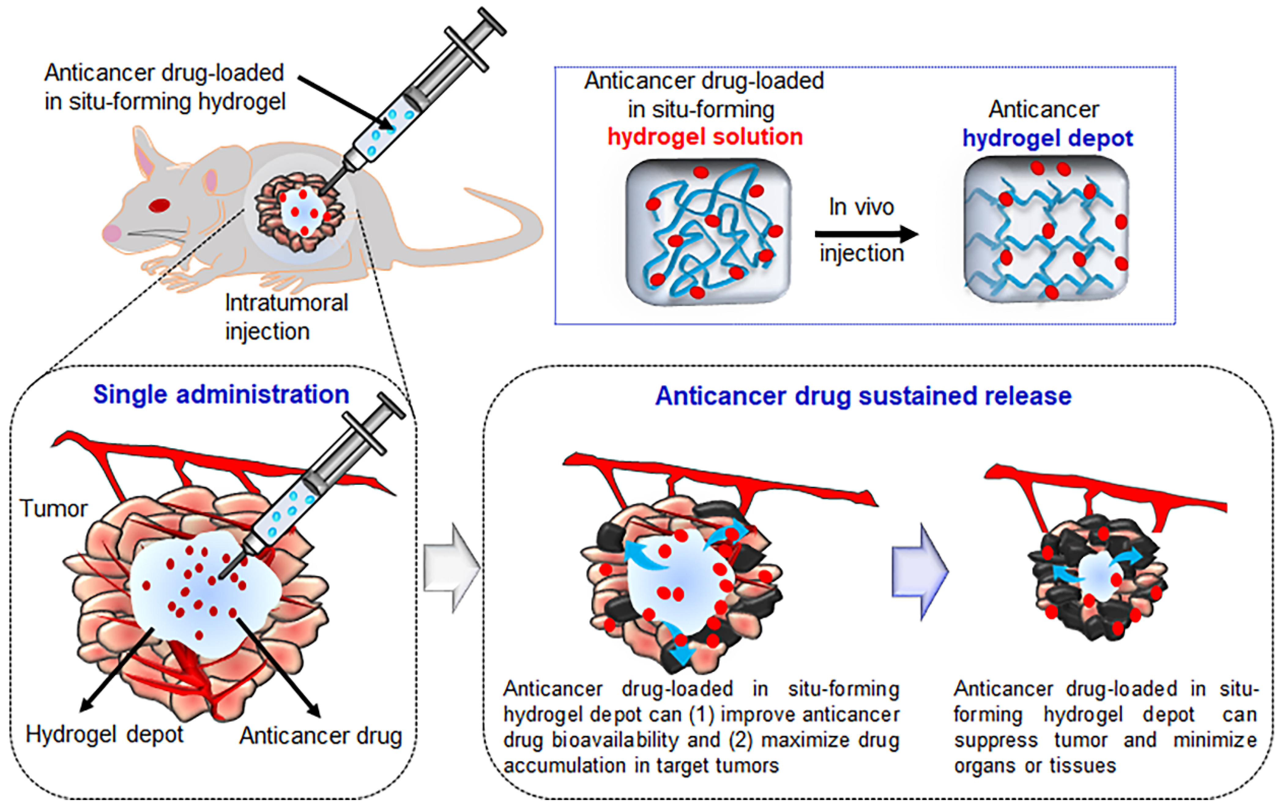
Intratumoral injection can maximize the efficiency of chemotherapeutic agents and minimize their toxicity to normal organs and tissues other than the target tumor. Additionally, the administration of chemotherapeutic compounds via intratumoral injection could greatly reduce the amount of a drug required for a single administration compared with conventional approaches in which anticancer drugs are repeatedly administered (
Figure 2). Therefore, the injectable in situ-forming hydrogels described in the previous sections can easily be prepared as anticancer drug-loaded hydrogel formulations and could be highly effective as a non-invasive method to treat cancer via direct intratumoral injection [
75,
76].
Table 1 and
Table 2 summarize a selection of recent studies that evaluated the applicability of anticancer drug-loaded in situ-forming hydrogels for intratumoral injection, which will be described in the following section.
3.1. Intratumoral Injection Using Hydrogels Prepared via Electrostatic Interactions
As discussed above, anticancer drugs can easily be mixed into electrostatic interaction hydrogels prepared using several electrostatic biomaterials.
Huayamares et al. prepared electrostatic interaction hydrogels using glatiramer acetate (GA) and hyaluronic acid (HA) [
77]. In a comparative experiment, the electrostatic interacted GA/HA hydrogel was injected into solid tumors using a hydrogel prepared from GA and nonionic PEG. After intratumoral injection, the GA/HA exhibited a higher tumor retention rate than the GA/PEG due to electrostatic interaction, and the drug release rate of these different hydrogels also varied when administered to treat tumor fibrosis.
Chitosan (CS) is enzymatically degradable, generally non-cytotoxic, and biocompatible, and therefore does not cause adverse effects on healthy organs and tissues near the injected tumor site. CS is a cationic polymer with two functional groups, hydroxyl and amine, and an overall positive charge. Thus, the cationic nature of chitosan (CS) enables this biopolymer to electrostatically interact with anionic molecules such as β-glycerophosphate (β-GP). Particularly, a single intratumoral injection of this CS/β-GP system showed good efficacy in mammary-tumor-bearing mice [
78,
79]
Previous studies sought to increase the efficacy of other drug delivery carriers by taking advantage of the electrostatic interactions with CS. For example, some studies reported on the applicability of a CS/β-GP-based material coupled with carboplatin-loaded PCL nanoparticles and paclitaxel (PTX)-loaded PLGA microparticles. Further, carboxymethylcellulose is an anionic (poly)electrolyte, and therefore hydrogels can be produced via the electrostatic interactions between this compound and chitosan (CCS). Kim et al. prepared curcumin-loaded microspheres to increase the in vivo half-life of curcumin. The authors reported that electrostatically interacting CCS hydrogels with curcumin-loaded microspheres showed good antitumor efficacy in animal models after intratumoral injection [
80].
Collectively, these studies demonstrated that electrostatically interacting hydrogels with/without drug-loaded nano- and micro-particles can synergistically enhance the anticancer activity. Although electrostatically interacting hydrogels can easily be formed using anionic and cationic biomaterials, they tend to be sensitive to changes in pH and are thus easily affected by the protonation and deprotonation of anionic and cationic biomaterials [
81]. These properties can be a disadvantage; however, pH-responsive electrostatic interacted hydrogels can be prepared via the protonation and deprotonation of ionic biomaterials. Furthermore, electrostatically interacting hydrogels are prone to in vivo degradation due to erosion and cracking, thus impairing their mechanical properties and controlled drug release.
3.2. Intratumoral Injection Using Hydrophobically Interacting Hydrogel
As described above, several studies have evaluated the applicability of hydrophobically interacting PEG–polyester block copolymers for the preparation of hydrogels. The most widely used polyesters are PLA, PLGA, and PCL.
Shi et al. used a poly(D,L-lactide)–PEG–poly(D,L-lactide) (PDLLA–PEG–PDLLA) triblock copolymer co-loaded with gemcitabine and cisplatin for a synergistic combination therapy to treat pancreatic cancer [
82]. Kim et al. prepared injectable in situ-forming hydrogels using a MPEG–PCL diblock copolymer for intratumoral injection of doxorubicin (DOX), PTX, and 5-fluorouracil (5-Fu) [
83,
84]. Additionally, the intratumoral injection of PLGA–PEG–PLGA triblock copolymers enabled the sustained release of tamoxifen and Herceptin [
85,
86].
Recently, OncoGel
TM (MacroMed Inc., Salt Lake City, UT, USA) has employed commercial formulations of a PLGA–PEG–PLGA triblock copolymer to deliver PTX to the tumor site in solid malignancies [
87]. The efficacy of hydrophobically interacting hydrogels has been preliminarily demonstrated in a Phase IIa study for intratumoral chemotherapy; however, no significant effects on the overall tumor response were observed.
3.3. Intratumoral Injection Using Photo-Irradiated Hydrogels
Some hydrogels with photoresponsive agents can be utilized for intratumoral injection. The o-nitrobenzyl or azobenzene groups in PEG- or PEG–polyester-based hydrogels undergo irreversible or reversible reactions when irradiated with UV light, respectively [
88,
89]. Another study examined the applicability of a hydrogel composed of indocyanine green–alginate (i.e., two near-infrared (NIR)-responsive agents) and Ca
2+/Mg
2+ for localized tumor ablation [
90]. This injectable indocyanine green–alginate hydrogel has been successfully applied as a highly efficient photothermal therapy in vivo without NIR-induced side effects. Nevertheless, NIR-I light (650–950 nm) penetrates <1 cm into the tissues, and therefore this approach is only suitable to treat superficial and thin tumors. NIR-II light (1000–1700 nm) provides a more feasible means for the treatment of large superficial tumors due to its tissue penetration depth of approximately 3–5 cm; however, its clinical application to the treatment of deep tumors remains restricted [
91].
Mukerji et al. developed a photoradiation-controlled intratumoral depot (PRCITD) driven by convection-enhanced delivery (CED) to spatiotemporally control tumors and anticancer drug coverage [
92]. This intratumoral depot consisted of a recombinant elastin-like polypeptide (ELP) containing periodic cysteine residues and was conjugated with a photosensitizer [chlorin-e6 (Ce6)] at the N-terminus of the ELP. The photodynamic therapy provided by the PRCITD caused significant tumor inhibition in a Ce6 dose-dependent manner. Additionally, the combination of photodynamic and intratumoral radionuclide therapy co-delivered by the PRCITD provided a greater antitumor effect than either monotherapy alone. These findings suggest that the PRCITD could provide a stable platform for the co-delivery of anticancer drugs to induce synergistic effects.
In intratumoral injection using photo-irradiated hydrogels, increasing the power density (W/cm2) and the exposure time to irradiation leads to greater penetration. Given the substantial limitations of UV light (e.g., poor penetration and risk of damaging tissues), approaches involving upconversion of low-energy photons (e.g., near-infrared; NIR) into high-energy photons (e.g., UV) are promising alternatives.
3.4. Intratumoral Injection Using Click Crosslinking Hydrogel
Several biomaterials with click crosslinking molecules can be prepared as a solution and have been used to rapidly form injectable hydrogels in physiological environments. The click crosslinking between functionalized molecules has a high efficiency and excellent specificity at high reaction rates. These properties enable the quick and facile formation of intratumoral depots of anticancer drugs after intratumoral injection.
Xu et al. prepared PEG-based dendrimer hydrogels consisting of alkyne dibenzocyclooctyne and PEG-bisazide. Bioorthogonal depots formed via azide–alkyne cycloaddition after intratumoral injection and exhibited a high cytocompatibility, thus enabling the sustained release of the anticancer drug 5-Fu and suppressing the tumor growth [
93].
Emoto et al. used an HA-aldehyde and HA-adipic dihydrazide with cisplatin for intratumoral injection. The time of gelation via cross-linking was modified by changing the concentration of the HA-aldehyde and HA-adipic dihydrazide. Click crosslinked cisplatin-loaded HA was successfully implemented to locally deliver cisplatin to live mice [
94].
Kim et al. described an in situ-forming PEG hydrogel produced via cross-linking between thiol and maleimide [
95]. The gelation time of this material could be controlled from 15 s to 5 min by modifying the thiol and maleimide concentrations. The TRAIL protein-loaded hydrogel quickly formed a depot and exerted anticancer effects on tumors of Mia Paca-2 cell-xenografted BALB mice.
Although click crosslinking can form a stable depot system for the intratumoral delivery of anticancer drugs, the utilized cross-linking agents can be toxic and have poor biocompatibility, which greatly limits the applicability of this approach. Additional studies are thus required to develop non-toxic crosslinking reagents [
96].
3.5. Intratumoral Injection Using Enzyme-Mediated Crosslinking Hydrogels
Enzyme-reactive hydrogels are typically obtained through enzyme-mediated crosslinking. The preparation of enzyme-reactive hydrogels for intratumoral injection requires the incorporation of an enzyme-specific substrate or a substrate-mimicking material into the hydrogel.
Tang et al. prepared cytarabine HA-tyramine (Ara-HA-Tyr) hydrogel conjugates [
97]. The enzyme-reactive hydrogels were formed through the oxidative coupling of tyramines by H
2O
2 and HRP. These enzyme-reactive hydrogels exhibited a robust synergistic antitumor efficacy when combined with radiotherapy in the Lewis lung cancer xenograft model.
Oh et al. investigated a gelatin-hydroxyphenyl propionic acid (GHPA)-based hydrogel composed of hydroxyphenyl propionic acid conjugated to gelatin obtained via HRP- and H
2O
2-mediated cross-linking [
98]. The enzymatic cross-linking reaction of this system can easily be manipulated to achieve hydrogels with desired properties such as gelation time, mechanical stiffness, and degradation rate. More importantly, the authors demonstrated a synergistic antitumor effect and induction of tumor-specific immune responses via the hydrogel-mediated sustained release of oncolytic adenovirus and dendritic cells in solid tumors.
Xu et al. synthesized an
L-phenylalanine-based low-molecular-weight gelator containing thioketal and a control gelator without reactive oxygen species (ROS)-cleavable bonds [
99]. Enzyme-reactive hydrogels co-loaded with DOX and a photosensitizer were intratumorally injected into 4T1-breast-tumor-bearing mice and rendered antitumor effects in vivo.
Collectively, the above-described findings demonstrate that enzyme-reactive hydrogels exhibit a high substrate specificity (both regioselectivity and stereoselectivity) and prevent the expensive and time-consuming separation of by-products and intermediates. Nevertheless, enzyme reactive hydrogels have recently been linked to cross-reactivity problems in vivo, thus highlighting the need for further research to enhance the specificity of enzymes to their target substrates [
100].
Table 2.
Intratumoral injection using an injectable hydrogel through covalent bonding with anticancer drugs.
Table 2.
Intratumoral injection using an injectable hydrogel through covalent bonding with anticancer drugs.
| | Hydrogel Materials | Drugs or Agents | Cells | Cancer Type | Ref. |
|---|
Photo
Irradiation | Photothermal Ca2+/Mg2+ stimuli-responsive ICG–alginate hydrogel | ICG | 4T1 | Breast cancer cell | [90] |
| cE6 | cELP [A14VC]16 gene | FaDu | Squamous cell carcinoma | [92] |
Click
reaction | DH-P-G4-PDBCO (P-G4-PDBCO + PEG-BA) | 5-Fu | HN12 | HNSCC | [93] |
| HA–ADH, HA-CHO | Cisplatin | MKN45P | Human gastric cancer | [94] |
| HAS-SH/PEG-MAL hydrogel | TRAIL protein | Paca-2 | - | [95] |
Active
enzyme | Ara-HA-Tyr hydrogel; H2O2, HRP | Cytarabine | LLC | Lung cancer | [97] |
| GHPA-based hydrogel | Ad, DC | MLLC | - | [98] |
| DOX–ZnPCS4-coloaded gel | DOX | 4T1 | Breast cancer cell | [99] |
4. Conclusions and Outlook
Research on in situ-forming hydrogels has recently garnered increasing attention, as this technology could enable the localized delivery of anticancer drugs via intratumoral injection. Here, we discussed the preparation of anticancer-drug-loaded injectable in situ-forming hydrogels via chemical and physical interactions for intratumoral injection in cancer therapy. However, this review did not describe the release behavior of anticancer drugs with different lipophilic properties or multiple drugs from hydrogel depots [
101,
102].
The properties of in situ-forming hydrogels are highly associated with their chemical and physical interactions, and therefore the functional improvement of these materials is extremely dependent on innovation of fabrication strategies. Normally, in situ-forming hydrogels generated via chemical interactions exhibit desirable mechanical properties, whereas in situ-forming hydrogel formulations obtained via physical interactions are more biocompatible due to the absence of chemical crosslinking agents in their composition. Therefore, novel and more comprehensive fabrication strategies for situ-forming hydrogels could pave the way for the development of hydrogel formulations containing various anticancer drugs.
Intratumoral injection technology has recently undergone tremendous progress, including the design of in situ-forming hydrogels optimized for the effective delivery of anticancer drugs. Further, we anticipate that the applications and adoption of this novel drug-delivery technology will continue to grow. The primary objective of intratumoral injection is the effective delivery of anticancer drugs to the tumor site with minimal or no systemic drug bioavailability and no toxicity to healthy organs and tissues. Therefore, in situ-forming hydrogels must enable the sustained release of anticancer drugs from depots after intratumoral injection, thus localizing the anticancer drug exclusively to the tumor site. Future studies on intratumoral injection using in situ-forming hydrogels must address the applicability of this technology to different cancer-affected tissues or organs in clinical environments.
Although the efficacy of in situ-forming hydrogels for cancer treatment has been repeatedly demonstrated in several proof-of-concept experiments using various animal models and cancer types, translating this technology to clinical applications in humans will pose several important challenges.
Despite the recent progress in the development of in situ-forming hydrogels for intratumoral injection, several challenges still need to be considered. The first major challenge is the immunogenicity of in situ-forming hydrogel materials to healthy organs and tissues near the injected tumor site, as most injected depots can cause serious inflammation at the injected site. Additionally, in situ-forming hydrogels must be biodegradable under the tumor microenvironment conditions, ideally at a similar rate to the tumor’s decrease. Moreover, the development of intratumoral injection technology requires a thorough understanding of the responses of tumor tissues to in situ-forming hydrogels, as many situ-forming hydrogels are designed and tested in hypothetical conditions and not in the tumor microenvironment. Finally, the spatiotemporal release of anticancer drugs from the depot is another important challenge that must be overcome in order to maximize the efficacy of the hydrogels for the desired treatment period.
Therefore, the following factors must be considered for clinical translation: (1) biomaterials for in situ-forming hydrogels must meet perfect biocompatibility and biodegradability standards; (2) in situ-forming hydrogels must be highly responsive to human tumors; and (3) drug loaded-in situ-forming hydrogels must be evaluated in human trials, paying special attention to the surrounding tumor processes, age, and physical activity.
A more comprehensive understanding of the properties of injectable in situ-forming hydrogels would contribute to the improvement of patients’ convenience while reducing drugs’ systemic toxicity and also allowing for the programmable delivery and sustained release of anticancer drugs from the hydrogel depot—all of which would facilitate clinical translation. In summary, a joint multidisciplinary effort is urgently needed to develop and apply novel strategies that might help to materialize the tremendous potential of injectable in situ-forming hydrogel technology for intratumoral injection. Achieving this would greatly contribute to the advancement of cancer therapy in the near future.
Author Contributions
Conceptualization, G.R.S., H.E.K. and M.S.K.; investigation, G.R.S., H.E.K., J.H.K., S.C. and M.S.K.; supervision, M.S.K.; funding acquisition, S.C. and M.S.K.; writing—original draft, G.R.S.; writing—review and editing, M.S.K. All authors have read and agreed to the published version of the manuscript.
Funding
This study was supported by National Research Foundation of Korea (NRF) grants: Creative Materials Discovery Program (2019M3D1A1078938) and Priority Research Centers Program (2019R1A6A1A11051471).
Institutional Review Board Statement
Not applicable.
Informed Consent Statement
Not applicable.
Data Availability Statement
The data presented in this study are available on request from the corresponding author.
Conflicts of Interest
The authors declare that they have no known competing financial interests or personal relationships that could have appeared to influence the work reported in this paper.
References
- Sung, H.; Ferlay, J.; Siegel, L.; Laversanne, M.; Soerjomataram, I.; Jemal, A.; Bray, F. Global cancer statistics 2020: GLOBOCAN estimates of incidence and mortality worldwide for 36 cancers in 185 countries. CA Cancer J. Clin. 2021, 71, 209–249. [Google Scholar] [CrossRef]
- Qin, S.Y.; Zhang, A.Q.; Cheng, S.X.; Rong, L.; Zhang, X.Z. Drug self-delivery systems for cancer therapy. Biomaterials 2017, 112, 234–247. [Google Scholar] [CrossRef] [PubMed]
- Avramović, N.; Mandić, B.; Savić-Radojević, A.; Simić, T. Polymeric nanocarriers of drug delivery systems in cancer therapy. Pharmaceutics 2020, 12, 298. [Google Scholar] [CrossRef] [Green Version]
- Zhang, M.; Gao, S.; Yang, D.; Fang, Y.; Lin, X.; Jin, X.; Liu, Y.; Liu, X.; Su, K.; Shi, K. Influencing factors and strategies of enhancing nanoparticles into tumors in vivo. Acta Pharm. Sin. B 2021, 11, 2265–2285. [Google Scholar] [CrossRef]
- Vishnevskiy, D.A.; Garanina, A.S.; Chernysheva, A.A.; Chekhonin, V.P.; Naumenko, V.A. Neutrophil and nanoparticles delivery to tumor: Is it going to carry that weight? Adv. Healthc. Mater. 2021, 10, 2002071. [Google Scholar] [CrossRef]
- Wang, G.Y.; Rayner, S.L.; Chung, R.; Shi, B.Y.; Liang, X.J. Advances in nanotechnology-based strategies for the treatments of amyotrophic lateral sclerosis. Mater. Today Biol. 2020, 6, 100055. [Google Scholar] [CrossRef]
- Fahmy, S.A.; Brüßler, J.; Alawak, M.; El-Sayed, M.M.H.; Bakowsky, U.; Shoeib, T. Chemotherapy based on supramolecular chemistry: A promising strategy in cancer therapy. Pharmaceutics 2019, 11, 292. [Google Scholar] [CrossRef] [Green Version]
- Lim, W.; Kim, H.S. Exosomes as therapeutic vehicles for cancer. Tissue Eng. Regen. Med. 2019, 16, 213–223. [Google Scholar] [CrossRef] [PubMed]
- Low, L.E.; Wu, J.; Lee, J.; Tey, B.T.; Goh, B.H.; Gao, J.; Li, F.; Ling, D. Tumor-responsive dynamic nanoassemblies for targeted imaging, therapy and microenvironment manipulation. J. Control Release 2020, 324, 69–103. [Google Scholar] [CrossRef] [PubMed]
- Yoon, M.S.; Lee, Y.J.; Shin, H.J.; Park, C.-W.; Han, S.-B.; Jung, J.-K.; Kim, J.-S.; Shin, D.H. Recent advances and challenges in controlling the spatiotemporal release of combinatorial anticancer drugs from nanoparticles. Pharmaceutics 2020, 12, 1156. [Google Scholar] [CrossRef]
- Waks, A.G.; Winer, E.P. Breast cancer treatment a review. JAMA 2019, 321, 228–300. [Google Scholar]
- Sood, N.; Bhardwaj, A.; Mehta, S.; Mehta, A. Stimuli-responsive hydrogels in drug delivery and tissue engineering. Drug Deliv. 2016, 23, 758–780. [Google Scholar] [CrossRef] [Green Version]
- Mathew, A.P.; Uthaman, S.; Cho, K.H.; Cho, C.S.; Park, I.K. Injectable hydrogels for delivering biotherapeutic molecules. Int. J. Biol. Macromol. 2018, 110, 17–29. [Google Scholar] [CrossRef] [PubMed]
- Norouzi, M.; Nazari, B.; Miller, D.W. Injectable hydrogel-based drug delivery systems for local cancer therapy. Drug Discov. 2016, 21, 1835–1849. [Google Scholar] [CrossRef]
- Sapino, S.; Chirio, D.; Peira, E.; Abellán Rubio, E.; Brunella, V.; Jadhav, S.A.; Chindamo, G.; Gallarate, M. Ocular drug delivery: A special focus on the thermosensitive approach. Nanomaterials 2019, 9, 884. [Google Scholar] [CrossRef] [PubMed] [Green Version]
- Meng, Q.Y.; Cong, H.L.; Hu, H.; Xu, F.J. Rational design and latest advances of codelivery systems for cancer therapy. Mater. Today Biol. 2020, 7, 100056. [Google Scholar] [CrossRef]
- Fakhari, A.; Subramony, J.A. Engineered in-situ depot-forming hydrogels for intratumoral drug delivery. J. Controlled Release 2015, 220, 465–475. [Google Scholar] [CrossRef] [PubMed]
- Rizzo, F.; Kehr, N.S. Recent advances in injectable hydrogels for controlled and local drug delivery. Adv. Healthc. Mater. 2021, 10, 2001341. [Google Scholar] [CrossRef]
- Das, B.; Chattopadhyay, D.; Rana, D. The gamut of perspectives, challenges, and recent trends for in situ hydrogels: A smart ophthalmic drug delivery vehicle. Biomater. Sci. 2020, 8, 4665–4691. [Google Scholar] [CrossRef]
- Park, K.M.; Park, K.D. In situ cross-linkable hydrogels as a dynamic matrix for tissue regenerative medicine. Tissue Eng. Regen. Med. 2018, 15, 547–557. [Google Scholar] [CrossRef]
- Wells, L.A.; Lasowski, F.; Fitzpatrick, S.D.; Sheardown, H. Responding to change: Thermo—and photo-responsive polymers as unique biomaterials. Crit. Rev. Biomed. Eng. 2010, 38, 487–509. [Google Scholar] [CrossRef]
- Li, J.; Chen, G.; Xu, X.; Abdou, P.; Jiang, Q.; Shi, D.; Gu, Z. Advances of injectable hydrogel-based scaffolds for cartilage regeneration. Regen. Biomater. 2019, 6, 129–140. [Google Scholar] [CrossRef] [Green Version]
- Leach, D.G.; Young, S.; Hartgerink, J.D. Advances in immunotherapy delivery from implantable and injectable biomaterials. Acta Biomater. 2019, 88, 15–31. [Google Scholar] [CrossRef] [PubMed]
- Akindoyo, J.O.; Mariatti, M.; Hamid, Z.A.A.; Nurul, A.A.; Teramoto, N. Injectable hydrogel scaffold from natural biomaterials—An overview of recent studies. In Proceedings of the AIP Conference Proceedings, Penang, Malaysia, 31 October–1 November 2019; AIP Publishing LLC: Huntington, NY, USA, 2020; Volume 2267, p. 020068. [Google Scholar]
- Lee, J.H. Injectable hydrogels delivering therapeutic agents for disease treatment and tissue engineering. Biomater. Res. 2018, 22, 27. [Google Scholar] [CrossRef] [PubMed] [Green Version]
- Parhi, R. Cross-linked hydrogel for pharmaceutical applications: A review. Adv. Pharm. Bull. 2017, 7, 515–530. [Google Scholar] [CrossRef] [PubMed]
- Hoffman, A.S. Hydrogels for biomedical applications. Adv. Drug Deliv. Rev. 2002, 54, 3–12. [Google Scholar] [CrossRef]
- Seo, J.Y.; Lee, B.; Kang, T.W.; Noh, J.H.; Kim, M.J.; Ji, Y.B.; Ju, H.J.; Min, B.H.; Kim, M.S. Electrostatically interactive injectable hydrogels for drug delivery. Tissue Eng. Regen. Med. 2018, 15, 513–520. [Google Scholar] [CrossRef]
- Akther, F.; Little, P.; Li, Z.; Nguyen, N.T.; Ta, H.T. Hydrogels as artificial matrices for cell seeding in microfluidic devices. RSC Adv. 2020, 10, 43682–43703. [Google Scholar] [CrossRef]
- Yadav, S.K.; Khan, G.; Bansal, M.; Vardhan, H.; Mishra, B. Screening of ionically crosslinked chitosan-tripolyphosphate microspheres using Plackett–Burman factorial design for the treatment of intrapocket infections. Drug Dev. Ind. Pharm. 2017, 43, 1801–1816. [Google Scholar] [CrossRef]
- Shitrit, Y.; Bianco-Peled, H. Insights into the formation mechanisms and properties of pectin hydrogel physically cross-linked with chitosan nanogels. Carbohydr. Polym. 2021, 269, 118274. [Google Scholar] [CrossRef]
- Jung, S.W.; Oh, S.H.; Lee, I.S.; Byun, J.H.; Lee, J.H. In situ gelling hydrogel with anti-bacterial activity and bone healing property for treatment of osteomyelitis. Tissue Eng. Regen. Med. 2019, 16, 479–490. [Google Scholar] [CrossRef]
- Lim, J.Y.C.; Lin, Q.; Xue, K.; Loh, X.J. Recent advances in supramolecular hydrogels for biomedical applications. Mater. Today Adv. 2019, 3, 100021. [Google Scholar] [CrossRef]
- Perera, M.M.; Ayres, N. Dynamic covalent bonds in self-healing, shape memory, and controllable stiffness hydrogels. Polym. Chem. 2020, 11, 1410–1423. [Google Scholar] [CrossRef]
- Choi, J.S.; Heang, O.S.; Kim, Y.M.; Lim, J.Y. Hyaluronic acid/alginate hydrogel containing hepatocyte growth factor and promotion of vocal fold wound healing. Tissue Eng. Regen. Med. 2020, 17, 651–658. [Google Scholar] [CrossRef]
- Sun, M.T.; O’Connor, A.J.; Milne, I.; Biswas, D.; Casson, R.; Wood, J.; Selva, D. Development of macroporous chitosan scaffolds for eyelid tarsus tissue engineering. Tissue Eng. Regen. Med. 2019, 16, 595–604. [Google Scholar] [CrossRef]
- Kissel, T.; Li, Y.; Unger, F. ABA-triblock copolymers from biodegradable polyester A-blocks and hydrophilic poly (ethylene oxide) B-blocks as a candidate for in situ forming hydrogel delivery systems for proteins. Adv. Drug Deliv. Rev. 2002, 54, 99–134. [Google Scholar] [CrossRef]
- Chang, H.; Li, C.; Huang, R.; Su, R.; Qi, W.; He, Z. Amphiphilic hydrogels for biomedical applications. J. Mater. Chem. B 2019, 7, 2899–2910. [Google Scholar] [CrossRef]
- Yuba, E. Development of functional liposomes by modification of stimuli-responsive materials and their biomedical applications. J. Mater. Chem. B 2020, 8, 1093–1107. [Google Scholar] [CrossRef] [PubMed]
- Ju, H.; Lai, G.; Yan, F. Immunosensing for Detection of Protein Biomarkers; Elsevier: Amsterdam, The Netherlands, 2017. [Google Scholar]
- Liu, H.Y.; Korc, M.; Lin, C.C. Biomimetic and enzyme-responsive dynamic hydrogels for studying cell-matrix interactions in pancreatic ductal adenocarcinoma. Biomaterials 2018, 160, 24–36. [Google Scholar] [CrossRef] [PubMed]
- Ahmed, E.M. Hydrogel: Preparation, characterization, and applications: A review. J. Adv. Res. 2015, 6, 105–121. [Google Scholar] [CrossRef] [Green Version]
- Marco Dufort, B.; Tibbitt, M. Design of moldable hydrogels for biomedical applications using dynamic covalent boronic esters. Mater. Today Chem. 2019, 12, 16–33. [Google Scholar] [CrossRef]
- Nicol, E. Photopolymerized porous hydrogels. Biomacromolecules 2021, 22, 1325–1345. [Google Scholar] [CrossRef]
- Pillarisetti, S.; Uthaman, S.; Huh, K.M.; Koh, Y.S.; Lee, S.; Park, I.K. Multimodal composite iron oxide nanoparticles for biomedical applications. Tissue Eng. Regen. Med. 2019, 16, 451–465. [Google Scholar] [CrossRef]
- Mantha, S.; Pillai, S.; Khayambashi, P.; Upadhyay, A.; Zhang, Y.; Tao, O.; Pham, H.M.; Tran, S.D. Smart hydrogels in tissue engineering and regenerative medicine. Materials 2019, 12, 3323. [Google Scholar] [CrossRef] [PubMed] [Green Version]
- Zhu, H.; Yang, H.; Ma, Y.; Lu, T.J.; Xu, F.; Genin, G.M.; Lin, M. Spatiotemporally controlled photoresponsive hydrogels: Design and predictive modeling from processing through application. Adv. Funct. Mater. 2020, 30, 2000639. [Google Scholar] [CrossRef]
- Yang, D.H.; Chun, H.J. Visible light-curable hydrogel systems for tissue engineering and drug delivery. Adv. Exp. Med. Biol. 2020, 1249, 85–93. [Google Scholar] [PubMed]
- Lee, C.; O’Connell, C.D.; Onofrillo, C.; Choong, P.F.M.; Di Bella, C.; Duchi, S. Human articular cartilage repair: Sources and detection of cytotoxicity and genotoxicity in photo-crosslinkable hydrogel bioscaffolds. Stem Cells Transl. Med. 2020, 9, 302–315. [Google Scholar] [CrossRef] [Green Version]
- Ji, W.; Wu, Q.; Han, X.; Zhang, W.; Wei, W.; Chen, L.; Li, L.; Huang, W. Photosensitive hydrogels: From structure, mechanisms, design to bioapplications. Sci. China Life Sci. 2020, 12, 1813–1828. [Google Scholar] [CrossRef]
- Choi, G.; Cha, H.J. Recent advances in the development of nature-derived photocrosslinkable biomaterials for 3D printing in tissue engineering. Biomater. Res. 2019, 23, 18. [Google Scholar] [CrossRef] [PubMed] [Green Version]
- Seo, J.W.; Shin, S.R.; Park, Y.J.; Bae, H. Hydrogel production platform with dynamic movement using photo-crosslinkable/temperature reversible chitosan polymer and stereolithography 4d printing technology. Tissue Eng. Regen. Med. 2020, 17, 423–431. [Google Scholar] [CrossRef]
- Yao, H.; Wang, J.; Mi, S. Photo processing for biomedical hydrogels design and functionality: A review. Polymers 2017, 10, 11. [Google Scholar] [CrossRef] [Green Version]
- Arslan, M.; Tasdelen, M.A. Polymer nanocomposites via click chemistry reactions. Polymers 2017, 9, 499. [Google Scholar] [CrossRef]
- Gopinathan, J.; Noh, I. Click chemistry-based injectable hydrogels and bioprinting inks for tissue engineering applications. Tissue Eng. Regen. Med. 2018, 15, 531–546. [Google Scholar] [CrossRef]
- Uliniuc, A.; Popa, M.; Hamaide, T.; Dobromir, M. New approaches in hydrogel synthesis—click chemistry: A review. Cellul. Chem. Technol. 2012, 46, 1–11. [Google Scholar]
- Xu, Z.; Bratli, K. Click chemistry and material selection for in situ fabrication of hydrogels in tissue engineering applications. ACS Biomater. Sci. Eng. 2018, 4, 2276–2291. [Google Scholar] [CrossRef] [PubMed]
- Nwe, K.; Brechbiel, M.W. Growing applications of “click chemistry” for bioconjugation in contemporary biomedical research. Cancer Biother. Radiopharm. 2009, 24, 289–302. [Google Scholar] [CrossRef] [PubMed]
- Kasiński, A.; Zielińska-Pisklak, M.; Oledzka, E.; Sobczak, M. Smart hydrogels—synthetic stimuli-responsive antitumor drug release systems. Int. J. Nanomed. 2020, 15, 4541–4572. [Google Scholar] [CrossRef]
- Wang, X.; Wang, Q. Enzyme-laden bioactive hydrogel for biocatalytic monitoring and regulation. Acc. Chem. Res. 2021, 54, 1274–1287. [Google Scholar] [CrossRef] [PubMed]
- Sakai, S.; Nakahata, M. Horseradish peroxidase catalyzed hydrogelation for biomedical, biopharmaceutical, and biofabrication applications. Chem. Asian J. 2017, 12, 3098–3109. [Google Scholar] [CrossRef]
- Lee, F.; Bae, K.H.; Kurisawa, M. Injectable hydrogel systems crosslinked by horseradish peroxidase. Biomed. Mater. 2016, 11, 014101. [Google Scholar] [CrossRef]
- Eleftheriadou, D.; Kesidou, D.; Moura, F.; Felli, E.; Song, W. Redox-responsive nanobiomaterials-based therapeutics for neurodegenerative diseases. Small 2020, 16, 1907308. [Google Scholar] [CrossRef]
- Zhao, L.; Li, X.; Zhao, J.; Ma, S.; Ma, X.; Fan, D.; Zhu, C.; Liu, Y. A novel smart injectable hydrogel prepared by microbial transglutaminase and human-like collagen: Its characterization and biocompatibility. Mater. Sci. Eng. 2016, 68, 317–326. [Google Scholar] [CrossRef]
- Nezhad-Mokhtari, P.; Ghorbani, M.; Roshangar, L.; Rad, J.S. Chemical gelling of hydrogels-based biological macromolecules for tissue engineering: Photo—and enzymatic-crosslinking methods. Int. J. Biol. Macromol. 2019, 139, 760–772. [Google Scholar] [CrossRef] [PubMed]
- Gohil, S.V.; Brittain, S.; Kan, H.; Drissi, H.; Rowe, D.; Nair, L. Evaluation of enzymatically crosslinked injectable glycol chitosan hydrogel. J. Mater. Chem. B 2015, 3, 5511–5522. [Google Scholar] [CrossRef] [PubMed]
- Wang, R.; Leber, N.; Buhl, C.; Verdonschot, N.; Dijkstra, P.J.; Karperien, M. Cartilage adhesive and mechanical properties of enzymatically crosslinked polysaccharide tyramine conjugate hydrogels. Polym. Adv. Technol. 2014, 25, 568–574. [Google Scholar] [CrossRef]
- Bae, J.W.; Choi, J.H.; Lee, Y.; Park, K.D. Horseradish peroxidase-catalysed in situ-forming hydrogels for tissue-engineering applications. J. Tissue Eng. Regen. Med. 2015, 9, 1225–1232. [Google Scholar] [CrossRef]
- Darakhshan, S.; Pour, A.B.; Kowsari-Esfahan, R.; Vosough, M.; Montazeri, L.; Ghanian, M.H.; Baharvand, H.; Piryaei, A. Generation of scalable hepatic micro-tissues as a platform for toxicological studies. Tissue Eng. Regen. Med. 2020, 17, 459–475. [Google Scholar] [CrossRef]
- Wemmer, J.; Holtgrave, S.; Wiest, L.; Michel, M.; Leser, M.E.; Windhab, E.J. Enzymatic cross-linking of pectin in a high-pressure foaming process. Food Func. 2020, 11, 2040–2047. [Google Scholar] [CrossRef] [Green Version]
- Kim, Y.H.; Park, K.D. Synthesis and characterization of enzyme-mediated injectable carrageenan hydrogels. Polym. Korea 2019, 43, 309–315. [Google Scholar] [CrossRef]
- Le Thi, P.; Lee, Y.; Nguyen, D.H.; Park, K.D. In situ forming gelatin hydrogels by dual-enzymatic cross-linking for enhanced tissue adhesiveness. J. Mater. Chem. B 2017, 5, 757–764. [Google Scholar] [CrossRef] [PubMed]
- Su, T.; Zhang, D.; Tang, Z.; Wu, Q.; Wang, Q. HRP-mediated polymerization forms tough nanocomposite hydrogels with high biocatalytic performance. Chem. Commun. 2013, 49, 8033–8035. [Google Scholar] [CrossRef]
- Song, W.; Ko, J.; Choi, Y.H.; Hwang, N.S. Recent advancements in enzyme-mediated crosslinkable hydrogels: In vivo-mimicking strategies. APL Bioeng. 2021, 5, 021502. [Google Scholar] [CrossRef]
- Cirillo, G.; Spizzirri, U.G.; Curcio, M.; Nicoletta, F.P.; Iemma, F. Injectable hydrogels for cancer therapy over the last decade. Pharmaceutics 2019, 11, 486. [Google Scholar] [CrossRef] [PubMed] [Green Version]
- Park, S.H.; Ji, Y.B.; Park, J.Y.; Ju, H.J.; Lee, M.; Lee, S.; Kim, J.H.; Min, B.H.; Kim, M.S. Injectable in situ-forming hydrogels for protein and peptide delivery. Adv. Exp. Med. Biol. 2020, 1250, 35–48. [Google Scholar] [PubMed]
- Huayamares, S.G.; Song, J.Y.; Huang, A.; Crowl, S.; Groer, C.; Forrest, M.; Berkland, C. Constructing a biomaterial to simulate extracellular drug transport in solid tumors. Macromol. Biosci. 2020, 20, 2000251. [Google Scholar] [CrossRef]
- Bragta, P.; Sidhu, R.K.; Jyoti, K.; Baldi, A.; Jain, U.K.; Chandra, R.; Madan, J. Intratumoral administration of carboplatin bearing poly (ε-caprolactone) nanoparticles amalgamated with in situ gel tendered augmented drug delivery, cytotoxicity, and apoptosis in melanoma tumor. Colloids Surf. B 2018, 166, 339–348. [Google Scholar] [CrossRef]
- Pesoa, J.I.; Rico, M.J.; Rozados, V.R.; Scharovsky, O.G.; Luna, J.A.; Mengatto, L.N. Paclitaxel delivery system based on poly(lactide-co-glycolide) microparticles and chitosan thermo-sensitive gel for mammary adenocarcinoma treatment. J. Pharm. Pharmacol. 2018, 70, 1494–1502. [Google Scholar] [CrossRef]
- Park, S.H.; Kim, D.Y.; Panta, P.; Heo, J.Y.; Lee, H.Y.; Kim, J.H.; Min, B.H.; Kim, M.S. An intratumoral injectable, electrostatic, cross-linkable curcumin depot and synergistic enhancement of anticancer activity. NPG Asia Mater. 2017, 9, e397. [Google Scholar] [CrossRef] [Green Version]
- Mekonnen, T.W.; Andrgie, A.T.; Darge, H.F.; Birhan, Y.S.; Hanurry, E.Y.; Chou, H.-Y.; Lai, J.-Y.; Tsai, H.-C.; Yang, J.M.; Chang, Y.-H. Bioinspired composite, pH-responsive sodium deoxycholate hydrogel and generation 4.5 poly (amidoamine) dendrimer improves cancer treatment efficacy via doxorubicin and resveratrol co-delivery. Pharmaceutics 2020, 12, 1069. [Google Scholar] [CrossRef]
- Shi, K.; Xue, B.; Jia, Y.; Yuan, L.; Han, R.; Yang, F.; Peng, J.; Qian, Z. Sustained co-delivery of gemcitabine and cis-platinum via biodegradable thermo-sensitive hydrogel for synergistic combination therapy of pancreatic cancer. Nano Res. 2019, 12, 1389–1399. [Google Scholar] [CrossRef]
- Seo, H.W.; Kim, D.Y.; Kwon, D.Y.; Kwon, J.S.; Jin, L.M.; Lee, B.; Kim, J.H.; Min, B.H.; Kim, M.S. Injectable intratumoral hydrogel as 5-fluorouracil drug depot. Biomaterials 2013, 34, 2748–2757. [Google Scholar] [CrossRef]
- Kang, Y.M.; Kim, G.H.; Kim, J.I.; Kim, D.Y.; Lee, B.N.; Yoon, S.M.; Kim, J.H.; Kim, M.S. In vivo efficacy of an intratumorally injected in situ-forming doxorubicin/poly (ethylene glycol)-b-polycaprolactone diblock copolymer. Biomaterials 2011, 32, 4556–4564. [Google Scholar] [CrossRef]
- Meng, D.; Lei, H.; Zheng, X.; Han, Y.; Sun, R.; Zhao, D.; Liu, R. A Temperature-sensitive phase-change hydrogel of tamoxifen achieves the long-acting antitumor activation on breast cancer cells. Onco. Targets Ther. 2019, 12, 3919–3931. [Google Scholar] [CrossRef] [Green Version]
- Chen, X.; Wang, M.; Yang, X.; Wang, Y.; Yu, L.; Sun, J.; Ding, J. Injectable hydrogels for the sustained delivery of a her2-targeted antibody for preventing local relapse of her2+ breast cancer after breast-conserving surgery. Theranostics 2019, 9, 6080–6098. [Google Scholar] [CrossRef] [PubMed]
- Elstad, N.L.; Fowers, K.D. OncoGel (Regel/paclitaxel)—Clinical applications for a novel paclitaxel delivery system. Adv. Drug Deliv. Rev. 2009, 61, 785–794. [Google Scholar] [CrossRef]
- Li, L.; Scheiger, J.M.; Levkin, P.A. Design and applications of photoresponsive hydrogels. Adv. Mater. 2019, 31, 1807333. [Google Scholar] [CrossRef] [PubMed] [Green Version]
- Jia, Y.P.; Shi, K.; Yang, F.; Liao, J.F.; Han, R.X.; Yuan, L.P.; Hao, Y.; Pan, M.; Xiao, Y.; Qian, Z.Y.; et al. Multifunctional nanoparticle loaded injectable thermoresponsive hydrogel as nir controlled release platform for local photothermal immunotherapy to prevent breast cancer postoperative recurrence and metastases. Adv. Funct. Mater. 2020, 30, 2001059. [Google Scholar] [CrossRef]
- Pan, H.; Zhang, C.; Wang, T.; Chen, J.; Sun, S. In situ fabrication of intelligent photothermal indocyanine green–alginate hydrogel for localized tumor ablation. ACS Appl. Mater. Interfaces 2019, 11, 2782–2789. [Google Scholar] [CrossRef] [PubMed]
- Wang, X.; Xuan, Z.; Zhu, X.; Sun, H.; Li, J.; Xie, Z. Near-infrared photoresponsive drug delivery nanosystems for cancer photo-chemotherapy. J. Nanobiotechnol. 2020, 18, 108. [Google Scholar] [CrossRef]
- Mukerji, R.; Schaal, J.; Li, X.; Bhattacharyya, J.; Asai, D.; Zalutsky, M.R.; Chilkoti, A.; Liu, W. Spatiotemporally photoradiation-controlled intratumoral depot for combination of brachytherapy and photodynamic therapy for solid tumor. Biomaterials 2016, 79, 79–87. [Google Scholar] [CrossRef] [Green Version]
- Xu, L.; Cooper, R.C.; Wang, J.; Yeudall, W.A.; Yang, H. Synthesis and application of injectable bioorthogonal dendrimer hydrogels for local drug delivery. ACS Biomater. Sci. Eng. 2017, 3, 1641–1653. [Google Scholar] [CrossRef]
- Emoto, S.; Yamaguchi, H.; Kamei, T.; Ishigami, H.; Suhara, T.; Suzuki, Y.; Ito, T.; Kitayama, J.; Watanabe, T. Intraperitoneal administration of cisplatin via an in situ cross-linkable hyaluronic acid-based hydrogel for peritoneal dissemination of gastric cancer. Surg. Today 2014, 44, 919–926. [Google Scholar] [CrossRef]
- Kim, I.; Choi, J.S.; Lee, S.; Byeon, H.J.; Lee, E.S.; Shin, B.S.; Choi, H.G.; Lee, K.C.; Youn, Y.S. In situ facile-forming PEG cross-linked albumin hydrogels loaded with an apoptotic TRAIL protein. J. Control. Release 2015, 214, 30–39. [Google Scholar] [CrossRef]
- Kempe, S.; Mäder, K. In situ forming implants—An attractive formulation principle for parenteral depot formulations. J. Control. Release 2012, 161, 668–679. [Google Scholar] [CrossRef] [PubMed]
- Tang, J.; Wang, N.; Wu, J.B.; Ren, P.R.; Li, J.Y.; Yang, L.S.; Shi, X.X.; Chen, Y.; Fu, S.Z.; Lin, S. Synergistic effect and reduced toxicity by intratumoral injection of cytarabine-loaded hyaluronic acid hydrogel conjugates combined with radiotherapy on lung cancer. Investig. New Drugs 2019, 37, 1146–1157. [Google Scholar] [CrossRef]
- Oh, E.; Oh, J.E.; Hong, J.W.; Chung, Y.H.; Lee, Y.; Park, K.D.; Kim, S.; Yun, C.O. Optimized biodegradable polymeric reservoir-mediated local and sustained co-delivery of dendritic cells and oncolytic adenovirus co-expressing IL-12 and GM-CSF for cancer immunotherapy. J. Control. Release 2017, 259, 115–127. [Google Scholar] [CrossRef] [PubMed]
- Xu, L.; Zhao, M.; Yang, Y.; Sun, C.; Gao, W.; Li, S.; He, B.; Pu, Y. A Reactive Oxygen Species (ROS)-responsive low molecular weight gel co-loaded with doxorubicin and Zn(II) phthalocyanine tetrasulfonic acid for combined chemo-photodynamic therapy. J. Mater. Chem. B 2017, 5, 9157–9164. [Google Scholar] [CrossRef] [PubMed]
- Claaßen, C.; Gerlach, T.; Rother, D. Stimulus-responsive regulation of enzyme activity for one-step and multi-step syntheses. Adv. Synth. Catal. 2019, 361, 2387–2401. [Google Scholar] [CrossRef] [Green Version]
- Fiorica, C.; Palumbo, F.S.; Pitarresi, G.; Puleio, R.; Condorelli, L.; Collura, G.; Giammona, G. A hyaluronic acid/cyclodextrin based injectable hydrogel for local doxorubicin delivery to solid tumors. Int. J. Pharm. 2020, 589, 119879. [Google Scholar] [CrossRef] [PubMed]
- Wang, J.; Williamson, G.S.; Yang, H. Branched polyrotaxane hydrogels consisting of alpha-cyclodextrin and low-molecular-weight four-arm polyethylene glycol and the utility of their thixotropic property for controlled drug release. Colloids Surf. B Biointerface 2018, 165, 144–149. [Google Scholar] [CrossRef]
| Publisher’s Note: MDPI stays neutral with regard to jurisdictional claims in published maps and institutional affiliations. |
© 2021 by the authors. Licensee MDPI, Basel, Switzerland. This article is an open access article distributed under the terms and conditions of the Creative Commons Attribution (CC BY) license (https://creativecommons.org/licenses/by/4.0/).


