Article

Anti-Inflammatory Effect of Tacrolimus/Hydroxypropyl-β-Cyclodextrin Eye Drops in an Endotoxin-Induced Uveitis Model

by
Xurxo García-Otero
1,2,†,
Cristina Mondelo-García
3,4,†,
Francisco González
5,6,†,
Roman Perez-Fernandez
7,
Leandro Avila
7,
Jose Ramón Antúnez-López
8,
Miguel González-Barcia
3,4,
Alfredo Adan
9,
Pablo Aguiar
2,
Francisco J. Otero-Espinar
1,10,*,
Maria A. Bermúdez
7,* and
Anxo Fernández-Ferreiro
3,4,*
1
Pharmacology, Pharmacy and Pharmaceutical Technology Department, Faculty of Pharmacy, University of Santiago de Compostela (USC), 15705 Santiago de Compostela, Spain
2
Molecular Imaging Group, University Clinical Hospital, Health Research Institute of Santiago de Compostela (IDIS), 15706 Santiago de Compostela, Spain
3
Pharmacy Department, University Clinical Hospital of Santiago de Compostela (SERGAS), 15706 Santiago de Compostela, Spain
4
Clinical Pharmacology Group, University Clinical Hospital, Health Research Institute of Santiago de Compostela (IDIS), 15706 Santiago de Compostela, Spain
5
Surgery Department–CIMUS, University of Santiago de Compostela (USC), 15706 Santiago de Compostela, Spain
6
Ophthalmology Service, University Clinical Hospital, Health Research Institute of Santiago de Compostela (IDIS), 15782 Santiago de Compostela, Spain
7
Department of Physiology, Center for Research in Molecular Medicine and Chronic Diseases (CIMUS), University of Santiago de Compostela, 15706 Santiago de Compostela, Spain
8
Pathological Anatomy Department, University Clinical Hospital of Santiago de Compostela (SERGAS), 15706 Santiago de Compostela, Spain
9
Department of Ophthalmology, Hospital Clinic of Barcelona, 08036 Barcelona, Spain
10
Paraquasil Group, Health Research Institute of Santiago de Compostela (IDIS), 15706 Santiago de Compostela, Spain
*
Authors to whom correspondence should be addressed.
The authors contributed equally to this work.
Pharmaceutics 2021, 13(10), 1737; https://doi.org/10.3390/pharmaceutics13101737
Submission received: 1 September 2021 / Revised: 11 October 2021 / Accepted: 14 October 2021 / Published: 19 October 2021
(This article belongs to the Special Issue Ophthalmic Drug Delivery, 2nd Edition)

Abstract

:
Background: Uveitis is an infrequent disease which constitutes a major cause of ocular morbidity. Correct management is essential, being corticosteroids its cornerstone. In case of contraindication to corticosteroids or treatment failure, the use of topical tacrolimus (TAC) could be an alternative which has already demonstrated safety and effectiveness in other ocular pathologies. However, TAC eye drops are not marketed, thus their elaboration must be carried out in Hospital Pharmacy Departments (HPDs). Methods: 32 Sprague-Dawley rats were divided into 4 groups of 8 rats each: (a) untreated healthy rats (Healthy); (b) untreated Endotoxin-Induced Uveitis model-rats (EIU); (c) EIU-rats treated with standard treatment of dexamethasone ophthalmic drops (DXM) and (d) EIU-rats treated with TAC-hydroxypropyl-β-cyclodextrin eye drops previously developed by our group (TAC-HPβCD). The mRNA expression levels of IL-6, IL-8, MIP-1α and TNF-α, quantitative analysis of leucocytes in aqueous humor and histological evaluation were performed. Results: TAC-HPβCD eye drops demonstrated to reduce ocular inflammation, expression of IL-6, TNF-α, MIP-1α and leukocyte infiltration in aqueous humor. Conclusions: TAC-HPβCD eye drops showed beneficial effect in EIU model in rats, positioning as an alternative for uveitis treatment in case of corticosteroids resistance or intolerance.

Graphical Abstract

1. Introduction

Uveitis constitutes a heterogeneous group of intraocular inflammatory diseases which is defined as inflammation of the uveal tract that can secondarily affect adjacent structures, such as the retina or the optical nerve [1,2]. The overall prevalence and incidence of these conditions range from 5.81 to 19.42 per 10,000 subjects, and 2.51 to 11.12 per 10,000 person-years, respectively. [3,4]. Even though it is an infrequent disease, it constitutes a major cause of ocular morbidity, since it is the third cause of blindness in developed countries, representing an important health problem [5,6].
From the pathophysiological point of view, the immune system plays a fundamental role in the development of uveitis, whether or not it is secondary to infectious processes [7]. In this sense, the origin of inflammation can be attributed to an endogenous mechanism, either as part of autoimmune systemic diseases or as an isolated ocular condition [8]. Concerning the etiology of non-infectious uveitis, in recent years there have been significant advances in the understanding of their pathogenic mechanisms, pointing to the interaction between environmental factors and a complex genetic background, triggering a deregulated immune response able to overcome the ocular immune privilege [9,10]. In this regard, different proinflammatory molecules are increased depending on the specific origin of the disease [11,12]. In addition, several genetic determinants which might predispose to uveitis have been reported, either focusing on a particular disease [9,13,14], or analyzing the shared genomic basis, regardless the diagnosis [15].
Uveitis correct management is essential for preserving visual function and avoiding ocular and extra-ocular morbidity [16]. In this way, patients are usually treated based on the presence of signs of inflammatory activity, being corticosteroids its cornerstone, due to their immediate efficacy [17]. Location and severity of the inflammation will influence the change from topical to oral administration route. However, long-term administration of these agents can cause serious systemic and ocular adverse effects, such as hypertension, diabetes, and glaucoma [18]. In addition, when patients do not respond to this therapy or have a contraindication for the use of corticosteroids, it is necessary to resort to ophthalmic topical immunosuppressants in order to achieve a sustained control of the inflammatory process [17,19]. In this context, the use of topical tacrolimus (TAC) could be an alternative which has already demonstrated safety and effectiveness in other ocular pathologies including corneal graft rejection [20], vernal keratoconjunctivitis (VKC) [21,22,23,24], dry eye [25] or scleritis [26,27] among others.
TAC is a calcineurin inhibitor with a potent immunosuppressive effect which has a mechanism of action similar to that of cyclosporine, but it is 10–50 times more potent, having demonstrated in clinical studies more effectiveness at lower concentrations and a better safety profile [19,28,29].
Regarding uveitis, TAC has showed a positive effect on the progression of the disease through reduction of inflammatory activity and suppression of the entire pathogenic pathway of the disease [28]. It inhibits the expression of Toll-like receptors (TLRs), macrophage inflammatory protein-1 alpha (MIP-1α), T-cell proliferation and the release of inflammatory cytokines such as interleukine-6 (IL-6), interleukine-8 (IL-8), and tumor necrosis factor alpha (TNFα), which have a key role in the development of uveitis [7,28,30,31,32]. Even though TAC has demonstrated to be effective in patients with refractory uveitis through oral [33] or intravitreal administration [34,35], topical application would be preferred because it avoids adverse effects related to systemic exposition to this drug and local complications after intraocular injections.
Despite all the above, the use of topical TAC for the treatment of uveitis has been limited because, nowadays TAC eye drops are not globally marketed. Consequently, this elaboration must be carried out in Hospital Pharmacy Departments (HPDs). Due to the deficient physicochemical properties of TAC, including low aqueous solubility and high molecular weight (804.02 g/mol), TAC eye drops must be prepared by reformulation from intravenous drug presentation (Prograf®) which contains ethanol and other compounds which usually cause discomfort and unpleasantness to patients [22].
In the last decade, several formulations of TAC for topical ophthalmic administration have been synthetized, including niosomes [36], nanovesicles [37], micelles [38] and nanocapsules [39]. However, it is not possible their translation to clinical practice because their synthesis is very complex to be carried out in HPDs. Most services do not have the necessary infrastructure to be able to develop these systems. The absence of specific equipment in production and quality control makes it impossible to produce these formulations optimally, and the personnel in charge of their production are not adequately trained to perform the necessary techniques in these processes.
In this sense, our group has previously developed and characterized a topical ophthalmic formulation of tacrolimus-hydroxypropyl-β-cyclodextrin (TAC-HPβCD) eye drops containing a 0.02% (w/v) of tacrolimus and a 40% (w/v) of HPβCD in Liquifilm®, demonstrating positive effects in terms of drug solubility, stability in aqueous solution and retention time on the ocular surface [40]. This formulation improves the currently prepared eyedrops in HPDs by replacing the irritating compounds of the intravenous drug presentation for HPβCD, which is the most appropriated cyclodextrin for topical ophthalmic administration according to the European Medicines Agency (EMA) [40,41].
The aim of this work is to demonstrate the anti-inflammatory effect of a TAC-HPβCD eye drops formulation, previously developed by our group, in an endotoxin-induced uveitis model in rats to provide for the first time a translational alternative for the elaboration of TAC eyedrops in HPDs.

2. Materials and Methods

Tacrolimus powder was acquired from Guinama® S.L.U. (La Pobla de Vallbona, Spain), 2-hydroxypropyl-β-cyclodextrin Kleptose® HPB (HPCD; MW = 1399 Da, substitution degree = 0.65 molar ratio) was provided from Roquette Laisa S.A.® (Valencia, Spain), Liquifilm® was purchased from Allergan® Pharmaceuticals Ireland (Mayo, Ireland), Maxidex® (1 mg/mL, eye drops) was purchased from Alcon Cusí, S.A.® (Barcelona, Spain) and Lipopolysaccharide endotoxin (LPS; Escherichia coli.) was obtained from Sigma-Aldrich (San Louis, MO, USA). All other chemicals and reagents were of the highest purity grade commercially available.

2.1. Formulation Elaboration Procedure

The entire elaboration process of TAC-HPβCD ophthalmic eyedrops was carried out under sterile conditions in a flow laminar cabinet in the HPD of the University Clinical Hospital of Santiago de Compostela. First, the indicated proportion of hydroxypropyl-β-cyclodextrin (40% w/v) is dissolved in Liquifilm® by stirring, when it was completely solubilized tacrolimus powder was added at a proportion of 0.02% (w/v) and left for 72 h under agitation (>750 rpm). Once this time has elapsed, the formulation is filtered through a 0.2 µm polyether sulfone (PES) filter (Stericup filtration system), and then packaged in sterilized polypropylene eyedropper bottles, showing to be stable for at least 3 months under refrigeration [40].

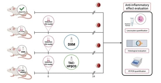
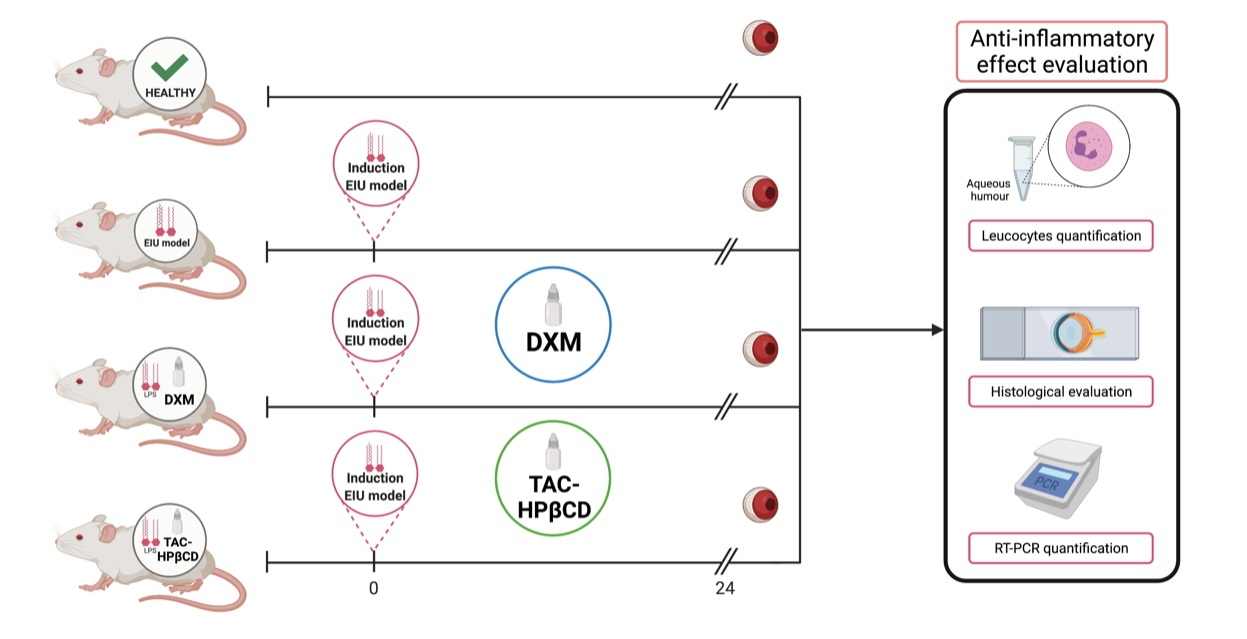
2.2. In Vivo Endotoxin-Induced Uveitis Model (EIU Model)

In vivo studies were carried out on male Sprague-Dawley rats with an average weight of 250 g supplied by the animal facility at the University of Santiago de Compostela (CEBEGA) (Santiago de Compostela, Spain). The animals were kept in cages under controlled temperature (22 ± 1 °C) and humidity (60 ± 5%), with day–night cycles regulated by artificial light (12/12 h) and feeding ad libitum. The animals were treated according to the ARVO statement for the use of animals in ophthalmic and vision research as well as the approved guidelines for laboratory animals [42,43].
The EIU was induced inoculating 1 mg/kg Escherichia coli. LPS diluted in 0.1 mL balance salt solution (BSS) into the rat’s right paw as previously described by other authors [44,45]. Once injected, the ocular inflammation peak reaches the maximum after 24 h [46].
In this case, 32 rats were divided into 4 groups of 8 rats each, as indicated in Figure 1. (a) untreated healthy rats (Healthy); (b) untreated EIU-rats (EIU); (c) EIU-rats treated with standard treatment of dexamethasone ophthalmic drops (DXM) (Maxidex®, dexamethasone 1 mg/mL, Alcon Cusí, Barcelona, Spain) and d) EIU-rats treated with TAC-HPβCD eye drops (TAC-HPβCD). Animals were anesthetized with 2.5% (v/v) isoflurane/oxygen and treatments were topically instilled (20 µL) on each eye 3 h before EIU model induction and every 3 h until 24 h later. Then, rats were euthanatized with carbon dioxide and all required samples were extracted.

2.3. RNA Isolation and Real-Time PCR Analysis

The mRNA expression levels of IL-6, IL-8, MIP-1α and TNF-α were evaluated in both eyes of the remaining 5 rats of each group by real time-PCR. After extraction, eyeballs were frozen at −20 °C and total RNA was isolated using TRIzol reagent (Invitrogen, Barcelona, Spain). Real-time PCR was made with 2 µg cDNA in a 20 µL volume using Luminaris Color HiGreenqPCR Master Mix (Fisher Scientific, Rockford, IL, USA). Samples were denatured at 94 °C for 10 s, annealed at 58 °C for 10 s and extended at 72 °C for 10 s for a total of 35 cycles. The amount of PCR products formed in each cycle was evaluated based on SYBR Green fluorescence with 18S as the endogenous control. Oligonucleotide sequences are detailed in Table 1.

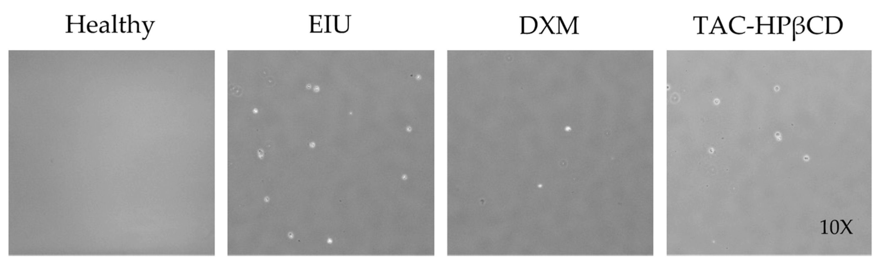
2.4. Quantitative Analysis of Leucocytes in Aqueous Humor

For leucocyte determination, an expert ophthalmologist collected aqueous humor (10 µL) from the anterior chamber of the left eye of 3 rats from each group using a 28-gauge needle. Aqueous humor was stained (1:5) with Turk’s solution (Merk, Darmstadt, Germany) for leucocyte counting in a Neubauer chamber (Brand GMBH þ CO KG, Wertheim, Germany). Each aqueous humor sample was evaluated in triplicate.

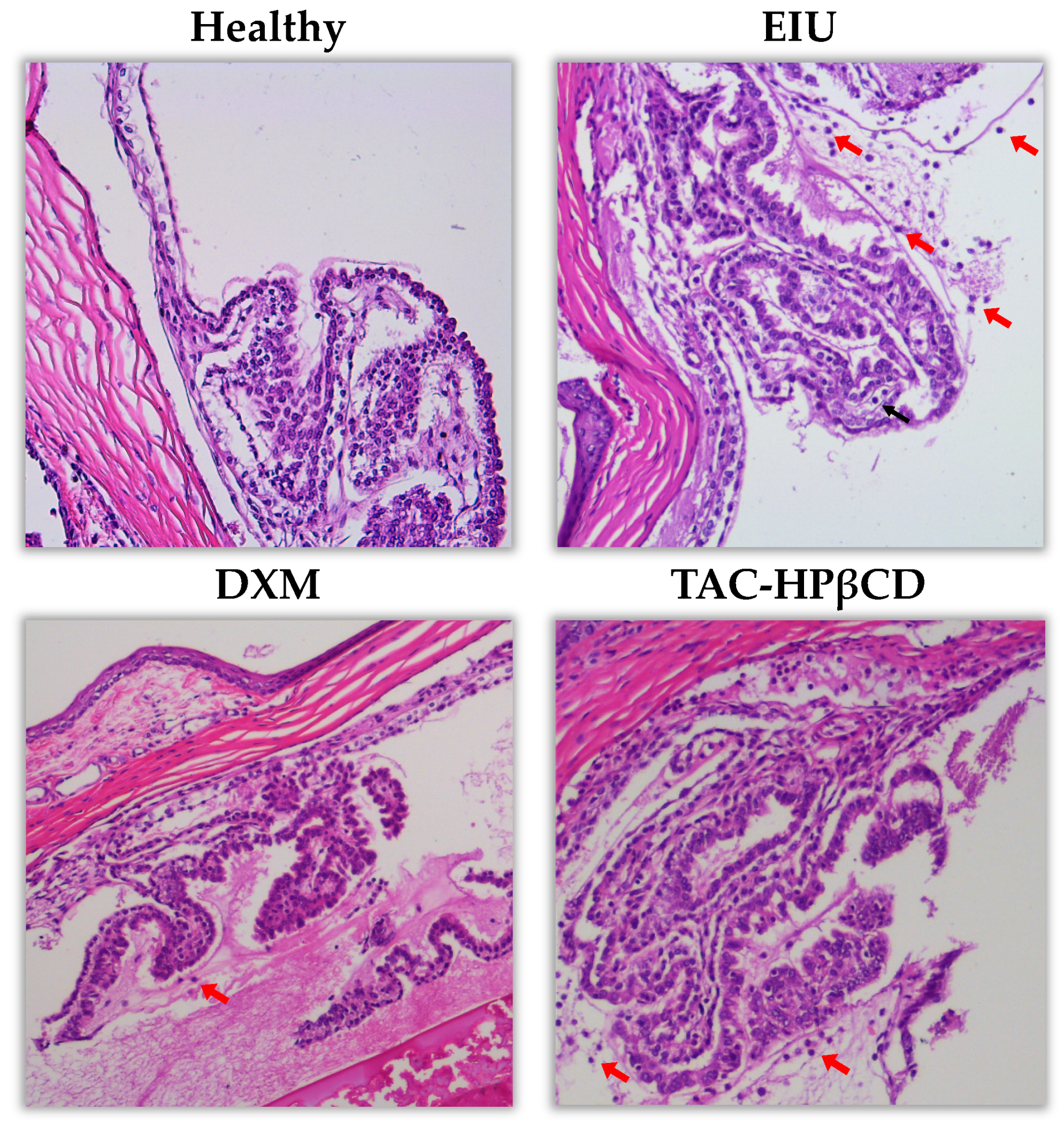
2.5. Histological Evaluation

For histological evaluation, right eyes of 3 rats of each group were fixed in 10% neutral buffered formalin for 24 h and embedded in paraffin routinely. Sections of 4 µm thick were stained with hematoxylin and eosin using a CoverStainer system (Dako-Agilent, Glostrup, Denmark). Six sections from each eye were examined to perform semiquantitative analysis by an anatomopathologist expert. Leucocytes surrounded ciliary processes were taken into account for counting and photographed using an Olympus BX51 microscope equipped with a MC170 camera (Leica, Wetzlar, Germany).

2.6. Statistical Analysis

Values were expressed as mean ± standard error of the mean (SEM) or standard deviation (SD). Average values were compared using one-way ANOVA with Tukey’s multiple comparison test. p values of less than 0.05 were considered statistically significant. Graph Pad Prism (Version 8.0.1) software (GraphPad Software, San Diego, CA, USA) was used for all calculations.

3. Results

3.1. Evaluation of the Effect of Tacrolimus on IL-6, IL-8, MIP-1α, and TNF-α

The mRNA presence of the pro-inflammatory cytokines (IL-6, IL-8, MIP-1α, and TNF-α) in the eye tissues was assessed by real time-PCR in order to evaluate the effect of TAC-HPβCD on factors involved in the inflammatory process. The levels of cytokines in the eye tissues of healthy, EIU, DXM and TAC- HPβCD groups are shown in Figure 2 and the statistical significance of the different levels detected among groups is depicted in Table 2.
EIU group had considerable high levels of IL-6 (138.89 ± 89.71), IL-8 (6.53 ± 5.36), MIP-1α (217.37 ± 113.15), and TNF-α (12.58 ± 3.25) compared to healthy group [IL-6 (0.78 ± 0.68), IL-8 (1.14 ± 0.14), MIP-1α (1.20 ± 0.18) and TNF-α (1.22 ± 0.34)]. Consequently, the augmentation of all these factors was statistically significant in the untreated EIU group as compared with healthy rats, which means that the EIU model was properly induced.
On the other hand, DXM group [IL-6 (1.29 ± 1.59), IL-8 (5.18 ± 5.09), MIP-1α (56.88 ± 54.70); TNF-α (7.31 ± 5.52)] and TAC-HPβCD group [IL-6 (28.69 ± 21.24); IL-8 (2.92 ± 2.73); MIP-1α (59.26 ± 41.43); TNF-α (3.57 ± 2.74)] had substantial fewer values of all the analyzed pro-inflammatory cytokines compared to EIU group. Hence, treatment with TAC-HPβCD and DMX significantly decreased levels of all these factors as compared with EIU group except for IL-8 whose reduction did not achieve statistical significance neither TAC-HPβCD group nor DMX.
Regarding the comparison of the detected levels of pro-inflammatory cytokines between healthy rats and TAC-HPβCD no statistically significant differences were found for IL-6, IL-8, MIP-1α levels, as well as between healthy rats and DMX group. This, along with the fact that no statistically significant differences were neither found among pro-inflammatory cytokines levels of TAC-HPβCD and DXM group demonstrate that TAC-HPβCD eye drops could be an alternative to topical corticosteroid therapy in the treatment of this model of EIU and their clinical translational use should be studied in future studies. In addition, it should be highlighted that with regard to TNF-α, statistically significant differences were detected between healthy rats and DXM group but no between healthy rats and TAC-HPβCD group. This fact suggest that TAC could be a better option than DXM when the uveitis is related with an elevation of TNF-α.

3.2. Quantitative Analysis of Leucocytes in Aqueous Humor

Leukocyte counts in aqueous humor of healthy, EIU, DXM and TAC-HPβCD groups are depicted in Figure 3 and Figure 4. In this sense, EIU group showed a significantly higher number of leukocytes (44.9 ± 11.0 cells/mL) compared to healthy group (0.2 ± 0.4 cells/mL) (p < 0.005), supporting the adequate development of the uveitis model. On the other hand, regarding leucocytes count of TAC-HPβCD group (27.55 ± 5.19 cells/mL), there is a considerable reduction in the number of leucocytes as compared with EIU group, being this difference statistically significant (p < 0.005) and positioning this therapy as a possible alternative to DXM treatment in uveitis. However, the decrease in the number of leucocytes is significantly greater in DXM group with regard to TAC-HPβCD group (p < 0.005), achieving a number of leucocytes comparable to healthy group.

3.3. Histological Evaluation

As it can be seen in Figure 5, the histological evaluation of retinas and ciliary processes showed no infiltration of leucocytes in healthy rats with no LPS injection. Very few or no leucocytes were found in DXM group while some leukocytes can be observed in TAC-HPβCD group. However, there was a considerable higher infiltration of leucocytes in untreated EIU-rats as compared with DXM and TAC-HPβCD groups.

4. Discussion

Nowadays, TAC is commercially available in oral and intravenous formulations, used in transplanted patients, and as an ointment for the treatment of atopic dermatitis [47]. TAC eyedrops can constitute an alternative in the treatment of uveitis [48,49] because it has been successfully used for various immune-mediated ocular disorders such as graft rejection [20], vernal keratoconjunctivitis (VKC) [21,22,23,24], dry eye [25] or scleritis [26,27]. However, TAC eyedrops are not marketed and they must be elaborated in HPDs. In this sense, it is necessary to highlight that TAC is a highly lipophilic macrolide lactone with a very poor water-solubility (1–2 μg/mL) [50]. This fact constitutes an important hurdle because TAC must be dissolved to allow efficient transport or permeation and reach the target site to become therapeutically effective [51].
Another inconvenience of TAC is that it has a high susceptibility to hydrolysis which leads to a very low stability in aqueous solutions [28,29]. For these reasons, TAC eye drops must be elaborated in HPDs by reformulation of the intravenous drug presentation (Prograf®), containing ethanol which have showed to disrupt the integrity of corneal epithelium and induce inflammation in corneal cells [52]. Based on this situation, in recent years several formulations of TAC for topical ophthalmic administration have been developed. In this sense, niosomes [29] and micelles [31] have been synthetized but they were not tested in vivo. However, positive effects of ophthalmic TAC were recently reported for nanovesicles [30] and nanocapsules [32].
Our group has previously developed a formulation of TAC-HPβCD eye drops containing a 0.02% (w/v) of TAC and a 40% (w/v) of HPβCD in Liquifilm®, which can be safely prepared in HPDs and showed to be stable for at least 3 months under refrigeration [40]. The major advantage of TAC-HβCD eye drops is that there is no need of using irritating excipients since our formulation strategy increases the solubilization of poorly soluble active ingredients and reduces the use of toxic products. In order to evaluate our formulation, a EIU model was selected because it is easily reproducible, quantifiable and clinical changes are similar to those of human acute uveitis [37]. Some authors stated that after LPS administration there is an activation of TLRs, increasing levels of proinflammatory mediators, developing inflammation of anterior uvea, choroid and retina and producing the breakdown of blood-humor barrier, with exudation of leukocytes into the aqueous humor [53,54]. In this sense, significantly higher levels of TNF-α, IL-6, IL-8, MIP-1α and leukocytes, as well as histological evaluation of EIU group, compared to healthy group confirmed the correct development of the uveitis model.
According to its mechanism of action, TAC inhibits calcineurin, blocks the production of proinflammatory cytokines and leads to inhibition of Th1 and Th2 cell activation [28,55]. In this regard, significantly lower levels of TNF-α, IL-6, MIP-1α and leukocytes, as well as histological evaluation of TAC-HPβCD compared to EIU group confirms the effectiveness of this formulation in the treatment of uveitis. Other authors stated that ophthalmic treatment with TAC solution in light mineral oil, a vehicle where this active substance is not adequately dissolved, was not able to improve any parameter related with clinical manifestation of uveitis comparing to a EIU model [37]. Consequently, our positive therapeutic effects using TAC-HPβCD demonstrate the proper solubilization of TAC in our formulation.
Similarly, TAC proglycosome nanovesicles were synthetized by Garg et al., showing promising results in a rabbit EIU model since they achieved suppression of expression of TNF-α and IL-6 in aqueous humor, and reduction in ocular inflammation, leukocyte infiltration and protein leakage into aqueous humor comparing with the administration of ophthalmic TAC solution [30]. Furthermore, Rebibo et al. elaborated TAC-loaded nanocapsules enabling a superior anti-inflammatory effect on an anterior chamber LPS-induced murine model of keratitis in comparison to the drug in oil solution and with a significant decrease in macrophage inflammatory protein 2 and IL-6, among other evaluated inflammatory markers [32].
Regarding the comparison between the efficacy of our formulation and the standard treatment with dexamethasone, no statistically significant differences were found among pro-inflammatory cytokines levels of TAC-HPβCD and DXM group demonstrating that TAC-HPβCD eye drops could be an alternative to topical corticosteroid therapy in the treatment of this model of EIU and their clinical translational use should be studied in future studies. It is necessary to highlight that Rebibo et al. and Garg et al., in contrast with us, did not compared their formulations with corticosteroid standard treatment [30,32].
In addition, concerning TNF-α mRNA expression, its reduction is statistically significant only between TAC-HPβCD and EIU group but no between DXM and EIU group, which is consistent with TAC mechanism of action [56] and could be especially beneficial in uveitis related to the augmentation of this factor, such as HLA-B27-associated uveitis, sarcoid uveitis and uveitis associated with Behcet’s disease among other noninfectious uveitis entities [11,12,57]. This possible personalization of the therapy based on the elevation of certain proinflammatory molecules could constitute an important advance, above all if this local increase could be correlated with systemic increases and detected through a blood test.
With respect to the limitations of our study, the fact that EIU model achieves the maximum inflammation 24 h after injection [36,37] makes necessary to carry out an accelerated dosage regimen and only permits a short-term evaluation which could constitute a hurdle regarding the translation of these results to the clinical setting. Another limitation is the lack of knowledge of the amount and rate of tacrolimus transcorneal permeation. Therefore, it will be necessary to study the intraocular pharmacokinetics of the TAC-HPβCD formulation with a suitable analytical method.
In spite of the fact that other ophthalmic TAC formulations have shown in vivo efficacy [37,39], as long as they are not commercialized, their translation to clinical practice is very difficult because of the high complexity of their synthesis. However, our TAC-HPβCD formulation has demonstrated in vivo efficacy and it can be easily elaborated in HPDs due to the simplicity of the elaboration process. Regarding the difference of cost associated between TAC eye drops usually prepared in HPDs by reformulation of Prograf® and TAC-HPβCD eye drops, an estimate of the expenditure has been made in the Hospital Pharmacy Service for the two formulations in which the cost would be reduced by 200 euros for each patient and year with the implementation of the TAC-HPβCD formulation.
Future studies are needed to confirm the clinical effectiveness of TAC-HPβCD in order to consider this formulation as an alternative for the treatment of some forms of uveitis. The next step will be to conduct a study with patients in which the TAC-HPβCD formulation will be tested by comparing it with the results obtained in this group’s previous work [22] with the TAC formulation used in HPDs.

5. Conclusions

TAC-HPβCD eyedrops showed beneficial effect in EIU model in rats, thus they could be considered an alternative for uveitis treatment in case of corticosteroids resistance or intolerance. This formulation demonstrated to reduce ocular inflammation, expression of IL-6, TNF-α, MIP-1α and leukocyte infiltration in aqueous humor. In addition, the simple elaboration process of TAC-HPβCD makes possible to prepare this formulation in HDPs, improving in terms of safety the current elaborated TAC eyedrops by reformulation from intravenous drug presentation which contains ethanol and other irritating excipients. Further clinical studies are needed in order to evaluate long-term effectiveness of TAC-HPβCD.

Author Contributions

Conceptualization, A.F.-F., F.J.O.-E. and M.A.B.; methodology, X.G.-O., C.M.-G., F.G., R.P.-F., J.R.A.-L. and L.A.; validation, A.A., M.G.-B. and R.P.-F.; formal analysis, J.R.A.-L. and A.A.; investigation, X.G.-O., C.M.-G., L.A. and J.R.A.-L., resources, X.G.-O., C.M.-G., and M.A.B.; data curation, F.G., A.A., R.P.-F. and L.A.; writing—original draft preparation, X.G.-O., C.M.-G. and F.G.; writing—review and editing, M.G.-B., P.A., A.F.-F., F.J.O.-E. and M.A.B.; visualization, F.G., M.G.-B., J.R.A.-L., A.A. and P.A.; supervision, P.A., A.F.-F., F.J.O.-E. and M.A.B.; project administration, A.F.-F., P.A. and F.J.O.-E.; funding acquisition, A.F.-F. and F.G. All authors have read and agreed to the published version of the manuscript.

Funding

This project was partially funded by Health Research Institute Carlos III (PI20/00719, RETICS RD16/0008/0003), FEDER, Axencia Galega Innovación (Grupos de Potencial Crecimiento IN607B2020/11 y Grupo de Referencia Competitiva ED431C2021/01) and Spanish Ministry of Science, Innovation and Universities (RTI2018-099597-B-100). X. García-Otero is grateful to the IDIS (Health Research Institute of Santiago de Compostela) for financing his predoctoral research fellowship. C. Mondelo-García and A. Fernández-Ferreiro are grateful to the Carlos III Health Institute for financing their personnel contracts: JR20/00026 and JR18/00014.

Institutional Review Board Statement

The study was conducted according to the ARVO statement for the use of animals in ophthalmic and vision research and the approved guidelines for laboratory animals. It was approved by the (Health Research Institute of Santiago de Compostela’s (IDIS) Animal Experimentation Ethics Committee (15010/2019/006, approval date: 25 September 2019).

Informed Consent Statement

Not applicable.

Data Availability Statement

Not applicable.

Acknowledgments

Not applicable.

Conflicts of Interest

The authors declare no conflict of interest. The funders had no role in the design of the study; in the collection, analyses, or interpretation of data; in the writing of the manuscript, or in the decision to publish the results.

References

  1. Tsirouki, T.; Dastiridou, A.; Dastiridou, O.; Brazitikou, I.; Kalogeropoulos, C.; Androudi, S. A Focus on the Epidemiology of Uveitis. Ocul. Immunol. Inflamm. 2018, 26, 2–16. [Google Scholar] [CrossRef] [PubMed]
  2. Fonollosa, A.; Adán, A. Uveitis: A multidisciplinary approach. Arch. Soc. Esp. Oftalmol. 2011, 86, 393–394. [Google Scholar] [CrossRef]
  3. Acharya, N.R.; Tham, V.M.; Esterberg, E.; Borkar, D.S.; Parker, J.V.; Vinoya, A.C.; Uchida, A. Incidence and Prevalence of Uveitis: Results from the Pacific Ocular Inflammation Study. JAMA Ophthalmol. 2013, 131, 1405–1412. [Google Scholar] [CrossRef] [Green Version]
  4. Hwang, D.-K.; Chou, Y.-J.; Pu, C.-Y.; Chou, P. Epidemiology of Uveitis among the Chinese Population in Taiwan: A Population-Based Study. Ophthalmology 2012, 119, 2371–2376. [Google Scholar] [CrossRef]
  5. Hart, C.T.; Zhu, E.Y.; Crock, C.; Rogers, S.L.; Lim, L.L. Epidemiology of Uveitis in Urban Australia. Clin. Exp. Ophthalmol. 2019, 47, 733–740. [Google Scholar] [CrossRef] [PubMed]
  6. Fanlo, P.; Heras, H.; Espinosa, G.; Adan, A. Complications and Visual Acuity of Patients with Uveitis: Epidemiological Study in a Reference Unit in Northern Spain. Arch. Soc. Esp. Oftalmol. 2019, 94, 419–425. [Google Scholar] [CrossRef]
  7. Weinstein, J.E.; Pepple, K.L. Cytokines in Uveitis. Curr. Opin. Ophthalmol. 2018, 29, 267–274. [Google Scholar] [CrossRef]
  8. De Smet, M.D.; Taylor, S.R.J.; Bodaghi, B.; Miserocchi, E.; Murray, P.I.; Pleyer, U.; Zierhut, M.; Barisani-Asenbauer, T.; LeHoang, P.; Lightman, S. Understanding Uveitis: The Impact of Research on Visual Outcomes. Prog. Retin. Eye Res. 2011, 30, 452–470. [Google Scholar] [CrossRef] [PubMed]
  9. Lee, R.W.; Nicholson, L.B.; Sen, H.N.; Chan, C.-C.; Wei, L.; Nussenblatt, R.B.; Dick, A.D. Autoimmune and Autoinflammatory Mechanisms in Uveitis. Semin. Immunopathol. 2014, 36, 581–594. [Google Scholar] [CrossRef] [PubMed] [Green Version]
  10. Taylor, A.W. Ocular Immune Privilege. Eye 2009, 23, 1885–1889. [Google Scholar] [CrossRef] [Green Version]
  11. Balamurugan, S.; Das, D.; Hasanreisoglu, M.; Toy, B.C.; Akhter, M.; Anuradha, V.K.; Anthony, E.; Gurnani, B.; Kaur, K. Interleukins and Cytokine Biomarkers in Uveitis. Indian J. Ophthalmol. 2020, 68, 1750–1763. [Google Scholar] [CrossRef]
  12. Curnow, S.J.; Falciani, F.; Durrani, O.M.; Cheung, C.M.G.; Ross, E.J.; Wloka, K.; Rauz, S.; Wallace, G.R.; Salmon, M.; Murray, P.I. Multiplex Bead Immunoassay Analysis of Aqueous Humor Reveals Distinct Cytokine Profiles in Uveitis. Investig. Ophthalmol. Vis. Sci. 2005, 46, 4251–4259. [Google Scholar] [CrossRef]
  13. Kirino, Y.; Bertsias, G.; Ishigatsubo, Y.; Mizuki, N.; Tugal-Tutkun, I.; Seyahi, E.; Ozyazgan, Y.; Sacli, F.S.; Erer, B.; Inoko, H.; et al. Genome-Wide Association Analysis Identifies New Susceptibility Loci for Behçet’s Disease and Epistasis between HLA-B*51 and ERAP1. Nat. Genet. 2013, 45, 202–207. [Google Scholar] [CrossRef] [Green Version]
  14. Medawar, P.B. Immunity to Homologous Grafted Skin; the Fate of Skin Homografts Transplanted to the Brain, to Subcutaneous Tissue, and to the Anterior Chamber of the Eye. Br. J. Exp. Pathol. 1948, 29, 58–69. [Google Scholar] [PubMed]
  15. Hou, S.; Du, L.; Lei, B.; Pang, C.P.; Zhang, M.; Zhuang, W.; Zhang, M.; Huang, L.; Gong, B.; Wang, M.; et al. Genome-Wide Association Analysis of Vogt-Koyanagi-Harada Syndrome Identifies Two New Susceptibility Loci at 1p31.2 and 10q21.3. Nat. Genet. 2014, 46, 1007–1011. [Google Scholar] [CrossRef]
  16. Dick, A.D.; Rosenbaum, J.T.; Al-Dhibi, H.A.; Belfort, R.; Brézin, A.P.; Chee, S.P.; Davis, J.L.; Ramanan, A.V.; Sonoda, K.-H.; Carreño, E.; et al. Guidance on Noncorticosteroid Systemic Immunomodulatory Therapy in Noninfectious Uveitis. Ophthalmology 2018, 125, 757–773. [Google Scholar] [CrossRef] [PubMed] [Green Version]
  17. Shahab, M.A.; Mir, T.A.; Zafar, S. Optimising Drug Therapy for Non-Infectious Uveitis. Int. Ophthalmol. 2019, 39, 1633–1650. [Google Scholar] [CrossRef] [PubMed]
  18. Valdes, L.M.; Sobrin, L. Uveitis Therapy: The Corticosteroid Options. Drugs 2020, 80, 765–773. [Google Scholar] [CrossRef] [PubMed]
  19. Jabs, D.A.; Rosenbaum, J.T.; Foster, C.S.; Holland, G.N.; Jaffe, G.J.; Louie, J.S.; Nussenblatt, R.B.; Stiehm, E.R.; Tessler, H.; Gelder, R.N.V.; et al. Guidelines for the Use of Immunosuppressive Drugs in Patients with Ocular Inflammatory Disorders: Recommendations of an Expert Panel. Am. J. Ophthalmol. 2000, 130, 492–513. [Google Scholar] [CrossRef]
  20. Joseph, A.; Raj, D.; Shanmuganathan, V.; Powell, R.J.; Dua, H.S. Tacrolimus Immunosuppression in High-Risk Corneal Grafts. Br. J. Ophthalmol. 2007, 91, 51–55. [Google Scholar] [CrossRef] [PubMed] [Green Version]
  21. Caputo, R.; Marziali, E.; de Libero, C.; Di Grande, L.; Danti, G.; Virgili, G.; Villani, E.; Mori, F.; Bacci, G.M.; Lucenteforte, E.; et al. Long-Term Safety and Efficacy of Tacrolimus 0.1% in Severe Pediatric Vernal Keratoconjunctivitis. Cornea 2021, 40, 1395–1401. [Google Scholar] [CrossRef]
  22. Luaces-Rodríguez, A.; Touriño-Peralba, R.; Alonso-Rodríguez, I.; García-Otero, X.; González-Barcia, M.; Rodríguez-Ares, M.T.; Martínez-Pérez, L.; Aguiar, P.; Gómez-Lado, N.; Silva-Rodríguez, J.; et al. Preclinical Characterization and Clinical Evaluation of Tacrolimus Eye Drops. Eur. J. Pharm. Sci. Off. J. Eur. Fed. Pharm. Sci. 2018, 120, 152–161. [Google Scholar] [CrossRef] [PubMed]
  23. Shoughy, S.S.; Jaroudi, M.O.; Tabbara, K.F. Efficacy and Safety of Low-Dose Topical Tacrolimus in Vernal Keratoconjunctivitis. Clin. Ophthalmol. Auckl. NZ 2016, 10, 643–647. [Google Scholar] [CrossRef] [PubMed] [Green Version]
  24. Astellas Pharma Inc A Randomized, Placebo-Controlled, Double-Masked Study of 0.1% Tacrolimus (FK506) Ophthalmic Suspension in Vernal Keratoconjunctivitis. Available online: clinicaltrials.gov (accessed on 17 August 2021).
  25. Moawad, P.; Shamma, R.; Hassanein, D.; Ragab, G.; El Zawahry, O. Evaluation of the Effect of Topical Tacrolimus 0.03% versus Cyclosporine 0.05% in the Treatment of Dry Eye Secondary to Sjogren Syndrome. Eur. J. Ophthalmol. 2021. [Google Scholar] [CrossRef] [PubMed]
  26. Young, A.L.; Wong, S.M.; Leung, A.T.S.; Leung, G.Y.S.; Cheng, L.L.; Lam, D.S.C. Successful Treatment of Surgically Induced Necrotizing Scleritis with Tacrolimus. Clin. Exp. Ophthalmol. 2005, 33, 98–99. [Google Scholar] [CrossRef]
  27. Lee, Y.J.; Kim, S.W.; Seo, K.Y. Application for Tacrolimus Ointment in Treating Refractory Inflammatory Ocular Surface Diseases. Am. J. Ophthalmol. 2013, 155, 804–813. [Google Scholar] [CrossRef] [PubMed]
  28. Thomson, A.W.; Bonham, C.A.; Zeevi, A. Mode of Action of Tacrolimus (FK506): Molecular and Cellular Mechanisms. Ther. Drug Monit. 1995, 17, 584–591. [Google Scholar] [CrossRef]
  29. Murphy, C.C.; Greiner, K.; Plskova, J.; Duncan, L.; Frost, N.A.; Forrester, J.V.; Dick, A.D. Cyclosporine vs. Tacrolimus Therapy for Posterior and Intermediate Uveitis. Arch. Ophthalmol. Chic. 2005, 123, 634–641. [Google Scholar] [CrossRef] [PubMed] [Green Version]
  30. Horai, R.; Caspi, R.R. Cytokines in Autoimmune Uveitis. J. Interferon Cytokine Res. Off. J. Int. Soc. Interferon Cytokine Res. 2011, 31, 733–744. [Google Scholar] [CrossRef]
  31. Sakamoto, T. Cell biology of hyalocytes. Nippon Ganka Gakkai Zasshi 2003, 107, 866–882. [Google Scholar]
  32. Mesquida, M.; Molins, B.; Llorenç, V.; de la Maza, M.S.; Adán, A. Targeting Interleukin-6 in Autoimmune Uveitis. Autoimmun. Rev. 2017, 16, 1079–1089. [Google Scholar] [CrossRef] [PubMed]
  33. Sloper, C.M.; Powell, R.J.; Dua, H.S. Tacrolimus (FK506) in the Treatment of Posterior Uveitis Refractory to Cyclosporine. Ophthalmology 1999, 106, 723–728. [Google Scholar] [CrossRef]
  34. Oh-i, K.; Keino, H.; Goto, H.; Yamakawa, N.; Murase, K.; Usui, Y.; Kezuka, T.; Sakai, J.-I.; Takeuchi, M.; Usui, M. Intravitreal Injection of Tacrolimus (FK506) Suppresses Ongoing Experimental Autoimmune Uveoretinitis in Rats. Br. J. Ophthalmol. 2007, 91, 237–242. [Google Scholar] [CrossRef] [Green Version]
  35. Ishikawa, T.; Hokama, H.; Katagiri, Y.; Goto, H.; Usui, M. Effects of Intravitreal Injection of Tacrolimus (FK506) in Experimental Uveitis. Curr. Eye Res. 2005, 30, 93–101. [Google Scholar] [CrossRef] [PubMed]
  36. Zeng, W.; Li, Q.; Wan, T.; Liu, C.; Pan, W.; Wu, Z.; Zhang, G.; Pan, J.; Qin, M.; Lin, Y.; et al. Hyaluronic Acid-Coated Niosomes Facilitate Tacrolimus Ocular Delivery: Mucoadhesion, Precorneal Retention, Aqueous Humor Pharmacokinetics, and Transcorneal Permeability. Colloids Surf. B Biointerfaces 2016, 141, 28–35. [Google Scholar] [CrossRef]
  37. Garg, V.; Nirmal, J.; Riadi, Y.; Kesharwani, P.; Kohli, K.; Jain, G.K. Amelioration of Endotoxin-Induced Uveitis in Rabbit by Topical Administration of Tacrolimus Proglycosome Nano-Vesicles. J. Pharm. Sci. 2021, 110, 871–875. [Google Scholar] [CrossRef] [PubMed]
  38. Siegl, C.; König-Schuster, M.; Nakowitsch, S.; Koller, C.; Graf, P.; Unger-Manhart, N.; Schindlegger, Y.; Kirchoff, N.; Knecht, C.; Prieschl-Grassauer, E.; et al. Pharmacokinetics of Topically Applied Tacrolimus Dissolved in Marinosolv, a Novel Aqueous Eye Drop Formulation. Eur. J. Pharm. Biopharm. 2019, 134, 88–95. [Google Scholar] [CrossRef] [PubMed]
  39. Rebibo, L.; Tam, C.; Sun, Y.; Shoshani, E.; Badihi, A.; Nassar, T.; Benita, S. Topical Tacrolimus Nanocapsules Eye Drops for Therapeutic Effect Enhancement in Both Anterior and Posterior Ocular Inflammation Models. J. Control. Release 2021, 333, 283–297. [Google Scholar] [CrossRef]
  40. García-Otero, X.; Díaz-Tomé, V.; Varela-Fernández, R.; Martín-Pastor, M.; González-Barcia, M.; Blanco-Méndez, J.; Mondelo-García, C.; Bermudez, M.A.; Gonzalez, F.; Aguiar, P.; et al. Development and Characterization of a Tacrolimus/Hydroxypropyl-β-Cyclodextrin Eye Drop. Pharmaceutics 2021, 13, 149. [Google Scholar] [CrossRef] [PubMed]
  41. European Medicines Agency (EMA). Cyclodextrins Used as Excipients. Available online: https://www.ema.europa.eu/en/cyclodextrins (accessed on 17 August 2021).
  42. The Association for Research in Vision and Ophthalmology-Statement for the Use of Animals in Ophthalmic and Vision Research. Available online: https://www.arvo.org/About/policies/statement-for-the-use-of-animals-in-ophthalmic-and-vision-research/ (accessed on 5 May 2021).
  43. National Research Council (US). Committee for the Update of the Guide for the Care and Use of Laboratory Animals Guide for the Care and Use of Laboratory Animals. In The National Academies Collection: Reports Funded by National Institutes of Health, 8th ed.; National Academies Press (US): Washington, DC, USA, 2011; ISBN 978-0-309-15400-0. [Google Scholar]
  44. Da Silva, P.S.; Girol, A.P.; Oliani, S.M. Mast Cells Modulate the Inflammatory Process in Endotoxin-Induced Uveitis. Mol. Vis. 2011, 17, 1310–1319. [Google Scholar]
  45. Girol, A.P.; Mimura, K.K.O.; Drewes, C.C.; Bolonheis, S.M.; Solito, E.; Farsky, S.H.P.; Gil, C.D.; Oliani, S.M. Anti-Inflammatory Mechanisms of the Annexin A1 Protein and Its Mimetic Peptide Ac2-26 in Models of Ocular Inflammation in Vivo and in Vitro. J. Immunol. 2013, 190, 5689–5701. [Google Scholar] [CrossRef] [PubMed]
  46. Bermudez, M.A.; Sendon-Lago, J.; Seoane, S.; Eiro, N.; Gonzalez, F.; Saa, J.; Vizoso, F.; Perez-Fernandez, R. Anti-Inflammatory Effect of Conditioned Medium from Human Uterine Cervical Stem Cells in Uveitis. Exp. Eye Res. 2016, 149, 84–92. [Google Scholar] [CrossRef]
  47. Cury Martins, J.; Martins, C.; Aoki, V.; Gois, A.F.T.; Ishii, H.A.; da Silva, E.M.K. Topical Tacrolimus for Atopic Dermatitis. Cochrane Database Syst. Rev. 2015, CD009864. [Google Scholar] [CrossRef]
  48. Gamalero, L.; Simonini, G.; Ferrara, G.; Polizzi, S.; Giani, T.; Cimaz, R. Evidence-Based Treatment for Uveitis. Isr. Med. Assoc. J. IMAJ 2019, 21, 475–479. [Google Scholar]
  49. Hogan, A.C.; McAvoy, C.E.; Dick, A.D.; Lee, R.W.J. Long-Term Efficacy and Tolerance of Tacrolimus for the Treatment of Uveitis. Ophthalmology 2007, 114, 1000–1006. [Google Scholar] [CrossRef] [PubMed]
  50. Kwon, M.; Yeom, D.; Kim, N.A.; Choi, D.H.; Park, J.; Wang, H.; Yoo, S.D.; Jeong, S.H. Bioequivalence of Tacrolimus Formulations with Different Dynamic Solubility and In-Vitro Dissolution Profiles. Arch. Pharm. Res. 2015, 38, 73–80. [Google Scholar] [CrossRef]
  51. Loftsson, T.; Muellertz, A.; Siepmann, J. For the Special IJP Issue “Poorly Soluble Drugs”. Int. J. Pharm. 2013, 453, 1–2. [Google Scholar] [CrossRef] [PubMed]
  52. Oh, J.Y.; Yu, J.M.; Ko, J.H. Analysis of Ethanol Effects on Corneal Epithelium. Investig. Ophthalmol. Vis. Sci. 2013, 54, 3852–3856. [Google Scholar] [CrossRef] [Green Version]
  53. Yadav, U.C.S.; Ramana, K.V. Endotoxin-Induced Uveitis in Rodents. Methods Mol. Biol. 2019, 1960, 161–168. [Google Scholar] [CrossRef]
  54. Fox, A.; Hammer, M.E.; Lill, P.; Burch, T.G.; Burrish, G. Experimental Uveitis. Elicited by Peptidoglycan-Polysaccharide Complexes, Lipopolysaccharide, and Muramyl Dipeptide. Arch. Ophthalmol. Chic. 1984, 102, 1063–1067. [Google Scholar] [CrossRef] [PubMed]
  55. Vafadari, R.; Kraaijeveld, R.; Weimar, W.; Baan, C.C. Tacrolimus Inhibits NF-ΚB Activation in Peripheral Human T Cells. PLoS ONE 2013, 8, e60784. [Google Scholar] [CrossRef] [PubMed] [Green Version]
  56. Hikita, N.; Chan, C.C.; Whitcup, S.M.; Nussenblatt, R.B.; Mochizuki, M. Effects of Topical FK506 on Endotoxin-Induced Uveitis (EIU) in the Lewis Rat. Curr. Eye Res. 1995, 14, 209–214. [Google Scholar] [CrossRef] [PubMed]
  57. Mesquida, M.; Molins, B.; Llorenç, V.; Sainz de la Maza, M.; Hernandez, M.V.; Espinosa, G.; Adán, A. Proinflammatory Cytokines and C-Reactive Protein in Uveitis Associated with Behçet’s Disease. Mediat. Inflamm. 2014, 2014, e396204. [Google Scholar] [CrossRef] [PubMed]
Figure 1. (A) Study groups scheme and distribution of eyes for their posterior analysis. (B) Study timeline: Induction EIU model, treatments, euthanasia and eyes removal. Created with BioRender.com.
Pharmaceutics 13 01737 g001
Figure 2. Anti-inflammatory in vivo effect (a) IL-6 (b) IL-8 (c) MIP-1α and (d) TNF-α mRNA expression in healthy eyes (Healthy), EIU untreated rats (EIU), EIU rats treated with dexamethasone (DXM) and EIU rats treated with TAC-HPβCD (TAC-HPβCD). (n = 10 eyes per group). Bars represent mean ± SD (* p < 0.05; ** p < 0.005).
Pharmaceutics 13 01737 g002
Figure 3. Leucocyte counting in aqueous humor 24 h after LPS injection in a Neubauer chamber. Groups were healthy eyes (Healthy), EIU untreated rats (EIU), EIU rats treated with dexamethasone (DXM) and EIU rats treated with TAC-HPβCD (TAC-HPβCD) (n = 3 eyes).
Pharmaceutics 13 01737 g003
Figure 4. Quantitative analysis of leucocytes in aqueous humor. Groups were healthy eyes (Healthy), EIU untreated rats (EIU), EIU rats treated with dexamethasone (DXM) and EIU rats treated with TAC-HPβCD (TAC-HPβCD) (n = 3 eyes per group measured in triplicate). Bars represent mean ± SD. There are statistically significant differences among all groups except between Healthy and DXM group whose difference is non-significant (ns).
Pharmaceutics 13 01737 g004
Figure 5. Representative hematoxylin-eosin staining showing infiltrated leucocytes (marked with arrows) in retina and ciliary processes of healthy, EIU, DXM and TAC-HPβCD groups. The histology study was carried out 24 h after LPS injection.
Pharmaceutics 13 01737 g005
Table 1. Primer sets for real-time PCR.
PrimersForward (5′-3′)Reverse (5′-3′)
IL-6CTTCAGGCCAAGTTCAGGAGAGTGG ATCGTGGTCGTCTTC
IL-8TCCAGCCAGTTGCCTTCTTGGGTCTGTTGTGGGTGGTATCC
MIP-1αCGTCCTCATCCTGATCACCTGATACATCCCCGTGAACACC
TNF-αCCAGATGGTCACCCTCAGATCCTTGACCG CTGAAGAGAAC
18SGTAACCGCTTGAACCCCATTCCATCCAATCGCTAGTAGCG
Table 2. Existence (YES) or absence (NO) of statistical significance calculated through a Tukey’s multiple comparison test among Healthy, EIU, DXM and TAC-HPβCD groups (* p < 0.05; ** p < 0.005).
Tukey’s Multiple
Comparison Test
IL-6IL-8MIP-1αTNF-α
Healthy vs. EIUYES **YES *YES **YES **
Healthy vs. TAC-HPβCDNONONONO
Healthy vs. DXMNONONOYES **
EIU vs. TAC-HPβCDYES ** NOYES **YES **
EIU vs. DXMYES **NOYES **YES *
TAC-HPβCD vs. DXMNONONONO
Publisher’s Note: MDPI stays neutral with regard to jurisdictional claims in published maps and institutional affiliations.

