Development of a Nonwoven Hemostatic Dressing Based on Unbleached Cotton: A De Novo Design Approach

Abstract
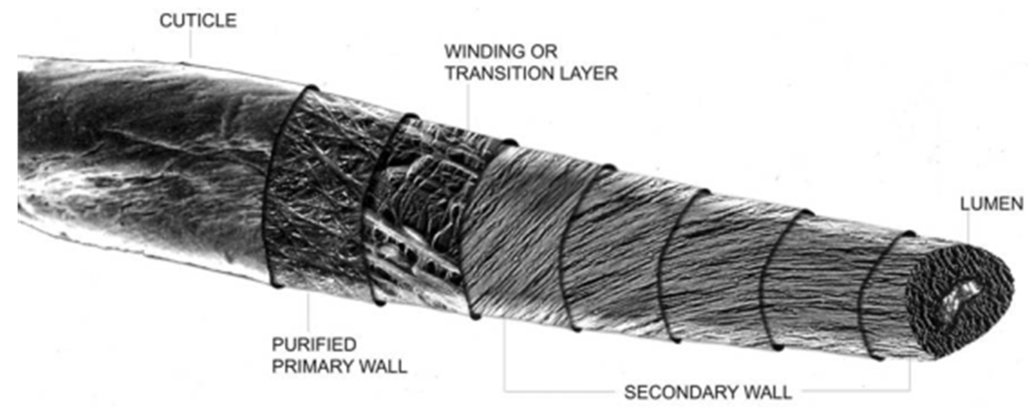
:1. Introduction
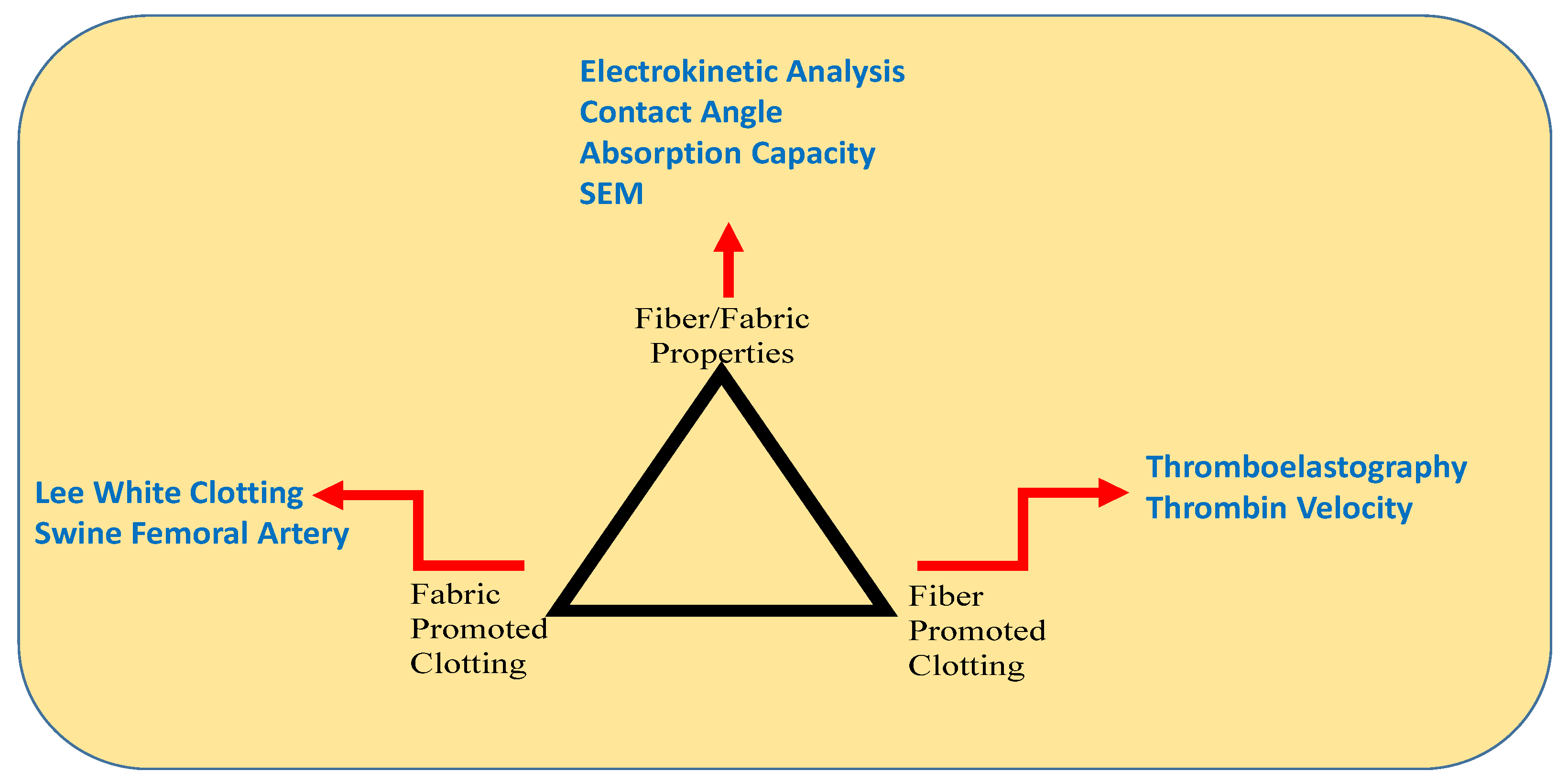
Triad Approach to Hemostatic Dressing Discovery
2. Materials and Methods
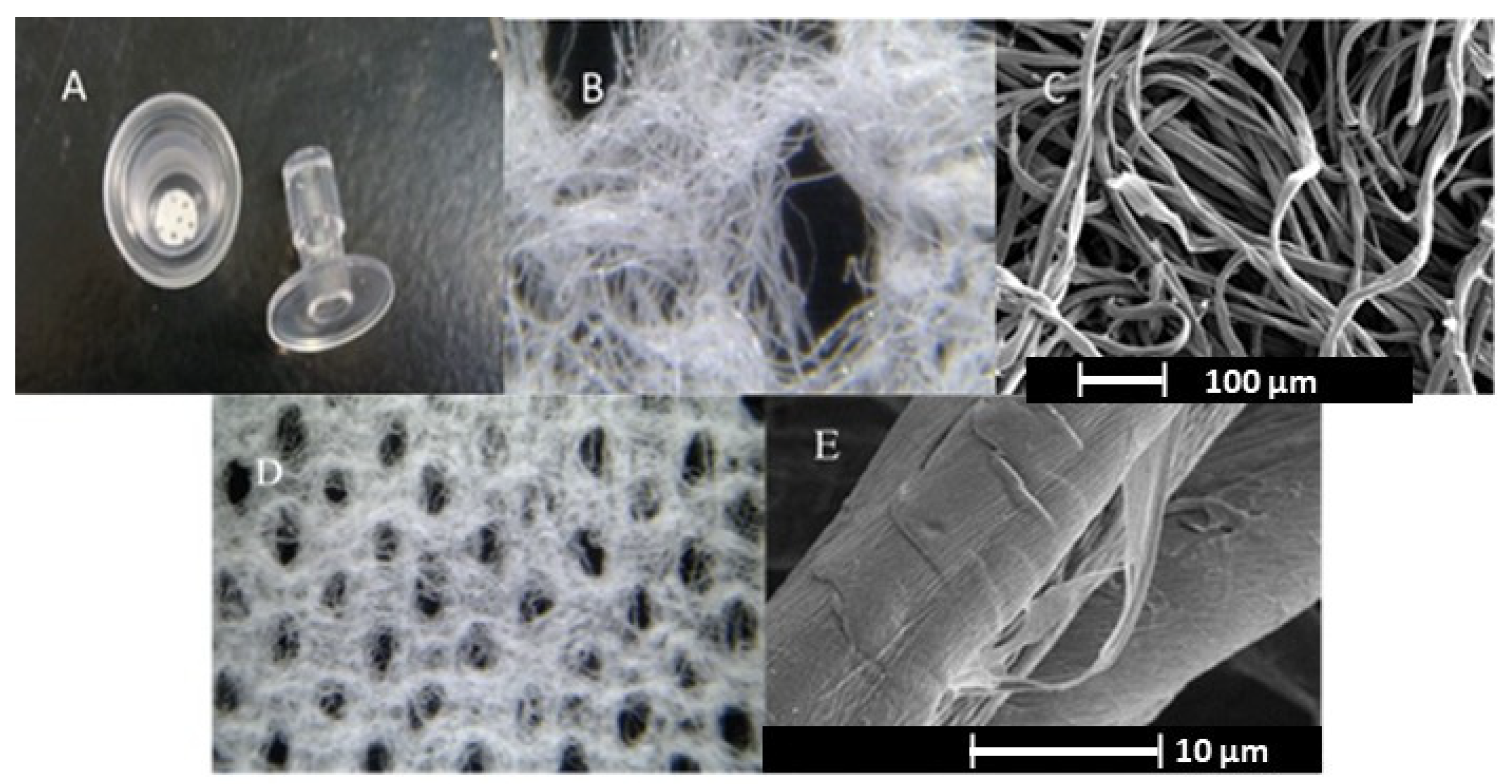
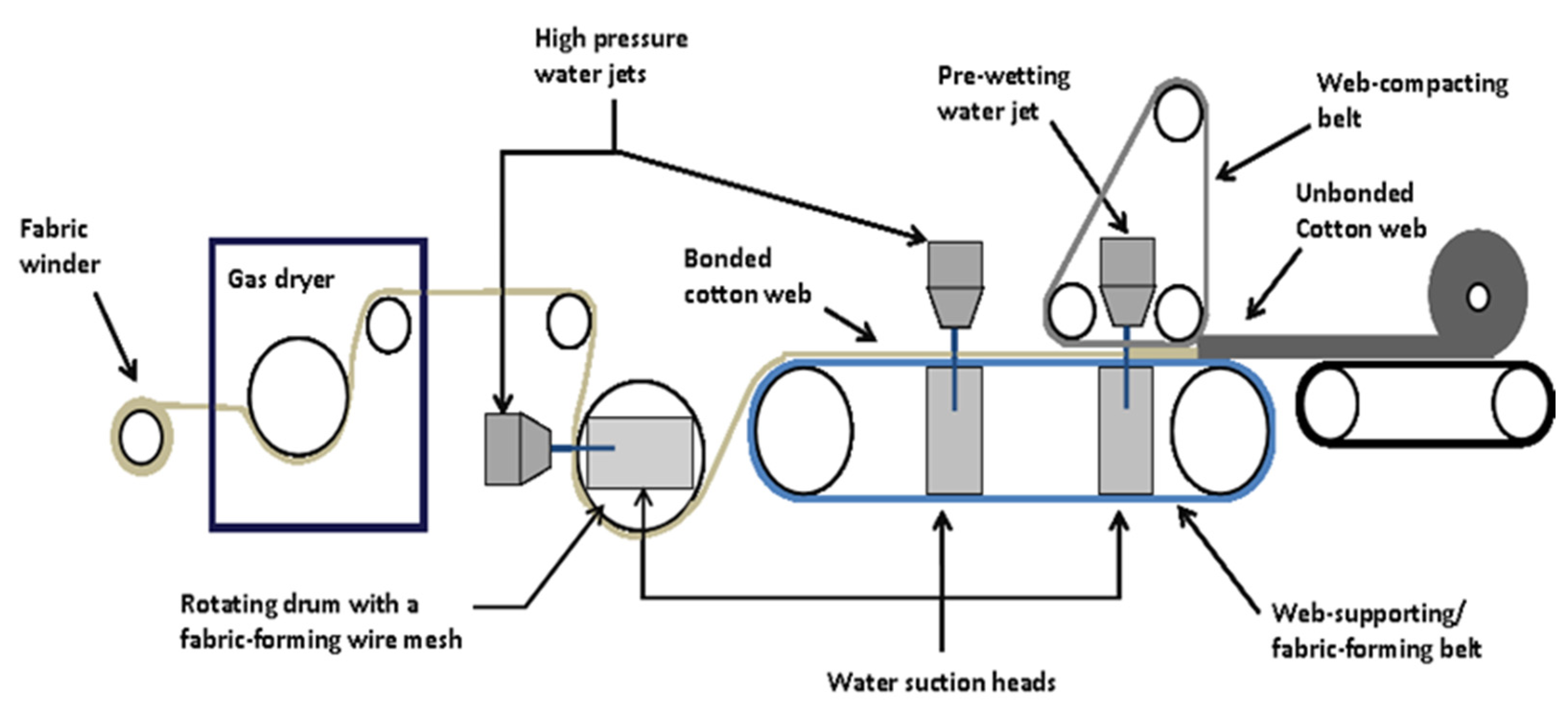
2.1. Hydroentanglement of Fibrous Webs into Nonwoven Fabric Structure
2.2. Thromboelastography
2.3. Modified Lee White Clotting Assay
2.4. Thrombin Assay
2.5. Zeta Potential Measurements
2.6. Absorbency
3. Results
3.1. Identification of Hemostatic Greige Cotton Nonwovens
3.2. Examination of Fabric Composition, Mesh and Density with a Model Fabric
3.3. Construction of Cotton-Based Nonwovens for Clotting Structure/Function Relations
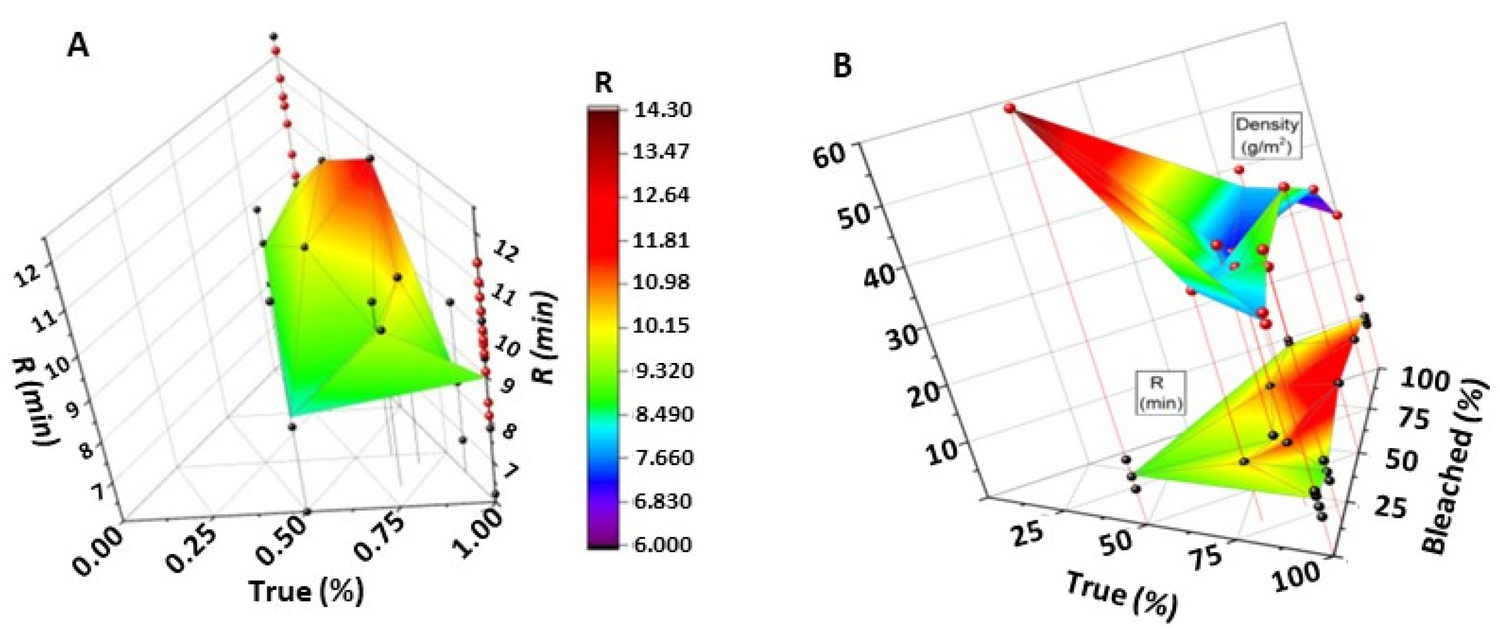
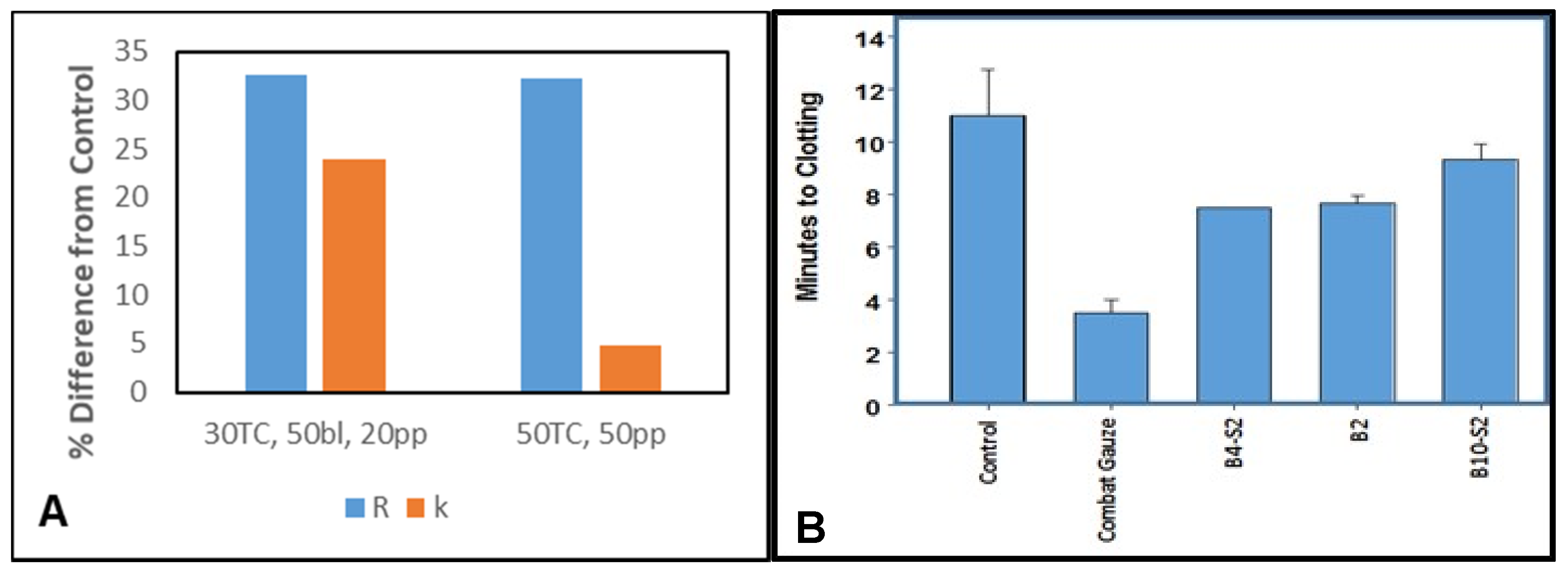
Modeling Fibrin Formation Based on Fabric Composition and Density
3.4. Electrokinetic Analysis
3.5. Absorption Capacity
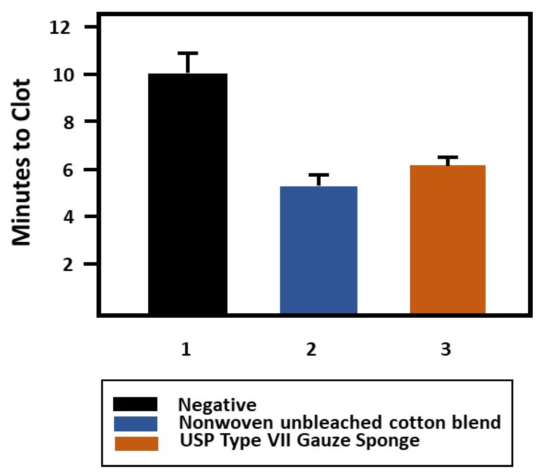
3.6. Lee Clotting and Thrombin Generation Analysis
4. Discussion
4.1. Identification of Hemostatic Greige Cotton Nonwovens
4.2. Absorption Capacity and Effect of Hydroentanglement Waterjet Pressure
4.3. Effect on Thrombin Release and Assessment with Lee White Clotting Assay
5. Conclusions
Author Contributions
Funding
Conflicts of Interest
References
- Dhivya, S.; Padma, V.V.; Santhini, E. Wound dressings—A review. BioMedicine 2015, 5, 24–28. [Google Scholar] [CrossRef] [PubMed]
- Mescher, V. Lint and Charpie: It’s not your dryer lint. J. Civil War Med. 2011, 15, 144–150. [Google Scholar]
- Wakelyn, P.J.; Bertoniere, N.R.; French, A.D.; Thibodeaux, D.P.; Triplett, B.A.; Rousselle, M.-A.; Goynes, W.R.; Edwards, J.V.; Hunter, L.; McAlister, D.D.; et al. Cotton Fiber Chemistry and Technology; International Fiber Science and Technology; Lewin, M., Ed.; CRC Press: Boca Raton, FL, USA, 2007; p. 162. [Google Scholar]
- Edwards, J.V.; Graves, E.; Bopp, A.; Prevost, N.; Santiago, M.; Condon, B. Electrokinetic and hemostatic profiles of nonwoven cellulosic/synthetic fiber blends with unbleached cotton. J. Funct. Biomater. 2014, 5, 273–287. [Google Scholar] [CrossRef] [PubMed] [Green Version]
- Edwards, J.V.; Mao, N.; Russell, S.; Carus, E.; Condon, B.; Hinchliffe, D.; Gary, L.; Graves, E.; Bopp, A.; Wang, Y. Fluid handling and fabric handle profiles of hydroentangled greige cotton and spunbond polypropylene nonwoven topsheets. Proc. IMechE Part L J. Mater. Des. Appl. 2015, 230, 847–859. [Google Scholar]
- Hickman, D.A.; Pawlowski, C.L.; Sekhon, U.D.S.; Marks, J.; Gupta, A.S. Biomaterials and advanced technologies for hemostatic management of bleeding. Adv. Mater. 2018, 30, 1700859. [Google Scholar] [CrossRef]
- Bennett, B.L.; Littlejohn, L. Review of new topical hemostatic dressings for combat casualty care. Mil. Med. 2014, 179, 497–514. [Google Scholar] [CrossRef] [Green Version]
- Peng, T. Biomaterials for hemorrhage control. Trends Biomater. Artif. Organs 2010, 24, 27–68. [Google Scholar]
- Fisher, T.H.; Vournakis, J.N.; Manning, J.E.; McCurdy, S.L.; Rich, P.B.; Nichols, T.C.; Scull, C.M.; McCord, M.G.; DaCorta, J.A.; Johnson, P.C.; et al. The design and testing of a dual fiber textile matrix for accelerating surface hemostasis. J. Biomed. Mater. Res. Part B Appl. Biomater. 2009, 91, 381–389. [Google Scholar] [CrossRef] [Green Version]
- Pogorielov, M.; Kalinkevich, O.; Deineka, V.; Garbuzova, V.; Solodovnik, A.; Kalinkevich, A.; Kalinichenko, T.; Gapchenko, A.; Sklyar, A.; Danilchenko, S. Haemostatic chitosan coated gauze: In vitro interaction with human blood and in-vivo effectiveness. Biomater. Res. 2015, 19, 1–10. [Google Scholar] [CrossRef] [Green Version]
- Fischer, T.H.; Thatte, H.S.; Nichols, T.C.; Bender-Neal, D.E.; Bellinger, A.D.; Vournakis, J.N. Synergistic platelet integrin signaling and factor xii activation in poly-n-acetyl glucosamine fiber-mediated hemostasis. Biomaterials 2005, 26, 5433–5443. [Google Scholar] [CrossRef]
- Gajjar, C.R.; McCord, M.G.; King, M.W. 19-Hemostatic wound dressings. In Biotextiles as Medical Implant; King, M.W., Gupta, B.S., Guidoin, R., Eds.; Woodhead Publishing Limited: Sawston, Cambridge, UK, 2013; pp. 563–589. [Google Scholar]
- Wagner, W.R.; Pachence, J.M.; Ristich, J.; Johnson, P.C. Comparative in vitro analysis of topical hemostatic agents. J. Surg. Res. 1996, 66, 100–108. [Google Scholar] [CrossRef] [PubMed]
- Khoshmohabat, H.; Paydar, S.; Kazemi, H.; Dalfardi, B. Overview of agents used for emergency hemostasis. Trauma Mon. 2016, 21, e26023. [Google Scholar] [CrossRef] [PubMed] [Green Version]
- Yang, X.; Liu, W.; Li, N.; Wang, M.; Liang, B.; Ullah, I.; Neve, A.L.; Feng, Y.; Chen, H.; Shi, C. Design and development of polysaccharide hemostatic materials and their hemostatic mechanism. Biomater. Sci. 2017, 5, 2357–2368. [Google Scholar] [CrossRef] [PubMed]
- Dowling, M.B.; Kumar, R.; Keibler, M.A.; Hess, J.R.; Bochicchio, G.V.; Raghavan, S.R. A self-assembling hydrophobically modified chitosan capable of reversible hemostatic action. Biomaterials 2011, 32, 3351–3357. [Google Scholar] [CrossRef] [PubMed]
- Behrens, A.M.; Sikorski, M.J.; Kofinas, P. Hemostatic strategies for traumatic and surgical bleeding. J. Biomed. Mater. Res. Part A 2014, 102, 4182–4194. [Google Scholar] [CrossRef] [Green Version]
- Ohta, S.; Nishiyama, T.; Sakoda, M.; Machioka, K.; Fuke, M.; Ichimura, S.; Inagaki, F.; Shimizu, A.; Hasegawa, K.; Kokudo, N.; et al. Development of carboxymethyl cellulose nonwoven sheet as a novel hemostatic agent. J. Biosci. Bioeng. 2015, 119, 718–723. [Google Scholar] [CrossRef]
- Hutchinson, R.W.; George, K.; Johns, D.; Craven, L.; Zhang, G.; Shnoda, P. Hemostatic efficacy and tissue reaction of oxidized regenerated cellulose hemostats. Cellulose 2013, 20, 537–545. [Google Scholar] [CrossRef]
- Cheng, W.; He, J.; Chen, M.; Li, D.; Li, H.; Chen, L.; Cao, Y.; Wang, J.; Huang, Y. Preparation, functional characterization and hemostatic mechanism discussion for oxidized microcrystalline cellulose and its composites. Fibers Polym. 2016, 17, 1277–1286. [Google Scholar] [CrossRef]
- Baumann, H.; Liu, C.; Faust, V. Regioselectively modified cellulose and chitosan derivatives for mono- and multilayer surface coatings of hemocompatible biomaterials. Cellulose 2003, 10, 65–74. [Google Scholar] [CrossRef]
- Liu, X.-J.; Xu, Y.-Y.; Zhou, J.; Zhang, J.-L.; Zhou, X.-T.; Tian, Y. Hemostatic effect of soluble hemostatic gauze on liver trauma in rabbits. Chin. J. Tissue Eng. Res. 2016, 20, 7070–7075. [Google Scholar]
- Yang, X.; Li, N.; Constantinesco, I.; Yu, K.; Kizhakkedathu, J.N.; Brooks, D.E. Choline phosphate functionalized cellulose membrane: A potential hemostatic dressing based on a unique bioadhesion mechanism. Acta Biomater. 2016, 40 (Suppl. C), 212–225. [Google Scholar] [CrossRef] [PubMed]
- Jayakumar, R.; Prabaharan, M.; Sudheesh Kumar, P.T.; Nair, S.V.; Tamura, H. Biomaterials based on chitin and chitosan in wound dressing applications. Biomaterials 2011, 29, 322–337. [Google Scholar] [CrossRef] [PubMed]
- Zielińska, D.; Struszczyk, M.H.; Madej-Kiełbik, L.; Chmal-Fudali, E.; Kucharska, M.; Wiśniewska-Wrona, M.; Brzoza-Malczewska, K. Design of new-generation usable forms of topical haemostatic agents containing chitosan. Molecules 2017, 22, 2240. [Google Scholar] [CrossRef] [Green Version]
- Peng, H.T. Thromboelastographic study of biomaterials. J. Biomed. Mater. Res. Part B Appl. Biomater. 2010, 94, 469–485. [Google Scholar] [CrossRef] [PubMed]
- Sperling, C.; Fischer, M.; Maitz, M.F.; Werner, C. Blood coagulation on biomaterials requires the combination of distinct activation processes. Biomaterials 2009, 30, 4447–4456. [Google Scholar] [CrossRef]
- Lyocell. How Products are Made Forum. Volume 5. Available online: http://www.madehow.com/Volume-5/Lyocell.html (accessed on 12 March 2020).
- Gupta, B.S. Porosity and its characterization in nonwovens. In Proceedings of the 2003 Beltwide Cotton Conferences, Nashville, TN, USA, 6–10 January 2003. [Google Scholar]
- Gupta, B.S.; Edwards, J.V. Textile materials and structures for wound care products. In Advanced Textiles for Wound Care; Rajendran, S., Ed.; CRC Press: Washington, DC, USA, 2009; pp. 48–96. [Google Scholar]
- Vaughn, E.A.; Carman, B.G. Expanded surface area fibers: A means for medical product enhancement. J. Ind. Text. 2001, 30, 303–310. [Google Scholar] [CrossRef]
- Zhuo, R.; Siedlecki, C.A.; Vogler, E.A. Autoactivation of blood factor XII at hydrophilic and hydrophobic surfaces. Biomaterials 2006, 27, 4325–4332. [Google Scholar] [CrossRef]
- Ribitsch, V.; Stana-Kleinscheck, K. Characterizing textile fiber surfaces with streaming potential measurements. Text. Res. J. 1998, 68, 701–707. [Google Scholar] [CrossRef]
- Renne, T.; Schmaier, A.H.; Nickel, K.F.; Blomback, M.; Maas, C. In vivo roles of factor XII. Blood 2012, 120, 4206–4303. [Google Scholar] [CrossRef] [Green Version]
- Persin, Z.; Stana-Kleinschek, K.; Kreze, T. Hydrophilic/Hydrophobic characteristics of different cellulose fibres monitored by tensiometry. Croat. Chem. ACTA 2002, 75, 271–280. [Google Scholar]
- Gupta, B.S.; Hong, C.J. Changes in web dimensions during fluid uptake and the impact on absorbency. Tappi J. 1994, 77, 181–188. [Google Scholar]
- Sayeb, S.; Hassen, M.B.; Sakli, F. Modelling absorption capacity performance of hygienic product. Open J. Appl. Sci. 2013, 3, 169–173. [Google Scholar] [CrossRef] [Green Version]
- Thomposn, A.L.; Paulis, D.; Tomasi, P.; Yurchenko, O.; Jenks, M.A.; Dyer, J.M.; Gore, M.A. Chemical variation for fiber cuticular wax levels in upland cotton (Gossypium hirsutum L.) evaluated under contrasting irrigation regimes. Ind. Crops Prod. 2017, 100, 153–162. [Google Scholar] [CrossRef] [Green Version]
- Cesar, J.M.; Vecino, A.M. Survival and function of transfused platelets. Studies in two patients with congenital deficiencies of platelet membrane glycoproteins. Platelets 2009, 20, 158–162. [Google Scholar] [CrossRef]
- Tripodi, A. Thrombin generation assay and its application in the clinical laboratory. Clin. Chem. 2016, 62, 699–707. [Google Scholar] [CrossRef] [Green Version]
- Rodrigues, S.N.; Gonçalves, I.C.; Martins, M.C.L.; Barbosa, M.A.; Ratner, B.D. Fibrinogen adsorption, platelet adhesion and activation on mixed hydroxyl-/methyl-terminated self-assembled monolayers. Biomaterials 2006, 27, 5357–5367. [Google Scholar] [CrossRef]
- Edwards, J.V.; Graves, E.; Reynolds, M.; Condon, B.; Yager, D.; Dacorta, J. Structure/Function relations of hemostatic nonwoven dressings based on greige cotton. In AATCC International Conference; American Association of Textile Chemists and Colorists: Greenville, SC, USA, 2018; pp. 265–278. [Google Scholar]
- Vogler, E.A.; Siedlecki, C.A. Contact activation of blood-plasma coagulation. Biomaterials 2009, 30, 1857–1869. [Google Scholar] [CrossRef] [Green Version]
- Smith, S.A.; Travers, R.J.; Morrissey, J.H. How it all starts: Initiation of the clotting cascade. Crit. Rev. Biochem. Mol. Biol. 2015, 50, 326–336. [Google Scholar] [CrossRef] [Green Version]
- Fathi, P.; Sikorski, M.; Christodoulides, K.; Langan, K.; Choi, Y.S.; Titcomb, M.; Ghodasara, A.; Wonodi, O.; Thaker, H.; Vural, M.; et al. Zeolite-loaded alginate-chitosan hydrogel beads as a topical hemostat. J. Biomed. Mater. Res. Part B Appl. Biomater. 2018, 106, 1662–1671. [Google Scholar] [CrossRef]
- Maas, C.; Renné, T. Regulatory mechanisms of the plasma contact system. Thromb. Res. 2012, 129, S73–S76. [Google Scholar] [CrossRef]
- Kozin, F.; Cochrane, C.G. The contact activation system of plasma-biochemistry and pathophysiology. In Inflammation: Basic Principles and Clinical Correlates, 2nd ed.; Gallin, J., Goldstein, I.M., Snyderman, R., Eds.; Raven Press: New York, NY, USA, 1992. [Google Scholar]
- Evans-Nguyen, K.M.; Tolles, L.R.; Gorkun, O.V.; Lord, S.T.; Schoenfisch, M.T. Interactions of thrombin with fibrinogen adsorbed on methyl-, hydroxyl-, amine-, and carboxyl-terminated self-assembled monolayers. Biochemistry 2005, 44, 15561–15568. [Google Scholar] [CrossRef]
- Johne, J.; Blume, C.; Benz, P.M.; Pozgajová, M.; Ullrich, M.; Schuh, K.; Nieswandt, B.; Walter, U.; Renné, T. Platelets promote coagulation factor XII-mediated proteolytic cascade systems in plasma. Biol. Chem. 2006, 387, 173–178. [Google Scholar] [CrossRef]
- Taylor, R.E.; French, A.D.; Gamble, G.P.; Himmelsbach, D.S.; Stipanovic, R.D.; Thibodeaux, D.P.; Wakelyn, P.J.; Dybowski, C. 1H and 13C solid-state NMR of Gossypium barbadense (Pima) cotton. J. Mol. Struct. 2008, 878, 177–184. [Google Scholar] [CrossRef] [Green Version]
- French, A.D.; Goynes, W.R.; Rousselle, A.-A.; Thibodeaux, D.P. Cotton Fiber and Moisture—Some of the Basics. In Proceedings of the 2004 Beltwide Cotton Conferences, San Antonio, TX, USA, 5–9 January 2004. [Google Scholar]
- Ramjee, M.J. The use of fluorogenic substrates to monitor thrombingeneration for the analysis of plasma and whole blood coagulation. Anal. Biochem. 2000, 277, 11–18. [Google Scholar] [CrossRef]


| Sample | Ra (min) | SD b | ka (min) | SD |
|---|---|---|---|---|
| Bovine blood | 14.6 | 7.8 | 7.3 | 3.2 |
| Bleached cotton | 9.6 | 1.6 | 6.6 | 2.2 |
| Greige cotton | 4.1 | 1.0 | 3.2 | 0.6 |
| Sample (Density a) | %Fibrin Rate (s) * | ζplateau | ∆ζ | R2 | ζ0 | ζ∞ | Swell Ratio |
|---|---|---|---|---|---|---|---|
| B1 [85gc/15bl] (41.9) | 26 | −31 | 0.14 | 0.902 | −32.58 | −29.76 | 1.05 |
| B2 [30gc/50bl/20pp] (34) | 32 | −37 | 0.09 | 0.946 | −40.62 | −37.18 | 1.04 |
| B3 [70gc/10bl/20pp] (37.8) | 6 | −35 | 0.09 | 0.97 | −36.80 | −33.72 | 1.04 |
| B4 (60gc/2bl5/15pp] (39.8) | 12 | −38 | 0.06 | 0.941 | −39.62 | −37.17 | 1.03 |
| B5 [20gc/80bl] (36.4) | 2 | −27 | 0.07 | 0.98 | −24.94 | −23.28 | 1.04 |
| B6 [100bl] (27.6) | 18 | −25 | 0.09 | 0.99 | −23.43 | −21.51 | 1.04 |
| B7 [100gc] (36.6) | 40 ** | −31 | 0.05 | 0.45 | −31.61 | −30.63 | 1.02 |
| B8 [80bl/20pp] (30.8) | 14 | −41 | 0.05 | 0.881 | −40.88 | −38.96 | 1.02 |
| B9 [50gc/50bl] (45.0) | 20 | −31 | 0.09 | 0.95 | −32.58 | −29.76 | 1.04 |
| B10 [50gc/50pp] (65.0) | 33 | −48 | 0.08 | 0.96 | −48.71 | −44.94 | 1.04 |
| Sample | SA a (mg/cm2) | Abs. Cap (g/g) | SD b | Intra (g/g) | SD | Inter (g/g) | SD |
|---|---|---|---|---|---|---|---|
| B1 [85gc/15bl] | 2.7 | 15.34 | 0.840 | 0.38 | 0.044 | 14.96 | 0.804 |
| B2 [30gc/50bl/20pp] | 3.2 | 17.88 | 1.130 | 0.34 | 0.017 | 17.54 | 1.119 |
| B3 [70gc/10bl/20pp] | 3.4 | 13.14 | 0.505 | 0.230 | 0.019 | 12.92 | 0.487 |
| B4 (60gc/2bl5/15pp] | 4.1 | 14.41 | 0.434 | 0.280 | 0.026 | 14.13 | 0.457 |
| B5 [20gc/80bl] | 3.2 | 14.15 | 0.220 | 0.034 | 0.019 | 13.81 | 0.220 |
| B6 [100bl] | 2.7 | 16.58 | 0.680 | 0.300 | 0.034 | 16.29 | 0.677 |
| B7 [100gc] c | 3.26 | 12.95 | 1.526 | 0.38 | 0.055 | 12.57 | 1.534 |
| B8 [80bl/20pp] | 7.58 | 14.87 | 1.554 | 0.32 | 0.143 | 14.56 | 1.412 |
| B9 [50gc/50bl] | 3.0 | 15.48 | 0.496 | 0.04 | 0.1757 | 15.09 | 0.323 |
| B10 [50gc/50pp] | 1.76 | 1.97 | 0.424 | 0.13 | 0.025 | 1.84 | 0.403 |
| Woven Cotton Gauze * | 9.7 | 5.45 | 0.581 | 0.032 | 0.090 | 5.13 | 0.051 |
| Sample | Time 1 (min) | SD | Thrombin (nM) | SD | Time 2 (min) | SD | Velocity-Index | SD |
|---|---|---|---|---|---|---|---|---|
| Plasma control | 2.0 | 0.0 | 124.5 | 19.9 | 14.5 | 2.1 | 10.2 | 3.3 |
| Positive Control * | 2.0 | 0.0 | 288.8 | 19.9 | 5.0 | 1.4 | 110.1 | 58.5 |
| HE0249-R4 | 2.0 | 0.0 | 294.2 | 17.1 | 7.5 | 0.7 | 54.1 | 10.1 |
| Mogul 5 | 2.0 | 0.0 | 294.2 | 30.1 | 8.0 | 0.0 | 49.0 | 5.0 |
© 2020 by the authors. Licensee MDPI, Basel, Switzerland. This article is an open access article distributed under the terms and conditions of the Creative Commons Attribution (CC BY) license (http://creativecommons.org/licenses/by/4.0/).
Share and Cite
Edwards, J.V.; Graves, E.; Prevost, N.; Condon, B.; Yager, D.; Dacorta, J.; Bopp, A. Development of a Nonwoven Hemostatic Dressing Based on Unbleached Cotton: A De Novo Design Approach. Pharmaceutics 2020, 12, 609. https://doi.org/10.3390/pharmaceutics12070609
Edwards JV, Graves E, Prevost N, Condon B, Yager D, Dacorta J, Bopp A. Development of a Nonwoven Hemostatic Dressing Based on Unbleached Cotton: A De Novo Design Approach. Pharmaceutics. 2020; 12(7):609. https://doi.org/10.3390/pharmaceutics12070609
Chicago/Turabian StyleEdwards, J. Vincent, Elena Graves, Nicolette Prevost, Brian Condon, Dorne Yager, Joseph Dacorta, and Alvin Bopp. 2020. "Development of a Nonwoven Hemostatic Dressing Based on Unbleached Cotton: A De Novo Design Approach" Pharmaceutics 12, no. 7: 609. https://doi.org/10.3390/pharmaceutics12070609